The short and midterm outcomes of the De
Vega annuloplasty of the tricuspid valve during
the surgical management of rheumatic valvular
heart disease
THESIS
PRESENTED AND PUBLICLY DEFENDED IN July 19th, 2018
BY
Ms.
Soukaina BENBAKH
Born in August 3rd, 1991in Marrakech
TO OBTAIN A MEDICAL DOCTORATE
KEYWORDS
Tricuspid valve – Tricuspid valve repair- De Vega annuloplasty
JURY
M.
M.
M.
M
PmeP.
M. EL HATTAOUI
Professor of Cardiology
D. BOUMZEBRA
Professor of Cardiovascular Surgery
R. EL HAOUATI
Associate professor of Cardiovascular Surgery
S. EL KARIMI
Associate professor of Cardiology
PRESIDENT
DIRECTOR
ِﱵﱠﻟا َﻚَﺘَﻤْﻌِﻧ َﺮُﻜْﺷَأ ْنَأ ِﲏْﻋِزْوَأ ِّب ر"
َﻞَﻤْﻋَأ ْنَأَو ﱠيَﺪِﻟاَو ٰﻰَﻠَﻋَو ﱠﻲَﻠَﻋ َﺖْﻤَﻌْـﻧَأ
ِﰲ َﻚِﺘَْﲪَﺮِﺑ ِﲏْﻠِﺧْدَأَو ُﻩﺎَﺿْﺮَـﺗ ﺎًِﳊﺎَﺻ
At the time of being admitted as a member of the medical profession:
I SOLEMNLY PLEDGE to dedicate my life to the service of humanity;
THE HEALTH AND WELL-BEING OF MY PATIENT will be my first
consideration;
I WILL RESPECT the autonomy and dignity of my patient;
I WILL MAINTAIN the utmost respect for human life;
I WILL NOT PERMIT considerations of age, disease or disability, creed, ethnic
origin, gender, nationality, political affiliation, race, sexual orientation, social
standing or any other factor to intervene between my duty and my patient;
I WILL RESPECT the secrets that are confided in me, even after the patient has
died;
I WILL PRACTICE my profession with conscience and dignity and in accordance
with good medical practice;
I WILL FOSTER the honour and noble traditions of the medical profession;
I WILL GIVE to my teachers, colleagues, and students the respect and gratitude
that is their due;
I WILL SHARE my medical knowledge for the benefit of the patient and the
advancement of healthcare;
I WILL ATTEND TO my own health, well-being, and abilities in order to provide
care of the highest standard;
I WILL NOT USE my medical knowledge to violate human rights and civil
liberties, even under threat;
LIST OF
PROFESSORS
MARRAKECH
Doyens Honoraires
: Pr. Badie Azzaman MEHADJI
: Pr. Abdelhaq ALAOUI YAZIDI
ADMINISTRATION
Doyen
: Pr. Mohammed BOUSKRAOUI
Vice doyen à la Recherche et la Coopération
: Pr. Mohamed AMINE
Vice doyen aux Affaires Pédagogiques
: Pr. Redouane EL FEZZAZI
Secrétaire Générale
: Mr. Azzeddine EL HOUDAIGUI
Professeurs de l’enseignement supérieur
Nom et Prénom Spécialité Nom et Prénom Spécialité ABOULFALAH Abderrahim Gynécologie-
obstétrique
FINECH Benasser Chirurgie – générale ADERDOUR Lahcen Oto- rhino-
laryngologie
FOURAIJI Karima Chirurgie pédiatrique B
ADMOU Brahim Immunologie GHANNANE Houssine
Neurochirurgie AIT BENALI Said Neurochirurgie KHALLOUKI
Mohammed
Anesthésie- réanimation AIT-SAB Imane Pédiatrie KHATOURI Ali Cardiologie AKHDARI Nadia Dermatologie KISSANI Najib Neurologie AMAL Said Dermatologie KOULALI IDRISSI
Khalid
Traumato- orthopédie AMINE Mohamed Epidémiologie-
clinique
KRATI Khadija Gastro- entérologie AMMAR Haddou
Oto-rhino-laryngologie
LAOUAD Inass Néphrologie ARSALANE Lamiae Microbiologie
-Virologie
LMEJJATI Mohamed Neurochirurgie ASMOUKI Hamid Gynécologie-
obstétrique B
LOUZI Abdelouahed Chirurgie – générale ASRI Fatima Psychiatrie MAHMAL Lahoucine Hématologie -
réanimation Mohammed
BOUKHIRA Abderrahman Biochimie - chimie MOUTAJ Redouane Parasitologie BOUMZEBRA Drissi Chirurgie
Cardio-Vasculaire
MOUTAOUAKIL Abdeljalil
Ophtalmologie BOURROUS Monir Pédiatrie A NAJEB Youssef Traumato-
orthopédie BOUSKRAOUI Mohammed Pédiatrie A NEJMI Hicham Anesthésie-
réanimation CHAKOUR Mohamed Hématologie NIAMANE Radouane Rhumatologie CHELLAK Saliha Biochimie- chimie OULAD SAIAD
Mohamed
Chirurgie pédiatrique CHERIF IDRISSI EL
GANOUNI Najat
Radiologie RAJI Abdelaziz Oto-rhino-laryngologie CHOULLI Mohamed
Khaled
Neuro pharmacologie SAIDI Halim Traumato- orthopédie DAHAMI Zakaria Urologie SAMKAOUI
Mohamed Abdenasser
Anesthésie- réanimation EL ADIB Ahmed Rhassane Anesthésie-
réanimation
SARF Ismail Urologie EL FEZZAZI Redouane Chirurgie pédiatrique SBIHI Mohamed Pédiatrie B EL HATTAOUI Mustapha Cardiologie SOUMMANI
Abderraouf
Gynécologie- obstétrique A/B EL HOUDZI Jamila Pédiatrie B TASSI Noura Maladies infectieuses ELFIKRI Abdelghani Radiologie YOUNOUS Said Anesthésie-
réanimation ESSAADOUNI Lamiaa Médecine interne ZOUHAIR Said Microbiologie ETTALBI Saloua Chirurgie réparatrice
et plastique
Professeurs Agrégés
Nom et Prénom Spécialité Nom et Prénom Spécialité ABKARI Imad Traumato-
orthopédie B
FADILI Wafaa Néphrologie ABOU EL HASSAN Taoufik Anésthésie-
réanimation
FAKHIR Bouchra Gynécologie- obstétrique A ABOUCHADI Abdeljalil Stomatologie et FAKHRI Anass Histologie-
Abdelhamid
ADALI Nawal Neurologie HAJJI Ibtissam Ophtalmologie AGHOUTANE El Mouhtadi Chirurgie
pédiatrique A
HAOUACH Khalil Hématologie biologique AISSAOUI Younes Anesthésie -
réanimation
HAROU Karam Gynécologie- obstétrique B AIT AMEUR Mustapha Hématologie
Biologique
HOCAR Ouafa Dermatologie AIT BENKADDOUR Yassir Gynécologie-
obstétrique A
JALAL Hicham Radiologie ALAOUI Mustapha Chirurgie-
vasculaire péripherique KAMILI El Ouafi El Aouni Chirurgie pédiatrique B
ALJ Soumaya Radiologie KHOUCHANI Mouna Radiothérapie AMRO Lamyae Pneumo-
phtisiologie
KRIET Mohamed Ophtalmologie ANIBA Khalid Neurochirurgie LAGHMARI Mehdi Neurochirurgie ATMANE El Mehdi Radiologie LAKMICHI Mohamed
Amine
Urologie BAIZRI Hicham Endocrinologie et
maladies métaboliques LAKOUICHMI Mohammed Stomatologie et Chirurgie maxillo faciale
BASRAOUI Dounia Radiologie LOUHAB Nisrine Neurologie BASSIR Ahlam Gynécologie-
obstétrique A
MADHAR Si Mohamed
Traumato- orthopédie A
BELBARAKA Rhizlane Oncologie médicale
MAOULAININE Fadl mrabih rabou
Pédiatrie (Neonatologie) BELKHOU Ahlam Rhumatologie MATRANE Aboubakr Médecine nucléaire BEN DRISS Laila Cardiologie MEJDANE Abdelhadi Chirurgie Générale BENCHAMKHA Yassine Chirurgie
réparatrice et plastique
MOUAFFAK Youssef Anesthésie - réanimation BENHIMA Mohamed Amine Traumatologie -
orthopédie B
MOUFID Kamal Urologie BENJELLOUN HARZIMI
Amine
Pneumo- phtisiologie
MSOUGGAR Yassine Chirurgie thoracique BENJILALI Laila Médecine interne NARJISS Youssef Chirurgie générale BENLAI Abdeslam Psychiatrie NOURI Hassan Oto rhino laryngologie BENZAROUEL Dounia Cardiologie OUALI IDRISSI Radiologie
obstétrique B
BOURRAHOUAT Aicha Pédiatrie B QAMOUSS Youssef Anésthésie- réanimation
BSISS Mohamed Aziz Biophysique RABBANI Khalid Chirurgie générale CHAFIK Rachid Traumato-
orthopédie A
RADA Noureddine Pédiatrie A DAROUASSI Youssef Oto-Rhino -
Laryngologie
RAFIK Redda Neurologie DRAISS Ghizlane Pédiatrie RAIS Hanane Anatomie
pathologique EL AMRANI Moulay Driss Anatomie RBAIBI Aziz Cardiologie EL ANSARI Nawal Endocrinologie et
maladies métaboliques
ROCHDI Youssef Oto-rhino- laryngologie EL BARNI Rachid Chirurgie-
générale
SAJIAI Hafsa Pneumo- phtisiologie EL BOUCHTI Imane Rhumatologie SAMLANI Zouhour Gastro- entérologie EL BOUIHI Mohamed Stomatologie et
chir maxillo faciale
SEDDIKI Rachid Anesthésie - Réanimation EL HAOUATI Rachid Chiru Cardio
vasculaire
SORAA Nabila Microbiologie - virologie EL HAOURY Hanane Traumato-
orthopédie A
TAZI Mohamed Illias Hématologie- clinique EL IDRISSI SLITINE Nadia Pédiatrie ZAHLANE Kawtar Microbiologie -
virologie
EL KARIMI Saloua Cardiologie ZAHLANE Mouna Médecine interne EL KHADER Ahmed Chirurgie générale ZAOUI Sanaa Pharmacologie EL KHAYARI Mina Réanimation
médicale
ZEMRAOUI Nadir Néphrologie EL MGHARI TABIB Ghizlane Endocrinologie et
maladies métaboliques
ZIADI Amra Anesthésie - réanimation EL OMRANI Abdelhamid Radiothérapie ZYANI Mohammed Médecine interne
Professeurs Assistants
Nom et Prénom Spécialité Nom et Prénom Spécialité ABDELFETTAH Youness Rééducation et Hammoune Nabil Radiologie
Cytogénéque ABIR Badreddine Stomatologie et
Chirurgie maxillo faciale
IHBIBANE fatima Maladies Infectieuses
ADARMOUCH Latifa Médecine Communautaire (médecine préventive, santé publique et hygiène)
JALLAL Hamid Cardiologie
AIT BATAHAR Salma Pneumo- phtisiologie
JANAH Hicham Pneumo- phtisiologie AKKA Rachid Gastro -
entérologie
KADDOURI Said Médecine interne ALAOUI Hassan Anesthésie -
Réanimation
LAFFINTI Mahmoud Amine
Psychiatrie
AMINE Abdellah Cardiologie LAHKIM Mohammed Chirurgie générale ARABI Hafid Médecine physique
et réadaptation fonctionnelle
LALYA Issam Radiothérapie
ARSALANE Adil Chirurgie Thoracique
LOQMAN Souad Microbiologie et toxicologie environnementale ASSERRAJI Mohammed Néphrologie MAHFOUD Tarik Oncologie médicale BAALLAL Hassan Neurochirurgie MARGAD Omar Traumatologie
-orthopédie BABA Hicham Chirurgie générale MILOUDI Mohcine Microbiologie -
Virologie BELARBI Marouane Néphrologie MLIHA TOUATI
Mohammed
Oto-Rhino - Laryngologie BELBACHIR Anass Anatomie-
pathologique
MOUHSINE Abdelilah Radiologie BELFQUIH Hatim Neurochirurgie MOUNACH Aziza Rhumatologie BELHADJ Ayoub Anesthésie
-Réanimation
MOUZARI Yassine Ophtalmologie BENNAOUI Fatiha Pédiatrie
(Neonatologie)
NADER Youssef Traumatologie - orthopédie BOUCHAMA Rachid Chirurgie générale NADOUR Karim Oto-Rhino -
Vasculaire CHETOUI Abdelkhalek Cardiologie OUERIAGLI NABIH
Fadoua
Psychiatrie CHRAA Mohamed Physiologie REBAHI Houssam Anesthésie -
Réanimation EL HARRECH Youness Urologie RHARRASSI Isam
Anatomie-patologique EL KAMOUNI Youssef Microbiologie
Virologie
SALAMA Tarik Chirurgie pédiatrique EL MEZOUARI El Moustafa Parasitologie
Mycologie
SAOUAB Rachida Radiologie ELBAZ Meriem Pédiatrie SEBBANI Majda Médecine
Communautaire (médecine préventive, santé publique et hygiène)
ELQATNI Mohamed Médecine interne SERGHINI Issam Anesthésie - Réanimation
ESSADI Ismail Oncologie Médicale TAMZAOURTE Mouna Gastro - entérologie FDIL Naima Chimie de
Coordination Bio-organique
TOURABI Khalid Chirurgie réparatrice et plastique
FENNANE Hicham Chirurgie Thoracique
YASSIR Zakaria Pneumo- phtisiologie GHAZI Mirieme Rhumatologie ZARROUKI Youssef Anesthésie -
Réanimation GHOZLANI Imad Rhumatologie ZIDANE Moulay
Abdelfettah
Chirurgie Thoracique HAMMI Salah Eddine Médecine interne ZOUIZRA Zahira Chirurgie
Cardio-Vasculaire LISTE ARRÉTÉÉ LE 12/02/2018
Words can never suffice to properly give them their merit.
This humble work is affectionately
dedicated to them…
You supported me unconditionally, and you continue to do so, in whatever path I took during
my very long academic journey
You relentlessly encouraged me to strive for excellence and you taught me that the sky is the
limit for a person’s dream
Whatever I achieve or hope to accomplish in the future, I know none of it is possible without
your love and endless support and I hope that someday I’ll make you proud …
Because of all the previously cited, I put you first in my list of dedications. Plus, I kind of had
to too knowing that you are going to be reading this manuscript cover to cover. I know because
that’s how dedicated you are to following my academic journey step by step
To my amazing mother
You are the best human being I know and the best mother I could have hoped for
With your strong and gentle soul, you taught me to trust Allah, believe in hard work, and that
so much can be accomplished with little
You have always been the rock of stability in my life. You supported me with your endless
wisdom and found the right words to lift up my spirits
I can’t thank you enough for putting up with my constant complaining, for being nothing but
supportive of my choices and for having enough Faith for both of us
I dedicate this work to you, for your interest in it, as in all my ventures, was never less than
mine
To my smart, mischievously creative sister
Continuous fighting is our thing apparently, but you know that I love you and I can’t imagine
my life without you. Thank you for your support and for speaking your mind without holding
back when I am being stupid. I hope you get everything you wish for.
To my troublesome little brother
Thank you for always being there to do my errands. I am highly indebted to you for providing
my usual sugar supply whenever stress took over. I love you. I hope you will grow up to be the
decent man I know you can be
To my dear grandmothers, grandfather, aunties, uncles and cousins
(maternal and paternal)
May ALLAH rest his soul in peace and grant him the best place in paradise
To my sister and best friend Fatima Zahra and her cutie son Mohamed
Anas (always Ahmed Anas for me)
Words cannot describe the hold you have on my heart. We share the most wonderful memories.
You are my best friend in the whole world. Neither time nor distance is capable of changing
that. Thank you for always being there for me. You deserve everything beautiful in this world. I
love you.
To my sweet cousin Fatima Ezzahra and her adorable son Ghassan
You have always been the gentle soul in the family. Thank you for your endless support and
countless prayers. May Allah grant all your wishes
To Fadwa, my best friend and the kindest person I know
Your big heart and generosity are both overwhelming and humbling. I cannot thank you enough
for being by my side in some of the hardest times of my life, as I cannot properly express my
gratitude for some of the best memories I have. I love you and I will always cherish our
friendship.
To my brother and friend Yassine J
You are still the same helpful and decent person I met twelve years ago. I wish you happiness
and success.
To all my friends Fatima B, Fatima Ezzahra T, Meryem B, Naila L,
Fatima Ezzahra El M, Chihab B, Layla B, Aissam G, Sara I, Yassine Ch,
Hajar J, Sanaa A, Rajaa B, Charaf Z, the two amazing Imane B and
Imane B, Hassan O, Wassima B, Aassim B …
I know I am not the most sensitive person in the world, but I do love you all. Thank you for
both the memories and the headaches
To all my internship colleagues
Thank you for the memorable two years.
To Mr Aouss
I am indebted to you for helping me improve my English and for reincarnating my love for
reading and books.
To all my other family members, big and small, to my friends and
colleagues who I inadvertently failed to mention
To all my teachers in primary, secondary and high school
To all my teachers in medical school of Marrakech
Professor of cardiology
You granted us a great honor by agreeing to preside over the jury of our thesis. I admire the
simplicity and the ease of your approach and the extent of your knowledge. Please accept the
expression of my deep gratitude and respect.
This work is an opportunity to our consideration and deep admiration for all of your scientific
and human qualities.
To our master and thesis director Professor BOUMZEBRA Drissi
Professor of cardiovascular surgery
You have done me a great honor by agreeing to entrust me with this work. I have nothing but
high esteem and admiration for your competence, your seriousness, your dynamism and your
kindness. I would always be grateful for your availability and your patience during this work
preparation despite your busy schedule and professional obligations. Mere words cannot express
my deep gratitude and admiration. Please accept my sincere acknowledgments and the
expression of my great respect.
To our master and thesis judge Professor EL HAOUATI Rachid
Professor of cardiovascular surgery
Thank you for honoring us with your presence and your interest in our thesis topic. Thank you
for your valuable participation in the development of this work. Allow me to express my
admiration for your human and professional qualities. Please accept the expression of my high
esteem, consideration and deep respect.
To our master and thesis judge Professor EL KARIMI Saloua
Professor of cardiology
It is a great honor for us that you have agreed to be a member of this honorable jury. Your
professional skills and your human qualities have been always an example to me. Please find
You have honored us with great sympathy when accepting to sit among our thesis jury. Please
find here the expression of my profound respect and acknowledgments.
To all the cardiovascular surgery department team
To Professor ZOUIZRA Zahira
I cannot express in words how thankful I am for you support, your guidance and your kindness.
This work couldn’t have possibly been accomplished without your help.
To Siham, Amal, Sanaa, Loubna and Khadija
For making my time in the department’s archives as smooth as can be.
For always being their when needed without ever complaining despite my constant annoyance.
Dr El Mardouli, Dr El Alaoui, Dr Issaka and Dr Kane
Thank you for your kindness and support. I owe you a lot for your understanding and help in
completing this work
The amazing nursing team: Sara Nabil, Hanane Hajji, Fatimzehra
Larhbira, Naima Ihend, Fatimzehra Samouni, Laila Bentajri, Touria
Berjoul, Hamza Znati, Ayoub El Ouadi, Mohamed Choukri, Oussama
Rouhy, Hidaya El Adrani, Mohamed El Alami, Mohamed Choukrat,
Houcine Hamni, Khadija Ismaoune, Hanane Outama, Hayat Tadili,
Meryem El Idrissi, Youness El Aouzi, Abdellatif Lagssir, Mohamed
Belkayed, Karim Elbyad
Your hard work and dedication are remarkable. Thank you for making working with you so
enjoyable
To Dr El Moktadir, Dr Boukaidi, Dr Fahd, Dr Gael
To all the cardiovascular medicine department team
Thank you for your undeniable efforts and contribution
And to all those who, in a way or another, contributed to the elaboration
of this work. Find here the expression of my endless gratitude.
Abbreviations list
AF : Atrial fibrillation AV : Aortic valve
AVR : Aortic valve replacement CPB : Cardiopulmonary bypass DVR : Double valve replacement ICU : Intensive care unit
LV : Left ventricle
LVEF : Left ventricle ejection fraction MV : Mitral valve
MVR : Mitral valve replacement NYHA : New York Heart Association PAH : Pulmonary artery hypertension PASP : Pulmonary artery systolic pressure RV : Right ventricle
TR : Tricuspid regurgitation TV : Tricuspid valve
I. Study design: 6 1. Hypothesis: 6 2. Aims: 6 3. Study type: 6 4. Setting: 6 II. Methods: 7 1. Inclusion criteria: 7 2. Exclusion criteria: 7 3. Subgroups: 7
4. Data collection and analyze: 8
RESULTS 9 I. Synthetic/descriptive study: 10 1. Demographic data: 10 2. Clinical data: 13 3. Chest X-ray: 15 4. Electrocardiogram: 16 5. Echocardiographic data: 17 6. Peroperative data: 24 7. Critical care data: 33 8. Immediate and short-term surgical results: (<3 months) 35 9. Mid-term surgical results: (6 months to 2 years) 39
II. Analytic study: 45
1. Mortality: 45
2. Factors of failure of De Vega tricuspid repair: 46 3. Data comparison by left-heart valvular procedures: 47
DISCUSSION 49
I. Demographic, clinical and paraclinical data: 51
1. Age: 51
2. Medical and surgical history: 52 3. Etiology of tricuspid regurgitation: 53
4. NYHA status: 56
5. Congestive heart failure signs: 56
6. Chest X-ray: 57
7. Electrocardiogram: 57
8. Echocardiography: 58
2. Comparison between De Vega annuloplasty and ring annuloplasty: 67 3. Predictive factors of tricuspid valve repair failure: 70
ANNEX 76
CONCLUSION 117
ABSTRACTS 119
Right-sided cardiac valvular disease has traditionally been considered less clinically important than mitral or aortic valve pathology. The tricuspid valve was relatively «forgotten» over the years, even though severe tricuspid regurgitation can cause relevant functional impairment. Lately, the once ‘‘forgotten valve’’ has garnered increasing attention (1).
With the advent of surgical options for rheumatic heart disease the general life expectancy of the patients has increased. This has resulted in increased diagnosis of tricuspid regurgitation (2).
There is no debate regarding the ‘malignant nature’ of this entity as is evidenced by the poor long-term outcomes associated with surgical management of left-sided heart disease without addressing the tricuspid valve.
Pathologic tricuspid regurgitation is divided into primary (organic) and secondary (functional). The most common mechanism is functional (80% to 90% of tricuspid regurgitation) caused by annular dilation, secondary to left-sided valvular disease and pulmonary hypertension (3–6). Even though structural pathology of the valve is not included in the definition of functional tricuspid regurgitation, leaflet tethering involvement in the pathogenesis of functional tricuspid regurgitation has been recently cited (7).
Outcome of isolated tricuspid valve surgery is poor because right ventricular dysfunction has already established. Therefore, tricuspid valve repair is often concomitant to left-heart valve surgery(1,7).
Recent years have seen a surge in surgery for functional tricuspid regurgitation and numerous techniques are available to correct tricuspid regurgitation. The decision whether or not to address the tricuspid valve at the time of primary surgery had been a debatable topic for
some time both in terms of timing, indication, surgical techniques and results. Whereas the short-term outcome of surgical therapy is satisfactory, the majority of surgical techniques are limited in the mid- and long term by unacceptably high rates of residual and/or recurrent TR (8– 10).
There is no discussion whether to treat the tricuspid valve in severe TR, but guidelines provided less firm recommendations for patients with moderate tricuspid regurgitation (10).
Another controversy is the technique of repair. In 1970s, Dr. Norberto De Vega introduced into clinical practice his tricuspid valve suture annuloplasty technique. Because of its simplicity and efficacy, the procedure became widespread (11).
The De Vega annuloplasty has been modified many times and widely used with accepted results by many surgeons. It is a simple and moreover economic procedure. The longevity of the outcome of this technique highly questioned the risk off its failure with subsequent development of significant tricuspid regurgitation requiring a redo surgery. This has led many surgeons to adopt the use of annuloplasty rings (12,13).
While transcatheter therapies for aortic, mitral and pulmonary valve disease are well established, interventional treatment of tricuspid valve disease is still in its early stages of development. The implementation of less invasive therapies in this field is therefore of major clinical interest. Percutaneous technologies might become an alternative option to treat selected tricuspid regurgitation patients with high surgical risk (14).
However, in a developing country such as ours, giving up an efficient and low-cost method like the De Vega annuloplasty for a method with an additional cost is not a luxury. The De Vega annuloplasty is still a gold standard technique for tricuspid valve repair.
Up-to-date ESC/EACTS guidelines for the management of valvular heart disease have recently been published in August 2017. The fact that once again new guidelines were published shows that this is interesting and timely topic.
Rheumatic fever is still a major health problem in our country with a growing number of young patients with rheumatic heart disease to manage. This unfortunate situation makes our health professionals (cardiologists and cardiac surgeons particularly) better placed to make guidelines based on results from rigorous randomized controlled trials on large cohorts.
PATIENTS
AND
I.
Study design:
1.
Hypothesis:
The De Vega annuloplasty is still a relevant tricuspid valve repair method with satisfactory results.
2.
Aims:
- Assessment of the short and mid-term outcomes of the De Vega annuloplasty in patients undergoing valve surgery in the context of rheumatic heart disease.
- Assessment of risk factor of failure of the De Vega annuloplasty
3.
Study type:
A retrospective, single center, observational study
4.
Setting:
Cardiovascular surgery department, Mohammed VI teaching Hospital, a tertiary referral center.
The preoperative echocardiographic assessment and postoperative follow-up was mainly realized by the cardiovascular medicine department of Mohammed VI teaching Hospital.
II.
Methods:
We retrospectively analyzed the perioperative and follow-up data of patients who underwent surgery for rheumatic heart disease with a De Vega annuloplasty in our institute from February 2007 to June 2017.
1.
- Patients with tricuspid regurgitation who underwent concomitant De Vega annuloplasty and another valve repair or replacement for rheumatic heart disease
Inclusion criteria:
- Patients with organic and functional tricuspid regurgitation were included as long as it was treated with the De Vega annuloplasty
- Patients who underwent other repair procedures on the tricuspid valve, besides the De Vega annuloplasty, were also included.
- Patients who underwent additional concomitant cardiac surgical procedures were also included.
2.
Exclusion criteria:
- Patients who underwent the De Vega annuloplasty for non-rheumatic tricuspid regurgitation
- Patients with incomplete or missing medical records
3.
Because of the high diversity in our study group, we identified some subgroups:
Subgroups:
o Patients who underwent double valvular procedures : mitral and tricuspid o Patients who underwent triple valvular procedures : mitral, aortic and tricuspid
4.
Data collection and analyze:
4.1.- The preoperative data (demographic, history, clinical examination and paraclinical exams) was collected through review of medical records in the cardiovascular surgery department archives.
Data collection
- Bidimensional and color doppler echocardiography were used to assess tricuspid
valve disease, other valvular abnormalities and their impact on cardiac cavities.
- The outcomes were analyzed by evaluating data from outpatient follow-up visits. In addition, telephonic follow-up was conducted in order to account for follow-up data
4.2.
- The data were recorded in a questionnaire sheet
Data analysis:
- Descriptive statistics were the primary focus of this analysis. Numeric data were presented by the following descriptive statistics as most appropriate: mean, median, standard deviation, range. Categorical data were presented by frequency tables: count and percentage.
- Descriptive data were analyzed using Microsoft Excel 2016.
- SPSS version 21 was used for multivariable analysis. A p value <0.05 was considered as statistically significant.
I.
Synthetic/descriptive study:
A total of 595 patients underwent concomitant De Vega annuloplasty and another cardiac procedure for rheumatic valvular heart disease from January 2007 to June 2017.
Only 356 patients were included in our case series. The remaining 239 were excluded due to missing or incomplete medical records.
1.
Demographic data:
1.1.
The mean age in our case series was 40.6 with a range from 10 years-old to 73 years-old and a standard deviation of 12.14.
Age:
The age of 73,6 % of our patients ranged from 20 to 50 years-old.
The detailed distribution of our patients by their age is presented in table I. Table I: Age distribution of our patients
AGE GROUP IN YEARS FREQUENCY PERCENTAGE
< 20 14 3.90% 20 - 30 56 15.70% 30 - 40 96 27% 40 - 50 110 30.90% 50 - 60 53 14.90% > 60 27 7.60% TOTAL 356 100.00%
Figure 1: Age distribution of our patients
1.2.
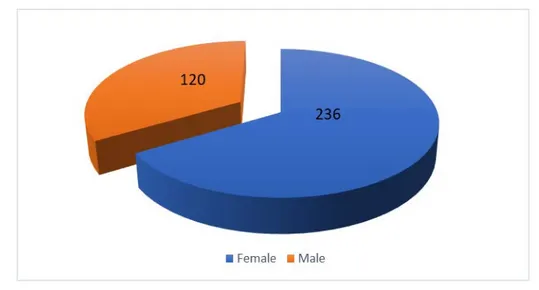
Out of the total of 356 patients in our study, 66.3% were female and 33.7% were male (figure 3).
Sex:
Figure 2: Sex distribution of our patients Table II: Age and sex distribution of our patients
MEAN AGE MINIMUM MAXIMUM
FEMALES 41 12 73
1.3.
A history of recurrent tonsillitis was present in 152 (42.7%) patients and 116 (32.6%) were previously treated for acute rheumatic fever. A total of 63 (17.7%) of our patients had at least one previous episode of heart failure.
Medical history:
The extent of our patients’ medical history is presented in Table 3.
Table III: Medical history in our case series
NUMBER OF PATIENTS PERCENTAGE RECURRENT TONSILLITIS 152 42.7% ACUTE RHEUMATIC FEVER 116 32.6%
HEART FAILURE 63 17.7%
ISCHEMIC STROKE 46 12.9%
CHRONIC SMOKING 36 10.1%
INFECTIVE ENDOCARDITIS 17 4.8% HIGH BLOOD PRESSURE 14 3.9% DIABETES MELLITUS 13 3.6% ACUTE LIMB ISCHEMIA 6 1.7%
DYSLIPIDEMIA 4 1.1%
CORONARY HEART DISEASE 2 0.6%
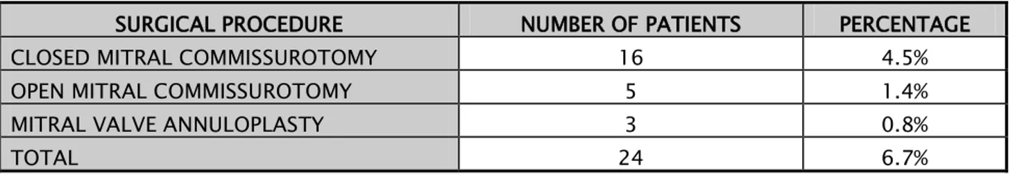
1.4.
A history of mitral valve surgery was present in 24 (6.7%) patients (Table 4). None of our patient had a history of tricuspid valve surgery.
Surgical history:
Table IV: History of cardiac surgery in our case series
SURGICAL PROCEDURE NUMBER OF PATIENTS PERCENTAGE CLOSED MITRAL COMMISSUROTOMY 16 4.5% OPEN MITRAL COMMISSUROTOMY 5 1.4% MITRAL VALVE ANNULOPLASTY 3 0.8%
2.
Clinical data:
2.1.The majority of our patients (82.9%) were in New York Heart Association (NYHA) functional classes III and IV. These results reflect the state of advanced heart failure in which our patients are usually operated on. Only 17.1% of our patients were in NYHA functional class II and none of them was in class II (figure 4).
NYHA status:
Figure 3: Distribution of our patients by their NYHA status
2.2.
Seventy-seven (21.6%) patients presented with congestive heart failure signs, ranging from jugular venous distension (Kussmaul sign) to edema, ascites and orthopnea (Table 5).
Congestive heart failure signs:
Table V: Distribution of heart failure signs in our patients
CONGESTIVE HEART FAILURE SIGNS NUMBER OF PATIENTS PERCENTAGE JUGULAR VEIN DISTENSION 77 21.60% HEPATOJUGULAR REFLUX 62 17.40%
EDEMA 45 12.60%
RIGHT UPPER QUADRANT PAIN 31 8.70%
HEPATOMEGALY 17 4.70%
2.3.
A diastolic murmur of mitral stenosis was the most commonly found heart murmur. It was present in 67.4% of patients.
Heart murmurs:
Cardiac auscultation found a systolic murmur of mitral regurgitation in 45.2%, a diastolic murmur of aortic regurgitation in 22.2% and a systolic murmur of aortic stenosis in 20.2%.
A systolic murmur of tricuspid regurgitation was present in only 29.2% of our patients and only 1.4% had a diastolic murmur of tricuspid stenosis.
Table VI: Distribution of cardiac auscultation findings in our case series
HEART MURMUR NUMBER OF PATIENTS PERCENTAGE MITRAL STENOSIS 240 67.4% MITRAL REGURGITATION 161 45.2% AORTIC REGURGITATION 79 22.2% AORTIC STENOSIS 72 20.2% TRICUSPID REGURGITATION 104 29.2% TRICUSPID STENOSIS 5 1.4% 2.4.
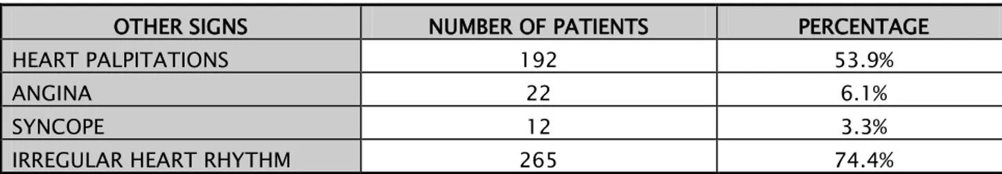
Other signs found in our case series included heart palpitations, angina, syncope and irregular heart rhythm (Table 7).
Other clinical signs:
Table VII: Distribution of other clinical signs in our case series
OTHER SIGNS NUMBER OF PATIENTS PERCENTAGE HEART PALPITATIONS 192 53.9%
ANGINA 22 6.1%
SYNCOPE 12 3.3%
IRREGULAR HEART RHYTHM 265 74.4%
2.5.
The majority of our patients (92.1%) were on a preoperative medical therapy (Table 8).
Table VIII: Distribution of our patients according to preoperative medication
PREOPERATIVE MEDICATION NUMBER OF PATIENTS PERCENTAGE
DIURETICS 295 82.8% ANTICOAGULANT 271 76.1% BETA BLOCKERS 67 18.8% ACE INHIBITOR 56 15.7% AMIODARONE 15 4.2% TOTAL 328 92.1%
Figure 4: Preoperative medication in our case series
3.
Chest X-ray:
Cardiomegaly was present in 312 patients (87.6%). The distribution of our patients according to their preoperative Cardiothoracic Index is presented in table 9.
Table IX: Distribution of patients according to cardiothoracic index
CARDIOTHORACIC INDEX NUMBER OF PATIENTS PERCENTAGE
<0.5 44 12.4%
0.5 - 0.7 250 70.2%
Figure 5: Cardiothoracic index in our case series
4.
Electrocardiogram:
4.1.In our case series, 268 (75.3%) patients had atrial fibrillation. Only 75 (21.1%) of our patients had a sinus rhythm (Table 10).
Rhythm abnormalities:
Table X: Distribution of rhythm abnormalities in our patients
RHYTHM ABNORMALITY NUMBER OF PATIENTS PERCENTAGE ATRIAL FIBRILLATION 268 75.3%
ATRIAL FLUTTER 8 2.2%
SUPRAVENTRICULAR EXTRASYSTOLES 5 1.4%
4.2.
The different conduction abnormalities found in our patients are presented in table 11.
Conduction abnormalities:
Table XI: Distribution of conduction abnormalities in our patients
CONDUCTION ABNORMALITY NUMBER OF PATIENTS PERCENTAGE RIGHT BUNDLE BRANCH BLOCK 25 7%
5.
Echocardiographic data:
5.1. Tricuspid valve:a. Leaflets:
The echocardiographic study of tricuspid valve anatomy showed normal leaflets in 268 (75.3%) patients and altered or thickened leaflets with suggesting organic lesions in 88 (24.7%) patients.
b. Tricuspid annulus:
The tricuspid annular diameter was measured in 316 patients. The mean was 37.7 mm +/- 6.8 mm with a minimal diameter of 23 and a maximal diameter of 58 mm (figure 9).
Figure 6: Distribution of patients by tricuspid annular diameter
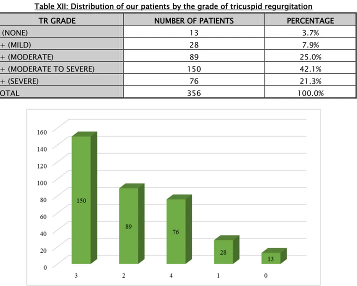
c. Tricuspid regurgitation:
Preoperative tricuspid regurgitation was found in 343 (96.3%) of our patients. The remaining 13 (3.7%) patients had no preoperative regurgitation and the decision of tricuspid repair was taken peroperatively because of the enlarged tricuspid annulus. This situation can be
explained by the long delay between the echocardiographic assessment and surgery, giving time for tricuspid regurgitation to develop.
A grade 3 tricuspid regurgitation or higher was found in 226 (63.4%) patients. Table XII: Distribution of our patients by the grade of tricuspid regurgitation
TR GRADE NUMBER OF PATIENTS PERCENTAGE
0 (NONE) 13 3.7% 1+ (MILD) 28 7.9% 2+ (MODERATE) 89 25.0% 3+ (MODERATE TO SEVERE) 150 42.1% 4+ (SEVERE) 76 21.3% TOTAL 356 100.0%
Figure 7: Distribution of our patients by the grade of tricuspid regurgitation
d. Tricuspid stenosis:
Nineteen patients had associated tricuspid stenosis which was mild in 8 cases, moderate in 6 and severe in 5.
We identified three mechanisms of tricuspid regurgitation in our case series as organic, functional and mixed (dilated tricuspid annulus with altered leaflets). The repartition of our patients according to these mechanisms is detailed in table 13.
Table XIII: Tricuspid regurgitation mechanisms in our case series
TR MECHANISM NUMBER OF PATIENTS PERCENTAGE
FUNCTIONAL 255 74.3%
ORGANIC 17 5.0%
MIXED 71 20.7%
TOTAL 343 100.00%
5.2.
All patients included in our case series had a pathological mitral valve in the echocardiographic study.
Mitral valve:
The majority of patients (53.1%) had mitral valve disease with an association of different degrees of mitral stenosis and regurgitation.
Isolated mitral stenosis interested 100 (28.1%) patients. Pure mitral regurgitation was found in 67 (18.8%) patients.
Table XIV: Mitral valve involvement in our case series
MITRAL ABNORMALITY NUMBER OF PATIENTS PERCENTAGE
NONE 0 0%
MITRAL DISEASE 189 53.1%
PURE MITRAL STENOSIS 100 28.1% PURE MITRAL REGURGITATION 67 18.8%
TOTAL 356 100%
a. Mitral stenosis:
Mitral stenosis was found in 289 (81.2%) of our patients. The degree of stenosis was graded as mild, moderate or severe. The distribution of our patient by the grade of mitral stenosis is detailed in table 14.
Table XV: Mitral stenosis grading in our patients GRADE OF MITRAL
STENOSIS NUMBER OF PATIENTS PERCENTAGE
NONE 67 18.8% MILD 19 5.3% MODERATE 126 35.4% SEVERE 144 40.5% TOTAL 356 100.0% b. Mitral regurgitation:
Among the patients included in our case series, 256 had mitral regurgitation. The preoperative assessment of mitral regurgitation was graded from 1 to 4 (from mild to severe).
Table XVI: Mitral regurgitation grading in our patients GRADE OF MITRAL REGURGITATION NUMBER OF PATIENTS PERCENTAGE NONE 100 28.1% 1+ 46 12.9% 2+ 96 27.0% 3+ 10 2.8% 4+ 104 29.2% TOTAL 356 100.0% 5.3.
An involvement of the aortic valve was identified in 217 (60.9%) of our patients.
Aortic valve:
Pure aortic regurgitation was predominant with 132 (37.1%) patients. Isolated aortic stenosis was found in 7 patients (1.9%) only.
The remaining 78 (21.9%) patients had an aortic valve disease with coexistent aortic regurgitation and stenosis.
Table XVII: Aortic valve involvement in our case series
AORTIC VALVE ABNORMALITY NUMBER OF PATIENTS PERCENTAGE
NONE 139 39.1%
ISOLATED AORTIC REGURGITATION 132 37.1%
AORTIC DISEASE 78 21.9%
ISOLATED AORTIC STENOSIS 7 1.9%
TOTAL 356 100.0%
a. Aortic stenosis:
Aortic stenosis was identified in 85 (23.9%) of patients. The distribution of patients by degree of stenosis is detailed in table 16.
Table XVIII: Aortic stenosis grading in our patients
GRADE OF AORTIC STENOSIS NUMBER OF PATIENTS PERCENTAGE
NONE 271 76.1% MILD 22 6.2% MODERATE 30 8.4% SEVERE 33 9.3% TOTAL 356 100.0% b. Aortic regurgitation:
A total of 210 (59%) patients had aortic regurgitation.
Table XIX: Aortic regurgitation grading in our patients GRADE OF AORTIC
REGURGITATION NUMBER OF PATIENTS PERCENTAGE
0 146 41% 1+ 58 16.3% 2+ 92 25.9% 3+ 21 5.9% 4+ 39 10.9% TOTAL 356 100.0%
5.4. Right ventricle:
a. Right ventricular size:
Right ventricular dilatation was present in 251 (70.5%) of our patients.
Figure 8: Right ventricular size in our case series
b. Right ventricular systolic function:
Right ventricular systolic dysfunction was present in 107 (30%) of our patients.
5.5. Left ventricle:
a. Left ventricular size:
Left ventricular dilatation was present in 58 (16.3%) of our patients.
b. Left ventricular systolic function:
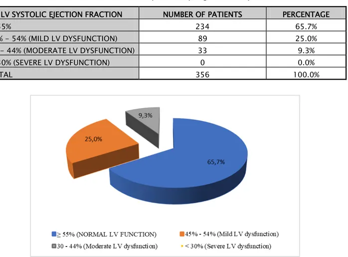
The mean ejection fraction of the right ventricle was 56.8% +/- 9.1% with a maximum of 78% and a minimum of 30%. The majority of our patients (65.7%) had a normal left ventricular systolic function. None of our patients was in severe dysfunction of the left ventricle.
Table XX: Distribution of our patients by degree of LV dysfunction
LV SYSTOLIC EJECTION FRACTION NUMBER OF PATIENTS PERCENTAGE
≥ 55% 234 65.7%
45% - 54% (MILD LV DYSFUNCTION) 89 25.0% 30 - 44% (MODERATE LV DYSFUNCTION) 33 9.3% < 30% (SEVERE LV DYSFUNCTION) 0 0.0%
TOTAL 356 100.0%
Figure 9: Left ventricular systolic function in our case series
5.6.
The mean pulmonary artery systolic pressure was 61.7 +/- 23.9 mmHg with a maximum of 146 mmHg and a minimum of 20 mmHg.
Pulmonary artery systolic pressure:
Pulmonary hypertension was found in 347 (97.5%) patients.
Severe pulmonary hypertension was present in 203 (57%) of our patients.
Figure 10: Pulmonary artery systolic pressure in our case series
5.7.
The left atrium was dilated in 193 (54.2%) of patients. An atrial thrombus was found in 23 (6.4%) patients.
Left atrium:
5.8.
The presumed etiology of valvular lesions was identified to be rheumatic in all our patients on echocardiographic study, confirmed later by surgical findings. Although, additional infectious endocarditis was present in 9 (2.5%) patients.
Etiology of valvular disease:
6.
Perioperative data:
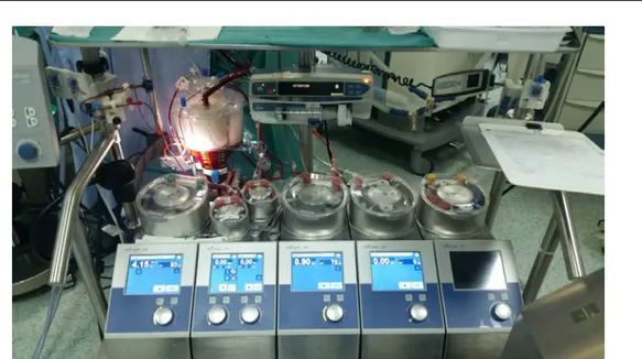
6.1.A median sternotomy was the surgical approach in the majority of our patients (352 which accounts for 98.9% of our patients). Only four surgeries were done mini invasively.
Surgical approach:
Figure 11: Cardiopulmonary bypass machine/ Cardiovascular Surgery Department – University Hospital Mohammed VI, Marrakech
Blood cardioplegia was used in 330 (92.7%) of patients while crystalloid cardioplegia was used in only 26 (7.3%) of patients. Crystalloid cardioplegia was used only in the beginning of our experience (before 2010). The surgeries were carried out in slight hypothermia in the majority of cases (88.2%).
The mean cardiopulmonary bypass time was 130 min ± 50.4 min with extremes ranging from a minimum of 42 min to a maximum of 412 min.
The mean aortic cross-clamp time was 92.7 min ± 38.4 min with a maximum of 28 min and a maximum of 300 min.
Weaning from cardiopulmonary bypass was possible without inotropic support in 58 (16.3%) patients. In the rest of cases, an inotropic (or more) drug was introduced to help coming off the bypass as detailed in table 21.
Table XXI: Use of inotropic agents when coming off cardiopulmonary bypass in our patients INOTROPIC DRUGS NUMBER OF PATIENTS PERCENTAGE
DOBUTAMINE 298 83.7%
6.3. Surgical exploration:
a. Tricuspid valve:
The tricuspid annulus was dilated in all patients.
An associated organic lesion of the tricuspid valve was found in 102 (28.6%) patients. Leaflets thickening was found in all these patients. The other associated lesions were commissural fusion or abnormal chordae tendinea.
Table XXII: Profile of peroperative tricuspid valve organic lesions found in our patients ORGANIC LESION NUMBER OF PATIENTS PERCENTAGE LEAFLETS THICKENING 102 28.6% COMMISSURAL FUSION 26 7.3% ABNORMAL CHORDAE 13 3.6%
b. Right heart geometry:
Right ventricular dilatation was reported in 161 (45.2%) patients. Right atrial dilatation was reported in 155 (43.5%) patients.
c. Pericardial adhesions:
Pericardial adhesions were found in only 17 (4.7%) cases.
6.4. Surgical procedures:
a. Tricuspid valve:
The De Vega annuloplasty:
De Vega annuloplasty was performed in all patients. Technique:
In our department, we used the classic technique of De Vega annuloplasty in the beginning of our experience, and then a modified technique was introduced.
In the modified technique, the suture is placed over a pledget from the anterior commissure to the posterior commissure using a 2.0 Ethibond (Contrary to the segmental technique, a single suture thread is used). The suture is doubled and the treads tips are met in the center of the annulus and a pledget is placed. This technique permits a more symmetrical reduction of the tricuspid annulus.
The tightening is controlled over two fingers of the surgeon. The result is controlled with a saline test.
Figure 12: Schematic representation of our modified De Vega annuloplasty technique used in our department
Figure 13: Placement of the first pledget
Figure 15: Placement of the second pledget
Figure 16: Placement of the third pledget where the sutures are met and tightened over two fingers
Figure 17: saline test
Concomitant procedures:
When the saline test is not satisfactory, an additional tricuspid repair is performed. This was necessary in 61 (17.1%) patients.
Eliminating leaflet tissue excess (secondary to leaflet tethering) is one of the most frequently performed procedures in our institution. Unfortunately, we don’t have the exact number of patients that needed this procedure in our case series as it was rarely mentioned on surgical reports.
Table XXIII: Distribution of concomitant tricuspid valve procedures
TRICUSPID PROCEDURE NUMBER OF PATIENTS PERCENTAGE
COMMISSUROTOMY 47 13.2%
KAY ANNULOPLASTY 14 3.9%
CHORDAE SHORTENING BY PLICATION 4 1.1%
Figure 19: Schematic representation of leaflet tissue excess (flail leaflet) Intraoperative assessment of tricuspid repair result:
A saline injection leak test was used to assess leaflet coaptation after tricuspid valve repair. The test was satisfactory in all patients.
The saline test is considered satisfactory when the right ventricle remains distended for a few seconds after filling it with only a small central leak of the tricuspid valve and with a clear
A residual central leak after tricuspid valve repair was noted in 31 (8.7%) patients.
We usually respect some degree of tricuspid regurgitation, especially in patient with severe pulmonary hypertension with right ventricular dysfunction, to relieve the dysfunctional ventricle.
b. Other valve procedures:
Major valve procedures in our case series interested left sided valves. An isolated mitral valve replacement was the most common procedure interesting 246 (69.1%) patients. There was 94 (26.4%) double valve replacements (mitral and aortic valves), 4 aortic valve replacements associated with a mitral valve repair, 6 mitral valve replacements with aortic valve repair and 6 isolated mitral repairs.
Table XXIV: Distribution of concomitant left-heart procedures
LEFT-HEART VALVE PROCEDURE NUMBER OF PATIENTS PERCENTAGE MITRAL VALVE SURGERY 252 70.8% AORTIC VALVE SURGERY 0 0% COMBINED MITRAL AND AORTIC SURGERIES 104 29.2%
There was a total of 444 valve replacements in our case series. A mechanical prosthesis was used in 440 patients. A bioprosthesis was used in only 4 patients.
Table XXV: Types of mechanical valve used in our case series
TYPE OF MECHANICAL PROSTHESIS NUMBER PERCENTAGE
ONYX 211 47.90%
SORIN 138 31.40%
SJM 71 16.10%
ATS 20 4.60%
TOTAL 440 100.00%
Mitral valve repair procedures included: - Ring annuloplasty: 6 patients - Kay annuloplasty: 4 patients
- Sub commissural annuloplasty: 1 patient - Chordae shortening: 1 patient
- Surgical excision of a vegetation: 1 patient Aortic valve repair procedures included:
- Sub commissural annuloplasty: 6 - Surgical excision of a vegetation: 2 - Leaflet plication: 1
- Chordae Shortening: 1
7.
Critical care data:
7.1.The mean ICU stay duration in our case series was 4.2 ± 2.4 days with a maximum of 28
7.2.
The average duration of respiratory assistance in our case series was 7.6 ± 15 hours with a maximum of 288 (accounting for 12 days). Only 13 patients (3.6%) were extubated after 24 hours.
Duration of intubation:
Reintubation was necessary in 14 (3.9%) patients.
Table XXVI: Estimated causes for reintubation in our case series
CAUSE OF REINTUBATION NUMBER OF PATIENTS PERCENTAGE ACUTE RESPIRATORY FAILURE 7 2% NEUROLOGICAL IMPAIRMENT 4 1.1% MULTIORGAN FAILURE 3 0.8%
7.3. Postoperative medication:
a. Antibiotic prophylaxis:
All patients were put postoperatively on an antibiotic prophylaxis using a first-generation cephalosporin (Cefalotin). The antibiotic is stopped after removing the chest drainage.
b. Anticoagulation:
Postoperative anticoagulation was prescribed in all patients using Heparin Sodium and Warfarin. The later was starter in the second postoperative day.
c. Inotropic agents:
Inotropic support was necessary in 304 (85.4%) patients.
Dobutamine was used in all these patients with a minimum duration of 3 hours and a maximum duration of 288 hours (12 days).
d. Antiarrhythmic agents:
The use of an antiarrhythmic drug during the ICU course was necessary in 170 (47.7%) of patients.
Table XXVII: Types of antiarrhythmic drugs used in our case series
ANTIARRHYTHMIC AGENT NUMBER OF PATIENTS PERCENTAGE
AMIODARONE 87 24.4% DIGOXIN 40 11.2% AMIODARONE + DIGOXIN 19 5.3% LIDOCAINE 16 4.5% BISOPROLOL 8 2.3% TOTAL 170 47.7% e. Other medications:
Diuretics (Furosemide) were used in all patients in immediate postoperative course. Sildenafil was used in 61 (17.1%) of our patients.
8.
Immediate and short-term surgical results: (<3 months)
8.1.Early postoperative death occurred in 4.8% (n=17) of patients in our case series.
Mortality:
Table XXVIII: Causes of early postoperative deaths in our case series CAUSE OF DEATH NUMBER OF PATIENTS LOW CARDIAC OUTPUT SYNDROME 6
CARDIAC TAMPONADE 3
MULTIPLE ORGAN FAILURE 3
SEPSIS 3
REFRACTORY HYPOVOLEMIC SHOCK 1 UNEXPLAINED CARDIAC ARREST 1
8.2.
During the ICU stay, 23.3% (n=79) of surviving patients presented various complications.
a. Cardiac complications:
a.1. Arrhythmias:
Table XXIX: Postoperative arrythmias in our case series
ARRHYTHMIA NUMBER OF PATIENTS PERCENTAGE ATRIAL FIBRILLATION 198 58.4% ATRIAL TACHYCARDIA 62 17.4% SINUS TACHYCARDIA 47 13.8% SINUS BRADYCARDIA 8 2.3% VENTRICULAR FIBRILLATION 12 3.5% EXTRASYSTOLES 9 2.7%
a.2. Conduction disturbances:
- Postoperative atrioventricular block occurred in 8 (2.3%) of our patients. It was transient in 5 patients and permanent in 3 patients who consequently underwent pacemaker implantation.
- A bundle branch block was observed in 3 (0.9%) patients.
a.3. Other cardiac complications:
- Hemopericardium: 18 (5.3%) patients. - Right sided heart failure: 24 (7.1%) patients. - Post pericardiotomy syndrome: 3 (0.9%) patients.
b. Hemorrhagic complications:
- Postoperative hemorrhage occurred in 58 (17.1%) patients. - The management of the hemorrhage was:
Medical in 49 (84.5%) patients using hemostatic agents and transfusion.
Surgical in 9 (15.5%) patients in which medical measures were not sufficient and reoperation was necessary.
c. Pulmonary complications:
Pulmonary complications occurred in 48 (14.1%) patients.
U
Table XXX: Postoperative pulmonary complications
PULMONARY COMPLICATION NUMBER OF PATIENTS PERCENTAGE
PNEUMONIA 24 7.1%
PLEURAL EFFUSION 13 3.8%
ACUTE PULMONARY EDEMA 16 4.7%
d. Neurological complications:
- Postoperative ischemic stroke occurred in 2 patients. The stroke was transient in both patient and they both had a full recovery.
- A transient delirium during the ICU stay was observed in 5 patients.
e. Infectious complications:
A total of 53 (15.3%) patients had infectious complications. Other than pneumonia, the other sites of infection were the surgical site and in a sepsis in 1 patient. We recorded no case of infectious endocarditis in our study.
U
Table XXXI: Postoperative infectious complications
INFECTION SITE NUMBER OF PATIENTS PERCENTAGE
PNEUMONIA 24 7.1% SURGICAL-SITE INFECTION - SUPERFICIAL - DEEP 28 23 5 8.3% 6.8% 1.5% SEPSIS 1 0.3% INFECTIOUS ENDOCARDITIS 0 0.0% f. Infectious complications:
- Postoperative acute renal failure occurred in 4 patients. Dialysis was necessary in 1 patient. Renal failure was reversible in all cases.
8.3.
We studied clinical evolution of our surviving patients (n=339) after surgery by evaluating their NYHA status and heart failure signs.
Clinical results:
a. NYHA status:
In the first month after surgery, 77.6% of patients were class I or II of the NYHA functional classification.
U
Table XXXII: Immediate postoperative NYHA status
NYHA STATUS NUMBER OF PATIENTS PERCENTAGE
I 39 11.5% II 224 66.1% III 65 19.2% IV 11 3.2% TOTAL 339 100.0% U
b. Heart failure signs:
Out of the 339 surviving patients, 30 (8.8%) had postoperative congestive heart failure. Thirteen patients did not have heart failure signs before surgery and only developed them in the first postoperative month.
8.4.
All surviving patients had an echocardiographic assessment during the first postoperative month. The average time delay after surgery was 7.2 ± 3.4 days with a minimum of 3 days and a maximum of 20 days.
Echocardiographic results:
a. De Vega annuloplasty results:
Postoperative tricuspid regurgitation was assessed in only 242.
The majority of patients (92.1%) had a class 2 or lower degree of tricuspid regurgitation (Moderate, mild or none). The remaining 7.9% of patients had a class 3 or 4 tricuspid regurgitation (moderate to severe or severe).
Tricuspid stenosis was present in 8 (2.3%) patients. It was mild in 5 and moderate in 3 patients.
b. Other echocardiographic data:
U
Table XXXIII: Comparison between preoperative and postoperative results
PREOPERATIVE DATA POSTOPERATIVE DATA
MEAN LVEF 56.8% 55.3%
MEAN PASP 61.7 mmHg 44.9 mmHg RV DILATATION 70.5% 32.7% RV DYSFUNCTION 30.0% 35.0%
9.
UMid-term surgical results: (6 months to 2 years)
9.1. UFallow-up:
The follow-up was insured with phone calls and outpatient consultations in a total of 273 patients while we lost track of 43 patients and late mortality occurred in 23 patients.
In the following segments, we only expose the results of the first 2 years following tricuspid repair.
9.2.
During the follow-up, we noted 23 deaths, accounting for 6.7% of our cohort patients.
Mortality:
The causes of death are exposed in table 30. The cause was undetermined in 18 patients, but we know that it is most likely to be Wafarin related accidents.
U
Table XXXIV: Causes of death in mid-term follow-up
CAUSE OF DEATH NUMBER OF PATIENTS VITAMIN K ANTAGONISTS-RELATED HEMORRHAGE 3
INFECTIOUS ENDOCARDITIS 2 UNDETERMINED CAUSE OF DEATH 18
9.3. UDevega annuloplasty mid-term results:
a. Clinical outcomes
The majority of our patients were asymptomatic in mid-term follow up with a total of 228 (83.5%) of patients having normal effort tolerance.
The remaining 58 patients had clinical signs as follows: - Dyspnea:
o NYHA II: 12 o NYHA III: 18 o NYHA IV: 15
- Right-sided heart failure signs were found in 34 patients ranging from jugular vein distension to recurrent ascites.
b. Echocardiographic data:
b.1. Tricuspid regurgitation
Table XXXV: Postoperative evolution of each grade of tricuspid regurgitation Preoperative tricuspid regurgitation Postoperative (Mid-term results)
None or mild (0, 1+): 41 patients 0, 1+ 25 61% 2+ 1 2.4% 3+ 1 2.4% 4+ 0 0% Unknown 14 34.2% Moderate (2+): 89 patients 0, 1+ 73 82% 2+ 4 4.5% 3+ 1 1.1% 4+ 0 0% Unknown 11 12.4% Moderate to severe (3+): 150 patients 0, 1+ 103 68.7% 2+ 11 7.3% 3+ 1 0.7% 4+ 3 2% Unknown 32 21.3% Severe (4+): 76 patients 0, 1+ 26 34.2% 2+ 12 15.7% 3+ 6 7.9% 4+ 8 10.6% Unknown 24 31.6%
b.2. Tricuspid valve stenosis:
In mid-term follow-up, tricuspid valve stenosis was found in 5 patients (1.4%). It was mild in 4 patients and moderate in 1 patient. All 5 patients had tricuspid stenosis in short-term follow up. The 3 remaining patients were lost in mid-term follow-ip.
b.3. Pulmonary arterial hypertension:
The mean postoperative pulmonary artery systolic pressure in mid-term follow-up is 38.5 mmHg.
Figure 23: Evolution of mean PASP in our case series
Impact of Sildenafil use on PASP evolution in our case series:
In our study, 61 patients were put on sildenafil in the post-operative course. We have no data on the treatment duration.
The indications of Sildenafil use in our study were: - Severe preoperative pulmonary hypertension
- Postoperative persistence of right-sided heart failure - Severe postoperative pulmonary hypertension
The mid-term follow-up data are available for 53 patients. We found no statistically significant difference in the evolution of PASP with or without Sildenafil use (p=0.7).
U
Table XXXVI: Evolution of the mean PASP in the Sildenafil group versus the rest of the cohort MEAN PASP SILDENAFIL NO SILDENAFIL
PREOPERATIVE 72.6 mmHg 61.7 mmHg SHORT-TERM FOLLOW-UP 49.3 mmHg 44.9 mmHg MID-TERM FOLLOW-UP 42.3 mmHg 38.5mmHg
b.4. Left ventricular systolic function:
II.
Analytic study:
1.
Mortality:
We had a total of 40 (11.2%) deceased patients during the 2 years period of fallow up: - 17 (4.8%) occurred in the first month.
- 23 (6.4%) occurred later on.
The clinical characteristics of this group are displayed bellow.
1.1. Preoperative data:
Table XXXVII: Preoperative data in deceased patients versus the total of the cohort Deceased group Cohort data Mean age 50.4 years 40.6 years Female predominance 72.5% 66.3% Heart failure signs 47.5% 21.6% NYHA status: - I - II - III - IV 0% 5% 50% 45% 0% 17.1% 24.2% 58.7% Atrial fibrillation 75% 75.3% Dilated right ventricle 70% 70.5% Right ventricle dysfunction 55% 30% Tricuspid regurgitation grade:
- 0, 1+ - 2+ - 3+ - 4+ 10% 12.5% 60% 17.5% 11.6% 25% 42.1% 21.3% Mean PASP 65.5 mmHg 61.7 mmHg Mean LVEF 56.6% 56.8%
1.2. Surgical data:
Table XXXVIII: Surgical data in deceased patients versus the total of the cohort Deceased group Cohort data Aortic cross clamp time 98.7 min 92.7 min CPB time 149.6 min 130 min Left-sided valvular procedures:
- MV and AV replacement - MV replacement - MV replacement + AV repair 72.5% 22.5% 5% 26.4% 39.1% 1.9% 1.3.
Only 27 out of the 40 deceased patients had a postoperative echocardiography done during the ICU stay.
Postoperative data:
The mean LVEF was 43.6% with 4 patients having a severe left ventricle dysfunction. The mean immediate postoperative PASP in these patients was 61 mmHg.
Only 10 patients had postoperative tricuspid valve regurgitation quantification: - Severe: 2 patients
- Moderate to severe: 1 patient - Moderate: 4 patients
- Mild: 3 patients
2.
Factors of failure of De Vega tricuspid repair:
With the use of the IBM SPSS Statistics 21 program, we analyzed risk factors for the De Vega annuloplasty failure. A p value <0.05 was considered as statistically significant.
We defined as failure a postoperative tricuspid regurgitation grade 3+ or 4+. In total, we had 23 cases (7.3%) of De Vega annuloplasty failure.
Table XXXIX: Different potential De Vega annuloplasty failure risk factors tested in our study
3+ and 4+ postop TR group data Cohort
data P value
Mean age 47.9 years 40.6 years 0.7
Female predominance 67.5% 66.3% 0.1
Preoperative heart failure signs 65.2% 21.6% 0.04
Preoperative NYHA status III or IV 86.9% 82.9% 0.07
Preoperative atrial fibrillation 91.3% 75.3% 0.3
Preoperative dilated right ventricle 78.3% 70.5% 0.03
Preoperative right ventricle dysfunction 82.6% 30% 0.01
Preoperative TR grade 4+ 69.6 % 21.3% 0.03
Preoperative TR grade 3+ 26 % 42.1 % 0.7
TV annulus diameter > 40 mm 91.3% 43.4% 0.01
Organic lesions of the TV 65.2% 25.7% 0.1
Severe preoperative PAH 91.3% 57% 0.002
Mean preoperative LVEF 54.3% 56.8% 0.07
Isolated mitral valve surgery 56.5% 70.8%
0.7
Combined MV and AV surgery 43.5% 29.2%
Severe postoperative PASP 69.6% 12.5% 0.004
Postoperative mean LVEF 55.6% 58.4% 0.3
The predictive factors of failure in our study were: - Preoperative heart failure signs (p=0.04) - Preoperative dilated right ventricle (p=0.03) - Preoperative right ventricular dysfunction (p=0.01) - Preoperative TR grade 4+ (p=0.03)
- Preoperative TV annulus diameter > 40 mm (p=0.01) - Severe preoperative pulmonary artery hypertension (0.002) - Severe postoperative pulmonary hypertension (0.004)
3.
We identified two sub-groups in our study:
- Group 2: Patients who underwent tricuspid repair + combined mitral valve and aortic valve surgery
Table XL: Data comparison between group 1 and 2
Group 1 (70.8%) Group 2 (29.2%) Mean age 39.6 years 43.7 years Female predominance 65.8% 68.1% Preoperative heart failure signs 26.2% 18.5% Preoperative NYHA - I - II - III - IV 0% 13.6% 66.7% 19.6% 0% 19.2% 49.3% 31.5% Preoperative atrial fibrillation 75.9% 74.6% Preoperative dilated right ventricle 72.2% 69.8% Preoperative right ventricle dysfunction 28.2% 34.4% Preoperative TR grading: - 0, 1+ - 2+ - 3+ - 4+ 6.6% 29.8% 41.9% 21.7% 15.2% 24.6% 43.8% 16.4% Mean TV annulus diameter 37.6 mm 39.9 mm Organic lesions of the TV 24.2% 27.4% Mean preoperative PASP 60.7 mmHg 61.9 mmHg Mean preoperative LVEF 57.3% 55.9% Postoperative NYHA - I - II - III - IV 83.8% 6.6% 4.6% 5% 80.8% 4.1% 8.2% 6.8% Postoperative TR grading: - 0, 1+ - 2+ - 3+ - 4+ 82.8% 9.6% 5.1% 2.5% 79.6% 10.9% 2.7% 6.8% Postoperative dilated right ventricle 21.2% 26% Postoperative right ventricle dysfunction 19.2% 23.2% Mean postoperative PAH 37.5 mmHg 41.5 mmHg
Functional TR was a forgotten entity for so long. When existing with concomitant left sided lesions such mitral or aortic valve disease, it is associated with high mortality and increased risk of adverse events (15).
Figure 26: Published articles on "tricuspid valve regurgitation" throughout the years – PubMed In patient with secondary TR concomitant with mitral or aortic valve(s) lesion(s), surgical treatment of left-sided valvular disease without addressing the tricuspid valve may improve mild TR (16,17). However, uncorrected moderate and severe TR may persist or even worsen after mitral valve surgery leading to progressive heart failure and death. In addition, reoperation for residual TR carries significant risks and may is associated with poor outcomes and (18,19).
Our study reports the short- and midterm results of 356 patients who underwent a De Vega tricuspid annuloplasty during the surgical treatment of left heart valvular disease. It was conducted in order to evaluate the effectiveness of this technique and to analyze the effects of preoperative, operative and postoperative parameters on outcomes. The limits of our study are:
- Our study is a retrospective study with all of the inherent limitations; some data were not mentioned on medical records or were lost in handwritten archives rendering many files unusable in our study.