i Université de Sherbrooke
DNA-PK sustains autophagy and pancreatic cancer cell growth
By
Rachita Chatterjee Cell Biology program
Thesis submitted at the Faculty of Medicine and Health Sciences for obtaining Master of Science degree (M.Sc) in cell biology
Sherbrooke, Québec, Canada January, 2021
Members of the jury of evaluation
Dr. Marie-Josée Boucher, Department of Medicine Dr. Steve Jean, Department of Immunology and Cell Biology
Dr. Brendan Bell, Department of Microbiology
ii
SOMMAIRE
DNA-PK supporte l’autophagie et la croissance des cellules pancréatiques tumorales Par
Rachita Chatterjee cell biology program
Mémoire présenté à la Faculté de médecine et des sciences de la santé en vue de l’obtention du diplôme de maitre ès sciences (M.Sc.) en biologie cellulaire, Faculté de médecine et des sciences de la santé, Université de Sherbrooke, Sherbrooke, Québec,
Canada, J1H 5N4
Selon les récentes statistiques, le cancer pancréatique est la quatrième cause de décès par cancer au Canada avec une survie à 5 ans de 8%. Le cancer du pancréas réfère à l'adénocarcinome canalaire pancréatique (PDAC) qui représente près de 85% des cas diagnostiqués. Actuellement, la gemcitabine demeure la première ligne de traitement du PDAC malgré une efficacité limitée. Il est connu que la gemcitabine provoque des dommages à l'ADN en provoquant des cassures double-brin. Ces cassures peuvent être réparées par la voie de réparation de jonction d'extrémités non-homologues, la DNA-PK y jouant un rôle clé. Il a été observé que les tissus PDAC présentent des niveaux d’expression élevés de la DNA-PKcs. De plus, il a été suggéré que la DNA-PK soutient la croissance des cellules PDAC. Les mécanismes impliqués demeurent toutefois mal étudiés. Récemment, il a été démontré que l'autophagie joue un rôle important dans la croissance des cellules PDAC. Dans certains types de cancer, la DNA-PK a été proposée comme une régulatrice de l’autophagie. Ainsi, nous avons entrepris cette étude en émettant l'hypothèse que la DNA-PK soutient l'autophagie et la croissance des cellules PDAC. Les lignées cellulaires PDAC MIA PaCa-2 et PANC1 ont été utilisées comme modèles d'étude. L'inhibiteur spécifique de la DNA-PK, le NU7441, a été exploitée pour explorer le rôle de la DNA-PK dans la croissance et l’autophagie des cellules PDAC. Une réduction, dépendante de la dose et du temps, de la croissance des cellules PDAC a été observée lors du traitement au NU7441. L'inhibition de la DNA-PK a également restreint la capacité des cellules PDAC à former des colonies lors d’essais de clonogénicité. Le traitement au NU7441 a provoqué un clivage de PARP et de la caspase-7 suggérant que l’inhibition de la DNA-PK induit l'apoptose. Le traitement au NU7441 a mené à une accumulation des niveaux de marqueurs d'autophagie soient LC3B II et p62. La mesure du flux autophagique a confirmé que cette accumulation était due au blocage de l'autophagie. Dans l'ensemble, l'étude révèle que la DNA-PK abroge la croissance des cellules PDAC corrélant avec un blocage de l'autophagie. Les mécanismes empruntés par la DNA-PK pour réguler l’autophagie demeurent à être identifiés, ce qui pourrait permettre de caractériser de nouvelles cibles visant à optimiser la réponse des cellules PDAC à la gemcitabine. Mots-clés: Cancer pancréatique, DNA-PK, Autophagie, NU7441, croissance cellulaire, traitement
iii
SUMMARY
DNA-PK sustains autophagy and pancreatic cancer cell growth By
Rachita Chatterjee CellBiology program
Thesis presented at the Faculty of medicine and health sciences in view of obtaining a Maitre ès Sciences (M.Sc.) diploma in cell biology, Faculty of medicine and health
sciences, Université de Sherbrooke, Sherbrooke, Québec, Canada, J1H 5N4 As per the latest Canadian Cancer Statistics, pancreatic cancer is the 4th leading cause of cancer-related deaths in Canada, with a net 5 year survival of 8%. Pancreatic cancer mostly refers to pancreatic ductal adenocarcinoma (PDAC) that accounts for almost 85% of the cases diagnosed. Currently, gemcitabine is the first line of treatment for PDAC. Gemcitabine is known to cause lethal DNA damage by inducing DNA double strand breaks (DSBs). DSB can be repaired by the non-homologous end joining repair (NHEJ) pathway, of which DNA-PK plays a key role. It was shown that PDAC tissues display elevated levels of DNA-PKcs. Besides supporting growth, DNA-PK was proposed to offer chemoprotection to the PDAC cells. Still, the mechanisms involved behind DNA-PK mediated PDAC growth sustenance remains poorly investigated. It is now accepted that a vital cellular process, autophagy, plays critical role in PDAC growth. A number of reports have suggested that autophagy could be regulated by DNA-PK in certain forms of cancer. As nothing is known about the regulation of autophagy by DNA-PK in the context of PDAC, we hypothesized that DNA-PK sustains autophagy and pancreatic cancer cell growth. The PDAC cell lines MIA PaCa-2 and PANC1 were used as study models. DNA-PK activity was targeted through a pharmacological strategy using the specific DNA-PK inhibitor NU7441 to analyse the impact of DNA-PK inhibition on PDAC cell growth and autophagy. We observed drastic reduction in PDAC cell growth upon NU7441 treatment, in both dose- and time dependent manner. DNA-PK inhibition also abrogated colony forming ability of PDAC cells. NU7441 treatment caused cleavage of PARP and caspase-7, suggesting an apoptotic response upon DNA-PK inhibition. Furthermore, DNA-PK inhibition via NU7441 led to an accumulation of autophagy markers LC3B II and p62. Measurement of autophagic flux confirmed that this accumulation in the levels of the autophagy markers is due to blockade of autophagy. Overall, the study reveals that DNA-PK abrogates PDAC cell growth that correlates with blockade in autophagy. The mechanisms exploited by DNA-PK to regulate autophagy remain to be identified. Their identification could lead to the characterization of novel targets that could optimize gemcitabine treatment in the context of pancreatic cancer. Keywords: Pancreatic cancer, DNA-PK, Autophagy, NU7441, cell growth, treatment
iv
TABLE OF CONTENTS
SOMMAIRE ... iiSUMMARY ... iii
TABLE OF CONTENTS ... iv
LIST OF FIGURES ... vii
LIST OF ABBREVIATIONS ... viii
ACKNOWLEDGEMENTS ... xi
1. INTRODUCTION ... 1
1.1. The Pancreas... 1
1.1.1 Pancreatic cancer ... 2
1.1.1.1 Pancreatic ductal adenocarcinoma (PDAC) ... 3
1.1.2 Pancreatic cancer: risk factors ... 4
1.1.3 Pancreatic cancer treatment... 5
1.1.3.1 Surgery: ... 5
1.1.3.2 Chemotherapy: ... 5
1.1.3.3 Radiotherapy: ... 6
1.1.4 Cell signalling pathways in pancreatic cancer ... 7
1.2. DNA-PK discovery ... 8
1.2.1 Structure of DNA-PKcs ... 8
1.2.2 Structure and function of KU heterodimer ... 10
1.3. Functions of DNA-PK ... 11
1.3.1 Role in DNA repair ... 11
1.3.2 Role in V(D)J recombination ... 12
1.4. Important regulators and substrates of DNA-PKcs ... 13
1.4.1 Autoregulation ... 13
1.4.2 Key substrates of DNA-PKcs ... 13
1.5. Role of DNA-PK in pancreatic cancer ... 14
1.6. Role of DNA-PK in other forms of cancer ... 14
1.7. Autophagy ... 15
v
1.7.2 Transcriptional regulation of p62/SQSTM1... 20
1.7.3 Signalling pathways regulating autophagy ... 22
1.7.4 Regulation of autophagy by DNA-PK ... 23
1.7.5 Role of autophagy in PDAC ... 24
1.8. Hypothesis and objectives ... 25
2. MATERIALS AND METHODS ... 26
2.1 Cell culture and passaging ... 26
2.2 Inhibitors and chemotherapeutic drugs ... 26
2.3. Measurement of cell growth ... 26
2.3.1 Growth curve assay ... 26
2.3.2 Clonogenic assay ... 27
2.4. Biochemical analysis ... 27
2.4.1 Preparation of cell lysates ... 27
2.4.2 Subcellular fractionation ... 28
2.4.3 Bicinchoninic acid (BCA) protein assay ... 29
2.4.4 Immunoblotting ... 29 2.4.5 Immunofluorescence ... 31 2.4.6 Immunoprecipitation ... 32 2.4.7 siRNA transfection ... 33 2.5 Autophagy monitoring ... 34 3. RESULTS ... 35
3.1 Subcellular localization of DNA-PKcs protein in pancreatic cancer cells ... 35
3.2. Pharmacological inhibition of DNA-PK modulates the phosphorylation of DNA-PKcs in PDAC cells ... 37
3.3. Interference with DNA-PK activity impairs the growth of pancreatic cancer cells ... 38
3.4. Inhibition of DNA-PK leads to dose- and time-dependent increase of apoptotic markers ... 39
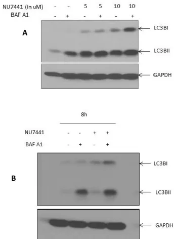
3.5. Inhibition of DNA-PK promotes the expression of autophagy markers ... 41
3.6. DNA-PK inhibition blocks autophagic flux in pancreatic cancer cells... 42
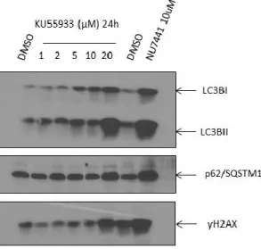
3.7. Comparison of the effect of DNA-PK inhibitor and ATM inhibitor on autophagy markers in PDAC cells ... 44
vi
3.9. Effect of DNA-PK inhibition on transcription factor EB (TFEB) ... 46
3.10. Impact of DNA-PK inhibition on signalling pathways known to regulate autophagy ... 47
3.11. siRNA-mediated knockdown of DNA-PK in PDAC cells ... 49
3.12. Impact of Gemcitabine on autophagy markers in pancreatic cancer cells ... 51
4. DISCUSSION ... 53
5. CONCLUSION ... 63
vii
LIST OF FIGURES
Figure 1. Structure, location and cell types of the pancreas ... 2 Figure 2. Proposed model of PanIN progression towards pancreatic ductal adenocarcinoma ... 4 Figure 3. Three-dimensional structure of DNA-PKcs depicting the open channels and openings of an enclosed cavity ... 9 Figure 4. Functional domains of DNA-PKcs ... 10 Figure 5. Schematic representation of the non-homologous end joining (NHEJ) repair pathway 12 Figure 6. The different domains of p62/SQSTM1 and its interacting partners during cargo
sequestration in selective autophagy ... 18 Figure 7. Schematic representation of the autophagy process in general ... 20 Figure 8. Mechanism of TFEB mediated transcriptional regulation of p62/SQSTM1 ... 21 Figure 9. Schematic representation of the three main pathways involved in autophagy
regulation and their crosstalk ... 23 Figure 10 Subcellualar localization of DNA-PKcs in MIA PaCa-2 pancreatic cancer
cells………....36 Figure 11 Modulation of DNA-PKcs phosphorylation upon pharmacological inhibition ... 37 Figure 12 Inhibition of DNA-PK activity impairs pancreatic cancer cell
growth……….38, 39 Figure 13 Impact of DNA-PK inhibition on apoptotic markers in PDAC
cells……….40 Figure 14 Impact of DNA-PK inhibition on autophagy markers in PDAC cells……….42 Figure 15 Autophagic flux blockade in DNA-PK inhibited PDAC
cells……….43 Figure 16 Comparison of the impact of DNA-PK inhibitor and ATM inhibitor on autophagy
markers in PDAC cells……….44 Figure 17 Immunoprecipitation of p62/SQSTM1………46 Figure 18 Impact of DNA-PK inhibition on TFEB in PDAC cells………..47 Figure 19 Impact of DNA-PK inhibition on signalling pathways modulating autophagy
in PDAC cells……….49 Figure 20 siRNA mediated knockdown of DNA-PK in PDAC cells………50, 51 Figure 21 Impact of Gemcitabine on autophagy markers in PDAC cells………..52
viii
LIST OF ABBREVIATIONS
ATM- Ataxia Telengiectasia Mutated protein ATP- Adenosine triphosphate
Atg-Autophagy-related gene BAF A1- Bafilomycin A1
BCA- Bicinchoninic acid protein assay B-CLL- B-cell chronic lymphocytic leukaemia bp- base pair
BRCA1/2-Breast cancer susceptibility genes 1 and 2 BSA- Bovine serum albumin
Cas9- CRISPR associated protein 9
CDKN2A- Cyclin-dependent kinase inhibitor 2A CIB- Cytoplasmic Isolation Buffer
CRISPR- Clustered regularly interspaced short palindromic repeats CTR- C-terminal region
CyNIB- Cytoskeletal/Nuclear Isolation Buffer DAB2IP- DOC-2/DAB2 interacting protein DAPI- 4′,6-diamidino-2-phenylindole
DMEM- Dulbecco’s Modification of Eagle’s Medium DMSO- Dimethyl sulfoxide DNA-deoxyribonucleic acid DNA-PK- DNA-dependent protein kinase
DNA-PKcs- DNA-dependent protein kinase catalytic subunitDNA-deoxyribonucleic acid DSB- Double strand break
EDTA- Ethylenediaminetetraacetic acid ERK2- Extracellular-signal-regulated kinase FAT- Focal adhesion kinase, targeting domain FATC- FAT domain at C-terminal
FBS- Fetal bovine serum
g- Relative Centrifugal Force (RCF) or G-Force
GAPDH- Glyceraldehyde 3-phosphate dehydrogenase GFP- Green fluorescent protein
GSK-3- Glycogen Synthase Kinase 3 H460- Lung cancer cell line
HEAT- Huntingtin Elongation Factor 3, PP2A and TOR1 HeLa- Henrietta Lacks (Cervical cancer cell line) HOPS- Homotypic fusion and protein sorting
HNPCC-Heriditary Non-Polyposis Colorectal Cancer gene 4 HPDE- Human pancreatic ductal epithelial cell line
ix IgG- Immunoglobulin
IORT-Intraoperative Radiotherapy
IPMN-Intraductal papillary-mucinous neoplasm JNK- c-jun N-terminal kinase
KCl- Potassium chloride
KEAP1- Kelch-like ECH-associated protein 1
KRAS-Kirtsen Rat Sarcoma Virus
KU80CTR- KU80 C-terminal region MAPK-Mitogen activated protein kinase
LC3- Microtubule associated proteins 1 light chain 3 LIR- LC3 interacting region
Lys- Lysine mA- Milliampere
MCF7- Breast cancer cell line mg- Milligram
MIB- Membrane Isolation Buffer ml-Millilitre
mm- Millimetre
mTORC1- Mammalian target of Rapamycin Complex 1 MUC1-Mucin 1
NaCl- Sodium chloride NaF- Sodium fluoride
NHEJ- Non-homologous end joining repair nm- Nanometre
nM- Nanomolar
NRF2- Nuclear factor erythroid 2-related factor 2 PALB2- Partner and localizer of BRCA2
PanINs-Pancreatic Intraepithelial Neoplasms PARP1- Poly (ADP ribose) Polymerase-1 PAS- Pre-autophagosomal structure PB1- Phox and Bem1p-1
PBS- Phosphate buffer saline
PDAC-Pancreatic ductal adenocarcinoma PE- Phosphatidylethanolamine
PFA- Paraformaldehyde
pH- Negative logarithm of hydrogen ion concentration PIKK- Phosphatidylinositol 3-kinase-related kinase PKB- Protein kinase B
PMSF- Phenylmethysulfonyl fluoride PNETs-Pancreatic neuroendocrine tumours PP-Pancreatic polypeptide
x RAG1/2- Recombination activating genes 1 and 2
RNA- Ribonucleic acid rpm- Revolutions per minute
RSS- Recombination signal sequences SAP- SAF-A/B, Acinus and PIAS SDS- Sodium dodecyl sulphate Ser-Serine
shRNA- Small hairpin RNA siRNA- Small interfering RNA
SMAD4- Mothers against decapentaplegic homolog 4
SNAP- Synaptosomal-Associated Protein
SNARE- Soluble N–ethylmaleimide sensitive factor (NSF) attachment protein receptor STX17- Syntaxin17
SW620- Colon cancer cell line TBK1- TANK binding kinase 1 TGF-Transforming Growth Factor Thr-Threonine
TP53-Tumour protein 53
TRRAP- Transformation/transcription associated protein9 TSC2- Tuberous Sclerosis Complex 2
TUNEL- Terminal deoxynucleotidyl transferase dUTP nick end labeling TFEB- Transcription factor EB
TGF-Tumour Growth Factor Thr-Threonine
TP53-Tumour protein 53
TRRAP- Transformation/transcription associated protein Ub-cargo- Ubiquitinated cargo
ULK1/2- Unc-51-like kinase 1/2
VAMP8- Vesicle-associated membrane protein WCL- Whole cell lysate
XRCC-X-ray repair cross complementing
XTT- 2,3-Bis-(2-Methoxy-4-Nitro-5-Sulfophenyl)-2H-Tetrazolium-5-Carboxanilide 3-MA-3-methyladenine 5-FU-5 Fluorouracil °C-Degree celcius μL- Microlitre
xi
ACKNOWLEDGEMENTS
‘Gratitude is the healthiest of all human emotions. The more you express gratitude for what you have, the more likely you will have even more to express gratitude for’- Zig Ziglar.
I would like to thank my supervisor Dr. Marie-Josée Boucher for providing me relentless guidance and support. She has been always very kind and has generously lent me the help and support I needed from time to time. I am truly grateful to her for that. She is an epitome of perfection when it comes to performing experiments and scientific writing, and I learnt a lot from her during my study tenure under her supervision. I would also like to extend my sincere gratitude to the professors in my mentoring committee, Dr. Steve Jean and Dr. Brendan Bell. Besides providing me valuable guidance, they have always encouraged me to excel and through their feedback and suggestions during committee meetings, helped me hone my scientific skills.
I am thankful to my colleague Dr. Benoît Marchand for helping me with certain experimental protocols and for being available to help me understand some concept or answer my questions regarding cell biology in general.
I would also like to take this opportunity to thank the administrative staff members of PRAC: Mme Susann Topping, Mme Carmen Labreque, Mme Jennifer Chambers, M. Yacine Tabet and also Mme Sophie Mailloux, for guiding me and supporting me whenever I needed their help.
I am sincerely grateful to Université de Sherbrooke for providing me this wonderful opportunity to pursue my Master’s studies from one of the best universities in Canada and my special gratitude and thanks to PRAC for providing me with all the resources that I needed during my research and also moulding me as a better researcher than I was in the past. I am thankful to the funding agencies: National Sciences and Engineering Research Council of Canada (NSERC-CRSNG) and Cancer Research Society of Canada for supporting my research.
Last but not the least I thank my family from the core of my heart for supporting me regardless of the situations. Thank you Maa (Mom) and Baba (Dad) for believing in me and picking me up during stress and sickness. Thank you for making me feel every day that I am the best and reminding me that I make you proud. I am blessed to have my little sister, Tina, in my life who has been there as a pillar of strength and whose maturity amazes me. Finally, I am indebted to my husband and my soulmate, Hardik, for being there beside me in my thick and thin and for loving me beyond limits. You all are the reason for my existence and you make my life enriched! Thank you!!
1. INTRODUCTION
1.1. The Pancreas
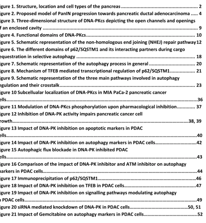
The word ‘pancreas’ originated from the Greek word ‘pankréas (παγκρέας)’ meaning ‘all-flesh’ (pan=all; kreas=flesh). It is a major gland that serves as an organ spanning the gastrointestinal and endocrine system in all vertebrates. It is often referred to as the heterocrine gland for its contribution in digestion and hormone production. The pancreas secretes a clear alkaline fluid or ‘pancreatic juice’ into the duodenum via the bile duct. This fluid contains bicarbonates for neutralizing stomach acid and also digestive enzymes for breaking down complex carbohydrates, fats and proteins. In the endocrine system, it contributes to maintaining blood sugar levels by secreting hormones like insulin, glucagon and somatostatin. Located in the upper left part of the abdominal cavity, the pancreas is approximately 5-6 inches long and is structurally divided into head, body and tail. Broadly the pancreas is made up of tissues that have exocrine function (digestion) and endocrine function (hormone secretion). Exocrine tissues of the pancreas comprise of acinar cells that secrete zymogens required for digestion, whereas endocrine tissue comprises of cell-clusters called ‘islets of langerhans’, which in turn comprise of alpha, beta and pancreatic polypeptide (PP) or gamma cells known to primarily secrete insulin and glucagon, besides other hormones (Röder et al, 2016); (see Fig.1). Malfunctioning of the organ could potentially lead to diseases such as pancreatitis, cancer or diabetes.
2 1.1.1 Pancreatic cancer
As per the latest Canadian Cancer Statistics, pancreatic cancer is the 4th leading cause of cancer related deaths in Canada with a net 5-year survival rate of 8%. Depending on the cells of origin, cancers of the pancreas are divided into two categories, exocrine pancreatic cancer and endocrine pancreatic cancer. Pancreatic ductal adenocarcinoma, acinar cell carcinoma, intraductal papillary-mucinous neoplasm (IPMN), adenosquamous carcinoma, colloid carcinoma arises from the exocrine part, whereas the pancreatic neuroendocrine tumours (PNETs) arise from the endocrine part and are named after the secreted hormone.
Figure 1. Structure, location and cell types of the pancreas
(Image source: https://www.cancer.org/cancer/pancreatic-cancer/about/what-is-pancreatic-cancer.html)
3
1.1.1.1 Pancreatic ductal adenocarcinoma (PDAC)
Pancreatic ductal adenocarcinoma (PDAC) arises from the exocrine cells of the pancreas and is the most common form of exocrine pancreatic cancer. PDAC accounts for almost 85% cases diagnosed (Hidalgo et al, 2015) and are therefore considered as the most common form of pancreatic cancer. PDAC cells could originate from ductal, acinar or pancreatic progenitor cells, although the exact cell of origin is still debatable (Yamaguchi et al, 2018). The key mutations driving PDAC progression are: KRAS, TP53, SMAD4,
CDKN2A and sometimes BRCA2 (Jones et al, 2008, Morris et al, 2010, Vincent et al, 2011).
Micro-anatomically, PDAC stems from pancreatic intraepithelial neoplasms (PanINs), which are microscopic lesions. Accumulation of several mutations will allow the progression of PanINs towards becoming PDAC (Pittman et al, 2017) (Fig.2). The journey starting from PanIN to PDAC takes several years which unfortunately leads to late diagnosis and poor prognosis. The transition comprises of stages starting from PanIN 1 to PanIN 3 (Pittman et al, 2017). Besides KRAS mutation, early PanIN-1 overexpresses Mucin-1 (MUC1), followed by inactivation of p16/CDKN2A during PanIN-2 stage. During PanIN-3, tumour protein (TP53), mothers against decapentaplegic homolog 4 (SMAD4) and breast cancer type 2 susceptibility protein BRCA2 are inactivated thereby progressing to PDAC (Khan et al, 2017). KRAS mutations occur in around 95% of pancreatic cancers (Bamford et al, 2004), whereas TP53 mutation occurs in 70% of pancreatic cancer. Inactivation of CDKN2A gene occurs in 98% of pancreatic cancer (Schutte et al, 1997) and mutation in SMAD4 leading to inactivation of the tumour suppressor protein occurs in 50% of the cases (Bosscher et al, 2004). Germline mutations in BRCA2 were found in 7.3% of Ashkenazi Jewish patients with sporadic pancreatic cancer (Goggins et al, 1996). A recent study has provided new information about the percentages of these mutations via whole genome sequencing of 150 PDAC specimens. They have found that KRAS mutations occurs in 93% of the population, TP53 mutation occurs in 72% of the population, whereas
CDKN2A and SMAD4 occurs in 30% and 32% of the population respectively (Raphael et al,
4 Figure 2. Proposed model of PanIN progression towards pancreatic ductal adenocarcinoma
(Image source: Wilentz et al, Loss of Expression of Dpc4 in Pancreatic Intraepithelial Neoplasia: Evidence That DPC4 Inactivation Occurs Late in Neoplastic Progression, Cancer Research, 2000;Image license number: 4785991497753)
1.1.2 Pancreatic cancer: risk factors
It is difficult to dissect specific cause(s) that lead to pancreatic carcinogenesis but some risk factors have been identified. Heredity plays a role in triggering the onset of the disease. Roughly 10% of PDAC arise due to genetic predisposition. A meta-analysis performed to analyse the risk posed by family history in causing the disease showed that people who had at least one relative suffering from the disease were at an estimated 80% higher risk to develop the disease (Permuth-Wey et al, 2009). People with mutation in breast cancer type 1 and 2 susceptibility proteins (BRCA1/2), PALB2 (partner and localizer of BRAC2)/FANCN, HNPCC or Lynch syndrome, are more susceptible to the disease (Becker et al, 2014). These gene mutations are associated with other forms of cancer but having these mutations increase the chances of developing PDAC in these patients.
5 Besides genetics, other factors like tobacco use, alcohol and pancreatitis play major role in driving pancreatic carcinogenesis.
1.1.3 Pancreatic cancer treatment
1.1.3.1 Surgery:
The treatment options available for pancreatic cancer are chemotherapy, radiotherapy and surgery. Only 20% of the patients are eligible for surgical resection, which leaves 80% of the patients diagnosed with the disease dependent on palliative chemotherapy and radiotherapy due to local invasion and metastasis (Gillen et al, 2010). Notably, neo-adjuvant therapy is suggested for patients with local invasion in order to reduce the tumour size before surgery. The therapy can help in tumour regression to a considerable extent and can be a ray of hope for patients who were declared non-eligible for surgery at diagnosis, although it is not a potential cure for PDAC patients.
1.1.3.2 Chemotherapy:
Majority of the patients depend on either chemotherapy or radiotherapy to improve their symptoms. To date, Gemcitabine and FOLFIRINOX stand as the first line of treatment for pancreatic cancer. Gemcitabine is also known as 2’ 2’-difluoro 2’ deoxycytidine and is sold under the tradename Gemzar®. Pharmacologically, it is a nucleoside analogue that replaces cytidine thereby causing a masked chain termination in replicating DNA strands. Hydrophilic Gemcitabine enters the cells with the help of nucleoside transporters (Belt et al, 1993, Mini et al, 2006). Inside the cell, Gemcitabine is activated by subsequent addition of phosphate group, thereby transitioning from mono-, di- and triphosphate. Addition of three phosphate groups helps the drug to attain deoxycytidine triphosphate configuration (dFdCTP) and gets incorporated in replicating DNA chains, thereby leading to termination of replication and cell death. In the late 90’s, Gemcitabine alone was prescribed to patients with advanced pancreatic cancer after it was found to be more effective than 5-FU. Gemcitabine offered better survival outcome (12 month survival=18%) versus 5-FU treatment which could only improve the 1 year
6 survival by 2% (Burris et al, 1997). Later, several patient cohort studies revealed that Gemcitabine in combination with other chemotherapeutic drugs like cisplatin or nab-paclitaxel (Abraxane®) showed more effective response in the patients, in terms of improving the overall as well as the progression-free survival (Ouyang et al, 2016; Fernandez et al, 2018).
FOLFIRINOX is another important chemotherapeutic regimen for pancreatic cancer treatment and is a drug cocktail comprising of folinic acid, fluorouracil, Irinotecan and oxaliplatin. Folinic acid is a vitamin-B derivative capable of lowering the side-effects of 5-FU, which is another component of the drug regime. Fluorouracil (5-FU) is a nucleoside (pyrimidine) analogue that blocks DNA replication. The topoisomerase inhibitor Irinotecan prevents DNA helix uncoiling essential for DNA replication. The antineoplastic drug oxaliplatin prevents DNA damage repair and/or synthesis essential for cancer cell proliferation. In a comparative study to analyse the efficacies of FOLFIRINOX and Gemcitabine in metastatic pancreatic cancer, Conroy and colleagues reported that the median overall survival was 11.1 months in FOLFIRINOX treated group compared to 6.8 in Gemcitabine treated group. The median progression-free survival was 6.4 and 3.3 months in FOLFIRINOX and Gemcitabine treated groups respectively (Conroy et al, 2011). In a recent study conducted to understand the outcomes of FOLFIRINOX treatment in locally advanced pancreatic cancer, tumour resectability was achieved for tumours that were otherwise considered non-resectable. Moreover, compared to the control group, the median overall survival was increased by two times in the FOLFIRINOX treated group (Lee et al, 2018).
1.1.3.3 Radiotherapy:
In principle, radiotherapy uses high-energy electron beams or ionizing radiation to kill cancer cells. It is often used in combination with chemotherapy, known as ‘chemoradiation’ that is advised to patients with non-resectable tumour, or to regress the tumour size of resectable tumours before surgery. Intraoperative radiotherapy (IORT)
7 helps to eliminate minor remains of malignant tissue post-surgery, by introducing high-beam radiation to the affected cells without harming the surrounding healthy cells. A systematic review to understand the effect of IORT to improve patient survival outcomes revealed that IORT could moderately increase the overall survival but the treatment is effective when used in combination and is not a potential cure for PDAC (Ruano-Ravina et al, 2007).
The current dismal situation indicates towards the need to develop better therapeutic options for the treatment of the disease. To devise effective therapies, root cause or the mechanisms that could potentially lead to the development of the disease need to be dissected. Aberrantly triggered cellular signalling pathways lead to uncontrolled cell proliferation, formation of malignant mass and therefore play a vital role in the development of any form of cancer.
1.1.4 Cell signalling pathways in pancreatic cancer
Diverse signalling pathways have been involved in the development of PDAC such as the RAS-MAPK, Wnt-β catenin, TGF-β and Notch signalling pathways (McCleary-Wheeler et al, 2013). Our laboratory is interested in investigating how deregulation of signaling pathways supports pancreatic cancer cell growth. Of interest, a protein involved in the Wnt-β catenin pathway called GSK-3 has been found to be involved in regulating various pathways associated with cell proliferation, and preventing apoptosis in PDAC cells (Ougulkov et al, 2005). Previous work in our lab demonstrated that inhibition of GSK-3 induces an apoptotic response in PDAC cells via JNK-dependent mechanism (Marchand et al, 2012). Additionally, a consecutive paper from our lab reported that GSK-3 inhibition could trigger autophagy as a prosurvival mechanism in PDAC cells to nullify the apoptotic response (Marchand et al, 2015). Further investigation in this area by our lab revealed that the protective autophagic signal is regulated by the transcription factor EB (TFEB). In an attempt to find out how GSK-3 inhibition impacts on TFEB, mass spectrometric analyses were performed to identify TFEB interacting partners. Interestingly, many DNA
8 damage repair proteins were found to be putative TFEB partners which associate with TFEB upon inhibition of GSK-3 activity including PARP1, KU70 and KU80 (Marchand et al, 2015). Therefore, we were interested to investigate further how DNA damage repair proteins might be involved in regulation of autophagy in PDAC cells. Notably, KU70 and KU80 are the subunits of an important DNA double strand break (DSB) repair enzyme complex known as DNA-PK.
1.2. DNA-PK discovery
DNA-PK or DNA-dependent protein kinase is a serine threonine kinase and belongs to the phosphatidylinositol 3-kinase-related kinase (PIKK) protein family. The catalytic subunit of the DNA-PK complex, DNA-PKcs, is a 460KDa protein and belongs to the DNA-PK holoenzyme along with KU70 and KU80 heterodimers. The DNA-PKcs protein is encoded by the PRKDC gene located on chromosome 8 (Sipley et al, 1995). In the year 1985, Walker and colleagues found that, when added to cellular extracts from HeLa cells, xenopus eggs or reticulocytes, double-stranded DNA could potentially lead to the phosphorylation of a lot of different proteins by transferring phosphate groups from ATP (Walker et al, 1985). This first indicated DNA-activated kinase activity and five years down the lane, three research groups independently purified and analysed the activity of DNA-PKcs (Carter et al, 1990, Jackson et al, 1990, Lees-Miller et al, 1990).
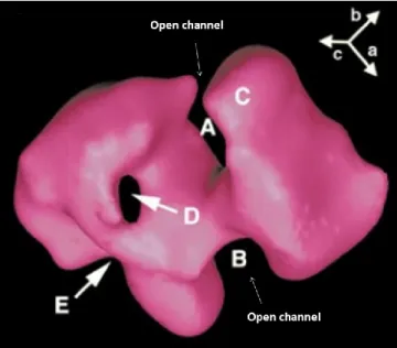
1.2.1 Structure of DNA-PKcs
Several research groups have carried out electron crystallography studies to understand the molecular structure of DNA-PKcs protein. The three-dimensional structure reveals that the protein has one open channel and one enclosed cavity which in turn has three openings to harbour DNA which is at least 12bp or more in length (Leuther et al, 1999); (Fig.3). This precise length of the DNA is required for activation of the protein. Broadly, the protein is made up of three major domains, the N-terminal domain, the circular cradle in the middle and the C-terminal domain (Goodwin et al, 2014; Wu et al,
9 2019). The large N-terminal domain comprises of many subdomains which are HEAT repeats (Huntingtin Elongation Factor 3, PP2A and TOR1) and phosphorylation clusters like JK, PQR and ABCDE (Goodwin et al, 2014) (Fig.4). The PQR and the ABCDE clusters harbour Ser2056 and Thr2609 respectively, which are essential for autophosphorylation of the protein. Another major domain is the kinase domain that also plays a vital role in autophosphorylation. The kinase domain is flanked by the FAT domain, which has homology with FAT, ATM and transformation/transcription associated protein (TRRAP), and at its C-terminal end, a domain called FATC (Goodwin et al, 2014).
Figure 3. Three-dimensional structure of DNA-PKcs depicting the open channels and openings of an enclosed cavity
(Adapted from Leuther et al, Structure of DNA-dependent protein kinase: implications for its regulation by DNA, 1999; Open access journal)
This figure illustrates different angles of the 3D-structure of the DNA-PKcs molecule. ‘A’ and ‘B’ are open channels that bind to double-stranded DNA, whereas ‘C’ denotes low protein density at the end of an arm-like structure that makes it flexible. The two openings of an enclosed cavity are depicted by the letters ‘D’ and ‘E’.
10 Figure 4. Functional domains of DNA-PKcs
(Adapted from: Goodwin and Knudsen, Beyond DNA repair: DNA-PK function in cancer, Cancer Discovery, 2014) Image License number: 4785980353552
1.2.2 Structure and function of KU heterodimer
The KU70/80 heterodimer is an important part of the DNA-PK complex and work in concert with DNA-PKcs to achieve double-strand break repair. The KU70 and KU80 subunits are encoded by XRCC6 and XRCC5 genes respectively. Cryo-electron microscopy studies have revealed that the subunits form ring shaped structures that cradle on the DNA strands. The subunits have N and C-terminal globular domains and the later plays important role in DNA binding. Particularly the C-terminal globular/SAP domain of KU80 also called KU80CTR binds to DNA end via weak interaction (Wu et al, 2019). Plausible role of KU80CTR is to temporarily recruit an associate with DNA-PKcs, thereby assisting it to tether to broken DNA ends (Walker et al, 2001).
11 The association of KU heterodimer with broken DNA ends plays an integral role in DNA-PKcs recruitment to the damage site (Gottlieb and Jackson, 1993). The association is also known to increase the kinase activity of the protein by 5-10 folds (Jette and Lees-Miller, 2015).
1.3. Functions of DNA-PK
1.3.1 Role in DNA repair
DNA double-strand breaks are the most lethal forms of DNA damage which, if left unrepaired, might lead to cell death. Therefore, the cells are equipped with repair mechanisms to sustain growth and proliferation. The two major double-strand break repair pathways are homologous recombination (HR) and non-homologous end joining repair (NHEJ) pathway. DNA-PK complex plays a pivotal role in the NHEJ pathway. The NHEJ pathway does not require homologous chromosomes or sister chromatids to repair the damage. NHEJ utilizes specific nucleases to modify the DNA ends in order to facilitate fusion of the ends by DNA ligase IV during the repair process (Lieber et al, 2011). Lack of complementarity often leads to faulty addition or deletion of base pairs thereby making the process error-prone. NHEJ is active during all phases of the cell cycle thereby making it the preferred pathway for cells which are not close to the S/G2 phase, as this is a requirement for HR pathway (Liebar et al, 2011). Mechanistically, upon DNA double-strand break, KU proteins form a ring/basket around the broken DNA ends thereby creating a scaffold to hold other proteins which are subsequently recruited to carry out the repair process. Once PKcs are recruited, the kinase domain of DNA-PKcs is activated and two DNA-DNA-PKcs molecules bring the two broken DNA ends close to each other for synapsis (Lees-Miller and Meek 2003). Subsequently, the DNA ends are modified via different nucleases to facilitate fusion of the broken ends. The XRCC4/DNA ligase 4 complex participates in sealing the gaps thereby healing the damage (Fig.5).
12 Figure 5. Schematic representation of the non-homologous end joining (NHEJ) repair pathway
1.3.2 Role in V(D)J recombination
DNA-PK is majorly involved in V(D)J recombination besides other proteins which help in the NHEJ process. V(D)J recombination is a genetic recombination that occurs in developing lymphocytes that leads to the rearrangement of segments of immunoglobulin genes and T-cell receptors that are found in B and T cells (Lees-Miller and Meek, 2003). The process is named after the different segments that are generally rearranged such as variable (V), joining (J) and only in some instances the diversity (D) segment. The recombination process is initiated by V(D)J recombinase which is composed of several enzymes. Some of the key enzymes are recombination activating genes 1 and 2 (RAG1/2), DNA-PKcs, Artemis. Generally, RAG1/2 recognizes specific sequences known as recombination signal sequences (RSS). RSS are located at the flank of coding variable region of immunoglobulin and T-cell receptor genes. They are made up of a heptamer of conserved nucleotides (CACAGTG), followed by a spacer region comprising of 12+/-1 or
13 23+/-1 base pairs, and a nonamer of conserved nucleotides (ACAAAAACC) (Ramsden et al, 1994). The recombination begins when RAG1/2 recognize, bind to specific RSS sites, and create a nick on one strand. This nick creates 3’ hydroxyl group that is used for transesterification that results in a DSB. This creates stem-loop or hairpin structure at the coding ends which is subsequently open, DNA ends are processed by DNA-PKcs and artemis, and repaired by the NHEJ repair pathway (Schatz and Swanson, 2011).
As a result of this recombination process, the antigen binding sites get novel amino acid sequences that could recognize a vast array of antigens thereby creating a diverse repertoire of antibodies and T-cell receptors in the developing lymphocytes.
1.4. Important regulators and substrates of DNA-PKcs
1.4.1 Autoregulation
Identifying the regulators and substrates of a protein is essential to monitor its intracellular activity and devise therapeutic strategies. The mode of activation of DNA-PKcs is still debatable, but a considerable number of studies have indicated that the protein is activated by autophosphorylation. Probably, the first indication of autophosphorylation was mentioned in the seminal work of Lees-Miller, Chen and
Anderson, where they purified DNA-PK and analyzed its activity (Lees-Miller et al, 1990).
Later, Block and colleagues found that autophosphorylation of DNA-PKcs is essential for DNA double-strand break repair and mutation in the phosphorylation sites might make the cells to succumb to radiation-induced DNA damage (Block et al, 2004). Many of the DNA-PKcs phosphorylation sites are present in the PQR and ABCDE regions (Fig.4) and the most common autophosphorylation sites are Ser2056, Ser2612, Ser2624, Thr2609 and Thr2620 (Douglas et al, 2002).
1.4.2 Key substrates of DNA-PKcs
The most important substrate of DNA-PKcs is the protein itself due to its autoregulatory properties. Besides, DNA-PKcs phosphorylates AKT at Ser473 thereby
14 activating it and phosphorylated AKT in turn promotes DNA-PKcs autophosphorylation, thereby creating a positive feedback loop (Toulany et al, 2011, 2012). In response to DNA damage, H2AX is phosphorylated at its Ser139 residue by DNA-PKcs. This phosphorylation event is used as a marker for the DNA damage response and the phosphorylated form of the protein is called H2AX. DNA damage-induced H2AX phosphorylation creates a foci where all other repair proteins like 53BP1, BRCA1, RAD50, etc., are recruited and they help to resolve the foci at the earliest to facilitate the passage of cells through cell cycle checkpoints (Podhorecka et al, 2010).
1.5. Role of DNA-PK in pancreatic cancer
Elevated expression of both DNA-PKcs and KU subunits was reported in PDAC by
Michael Osterman and colleagues (Osterman et al, 2014). In their study with normal and
pancreatic tumour tissues obtained from surgical biopsies, they observed that the tumour tissues display increased phosphorylation of DNA-PKcs than the normal tissues suggesting increased activity of DNA-PK in PDAC. Additionally, immunofluorescence experiments showed enhanced KU70 staining in pancreatic tumour tissues as compared to normal tissue (Osterman et al, 2014). In a paper describing the role of DNA-PK in pancreatic cancer, Li and colleagues observed that DNA-PK contributes to PDAC growth. Knockdown of DNA-PK regulatory subunits via RNA interference led to drastic reduction in growth of PDAC cell lines. Moreover, pharmacological inhibition of DNA-PK activity impeded colony forming ability of PDAC cells in culture (Li et al, 2012). These studies suggest a role for DNA-PK in PDAC cell growth and sustenance, but little is known about the mechanisms as to how DNA-PK supports PDAC cell growth.
1.6. Role of DNA-PK in other forms of cancer
The role of DNA-PK in driving different types of carcinogenesis has been studied by several research groups. The mRNA expression levels for DNA-PKcs are reported to be elevated in lung cancer (Xing et al, 2008). Pharmacological inhibition of DNA-PK was found
15 to sensitize lung cancer cells to doxorubicin and radiation, and caused tumour regression (Fok et al, 2019). Kotula and colleagues showed that RNA interference mediated depletion of DNA-PKcs could potentially impair melanoma cell invasion and metastasis (Kotula et al, 2015). High protein levels of DNA-PKcs have been reported in lymphoid malignancy like B-cell chronic lymphocytic leukaemia (B-CLL) which subsequently offers chemoprotection to the cancer cells. Pharmacological inhibition of DNA-PK activity could re-sensitize the cells to chemotherapy (Muller et al, 1998; Elliott et al, 2010). Cisewski and colleagues reported that abrogation of DNA-PK activity could sensitize breast cancer cells to doxorubicin and radiation (Cizewski et al, 2013). Yanai and co-workers have shown that pharmacological inhibition of DNA-PK can sensitize non-small cell lung cancer cells to topoisomerase inhibitors (Yanai et al, 2017). All these studies highlight the fact that DNA-PK is essential for supporting growth of cancer cells and also for providing chemoprotection. Still, the mechanisms sustaining such function by DNA-PK remain elusive.
A recent work on malignant glioma cell lines revealed that inhibition of DNA-PK activity could radiosensitize the cells by triggering autophagy (Daido et al, 2017). This is an indication towards a plausible role played by DNA-PK in regulating an important cellular process-autophagy.
1.7. Autophagy
The word ‘autophagy’ originated from the Greek word ‘autophagos’ which means self-eating. Autophagy is an intracellular dynamic and well-orchestrated catabolic degradation process where the cell degrades its own contents to generate energy via recycling macromolecules to survive during stressful situations. Autophagy can be of various types based on the type of cargo sequestered and degraded, such as microautophagy, chaperone-mediated autophagy, mitophagy (selective degradation of mitochondria), pexophagy (selective degradation of peroxisomes). But the most prevalent and distinct form of autophagy is macroautophagy, which is characterized by double-membrane
16 bound phagosomal structure (Feng et al, 2014) and it can employ both selective and non-selective modes of cargo degradation. In non-selective autophagy, specific ubiquitin-bound cargoes are recognized and degraded (Mizushima et al, 2007).
1.7.1 The process in general
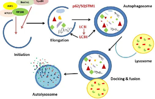
Induction: The process begins with budding of nascent phagophores upon induction of autophagy by various stress stimuli. The origin of autophagosomal membrane still remains debatable. Interestingly, the plasma membrane, Golgi complex, endoplasmic reticulum, mitochondria and endosomes might contribute in some way towards phagophore biogenesis and expansion. The naive vesicles could be considered as a mosaic model of membrane, considering the diverse range of organelles that contribute towards their origin (Mari et al, 2011). Various studies on yeast have suggested the occurrence of perivacuolar punctate structures also known as ‘pre-autophagosomal structure (PAS)’ as the origin of autophagosome biogenesis. The punctate structure as observed under microscope is actually the accumulation of Atg proteins that help in autophagy initiation (Kim et al, 2002). More specifically, in yeast upon stress, Atg1 is activated and its binding affinity with Atg13 and Atg17 gets intensified. The Atg1-Atg13-Atg17 creates a scaffold for recruiting other proteins instrumental in autophagosome formation. The mammalian homologues of Atg1 and Atg17 are ULK1/2 and FIP200 respectively and together with ATG13 and ATG 101 they form the ULK complex essential for autophagy initiation (He and Klionsky, 2009; Wesselborg and Stork, 2015; Zachari and Ganley, 2017).
Elongation/expansion: Especially during selective autophagy, p62/SQSTM1 (sequestosome), a 62KDa adaptor protein and a substrate of the process, recognizes and sequesters ubiquitinated cargoes to the growing autophagosome membrane. p62/SQSTM1 is regulated by specific post translational modifications that help in its functioning during the process. The N-terminal end of p62/SQSTM1 harbours the PB1 domain (Phox and Bem1p-1) and the C-terminal end harbours the UBA domain or the
17 ubiquitin binding domain (Fig.6). Several post-translational modifications in p62 impart cargo-binding abilities. The UBA domain generally has low affinity for ubiquitinated cargoes due to homodimerization, which is prevented by ubiquitination of Lys420 residue (Peng et al, 2017; Lee et al, 2017). Another mechanism by which the affinity of p62 towards ubiquitin is enhanced is by TBK1 mediated phosphorylation of Ser407 in the UBA domain (Pili et al, 2012; Lim et al, 2015). Besides these modifications, another important post translational modification is the head-to-tail self-interaction of p62 via electrostatic interaction between Lys7 and Asp69 and this polymerization is essential for the proper functioning of p62 in selective autophagy (Johansen and Lamark, 2017; Deng et al, 2017).
Selective autophagy requires tight attachment of the p62-bound ubiquitinated cargoes with the phagophore membrane during the elongation phase. The association with inner autophagosomal membrane takes place via LC3. Microtubule associated protein 1 light chain 3 (LC3) is a key protein in autophagy process and plays a vital role in autophagosome biogenesis. For its efficient functioning LC3-I undergoes a modification (lipidation) via covalent interaction with phosphatidylethanolamine (PE) and becomes LC3-II (Thukral et al, 2015). The interaction of p62 with LC3 occurs via its LC3 interacting domain (LIR). Specific post translational modification of LIR domain of p62 such as phosphorylation positively regulates its function and facilitates its effective interaction with LC3 (Lamark et al, 2017). Besides being regulated by post-transcriptional modifications, p62 expression is also regulated transcriptionally.
18 Figure 6. The different domains of p62/SQSTM1 and its interacting partners during cargo sequestration in selective autophagy
This figure illustrates the interaction of p62 bound cargo with LC3 which in turn facilitates attachment of the complex to the inner phagophore membrane. p62 interacts with LC3 via its LIR domain whereas it binds with ubiquitinated cargoes via UBA domain. The domain architecture of p62 presented in the illustration depicts the occurrence of PB1 domain at the N-terminal end followed by zinc finger domain, TRAF6 binding domain, LC3-interacting and KEAP-1 interacting regions respectively. The UBA domain is present at the C-terminal end.
Closure and docking: Following cargo recruitment the double membrane phagophore sac gradually begins the maturation process and closes forming an autophagosome that will dock on lysosome for fusion. Docking of autophagosomes on lysosomes and their subsequent fusion is mediated by SNAREs (Soluble N–ethylmaleimide sensitive factor (NSF) attachment protein receptor). During the early stage of autophagosome formation, Atg12 and Atg8 proteins are involved. Atg12 conjugates with its substrate Atg5 via covalent bonding, thereby forming Atg12-Atg5. Later this Atg12-Atg5 binds with Atg16 thereby forming an oligomeric complex, that aids in the autophagosome elongation
19 process (Kim et al, 2015). Open autophagosomes still retain the Atg proteins on their surface but gradually as they proceed towards maturation, the Atg proteins associated with the autophagosome are cleaved off by proteases (Atg4 protease family) (Zou et al, 2018). This marks a transitional phase when the autophagosomes acquire fusion machinery and proceed towards lysosome for the formation of autolysosomes.
Following closure of autophagosomes, the SNARE protein syntaxin17 (STX17) is recruited to the autophagosome and it recognizes and binds to VAMP8 on lysosomal membrane. Fusion is mediated by SNARE proteins that later help in autophagosome-lysosome fusion. Syntaxin17 (STX17) and YKT6 are two proteins from two different SNARE protein complexes that play an integral role in autophagosome fusion (Zhao et al, 2020). The glycine zipper like motif in STX17 and N-terminal longin domain in YKT6 are essential for their translocation to autophagosome (Zhao et al, 2020). Several other tethering factors such as homotypic fusion and protein sorting (HOPS) also help in autophagosome-lysosome fusion (Nakamura et al, 2017; Diao et al, 2015). Since the autophagosome-lysosomes and late endosomes reside at the perinuclear region, the autophagosomes traverse through the cytoplasm on specific tracks which are the microtubules and their directionality is mediated by dynein-dynectin motor protein complex, to reach the perinuclear site (Nakamura and Yoshimori, 2017; Gross et al, 2007). Autophagosomes sometimes fuse with late endosomes, thereby forming ‘amphisomes’, or else they fuse specifically with the lysosomes forming ‘autolysosomes’ (Berg et al, 1998).
Cargo degradation: Once the membranes of autophagosome and lysosome fuse together, the cargos from autophagosome are unloaded inside lysosome for degradation. Inside the newly formed autolysosome, the lysosomal hydrolases stepwise degrade the cargos (Eskelinen et al, 2008). The low pH of lysosomes is maintained by ion channels and transporters. Vacuolar ATPases are the proton pumps that help to import protons from cytoplasm across the lysosomal membrane and serve as targets for the potent autophagy inhibitor Bafilomycin A1 (BAF A1). Another important target of BAF A1 is SERCA pump. SERCA or sarco/endoplasmic reticulum Ca2+-ATPase, transport calcium to sarcoplasmic
20 reticulum from the cytoplasm. The activated SERCA pumps are known to mediate autophagosome-lysosome fusion and are sensitive to BAF A1 (Mauvezin et al, 2015).The acidic environment (pH 4-5) inside the lysosomal lumen is crucial for the activity of degradative enzymes like hydrolases and cathepsins thus allowing the cargos to be degraded. The resulting products can be reused for biosynthesis and/or energy production for cell survival. The entire process is illustrated in figure 7.
Figure 7. Schematic representation of the autophagy process in general
1.7.2 Transcriptional regulation of p62/SQSTM1
As mentioned earlier, p62 expression is often regulated via transcriptional control. Transcriptional regulation of p62 is instrumental in understanding the elevated levels of this protein during active autophagy, which may not be always due to inhibition of autophagic flux. During stress, p62 mRNA levels are elevated by transcriptional up-regulation via nuclear factor erythroid 2-related factor 2 (NRF2). NRF2 binds to antioxidant response element (ARE) in p62 promoter region, thereby strongly inducing gene expression (Lamark et al, 2017). Under normal conditions, NRF2 interacts with
21 like ECH-associated protein 1 (KEAP1) which promotes Cullin-E3 ubiquitin ligase-mediated degradation of NRF2 by the ubiquitin proteasome pathway. But under oxidative stress, NRF2-KEAP1 interaction is hindered and NRF2 travels to the nucleus where it transcriptionally up-regulates p62 expression.
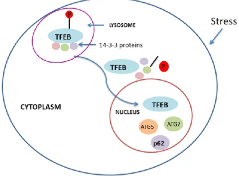
Another important regulator of p62 is transcription factor EB (TFEB). TFEB regulates genes involved in lysosomal biogenesis including genes involved in autophagy (Settembre et al, 2017). Protein kinases like ERK2 and mTORC1 phosphorylate TFEB and facilitate attachment with 14-3-3 proteins, thereby retaining it in the cytoplasm (Martina et al, 2012; Palmieri et al, 2017). During stress, TFEB gets dephosphorylated, dissociates from 14-3-3 proteins and translocates to the nucleus (Medina et al, 2015). Inside the nucleus, it triggers the expression of various genes including p62 (Fig.8).
Figure 8. Mechanism of TFEB mediated transcriptional regulation of p62/SQSTM1 This figure illustrates dephosphorylation-mediated activation and translocation of TFEB to the nucleus and its subsequent function (transcriptional regulation).
22 1.7.3 Signalling pathways regulating autophagy
Intracellular signalling pathways act as molecular switches for autophagy activation or inhibition depending on environmental cues. Some of the major pathways involved in autophagy regulation are the AMPK, mTORC1 and ULK1 pathways. AMPK pathway regulates cellular homeostasis by sensing nutrient depletion signals. During low energy level state, AMPK is phosphorylated at Thr172 and this reduces energy-intensive protein biosynthesis processes, thereby saving energy. AMPK phosphorylates TSC2, leading to mTORC1 inhibition. Inhibition of mTORC1 by AMPK also occurs directly via phosphorylation of raptor a member of the mTORC1 complex, at Ser (722) and Ser (792) sites (Tavakol et al, 2019). Mammalian target of rapamycin complex 1 (mTORC1), is a complex that functions quite similar to AMPK, in terms of functioning as a nutrient/energy sensor. Inhibition of mTORC1 can potentially trigger the activation of autophagy. Blocking mTORC1 and mTORC2 activity is usually achieved by using the inhibitor Torin1. This inhibitor is often used as a control for autophagy activation. As S6K1 is a downstream target of mTORC1, phosphorylation of S6K1 Thr (389) is used as a marker of mTORC1 activity.
Another important signalling pathway at the crossroads of AMPK and mTORC1 pathways, which is also known to regulate autophagy, is ULK1 pathway, which is known to activate autophagy. AMPK activates ULK1 directly via phosphorylation (Ser555) (Egan et al, 2011) and also via phosphorylation at sites Ser317, Ser777 (Tavakol et al, 2019). Mechanistically, autophagy is repressed by mTORC1 via Atg13 phosphorylation. This event prevents ATG13 from entering the ULK1 complex. As a consequence, the ULK1 kinase complex is not recruited to the pre-autophagosomal structure (PAS) and autophagy is repressed. Contrarily, upon inhibition of mTORC1, autophagy is activated via ULK1 (Fig.9).
23 Figure 9. Schematic representation of the three main pathways involved in autophagy regulation and their crosstalk
This figure represents two different ways of autophagy activation via AMPK. On the left, AMPK activation leads to repression of mTORC1 and subsequent trigger in autophagy. On the right, AMPK activation leads to phosphorylation of the proteins of the ULK1 complex, leading to autophagy activation.
1.7.4 Regulation of autophagy by DNA-PK
We came across studies that reported autophagy regulation by DNA-PK in different forms of cancer. A study on glioblastoma cells showed that inhibition of DNA-PKcs via RNA interference can radiosensitize the cells (Zhuang et al, 2011). They reported that the cell death is caspase-independent which signifies that the cells did not die due to apoptosis. Rather they found that the cell death was mediated by autophagy. Upon observing increased LC3B II levels by immunoblot and increased LC3 dots by immunofluorescence,
24 the authors concluded that DNA-PKcs knockdown induces autophagy. However the measurement of autophagic flux was rather unusual because the autophagy inhibitors 3-MA (3-methyladenine) or E46D were added 10 minutes before subjecting the cells to irradiation and then the cells were incubated for 48 hours. Conventionally during autophagic flux measurement the autophagy inhibitor is added for 2-8 hours. Moreover the comparison for LC3 dots was performed by comparing control cells with DNA-PK inhibited cells treated with the autophagy inhibitor. There was no treatment of the control cells with E64D to better appreciate the increase in LC3 dots/puncta upon DNA-PK inhibition. The study suffered this limitation; however it gave us a hint that there is a probability of regulation of autophagy by DNA-PK in cancer cells (Zhuang et al, 2011). Inhibition of DNA-PK activity by the pharmacological DNA-PK inhibitor was also found to trigger autophagy in DOC-2/DAB2 interacting protein (DAB2IP) deficient osteosarcoma cells (Yu et al, 2012). Yet, in another study, contradictory effects were observed as DNA-PK inhibition inhibited basal as well as etoposide induced autophagy in various cancer cell lines such as breast cancer, osteocarcinoma and cervical cancer cell lines respectively (Puustinen et al, 2020). These reports highlight a potential role for DNA-PK in autophagy regulation in cancer cells. Still, the impact of DNA-PK inhibition on autophagy remains to be clarified.
1.7.5 Role of autophagy in PDAC
Autophagy is known to provide cancer cells with degradative products that could be reused for metabolism and cell survival (Yang el al, 2011, Mizushima and Komatsu, 2011). Many research groups have explored the status of autophagic activity in cancer cells and its corresponding outcome in patient prognosis. In most cases, autophagy is found to act as a prosurvival mechanism in cancer cells leading to poor prognosis in patients (Dalby et al, 2010; Mathew et al, 2010).In the context of my project, it is important to understand the level, activity and subsequent role(s) of autophagy particularly in PDAC. Yang and
25 and colleagues performed GFP-LC3 reporter assay in an array of pancreatic cancer cell lines including non-transformed pancreatic epithelial cell line HPDE (normal cell line), to compare autophagic activity in normal versus tumour cell lines. They also performed the assay on breast cancer (MCF7) and lung cancer (H460) cell lines to compare autophagic activity between PDAC and other carcinomas. Interestingly, 70-90% of the PDAC cell lines displayed elevated GFP-LC3 puncta whereas only 3-18% of the population of cells from HPDE, MCF7 and H460 displayed GFP-LC3 puncta. In a parallel experiment, they showed that blocking autophagy in PDAC cells via RNA interference strategy (siRNA against ATG5 which is responsible for autophagosome expansion) could impede colony forming ability in culture suggesting that PDAC cells require autophagy for their growth (Yang et al, 2011; Yang et al, 2018).
1.8. Hypothesis and objectives
As mentioned earlier, reports suggested regulation of autophagy by DNA-PK in various types of cancers (Zhuang et al, 2011; Yu et al, 2012; Puustinen et al, 2020). However, the role of DNA-PK on autophagy in PDAC has never been addressed. Considering the high level of autophagy in PDAC (Yang et al, 2011) and the elevated expression of DNA-PKcs (Osterman et al, 2014), both shown to support PDAC cell growth, investigating the contribution of DNA-PK on autophagy in the context of PDAC is worth pursuing.
Taking into account the above information, the current project was initiated to test the hypothesis that:
DNA-PK sustains autophagy and supports pancreatic cancer cell growth
We specifically aimed to address the following objectives:
1) Elucidate the effect of DNA-PK inhibition on autophagy in pancreatic epithelial cells 2) Determine the impact of DNA-PK inhibition on the growth of pancreatic epithelial cells
26 2. MATERIALS AND METHODS
2.1 Cell culture and passaging
The human pancreatic cancer cell lines MIA PaCa-2 and PANC1 were obtained from American Type Culture Collection (ATCC). The cells were cultured in Dulbecco’s Modified Eagle’s Medium (DMEM) (Wisent Inc.), supplemented with 10% foetal bovine serum (FBS), 2mM GlutaMAX™ (35050061 Thermo Fisher Scientific) and 10mM Hepes (330-050-E2; Wisent Inc.). The cells were grown in a humidified 5% CO2 atmosphere at 37°C.
Medium was replaced every other day with fresh medium. The cells were trypsinized (Trypsin/EDTA; 325-043-CL Wisent Inc.) upon reaching 90-95% confluency.
2.2 Inhibitors and chemotherapeutic drugs
The pharmacological inhibitor NU7441 (Selleckchem) was used for DNA-PK inhibition. During the course of the study, different inhibitors were used as listed: ATM inhibitor- KU55933 (Selleckchem), PI3K inhibitor- LY294002 (Millipore Sigma), RAD51 inhibitor-B02 (Selleckchem), mTOR inhibitor- Torin I (Tocris). To study the impact of chemotherapeutic drugs on PDAC cells, the following drugs were used: Gemcitabine (LC Laboratories), and Bafilomycin A1 (Selleckchem).
2.3. Measurement of cell growth
2.3.1 Growth curve assay
For cell growth assay, 25 000 cells were plated in triplicate in 12-well plate. The following day (day 0), cells were treated with the DNA-PK inhibitor NU7441. The vehicle DMSO was used as negative control. Medium was replaced every 24 hours with fresh medium containing DMSO or the inhibitor. To count the number of cells, 200μl of trypsin was added to the wells, followed by addition of 800μl of DMEM to inhibit trypsin. Ten microliters of the 1mL cell suspension was added to both chambers of a haemocytometer
27 (Reichert Bright Line™) and cells were counted. Data were analysed by Prism 7 (GraphPad software). The average number of cells and standard deviation for every condition at each time-point is graphically represented.
2.3.2 Clonogenic assay
Two hundred cells per well were seeded in triplicate in a 12-well plate. The following day, the cells were treated with DMSO or different concentrations of NU7441. The cells were allowed to grow up to 10 days with replacement of medium and adding fresh medium with DMSO or NU7441 every day. After 10 days, colonies were stained with 0.5% crystal violet solution in 30% ethanol for 30 minutes and then washed several times with distilled water until the excess stain was removed. Colonies were counted manually and the plates were air-dried before being scanned with a HP Scanjet G4010 scanner.
2.4. Biochemical analysis
2.4.1 Preparation of cell lysates
Cells were lysed in high salt buffer containing 1% Nonidet P40 (NP40, Bio Basic Canada Inc.), 50mM Tris pH 7.5 (Bio Basic Canada Inc.), 300mM NaCl (Bio Basic Canada Inc.), 150mM KCl (Bio Basic Canada Inc.), 5mM EDTA (Wisent Bioproducts), 10mM sodium fluoride (NaF) (BDH), 10% Glycerol (Bio Basic Canada Inc.) and protease inhibitors were added extemporarily as follow: 200μM sodium orthovanadate (Sigma), 1mM phenylmethysulfonyl fluoride (PMSF), 0.5μg/ml each of apoprotinine, leupeptine and pepstatine.
Following treatment, cells were kept on ice and medium was aspirated out gently. Each well was washed twice with 1X cold PBS. After the second wash, high salt lysis buffer was added to each well, according to the surface area of the well/dish. The measurements are as follows:
28 35mm dish= 350-400μl
60mm dish= 500-600μl 100mm dish= 1000μl
The cells were then scraped out using a plastic scraper (Sarstedt # 83.1830) and the lysates were carefully transferred to mini centrifuge tubes. Samples were allowed to stand on ice for 10 minutes. After the incubation period, the lysates were sonicated (Thermo Fischer, Model CL-18) on ice for 5-10 seconds at 30% amplitude. After sonication, the samples were centrifuged (Micromax, Thermo Electron Corporation) for 10 minutes at 10,000 rpm at 4°C. The supernatants were transferred to fresh tubes and protein estimation was performed using the bicinchoninic acid method as described in section 2.4.3.
2.4.2 Subcellular fractionation
Subcellular fractionation was performed as per the instruction provided with the Cell Signalling technology kit (#9038). Cytoplasmic Isolation Buffer (CIB), Membrane Isolation Buffer (MIB), Cytoskeletal/Nuclear Isolation Buffer (CyNIB) are provided in the kit. Protease inhibitors (similar to the cell lysate preparation protocol mentioned in the previous section) were added to each buffer prior to use. Briefly, the cells were first rinsed with 1X cold PBS. The cells were scraped in a mini centrifuge tube (1.5ml) and pelleted down at 350g for 5 minutes. The pellet was rinsed with 1ml of 1X cold PBS before being resuspended in 500μl of cold 1X PBS. For whole cell lysate (WCL), 50μl of the cell suspension was aliquoted out. The remaining suspension (450μl) was centrifuged for 5 minutes at 500g at 4°C. The pellet was subsequently resuspended in 250μl of CIB, vortex for 5 seconds and incubated on ice for 5 minutes before centrifugation for 5 minutes at 500g at 4°C. The supernatant was saved as the cytoplasmic fraction whereas the supernatant was resuspended in 250μl of MIB, vortex for 15 seconds and incubated on ice for 5 minutes before centrifugation for 5 minutes at 8000g 4°C. The supernatant was saved as the membrane and organelle fraction. The pellet was resuspended in 125μl of CyNIB buffer and sonicated to obtain the cytoskeleton/nuclear fraction. Laemmli buffer