OATAO is an open access repository that collects the work of Toulouse researchers and makes it freely available over the web where possible
Any correspondence concerning this service should be sent
to the repository administrator: tech-oatao@listes-diff.inp-toulouse.fr This is an author’s version published in: http://oatao.univ-toulouse.fr/21349
To cite this version:
Barrick, Andrew and Manier, Nicolas and Lonchambon, Pierre and Flahaut, Emmanuel and Jrad, Nisrine and Mouneyrac, Catherine and Châtel,
Amélie Investigating a transcriptomic approach on marine mussel hemocytes exposed to carbon nanofibers: An in vitro/in vivo comparison. (2019) Aquatic Toxicology, 207. 19-28. ISSN 0166-445X
Official URL: https://doi.org/10.1016/j.aquatox.2018.11.020
-Investigating a Transcriptomic Approach on Marine Mussel Hemocytes Exposed to Carbon 1
Nanofibers: an in vitro/in vivo Comparison 2
Andrew Barrick1*, Nicolas Manier2, Pierre Lonchambon3, Emmanuel Flahaut3, Nisrine Jrad4, 3
Catherine Mouneyrac1 and Amélie Châtel1
4
1 : UBL (Université Bretagne et Loire), Mer Molécules Santé (MMS), Université Catholique de 5
l’Ouest, 3 place André Leroy, BP10808, 49008 Angers Cedex 01, France. 6
2: INERIS (Institut National de l’Environnement Industriel et des Risques),\ Expertise and assay 7
in ecotoxicology unit, Parc Technologique ALATA, 60550 Verneuil-en-Halatte, France. 8
3 :CIRIMAT, Université de Toulouse, CNRS, INPT, UPS, UMR CNRS-UPS-INP N°5085, 9
Université Toulouse 3 Paul Sabatier, Bât. CIRIMAT, 118, route de Narbonne, 31062 Toulouse 10
cedex 9, France 11
4 : LARIS (Laboratoire Angevin de Recherche en Ingénierie des Systèmes), EA-7315, Université 12
Catholique de l'Ouest - 3 place André Leroy, BP10808, 49008 Angers Cedex 01, France. 13
E-mail contact: andrew.barrick@etud.uco.fr 14
15 16 17
Key Words: Carbon nanofibers, Transcriptomics, in vitro/in vivo exposure, Mytilus edulis 18
Hemocytes 19
2
Abstract 20
Manufactured nanomaterials are an ideal test case of the precautionary principle due to their 21
novelty and potential environmental release. In the context of regulation, it is difficult to 22
implement for manufactured nanomaterials as current testing paradigms identify risk late into the 23
production process, slowing down innovation and increasing costs. One proposed concept, namely 24
safe(r)-by-design, is to incorporate risk and hazard assessment into the design process of novel 25
manufactured nanomaterials by identifying risks early. When investigating the manufacturing 26
process for nanomaterials, differences between products will be very similar along key 27
physicochemical properties and biological endpoints at the individual level may not be sensitive 28
enough to detect differences whereas lower levels of biological organization may be able to detect 29
these variations. In this sense, the present study used a transcriptomic approach on Mytilus edulis 30
hemocytes following an in vitro and in vivo exposure to three carbon nanofibers created using 31
different production methods. Integrative modeling was used to identify if gene expression could 32
be in linked to physicochemical features. The results suggested that gene expression was more 33
strongly associated with the carbon structure of the nanofibers than chemical purity. With respect 34
to the in vitro/in vivo relationship, results suggested an inverse relationship in how the 35
physicochemical impact gene expression. 36
37 38 39 40
41
1. Introduction 42
The assessment of environmental risk of manufactured nanomaterials (MNMs), or 43
nanomaterials designed for a specific purpose, in the marine environmental is more of a hypothesis 44
than an established risk (Matranga and Corsi, 2012). Ecosystems are also influenced by multiple 45
stressors (e.g. urban and industrial runoff) often from nonpoint sources limiting the ability to prove 46
that MNMs pose significant risks. To assess the potential environmental risk of emerging 47
contaminants, environmental programs, like the European Water Framework Directive (WFD) 48
which follows the precautionary principle, aim to focus on prevention rather than mitigation. In 49
this sense, hazard assessment aims to identify the associated environmental impact with MNMs 50
prior to their use and, in some cases, eventual release into the environment (Canesi et al., 2008a). 51
In the context of regulation, it is difficult to implement this approach as current testing paradigms 52
identify risk late into the production process, slowing down innovation and increasing costs. To 53
address this, one of the proposed approaches, safe(r)-by-design (SbD), aims to incorporate hazard 54
assessment into the design process of novel MNMs(Schwarz-plaschg et al., 2017). The application 55
of SbD to MNMs was developed in the European FP7 projects NANoREG and Prosafe and is 56
being expanded on in the European Horizon 2020 project NanoReg2. The objectives of an SbD 57
approach is to apply the precautionary principle early in the production/innovation process and in 58
this way, hazards and risks can be identified early and strategies to mitigate their effects without 59
placing significant burdens for industry(Kraegeloh et al., 2018). It is important to note however 60
that the aim of SbD is not to completely remove the risk but to find ways to lower the risk without 61
hindering the performance of the product. 62
4
To adequately implement a SbD approach, rapid and cost-effective techniques need to be 63
developed to quickly screen the potential hazards of products. Testing that focuses on sub-64
individual multi-endpoint responses may provide an ideal starting point for developing a rapid 65
prescreening strategy for MNMs (Moore, 2006). In the context of SbD, MNMs produced by a 66
company are also likely to have minimal differences which may make individual endpoints (e.g. 67
growth, mortality…) unsuitable in accurately detecting differences in the potential adverse effects 68
between MNMs. As a result, testing at lower levels (e.g. molecular or biochemical) of biological 69
organization may be more appropriate in discriminating between MNMs that are similar along 70
many key physicochemical properties. Due to the rapid rate that new nanomaterials are being 71
produced, as well as legislative and public concerns over the ethics of animal testing promoting 72
the use of the 3 R’s (Replacement, Reduction and Refinement), in vitro testing that can be adapted 73
to a high throughput screening (HTS) approach can provide a relatively low cost means of 74
screening a large number of chemical in a short amount of time (Barrick et al., 2017). One of the 75
challenges however, is that in vitro testing has not been adequately demonstrated to date to be a 76
suitable alternative to in vivo testing for marine environmental risk assessment. 77
Transcriptomic tools could be considered as a suitable “HTS approach” for ecotoxicity as 78
it allows for an improved understanding of the molecular mechanisms underlying responses to 79
environmental contaminants and can screen a large number of endpoints in a short amount of time 80
(Snape et al., 2004). However, it is important to note that for marine species there is limited 81
knowledge for genes, limiting the number of available endpoints for testing (Revel et al., 2017). 82
Transcriptomics could also provide knowledge on mode of actions (MoAs) for MNMs and 83
represent a way to predict toxicity before stronger effects occur at higher levels of biological 84
organization (Revel et al., 2017). In the context of integrating ecotoxicological hazards into aSbD 85
approach, this is could be ideal in discriminating between similar products. 86
In this sense, the present study implemented a transcriptomic approach through real-time 87
quantitative PCR (qPCR) on a primary cell culture of M. edulis hemocytes to measure the 88
ecotoxicity of 3 carbon nanofibers (CNFs), GANF, GATam and GANFg, under development by 89
an industrial partner. The objectives of the study were i) to identify the impact of these products 90
on gene expression, ii) identify if the CNFs could be discriminated from one another through gene 91
expression, and iii) if in vitro testing could be considered a suitable alternative testing strategy. To 92
determine if the in vitro exposure could adequately be used as an alternative testing strategy, an in 93
vivo exposure was also conducted for comparison. The aim was not to have equivalent values 94
between in vitro and in vivo testing but to determine if both testing strategies developed the same 95
conclusions. After a 24-hour exposure, expression levels of a battery of genes implicated in 96
xenobiotic transport/transformation, oxidative stress, metabolic activity, cell transport, 97
cytoskeleton and cell cycle control were investigated. These endpoints were selected have been 98
used previously in to investigate the effects of nanomaterials on gene expression in M. edulis 99
(Châtel et al., 2018). The 24-hour exposure was selected as previous studies have shown this to be 100
the optimal duration to maintain Mytilus hemocytes in cell culture and previous study have shown 101
hemocytes, in vitro and in vivo, to respond to MNMs during this time period (Barrick et al., 2018; 102
Canesi et al., 2008b; Gagné et al., 2008; Katsumiti et al., 2014). 103
2. Material and Methods 104
2.1 Nanomaterials used in the study
6
Three CNFs (GANF, GATam and GANFg) were provided by Grupo Antolin. The 106
CNFsare an industrial grade product for commercial use in automobile parts. Grupo Antolin 107
initially produced GANF through catalytic vapor deposition (CVD) using a natural gas and sulfur 108
feed stock at temperatures greater than 1100°C in a floating catalyst reactor (Vera-Agullo et al., 109
2007; Weisenberger et al., 2009). Deposition of graphene layers is promoted by metallic nickel 110
while catalytically inactive NiS allows for the formation of helical-ribbons with a stacked cup 111
structure. To scale up production, a new method was developed to produce higher volumes of 112
CNFs. This is the process used to create GATam, which has slight differences in physicochemical 113
properties when compared to GANF. GANFg CNFs are created by super heating GANF at 114
2500°C, which decreases the interlayer spacing in the CNF and removes Nickel and Sulfur 115
impurities (Weisenberger et al., 2009). Physicochemical differences measured by Grupo Antolin 116
are summarized in table 1. 117
2.2 Preparation and characterization of nanomaterials
118
Nanomaterial suspensions were prepared following the NANoREG Standard Operating 119
Procedure (SOP) (Jensen et al., 2011). Briefly, 15.36mg of nanomaterial powder was measured 120
into a 20mL Scint-Burk glass vial (WHEA989581; Wheaton Industries Inc.) which is prewet with 121
30µL of absolute ethanol. The volume was adjusted to 6mL with 0.05% Bovine Serum Albumin 122
(BSA)-water (w/v) to achieve a final concentration of 2.56mg/mL. The suspension was then placed 123
in an ice-water bath solution and sonicated using a Branson-S450 sonicator at 10% amplitude for 124
16 minutes. The suspension was left on ice for 10 minutes prior to use. 125
To characterize the ENM behavior, suspensions were diluted to 25.6mg/L in BSA stock 126
suspension, cell culture media and in 30 p.s.u. (practical salt units) of artificial sea water (ASW), 127
Tropic Marine, in 20mL Scint-Burk glass vials and maintained at test conditions. This 128
concentration was selected as it was found to achieve reliable results. Dynamic light scattering 129
(DLS) and zeta potential (Malvern ZS90) was measured at 0, 2, 4, 6 and 24 hours for each 130
suspension to characterize the behavior of the nanomaterials over the duration of the experiment. 131
Due to high ionic strength, zeta potential could not reliably be measured in cell culture media and 132
ASW. 133
Transmission Electron Microscopy (TEM) was also used for each suspension to 134
characterize the relative particle sizes of the ENMs. TEM (JEOL JEM 1400 plus @120kV, Japan) 135
was used to visualize morphology of CNFs in the stock suspensions, culture media and artificial 136
sea water. Carbon-coated grids were hydrophilized using a glow discharge apparatus (K100X, 137
Emitech, UK). The glow discharge was performed for 180 s at an air pressure of 10-1 mbar and an 138
electric current of 40 mA. This treatment was applied to TEM grids prior to the suspension 139
deposition as it prevents most of the artefactual agglomeration phenomenon during the drying of 140
the suspensions on the TEM grids (Dubochet et al., 1982). 141
CNFs suspensions were also analyzed using a Tecan Sunrise spectrometer to measure 142
optical density as an approximation of stability over the duration of the experiment . Each 143
suspension was measured across the visible light wavelength to determine which wavelength 144
yielded the highest value. It was identified that a 340nm wavelength yielded the highest 145
absorbance values for all three CNFS. 60µL of suspension was taken from the surface of the 146
liquids and triplicates were measured using a 96-well plate for all time points. The results were 147
then normalized using a blank for each media suspension and values at each time point were 148
adjusted relative to the start of the experiment. 149
8
Raman spectroscopy was performed using a confocal Jobin Yvon LABRAM HR800 150
spectrometer (red laser at 633 nm) using a maximum power of 5 mW with a spot size ofca. 1 µm. 151
Ten accumulations of 5s were acquired. Irradiation of the samples started 30s before acquisition 152
to limit the possible interference with fluorescence. 5 spectrums were obtained in 5 different areas 153
of each sample. 154
155
2.3 Hemocyte collection and establishment of primary cell culture
156
M. edulis individualsof the same size (4.2 ± 0.23cm) were collected from a relatively clean 157
sight, Saint-Cast-le-Guildo (48°37′48″N 2°15′24″W), previously identified as suitable for 158
experimental research (Chevé et al., 2014). Sampling was conducted in late fall to avoid the 159
reproductive period, which would potentially influence results. Mussels were placed in artificial 160
sea water (30 psu, at 15°C with a 12-hour light/day cycle) for a 2-day acclimation period prior to 161
testing. 162
A primary cell culture on M. edulis hemocytes was established following the methodology 163
described in (Barrick et al., 2018).A 23-guage, 2mL syringe containing 0.1mL of Alseve (ALS) 164
buffer (20.8 g.L-1 glucose, 8 g.L-1 sodium citrate, 3.36 g.L-1 EDTA, 22.5 g.L-1NaCl, pH 7.0) was 165
used to extract the hemolymph (Cao et al., 2003). After aspirating hemolymph from 5 organisms, 166
the needle was removed from the syringe and the contents were filtered through a 70µm filter into 167
a falcon tube maintained at 4°C. After extracting hemolymph from 40 mussels the total volume 168
was recorded. 169
Cell viability and cell concentration was then recorded through trypan blue exclusion. 170
Hemocyte concentration was diluted to 1x10⁶cells.mL-1 using the ALS solution. 200µL of
hemolymph was then seeded into a 96-well microplate (2x10⁵ cells/well). The plate was then 172
placed into an incubator at 18°C for 30 minutes, 3.5% CO2. After 30 minutes, hemolymph was
173
aspirated and replaced with adjusted Leibovitz L-15 medium (20.2 g.L-1NaCl, 0.54 g.L-1KCl, 0.6 174
g.L-1 CaCl2, 1 g.L-1 MgSO4, 3.9 g.L-1 MgCl2, 100 units.mL-1 penicillin G, 100µg.mL-1
175
streptomycin, 1% gentamycin, 10% glucose and 10% Fetal Bovine Serum (FBS), pH 7.0). Cells 176
were left to adhere overnight prior to exposure. 177
2.4 In vitro exposure
178
Cell quality and attachment was visually confirmed the next day prior to ENM exposure 179
using an inverted confocal microscope. Cell culture media was then refreshed with cell culture 180
media containing ENMs in suspension (0.01, 0.1 and 1mg.L-1) with three replicates per test
181
concentration. These concentrations were selected as they are within the range of previous test 182
concentrations used with Mytilus hemocytes(Canesi et al., 2008b). The cell culture was then 183
returned to the incubator for 24 hours. After 24 hours, the media was removed and the cells were 184
washed twice with PBS (1,100mOSM). 50µL of trypsin was then added for to each well to detach 185
the cells. After 5 minutes, detachment was confirmed using an inverted confocal microscope after 186
gently mixing the solution with a 20µL pipette. 150µL of cell culture media containing 10% FBS 187
was then added to arrest trypsin activity. The cells were then collected in an Eppendorf tube and 188
centrifuged at 500g for 5 minutes at 4°C to pellet the cells. Cell culture media was removed and 189
the cells were washed with PBS (1,100mOsm). This step was repeated twice, after which the cell 190
pellet was stored at -80°C prior to analysis. 191
2.5 In vivo exposure
10
60 Mussels were placed in four 12L aquariums (1.25 mussels.L-1) and maintained in the same 193
conditions as the acclimation period. ENMs were spiked once into each aquarium at the three test 194
concentrations (0.01, 0.1 and 1mg.L-1) with 15 organisms per test concentration. Organisms were 195
exposed for 24 hours, unfed and oxygenated using a glass Pasteur pipette, after which hemolymph 196
was pooled for each test concentration following the previously described method. 197
2.6 qPCR assay
198
2.6.1 RNA extraction 199
RNA extraction was conducted using a previously defined protocol (Châtel et al., 2018). 200
Briefly, the hemocytes were ground in TRIzol Reagent® (Ref: 15596026, Invitrogen ™) 1ml per 201
100 mg of cells. Centrifugation (12000g for 10 minutes at 4°C) was then used to suppress cellular 202
debris. 0.2 mL Chloroform per 1mL of TRIzol was added to the supernatant and shaken vigorously 203
prior to centrifugation (12000 g for 15 minutes at 4°C) to ensure a phase separation with the clear 204
upper aqueous phase, containing RNA, being collected. 0.5 mL of isopropanol per ml of TRIzol 205
Reagent® was then added and the solution was incubated for 10 minutes at room temperature, to 206
precipitate the RNA. Centrifugation (12000g for 10 minutes at 4°C) was then used to the pellet 207
RNA. The pellet was washed with 200µL of absolute ethanol and the RNA was then pellet again 208
through centrifugation (12000g for 5 minutes at 4°C). The ethanol was then evaporated and the 209
pellet was allowed to completely dry under a flow hood before adding 10µL of Diethyl 210
pyrocarbonate (DEPC) water. 211
2.6.2 Determination of total RNA and preparation of cDNA 212
Determination of total RNA of each extraction was carried out with a Nanodrop using 1µL 213
of RNA per sample (Thermo Scientific ™ NanoDrop 2000). First strand cDNA synthesis was 214
conducted using 0.2 μg of RNA extractand was mixed with oligo-dT primers following the 215
SuperScript ™ III First-Strand Synthesis SuperMix protocol supplied by Invitrogen™. 216
2.6.3 qRT-PCR analysis 217
cDNA amplification was performed using a LightCycler 480 Real Time PCR system 218
(Biorad) using SYBR Green Power Master Mix (Invitrogen) with specific primer pairs (see 219
supplemental material). Thermocycling was conducted using polymerase activation at 94°C for 2 220
minutes with an amplification and quantification cycle repeated for 50 cycles (94°C for 30 seconds, 221
58°C for 30 seconds, 72°C 30 seconds). The cq (Threshold cycle) values were recorded for analysis 222
using actin as a housekeeper gene. Each gene was analyzed in triplicate. 223
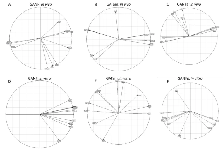
2.6.4 Statistical analysis and modelling 224
Statistical Analysis was conducted using “R Studio 3.3.1”(R Studio Team, 2015). The 225
measured values were compared among the different groups using nonparametric analysis through 226
Kruskall-Wallis and Dunn’s analysis (R package dunn.test) with a P<0.05 indicating statistical 227
significance. P-values were then corrected using false discovery rate (fdr) using the R-package 228
hmisc. To create an integrative analysis, partial least square discriminant analysis (PLS-DA) was 229
used to determine if test conditions could be discriminated from the control(Bertrand et al., 2017; 230
Cho et al., 2008). PLS-DA was selected as it has good performance when dealing with multi-231
collinear data with small samples and many variables (genes). ENMs and test condition (in vitro 232
or in vivo) were analyzed separately to identify which genes were significantly impacted by the 233
exposure conditions used. Variable Importance on Projection (VIP) was scored to identify which 234
genes were most important in group discrimination (Jaumot et al., 2015). For each CNF, both in 235
vitro and in vivo exposures were plotted together. For each test condition, a correlation circle was 236
12
plotted on the factorial plane combining the first two axes of the PLS-DA model. Vectors of the 237
correlation circle represent the variables (genes) used to generate the model. Vectors describe the 238
relationships between the genes and each of the axes. 239
To identify if production process influenced ecotoxicity, results were analyzed to 240
determine if product features could be linked to gene expression. As the CNF products were 241
similar in all aspects except for small differences due to the production process, the focus of the 242
analysis was to determine if these differences could be linked to gene expression results. Two 243
analyses of correlation were conducted to i)determine if gene expression could be correlated with 244
the ID/IG-band ratio from Raman spectroscopy and ii)determine if there is a relationship between
245
the reported purity of the CNFs. To achieve this, foreach test condition (in vitro or in vivo) results 246
were separated based on test concentration (0.01, 0.1 and 1 mg.L-1). Using these groups, Pearson’s 247
correlation coefficient was used to define their relationship between ID/IG ratio and chemical purity
248
with gene expressions. Each gene was analyzed at each test concentration to determine statistical 249 significance using at P <0.05. 250 251 3. Results 252 3.1 Physicochemical characterization 253
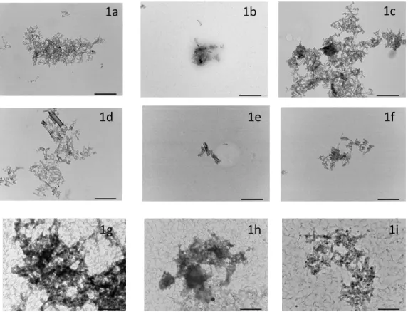
TEM results indicated that aggregation/agglomeration occurred in all media but no clear 254
differences between suspensions could be identified (Figures 1a-i). TEM results for ASW media 255
were difficult to analyze due to deposition of salt during the grid preparation. Raman spectroscopy 256
results indicated that the ratio between the Dband and Gband (ID/IG) could be used to discriminate
257
between the CNFs. GANF had a higher ID/IG-band ratio (1.26) than GATam (1.07) and
GANFg(0.78) indicating that GANF has the highest number of structural defects and GANFg has 259
the lowest number of structural defects (Figure 2a-c). 260
All suspensions could be analyzed through DLS with results being considered good quality 261
(Table 2). Stock suspensions for GANF (489.3 d-nm), GATam (414.3 d-nm) and GANFg (479.8 262
d-nm) were all comparable in the size of agglomerates. When prepared in culture media, GANF 263
(260.1 d-nm), GATam (197.8 d-nm), and GANFg (244.1 d-nm) were still comparable in e but 264
agglomerate sizes were notably smaller than the stock suspensions. This was also observed in 265
ASW where GANF (186.7 d-nm), GATam (223 d-nm) and GANFg (217.4 d-nm) agglomerates 266
were smaller in size when compared to the stock media but were similar in size to the 267
measurements obtained in the cell culture media. The size of agglomerates after 24 hours were 268
similar to the start of the experiment for all test media. 269
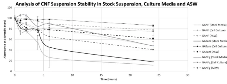
The optical density measured in the stock suspension suggested that CNFs remaining in 270
suspension were similar between GANF (11.5%) and GATam (17.62%) in the stock suspensions 271
whereas GANFg was higher (47.72%), suggesting more CNFs remained in suspension (Figure 3). 272
Optical density in cell culture media for GANF (47.88%), GATam (86.34%) and GANFg 273
(89.80%) was notably increased when compared to the stock suspension. For suspensions prepared 274
in ASW, GANF (40.67%) and GATam (77.79%) where similar in optical density to the cell culture 275
media. GANFg (40.67%) had lower optical density in ASW. 276
3.2 Gene expression Analysis 277
3.2.1 in vivo 278
14
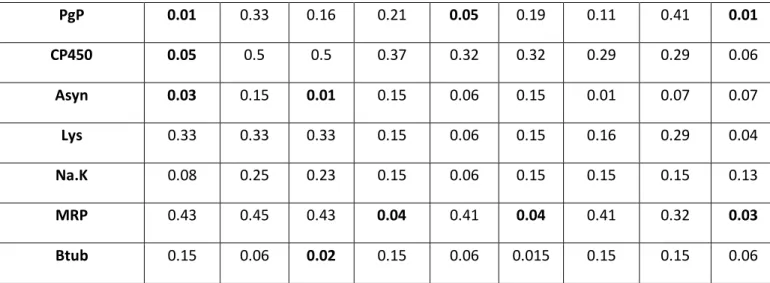
Mussels exposed to the CNFs showed limited statistical significant between the control 279
and the test concentrations for many of the genes suggesting little effects when exposed to the 280
CNFs (Table 3). Histograms associated with the results can be found in the supplemental material. 281
3.2.1.1 Oxidative stress/detoxification 282
SOD mRNA levels were not significantly impacted. Catalase gene was significantly 283
increased when exposed to GANF (0.01 and 1 mg.L-1) and GATam (0.01mg/L). . HSP70 was also 284
not significantly increased when exposed to GANF (0.01 mg.L-1). GST was significantly increased
285
by GATam (1mg.L-1). Cytochrome P450 was significantly MT was significantly increased when 286
exposed to GANFg (0.1 and 1mg.L-1). 287
3.2.1.2Cytoskeleton, Cell metabolism &Cell Cycle Control 288
B-tub was not significantly impacted. MRP was significantly decreased by GATam (0.01 and 289
1mg.L-1) and GANFg (1mg.L-1). Na/K ATPase was not significantly impacted. P53 was
290
significantly decreased by GANF (0.01 and 1mg.L-1). Lysozyme gene expression was not 291
significantly impacted. 292
3.2.2in vitro 293
Mussels exposed to the CNFs showed statistical significance with the in vitro exposure to 294
all three CNFs with more significant effects occurring than with the in vivo exposure (Table 4). 295
Histograms associated with the results can be found in the supplemental material. 296
3.2.2.1 Oxidative stress/detoxification 297
SOD mRNA levels were significantly decreased when mussels were exposed to GANF 298
(1mg.L-1), GATam (0.01 and 0.1 mg.L-1) and GANFg (0.01 and 0.1mg.L-1). Catalase gene had no 299
significant effects. HSP70 was also significantly increase for GANF (0.01 and 1mg.L-1), GATAM 300
(0.1 and 1mg.L-1) and GANFg (0.01 and 1mg.L-1). GST was significantly increased by GANF
301
(0.1 and 1mg.-1L), GATam (0.1mg.L-1) and GANFg (0.01 and 1 mg.L-1). Cytochrome P450 was 302
not statistically significant. MT was significantly increased when exposed to GANF (0.1 and 303
1mg.L-1), GATAM (1mg.L-1) and GANFg (0.01 and 1mg.L-1). 304
3.2.2.2 Cytoskeleton, Cell metabolism &Cell Cycle Control 305
B-tub was significantly decreased when exposed to GANF (0.1 and 1mg.L-1), GATam
306
(0.01 and 0.1mg.L-1) and GANFg (1mg.L-1). PgP was only significantly affected by GANF 307
(1mg.L-1). MRP was only affected by GATam (0.01mg.L-1). Na/K ATPase was only significantly 308
affected by GANF (0.1 and 1mg.L-1) and GATam (0.1 and 1 mg.L-1). P53 was significantly
309
increased by GATam (0.01mg.L-1). ATP synthase was significantly increased by GATam (1mg.L -310
1) and significant decreased by GANFg( 0.01 and 1 mg.L-1). Lysozyme gene expression was not
311
significantly impacted. 312
3.3 PLS-DA analysis 313
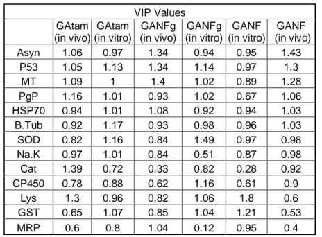
VIP values were identified for all 6 conditions and summarized in the table 5. When 314
analyzing the in vivo and in vitro exposure conditions most of the genes were identified as 315
important in describing variation between test conditions (>0.8). Of the genes used, consistently 316
high VIP values were found with ATP synthase, P53, metallothionein, lysozyme, catalase and 317
superoxide dismutase indicating these genes were essential in discriminating between test 318
concentrations. The results of the model were then plotted on a factorial plane describing the 319
relationship between genes (Figure 4). This was then used to plot the test conditions of each CNF 320
16
with their orientation dependent on which genes more effectively described the test condition 321
(Figure 5). 322
PLS-DA analysis for GANF in vivo showed that 0.01mg.L-1 and 1mg.L-1 test 323
concentrations were be discriminated from the control and showed a high degree of separation 324
(Figure 5A). 0.1 mg.L-1could be discriminated from the control but was similar in gene expression. 325
When analyzing the PLS-DA model for GANF in vitro results showed that all three test 326
concentrations could be discriminated from the control and showed a high degree of separation 327
between concentrations. 0.01 and 0.1 mg.L-1exposure times were similar in response. For both in 328
vivo and in vitro results the 1mg.L-1 test concentration was clearly separated.
329
The PLS-DA model for GATam in vivo results also showed discrimination from the control 330
at 0.01, 0.1mg.L-1 and at 1mg.L-1 (Figure 5B). This pattern was observed as well for the in vitro 331
test condition but little similarities were observed between the two test conditions. 332
The PLS-DA model GANFg in vivo had a clear separation of the test concentrations from 333
the control (Figure 5C). GANFg in vitro showed clear separation from the control for all three test 334
concentrations and 0.01mg.L-1 and 1mg.L-1exposure conditions were similar in response. 335
3.4 Correlating CNF form with gene expression 336
To determine if relationships between gene expression and differences in the CNF 337
production process could be established Pearson’s correlation coefficient and CNF structural 338
purity (as measured through Raman spectrometry) was used. Positive correlations would indicate 339
that as the ID/IG-band ratio increases, gene expression increases where as a negative correlation
340
would result in an inverse interpretation. Significant positive correlations in the relationship 341
between gene expression and CNF purity would imply that as purity increases, gene expression 342
increases. A negative correlation would suggest that as purity decreases, gene expression increases. 343
When test concentrations were controlled for strong correlations between gene expression 344
and ID/IG-band ratio could be identified (Table 6). For the in vivo exposure there were clear
345
differences with test concentration and relationship ID/IG-band ratio. At a concentration of 0.01
346
mg.L-1 HSP70, GST, SOD, ATP synthase and B-tubulin gene expressions had strong positive 347
correlations with the ID/IG-band ratio where at a dose of 0.1mg.L-1 only MRP gene had a significant
348
correlation. For mussels exposed to 1mg.L-1, metallothionein, lysozyme, PgP, ATP synthase, P53 349
and MRP gene expression levels had all positive correlations. However, mussels exposed in vitro 350
to 0.01mg.L-1,showed that HSP70, GST, metallothionein and ATP synthase mRNA levels had 351
significant negative correlations with the ID/IG-band ratio where Na/K ATPase and MRP genes
352
had significant positive correlations. At a concentration of 0.1 mg.L-1, SOD, lysozyme, PgP and
353
cytochromeP450 gene expression levels had negative correlations and GST, Na/K ATPase and 354
P53 genes had positive correlations. At a concentration of 1mg.L-1, SOD, lysozyme,
355
cytochromeP450 and ATP synthase mRNA levels had strong positive correlations. 356
When determining the relationship between gene expression and CNF chemical purity (as 357
reported in Table 1), there were some significant relationships were identified but there were less 358
correlations than when comparing gene expression to the D/G band ratio (Table 7). When looking 359
at the in vivo exposure condition, β-tubulin, Na/K ATPase and P53 genes had significant negative 360
correlations with purity of the CNF at 0.01 mg.L-1. At 0.1 mg.L-1 exposure condition, HSP70,
361
GST, SOD, β-tubulin and MRP mRNA expressions depicted significant negative correlations with 362
purity whereas catalase gene had a significant positive relationship. At 1mg.L-1, only MRP showed
363
a significant negative correlation to the purity. For the in vitro exposure condition, PgP, 364
18
cytochromeP450 and Na/K ATPase mRNA levels had negative correlations whereas GST and P53 365
genes had positive correlations. At 1mg.L-1, HSP70 and Na/K ATPase expressions had significant
366
negative relationships whereas GST and PgP mRNA levels had significant positive relationships 367 with purity of CNF. 368 369 4. Discussion 370
The aim of the study was to investigate whether or not differences in the production process 371
of CNFs would alter gene expression profiles as well as to establish an in vitro/in vivo comparison. 372
In this sense, the first objective was to establish if the three CNFs have similar physicochemical 373
and to determine differences between in vitro and in vivo stability and hydrodynamic diameters. 374
4.1 Physico-chemical characterization 375
To maintain a relevant comparison between the 3 CNFs, the physico-chemical properties 376
of the CNFs in suspensions need to be accounted for. In this sense, DLS is a commonly used 377
technique for measuring ENMs in suspension but it is limited in the sense that it assumes a 378
spherical shape for the particles. In the context of CNFs this does not hold true for the single fibers, 379
but agglomerates can be approximated as ovoid and the size of the particles can be approximated 380
(Reinert et al., 2015).The DLS results suggest that particle sizes are comparable between CNFs as 381
well as between in vitro and in vivo assays. In addition to this, the optical density results suggest 382
that the test suspensions are somewhat stable with slight differences between in vitro and in vitro 383
test media for GATam and GANF. As a result, the CNFs used in the study can be considered to 384
have similar physico-chemical properties between the two testing strategies, facilitating an in 385
vitro/in vivo comparison. It is however important note that differences between the media may 386
lead to differences in sedimentation and effectively change the dosimetry. 387
Another interesting observation is that the CNFs displayed poor stability in ultra-pure 388
water whereas in both culture media and ASW the stability was improved. One potential 389
explanation for this observation is that the ionic strength of these suspensions decreased the surface 390
potential of the CNFs, reducing rate at which particles interact with other another (Pavlin and 391
Bregar, 2012). This is interesting as it would imply that the behavior of the CNFs in the ocean will 392
be different than the behavior in fresh water ecosystem, which can lead to distinct differences in 393
toxicity profiles. 394
Of the methods used to characterize the CNFs in the present study, only Raman 395
spectroscopy demonstrated clear differences between the CNFs. The comparison of the Raman 396
spectra of the 3 samples illustrates the influence of the annealing treatment with a clear 397
improvement of the ID/IG intensity ratio, decreasing from 1.3 (Fig. 2(a)) to 0.8 (Fig. 2(c)) and
398
evidencing a significant improvement of the structure of the carbon network, also visible with the 399
increased intensity of the 2D band (2650 cm-1). Sample GATam (Fig. 2(b)) lies in between with 400
an intermediate value of the ID/IG intensity ratio of 1.1 but a noisier spectrum, although all data
401
were acquired exactly in the same experimental conditions. All 3 samples exhibited some 402
fluorescence leading to an important background signal. For this reason, irradiation of the samples 403
started 30s before acquisition to limit this phenomenon. It is important to note that Raman 404
spectroscopy does not allow a direct measurement of the chemical purity of the samples but more 405
of the level of structural defects (Ivanova et al., 2012). In the context of SbD and the case study 406
this is important because the structural quality of the CNFs may impact the performance of the 407
products and GATam may be a more desirable product than GANF as a result. In the context of 408
20
ecotoxicological hazards structural defects can lead to the functionalization of CNFs, providing 409
favorable binding sites for ions or molecules, which can lead to differences in hazard profiles. 410
4.2 Gene expression 411
The endpoints used in the present study provides broad spectrum response analysis of 412
hemocyte gene expression to CNF exposure, which allows for characterization of mechanisms of 413
action. To the authors’ best knowledge, this is the first time that carbon nanofibers have been 414
investigated for ecotoxicological effects on mussels. As a result, there are limited examples that 415
could be considered a suitable comparison. Of the information available mussel hemocytes, 416
exposed in vitro and in vivo for 24 hours, have been shown to display sublethal responses to carbon 417
black and fullerene (0.01-1mg.L-1) with little to no effects when exposed to a carbon nanotube
418
(0.01-1mg.L-1) (Canesi et al., 2010, 2008b; Moore et al., 2009). Previous studies in human 419
toxicology have shown induction of oxidative stress and inflammation when cells, IL-8, A549 and 420
HaCaT, were exposed to multi-walled carbon nanotubes at 1mg/L and 25mg.L-1for 24 hours 421
(Vitkina et al., 2016; Ye et al., 2009).When analyzing the in vivo responses, the gene expression 422
had little significant effects (which may be attributed to the small sample sizes) with all three CNFs 423
and little clear pattern is evident with the CNFs. There is a however a notable trend for GATam to 424
have higher fold expressions which could suggest more adverse effects may occur with this CNF. 425
When looking at the in vitro exposure however, hemocytes showed more significant effects 426
when compared to the in vivo exposure for all CNFs with many of the gene response associated 427
with oxidative and xenobiotic stress. This can be attributed to the simpler exposure scenario for 428
the in vitro exposure as the cells are directly exposed to the CNFs. When looking at the results for 429
approximated stability, GANF showed the least stability of the three CNFs in culture media. This 430
observation could suggest that due to the a sedimentation effect the effective dosage for GANF is 431
higher than the other two CNFs (Deloid et al., 2017; Hinderliter et al., 2010). This could also 432
suggest that these CNFs are aggregating at the bottom of the well and not interacting with the cells, 433
which can complicate the comparison between the three CNFs. Looking at the gene expression 434
holistically more significant effects occur with GANFg at 0.01mg.L-1 which could suggest this 435
product is more hazardous. The results of the in vitro approach may suggest that all three CNFs 436
could potentially cause oxidative and xenobiotic stress, which is not in agreement with the in vivo 437
approach. It is also important to note that the cell culture is a complex media consisting of a mixture 438
of proteins, which can lead to protein corona that does not occur naturally. As a result, further 439
analysis of dosimetry may be necessary when applying an in vitro approach for SbD in order to 440
demonstrate it is an accurate predictor of in vivo effects. 441
4.3 PLS-DA analysis 442
The integrative analysis through PLS-DA showed some unexpected results in that almost 443
all of the genes were important in the discrimination between test concentrations for all the CNFs 444
and for both in vivo and in vitro test conditions, highlighting its sensitivity in identifying sublethal 445
effects. Of these genes however, ATP synthase, P53, metallothionein, PgP and HSP70 were 446
identified as the most important discriminators for almost all test conditions. This suggests that 447
exposure to the CNFs is influencing a wide array of cellular functions (oxidative stress, 448
detoxification, cytoskeleton, cellular metabolism and cell cycle control) which can alter cell 449
functioning and potentially cascade into higher levels of biological organization. When comparing 450
the VIP values in vivo to in vitro it became apparent that in general there were some similarities in 451
the values between the two testing conditions. However, when analyzing the correlation circles the 452
relationship between genes was not consistent between the in vitro and in vivo exposures. The in 453
22
vivo/in vitro relationship is a challenge to many investigators as gene responses are often not 454
consistent between the two testing strategies (Heise et al., 2012). 455
4.4 Correlation CNF properties to Gene expression 456
In the context of applying ecotoxicology to a SbD for MNMs, there needs to be an 457
identification of what features of MNMs cause toxicity. The CNFs used in the present study 458
provide an ideal case study for the application of SbD as the products are very similar which allow 459
for an analysis on how the differences in production influence ecotoxicity. In this sense, structural 460
purity, measured through the ID/IG-band ratio, and overall purity were identified as potential
461
discriminators between the CNFs. Of these two measurements, the relationship between the ID/IG
-462
band ratio proved to be more strongly correlated with gene expression than chemical purity of 463
CNF. In vivo, all the genes showed a positive correlation with the ID/IG-band ratio indicating that
464
gene expression increased with structural impurities. With the in vitro results however, the inverse 465
is observed in the fact that gene expression was more strongly correlated with a decrease in the 466
ID/IG-band ratio. A previous study investigating the physicochemical relationship between toxicity
467
and MWNCT was able to demonstrate that the presence of proteins profoundly affects their 468
behavior in media and could alter their toxicity as a result, which may explain the difference in 469
interpretation(Allegri et al., 2016). The relationship between test media properties and CNFs 470
behavior requires further investigation, in particular the role of a protein corona in gene expression. 471
This does however provide insight that could be useful in linking ecotoxicity results in a way that 472
can promote SbD strategies. 473
474
5. Conclusion 475
Transcriptomic is a promising emerging tool to implement ecotoxicology into a SbD 476
approach for MNMs. The results in the present study demonstrate a way in which gene expression 477
can be used to provide information to improve the safety of MNM products. In the context of the 478
current study the in vitro and in vivo tests were not consistent in their interpretation of which CNF 479
was “least safe”, suggesting that in vitro and in vivo strategies need to be investigated in parallel 480
to verify that an in vitro approach using M. edulis hemocytes is a suitable alternative testing 481
strategy. It is however important to note that while in vitro/in vivo extrapolation remains a 482
challenge requiring additional research, the application of ecotoxicology in SbD may require the 483
use of in vitro testing to adequately screen products produced by industry in a cost effective and 484
timely manner. 485
486
Acknowledgements: The research contained within this publication was funded by the European 487
Union’s Horizon 2020 research and innovation program NANoREG2 under grant agreement 488
646221. 489
"The sole responsibility of this publication lies with the author. The European Union is not 490
responsible for any use that may be made of the information contained therein." 491 492 493 494 495 496
24 497 498 499 500 501 References 502
Allegri, M., Perivoliotis, D.K., Bianchi, M.G., Chiu, M., Pagliaro, A., Koklioti, M.A., Trompeta, 503
A.F.A., Bergamaschi, E., Bussolati, O., Charitidis, C.A., 2016. Toxicity determinants of 504
multi-walled carbon nanotubes: The relationship between functionalization and 505
agglomeration. Toxicol. Reports 3, 230–243. https://doi.org/10.1016/j.toxrep.2016.01.011 506
Barrick, A., Châtel, A., Bruneau, M., Mouneyrac, C., 2017. The role of high-throughput 507
screening in ecotoxicology and engineered nanomaterials. Environ. Toxicol. Chem. 9999, 508
1–11. https://doi.org/10.1002/etc.3811 509
Barrick, A., Guillet, C., Mouneyrac, C., Châtel, A., 2018. Investigating the establishment of 510
primary cultures of hemocytes from Mytilus edulis. Cytotechnology. 511
https://doi.org/10.1007/s10616-018-0212-x 512
Bertrand, C., Devin, S., Mouneyrac, C., Giambérini, L., 2017. Eco-physiological responses to 513
salinity changes across the freshwater-marine continuum on two euryhaline bivalves: 514
Corbicula fluminea and Scrobicularia plana. Ecol. Indic. 74, 334–342. 515
https://doi.org/10.1016/j.ecolind.2016.11.029 516
Canesi, Ciacci, C., Vallotto, D., Gallo, G., Marcomini, A., Pojana, G., 2010. In vitro effects of 517
suspensions of selected nanoparticles (C60 fullerene, TiO2, SiO2) on Mytilus hemocytes. 518
Aquat. Toxicol. 96, 151–158. https://doi.org/10.1016/j.aquatox.2009.10.017 519
Canesi, L., Borghi, C., Ciacci, C., Fabbri, R., Lorusso, L.C., Vergani, L., Marcomini, A., Poiana, 520
G., 2008a. Short-term effects of environmentally relevant concentrations of EDC mixtures 521
on Mytilus galloprovincialis digestive gland. Aquat. Toxicol. 87, 272–279. 522
https://doi.org/10.1016/j.aquatox.2008.02.007 523
Canesi, L., Ciacci, C., Betti, M., Fabbri, R., Canonico, B., Fantinati, A., Marcomini, A., Pojana, 524
G., 2008b. Immunotoxicity of carbon black nanoparticles to blue mussel hemocytes. 525
Environ. Int. 34, 1114–1119. https://doi.org/10.1016/j.envint.2008.04.002 526
Cao, A., Mercado, L., Ramos-Martinez, J.I., Barcia, R., 2003. Primary cultures of hemocytes 527
from Mytilus galloprovincialis Lmk.: Expression of IL-2Rα subunit. Aquaculture 216, 1–8. 528
https://doi.org/10.1016/S0044-8486(02)00140-0 529
Châtel, A., Lièvre, C., Barrick, A., Bruneau, M., Mouneyrac, C., 2018. Transcriptomic approach: 530
A promising tool for rapid screening nanomaterial-mediated toxicity in the marine bivalve 531
Mytilus edulis —Application to copper oxide nanoparticles. Comp. Biochem. Physiol. Part 532
C Toxicol. Pharmacol. 205, 26–33. https://doi.org/10.1016/j.cbpc.2018.01.003 533
Chevé, J., Bernard, G., Passelergue, S., Prigent, J.-L., 2014. Suivi bactériologique des gisements 534
naturels de coquillages de l’Ille-et-Vilaine et des Côtes- d’Armor fréquentés en pêche à pied 535
1–99. 536
Cho, H.-W., Kim, S.B., Jeong, M.K., Park, Y., Miller, N.G., Ziegler, T.R., Jones, D.P., 2008. 537
26
Discovery of metabolite features for the modelling and analysis of high-resolution NMR 538
spectra. Int. J. Data Min. Bioinform. 2, 176–92. 539
Deloid, G.M., Cohen, J.M., Pyrgiotakis, G., Demokritou, P., 2017. Preparation, characterization, 540
and in vitro dosimetry of dispersed, engineered nanomaterials. Nat. Protoc. 12, 355–371. 541
https://doi.org/10.1038/nprot.2016.172 542
Dubochet, J., Groom, M., Mueller-Neuteboom, S., 1982. The Mounting of Macromolecules for 543
Electron Microscopy with Particular Reference to Surface Phenomena and the Treatment of 544
Support Films by Glow Discharge. Adv. Opt. Electron Microsc. 8, 107–135. 545
Fenwick, N., Griffin, G., Gauthier, C., 2009. The welfare of animals used in science: How the 546
“Three Rs” ethic guides improvements. Can. Vet. J. 50, 523–30. 547
Gagné, F., Auclair, J., Turcotte, P., Fournier, M., Gagnon, C., Sauvé, S., Blaise, C., 2008. 548
Ecotoxicity of CdTe quantum dots to freshwater mussels: Impacts on immune system, 549
oxidative stress and genotoxicity. Aquat. Toxicol. 86, 333–340. 550
https://doi.org/10.1016/j.aquatox.2007.11.013 551
Heise, T., Schug, M., Storm, D., Ellinger-Ziegelbauer, H., J. Ahr, H., Hellwig, B., Rahnenführer, 552
J., Ghallab, A., Guenther, G., Sisnaiske, J., Reif, R., Godoy, P., Mielke, H., Gundert-Remy, 553
U., Lampen, A., Oberemm, A., G. Hengstler, J., 2012. In Vitro - In Vivo Correlation of 554
Gene Expression Alterations Induced by Liver Carcinogens. Curr. Med. Chem. 19, 1721– 555
1730. https://doi.org/10.2174/092986712799945049 556
Hinderliter, P.M., Minard, K.R., Orr, G., Chrisler, W.B., Thrall, B.D., Pounds, J.G., Teeguarden, 557
J.G., 2010. ISDD: A computational model of particle sedimentation, diffusion and target 558
cell dosimetry for in vitro toxicity studies. Part. Fibre Toxicol. 7, 36. 559
https://doi.org/10.1186/1743-8977-7-36 560
Ivanova, M. V., Lamprecht, C., Jimena Loureiro, M., Torin Huzil, J., Foldvari, M., 2012. 561
Pharmaceutical characterization of solid and dispersed carbon nanotubes as nanoexcipients. 562
Int. J. Nanomedicine 7, 403–415. https://doi.org/10.2147/IJN.S27442 563
Jaumot, J., Navarro, A., Faria, M., Barata, C., Tauler, R., Piña, B., 2015. qRT-PCR evaluation of 564
the transcriptional response of zebra mussel to heavy metals. BMC Genomics 16. 565
https://doi.org/10.1186/s12864-015-1567-4 566
Jensen, K.A., Kembouche, Y., Christiansen, E., E., J., N.R., W., Giot, C., Spalla, O., Witschger, 567
O., 2011. Final protocol for producing suitable manufactured nanomaterial exposure media. 568
NANoREG A common Eur. approach to Regul. Test. Nanomater. Web-Report. 569
Katsumiti, A., Berhanu, D., Howard, K.T., Arostegui, I., Oron, M., Reip, P., Valsami-Jones, E., 570
Cajaraville, M.P., 2014. Cytotoxicity of TiO2 nanoparticles to mussel hemocytes and gill 571
cells in vitro: Influence of synthesis method, crystalline structure, size and additive. 572
Nanotoxicology 5390, 1–11. https://doi.org/10.3109/17435390.2014.952362 573
Kraegeloh, A., Suarez-merino, B., Sluijters, T., Micheletti, C., 2018. Implementation of Safe-by-574
Design for Nanomaterial Development and Safe Innovation : Why We Need a 575
Comprehensive Approach. https://doi.org/10.3390/nano8040239 576
Matranga, V., Corsi, I., 2012. Toxic effects of engineered nanoparticles in the marine 577
environment: Model organisms and molecular approaches. Mar. Environ. Res. 76, 32–40. 578
https://doi.org/10.1016/j.marenvres.2012.01.006 579
28
Moore, M.N., 2006. Do nanoparticles present ecotoxicological risks for the health of the aquatic 580
environment? Environ. Int. 32, 967–976. https://doi.org/10.1016/j.envint.2006.06.014 581
Moore, M.N., Readman, J.A.J., Readman, J.W., Lowe, D.M., Frickers, P.E., Beesley, A., 2009. 582
Lysosomal cytotoxicity of carbon nanoparticles in cells of the molluscan immune system: 583
An in vitro study. Nanotoxicology 3, 40–45. https://doi.org/10.1080/17435390802593057 584
Pavlin, M., Bregar, V.B., 2012. Stability of nanoparticle suspensions in different biologically 585
relevant media. Dig. J. Nanomater. Biostructures 7, 1389–1400. 586
R Studio Team, 2015. RStudio: Integrated Development for R. 587
Reinert, L., Zeiger, M., Suárez, S., Presser, V., Mücklich, F., 2015. Dispersion analysis of carbon 588
nanotubes, carbon onions, and nanodiamonds for their application as reinforcement phase in 589
nickel metal matrix composites. RSC Adv. 5, 95149–95159. 590
https://doi.org/10.1039/C5RA14310A 591
Revel, M., Châtel, A., Mouneyrac, C., 2017. Omics tools: New challenges in aquatic 592
nanotoxicology? Aquat. Toxicol. 193, 72–85. https://doi.org/10.1016/j.aquatox.2017.10.005 593
Schwarz-plaschg, C., Kallhoff, A., Eisenberger, I., 2017. Making Nanomaterials Safer by 594
Design ? 277–281. 595
Snape, J.R., Maund, S.J., Pickford, D.B., Hutchinson, T.H., 2004. Ecotoxicogenomics: The 596
challenge of integrating genomics into aquatic and terrestrial ecotoxicology. Aquat. 597
Toxicol. 67, 143–154. https://doi.org/10.1016/j.aquatox.2003.11.011 598
Vitkina, T.I., Yankova, V.I., Gvozdenko, T.A., Kuznetsov, V.L., Krasnikov, D. V., Nazarenko, 599
A. V., Chaika, V. V., Smagin, S. V., Tsatsakis, A.M., Engin, A.B., Karakitsios, S.P., 600
Sarigiannis, D.A., Golokhvast, K.S., 2016. The impact of multi-walled carbon nanotubes 601
with different amount of metallic impurities on immunometabolic parameters in healthy 602
volunteers. Food Chem. Toxicol. 87, 138–147. https://doi.org/10.1016/j.fct.2015.11.023 603
Ye, S.F., Wu, Y.H., Hou, Z.Q., Zhang, Q.Q., 2009. ROS and NF-κB are involved in upregulation 604
of IL-8 in A549 cells exposed to multi-walled carbon nanotubes. Biochem. Biophys. Res. 605
Commun. 379, 643–648. https://doi.org/10.1016/j.bbrc.2008.12.137 606
607 608
Measured property Unit GANF GANFg GATam Fiber diameter (TEM) nm 20-80 20-80 20-80 Carbon purity (TGA) % >85 >99 >80 Apparent density g/cc ~0.06 ~0.08 ~0.08 Specific surface area (BET N2) m2/g
100-170 70-90 70-140 Graphitization degree (XRD) % ≈70 ≈90 ≈60 Electrical resistivity Ω-*m
1*10
-3 1*10-4 1*10-3Table 1: Physico-chemical properties reported by the industrial partner
Table 2: DLS results measured in stock suspension, cell culture media and ASW at the start of the experiment and at the end. Zeta potential was only able to measured in the stock suspensions due to
the high ionic strengths in the test media.
Statistical Significance in gene expression: in vivo
GANF GATam GANFg
0.01 mg/L 0.1 mg/L 1 mg/L 0.01 mg/L 0.1 mg/L 1 mg/L 0.01 mg/L 0.1 mg/L 1 mg/L Cat 0.01 0.09 0.05 0.04 0.37 0.5 0.15 0.15 0.15 HSP70 0.13 0.13 0.5 0.06 0.15 0.15 0.15 0.15 0.06 GST 0.29 0.29 0.29 0.19 0.46 0.03 0.06 0.13 0.13 SOD 0.11 0.11 0.45 0.13 0.15 0.13 0.33 0.23 0.08 MT 0.37 0.37 0.06 0.15 0.06 0.15 0.01 0.03 0.1541 P53 0.15 0.15 0.06 0.15 0.06 0.15 0.01 0.15 0.01
PgP 0.01 0.33 0.16 0.21 0.05 0.19 0.11 0.41 0.01 CP450 0.05 0.5 0.5 0.37 0.32 0.32 0.29 0.29 0.06 Asyn 0.03 0.15 0.01 0.15 0.06 0.15 0.01 0.07 0.07 Lys 0.33 0.33 0.33 0.15 0.06 0.15 0.16 0.29 0.04 Na.K 0.08 0.25 0.23 0.15 0.06 0.15 0.15 0.15 0.13 MRP 0.43 0.45 0.43 0.04 0.41 0.04 0.41 0.32 0.03 Btub 0.15 0.06 0.02 0.15 0.06 0.015 0.15 0.15 0.06
Table 3: P-values, measured through Kruskall-Wallis and Dunn, of M. edulis hemocytes exposed in vivo. Stars indicate p-values <0.05 with + indicating a up regulation and – indicating down regulation.
Statistical Significant in gene expression: in vitro
GANF GATam GANFg
mg/L 0.1 mg/L 1 mg/L 0.01 mg/L 0.1 mg/L 1 mg/L 0.01 mg/L 0.1 mg/L 1 mg/L 0.01 Cat +0.37 +0.37 +0.37 0.27 0.323 0.17 0.21 0.21 0.21 HSP70 -0.03* -0.15 -0.03* 0.15 0.03* 0.01* 0.01* 0.13 0.01* GST -0.15 -0.03 -0.01* 0.15 0.01* 0.10 0.01* 0.15 0.01* SOD -0.09 -0.07 -0.01* 0.01* 0.02* 0.15 0.01* 0.01* 0.15 MT -0.15 -0.03* -0.01* 0.06 0.06 0.01* 0.04* 0.13 0.01* Lys 0.06 0.15 0.15 0.16 0.29 0.04 0.15 0.15 0.08 PgP +0.33 +0.33 +0.03* 0.21 0.08 0.19 0.06 0.15 0.15 CP450 -0.33 +0.16 -0.16 0.11 0.11 0.25 0.29 0.19 0.19 Asyn +0.13 +0.13 +0.13 0.09 0.21 0.01* 0.01* 0.15 0.03* Btub -0.11 -0.05* -0.01* 0.01* 0.02* 0.15 0.07 0.07 0.01* Na.K -0.15 -0.03* -0.01* 0.15 0.02* 0.01* 0.15 0.15 0.08 P53 +0.13 +0.13 -0.15 0.01 0.07 0.37 0.06 0.15 0.15
MRP +0.33 -0.16 +0.16 0.33 0.16 0.16 0.04* 0.16 0.29 Table 4: P-values, measured through Kruskall-Wallis and Dunn, of M. edulis hemocytes exposed in vitro.
Stars indicate p-values <0.05 with + indicating a up regulation and – indicating down regulation.
VIP Values GAtam (in vivo) GAtam (in vitro) GANFg (in vivo) GANFg (in vitro) GANF (in vitro) GANF (in vivo) Asyn 1.06 0.97 1.34 0.94 0.95 1.43 P53 1.05 1.13 1.34 1.14 0.97 1.3 MT 1.09 1 1.4 1.02 0.89 1.28 PgP 1.16 1.01 0.93 1.02 0.67 1.06 HSP70 0.94 1.01 1.08 0.92 0.94 1.03 B.Tub 0.92 1.17 0.93 0.98 0.96 1.03 SOD 0.82 1.16 0.84 1.49 0.97 0.98 Na.K 0.97 1.01 0.84 0.51 0.87 0.98 Cat 1.39 0.72 0.33 0.82 0.28 0.92 CP450 0.78 0.88 0.62 1.16 0.61 0.9 Lys 1.3 0.96 0.82 1.06 1.8 0.6 GST 0.65 1.07 0.85 1.04 1.21 0.53 MRP 0.6 0.8 1.04 0.12 0.95 0.4
Table 5: VIP values of genes expression for each exposure condition to CNFs. VIP values below 0.8 were removed from the analysis.
Correlation with ID/IG-band ratio
0.01 mg/L 0.1 mg/L 1 mg/L
in vitro in vivo in vitro in vivo in vitro in vivo
Cat 0.52 0.06 -0.16 -0.44 0.56 0.51 HSP70 -0.93* 0.84* -0.3 0.26 0.32 0.23 GST -0.91* 0.69* 0.7* 0.59 -0.22 0.49 SOD -0.55 0.85* -0.74* 0.55 0.79* 0.57 MT -0.88* 0.54 -0.07 0.42 0.48 0.77* Lys -0.07 0.30 -0.92* 0.12 0.83* 0.86* PgP 0.69 -0.36 -0.93* 0.32 -0.29 0.94* CP450 0.46 -0.39 -0.94* 0.3 0.84* 0.66 Asyn -0.97* 0.87* -0.29 0.29 0.78* 0.89* Btub -0.62 1* -0.65 0.21 -0.18 0.51
Nak 0.94* 0.53 0.9* 0.19 0.32 0.39 P53 -0.34 0.15 0.96* 0.26 0.34 0.89* MRP 0.75* 0.5 0.06 0.82* 0.45 0.97* Table 6: Table showing the correlation between the D/G-band ratio, measured through Raman
spectroscopy, and gene expression. *Indicates statistical significant (P<0.05) with + indicating a positive relationship with purity and – indicating a negative relationship with purity.
Correlation with Reported CNF Purity
0.01 mg/L 0.1 mg/L 1 mg/L
in vitro in vivo in vitro in vivo in vitro in vivo
Cat -0.66 0.28 0.47 0.75* -0.2 -0.58 HSP70 0.57 -0.33 -0.35 -0.79* -0.76* 0.39 GST 0.74* -0.23 -0.92* -0.65 0.70* 0.13 SOD -0.02 -0.35 0.23 -0.92* -0.21 0.02 MT 0.67 -0.34 -0.12 0.22 0.1 -0.26 Lys -0.52 -0.37 0.64 0.5 -0.33 -0.38 PgP -0.89* -0.27 0.58 0.32 0.8* -0.54 CP450 -0.73* -0.03 0.68* 0.16 -0.54 -0.43 Asyn 0.64 -0.39 -0.30 0.36 -0.3 -0.43 Btub 0.03 -0.83* 0.06 -0.76* 0.64 0.11 Nak -0.85* -0.93* -0.84* -0.74* -0.69* 0.23 P53 0.82* -0.69* -0.64 0.39 0.05 -0.5 MRP -0.84* -0.11 -0.57 -0.77* 0.04 -0.78*
Table 7: Table showing the correlation between carbon nanofiber purity and gene expression. *indicate statistical significant (P<0.05) with + indicating a positive relationship with purity and – indicating a negative relationship with purity.
Figure 1a-i: TEM images of GANF (1a), GATam (1b) and GANFg (1c) prepared in stock suspensions compared with GANF (1d), GATam (1e) and GANFg (1f) in culture media and GANF (1g), GATam (1h) and
GANFg (1i) prepared in ASW. Black bars indicate 1µm.
Figure 2a-c: Raman spectra of carbon nanofibers are expressed as a function of intensity in relation to the raman shift with peaks representing the D-band and G-band. To measure the structural purity of
GANF (A), GATam (B) and GANFg (C) the ratio of intensity between the D-band and G-band (ID/IG) is used.
Figure 3: Optical density (340nm) as an approximation of suspension stability of the CNFs in the stock suspensions, cell culture media and in ASW over 24 hours.
Figure 4: PLS-DA correlation for all test conditions in vivo for GANF (A), GATam (B) and GANFg (C) as well as in vitro for GANF (D), GATam (E) and GANFg (F) plotted on a factorial plane. Orientation of the vectors describes how strongly each gene describes the axes as well as the relationship between genes.
Figure 5: PLS-DA analysis of M. edulis hemocytes exposed in vitro and in vivo to GANF (A), GAtam (B) and GANFg (C) at 0.01, 0.1 and 1mg.L-1.

