OATAO is an open access repository that collects the work of Toulouse
researchers and makes it freely available over the web where possible
Any correspondence concerning this service should be sent
to the repository administrator:
[email protected]
This is an author’s version published in:
http://oatao.univ-toulouse.fr/20620
To cite this version:
Banoub, Joseph H. and Delmas, Guo-Hua and Joly, Nicolas and
Mackenzie, Grahame and Cachet, Nadja and Benjelloun-Mlayah,
Bouchra and Delmas, Michel
A critique on the structural analysis of
lignins and application of novel tandem mass spectrometric strategies
to determine lignin sequencing. (2015) Journal of Mass Spectrometry,
50 (1). 5-48. ISSN 1076-5174
Introduction
The plant cell wall (CW) of all vascular plants and woods is com-posed of cellulose, hemicelluloses and lignins, which are formed in large quantities by means of solar energy. The CW structure is composed of primary and secondary CW parts. Both parts are com-posed of cellulose microfibrils (9–25%) and an interpenetrating ma-trix of hemicelluloses (25–50%), pectins (10–35%) and proteins (10%).[1]Cellulose is the main constituent of all skeletal constituents in plants[2]and forms the framework of the CWs, whereas hemicel-luloses cross-link non-cellulosic and cellulosic polymers.[3,4] Cellu-lose is composed of approximately 8 × 10³ D-glucopyranose residues linked byβ-D-(1→ 4)-glycosidic bonds.[5]Hydrogen bonds hold about 40 of these glycan chains together to form a cellulose microfibril. The cellulose microfibril arrangements in the primary wall are random. Cellulose microfibrils are linked to hemicellulosic polysaccharides that are mainly composed of xylans, mannans, galactans or combinations thereof, which are attached to the cellu-lose fibres by hydrogen and covalent bonding.[3,5]
In general, secondary walls are derived from the primary walls by thickening and inclusion of lignin into the CW matrices. The
* Correspondence to: Joseph Banoub, Department of Chemistry, Memorial University of Newfoundland, St John’s, Newfoundland, A1C 5X1, Canada. E-mail: [email protected]
* Correspondence to: Michel Delmas, Compagnie Industrielle de la Matière Végétale (CIMV), 109 Rue Jean Bart– Diapason A, 31670 Labège, France. E-mail: [email protected]
a Department of Chemistry, Memorial University of Newfoundland, St John’s, Newfoundland, A1C 5X1, Canada
b Science Branch, Special Projects, Fisheries and Oceans Canada, St John’s, NL, A1C 5X1, Canada
c Compagnie Industrielle de la Matière Végétale (CIMV), 109 Rue Jean Bart – Diapason A, 31670, Labège, France
d Centre Technologique des Agroressources, IUT de Béthune, Université d’Artois, 1230 rue de l’Université, BP 819, 62408, Béthune Cedex, France
e Department of Chemistry, University of Hull, Hull HU6 7RX, UK
f Inp-Ensiacet, Laboratoire de Génie Chimique, Université de Toulouse, 4 allée Emile Monso, 31432, Toulouse Cedex 4, France
DOI 10.1002/jms.3541
A
critique on the structural analysis of lignins
and
application of novel tandem mass
spectrometric
strategies to determine
lignin
sequencing
Joseph
Banoub,
a,b*
Guo-Hua Delmas Jr.,
cNicolas
Joly,
dGrahame
Mackenzie,
eNadja
Cachet,
cBouchra
Benjelloun-Mlayah
cand
Michel Delmas
c,f*
This review is devoted to the application of MS using soft ionization methods with a special emphasis on electrospray ionization, atmospheric pressure photoionization and matrix-assisted laser desorption/ionization MS and tandem MS (MS/MS) for the eluci-dation of the chemical structure of native and modified lignins. We describe and critically evaluate how these soft ionization methods have contributed to the present-day knowledge of the structure of lignins.
Herein, we will introduce new nomenclature concerning the chemical state of lignins, namely, virgin released lignins (VRLs) and processed modified lignins (PML). VRLs are obtained by liberation of lignins through degradation of vegetable matter by either chemical hydrolysis and/or enzymatic hydrolysis. PMLs are produced by subjecting the VRL to a series of further chemical trans-formations and purifications that are likely to alter their original chemical structures.
We are proposing that native lignin polymers, present in the lignocellulosic biomass, are not made of macromolecules linked to cellulose fibres as has been frequently reported. Instead, we propose that the lignins are composed of vast series of linear related oligomers, having different lengths that are covalently linked in a criss-cross pattern to cellulose and hemicellulose fibres forming the network of vegetal matter. Consequently, structural elucidation of VRLs, which presumably have not been purified and proc-essed by any other type of additional chemical treatment and purification, may reflect the structure of the native lignin.
In this review, we present an introduction to a MS/MS top–down concept of lignin sequencing and how this technique may be used to address the challenge of characterizing the structure of VRLs.
Finally, we offer the case that although lignins have been reported to have very high or high molecular weights, they might not exist on the basis that such polymers have never been identified by the mild ionizing techniques used in modern MS.
secondary plant CW contains hemicelluloses (10–40%) that are em-bedded in lignin (25%). Historically, this type of arrangement has been compared with steel rods embedded in concrete to form prestressed concrete.[6]Cellulose and hemicelluloses appear to be more structurally organized in the secondary CW than in that of the primary CW. Lignin occurs widely in the middle lamellae and secondary CWs of higher plants and plays a key role in constructive tissues as a building material, giving it its strength and rigidity and resistance to environmental factors.[1]The diverse and complex na-ture of lignin monomers and hemicellulosic moieties in lignohemicellulosic complexes make stereotypical conceptualiza-tion of secondary wall structures for all plants extremely difficult.[3,5]
History of lignin
Lignin was discovered by Anselme Payen (1839) and was first de-scribed as the encrusting material in wood. Researchers were puzzled by the nature of this very abundant material.[7]Although lignin was found to have higher carbon content than carbohy-drates, the chemical nature remained obscure for a long time. Accordingly, for almost the last century, the structure of lignin was described as a complex polymer composed of irregular branched units. After cellulose, lignin is the second most abun-dant biopolymer on Earth. It is found in all vascular plants, mostly between the cells, as well as within the cells and in the CWs. The term lignin is derived from the Latin word for wood: lignum. The polyphenolic structure of lignin is well known for its role in woody biomass to give resistance to biological and chemical degradation. This is due to their hydrophobic nature and insolu-bility in aqueous systems preventing access of degrading chemicals and organisms.
It is well accepted in our era that lignin is a very promising mas-sive renewable organic resource. The worldwide annual production of lignin, as a side product of wood processing industries, exceeds 50 million tons.[8]
Lignins in the CWs of vascular plants are intimately mixed with the carbohydrate components and form a complex glycolignin net-work. In recent years, it was shown that the chemical structure of the various lignin biomolecules depends on the botanical origin and chemical composition of the vegetal fibres.[9]It has also been proposed that the several different chemical, enzymatic and me-chanical extraction methods are accountable for the major struc-tural divergence that occurs after extraction and isolation. As a result, it appears that the only logic step to determine the natural structure of lignin is to isolate it from the vegetal matrix, without causing any structural change.[10]Therefore, preparing pure sam-ples of unchanged lignin is not an easy endeavour with the conse-quence that structural determination of lignin is perhaps more challenging than with other biopolymers.[11–13]
Proposed structure of lignin
During the last century, it was proposed that lignin existed as a cross-linked amorphous macromolecular material composed of phenylpropanoid monomers known as lignols. Lignin is relatively hydrophobic and aromatic in nature and consists of several types of substructures, which possess the same phenylpropanoid skele-ton, but differ in the degree of oxygen substitution on the phenyl ring. The monolignol H-structure consists of a 4-hydroxyphenyl ring. The monolignol guaiacyl G-structure contains one hydroxyl and one methoxyl group, whereas the monolignol syringyl
S-structure contains two methoxyl groups and one hydroxyl group (Fig. 1).[8]
The H, G and S constituent units are linked covalently, forming ether, ester and carbon–carbon bonds, which repeat in an apparently random manner and provide great complexity.[14]In general, the site of attachment of the covalent linkages can vary between two lignols. It is generally accepted that lignins are composed of H, G and S units attached byβ–O–4′, β–5′, β–β′, β–1′, 5–5′ and 5–O–4′ linkages that are relatively resistant towards degradation. These are designated according to the atomic cen-tres in the radicals that are coupled together during the final step of lignin biosynthesis.[15] The equivalent substructures in the presumed overall structure of the biopolymer consist of alkyl–aryl ethers, phenylcoumarans, resinols, dibenzodioxocins, biphenyls, tetrahydrofuran-spiro-cyclohexadienones and diaryl ethers (Fig. 2).[16]
In softwood lignin, the G structure is dominant. Hardwood lignin normally contains a mixture of S and G lignols, with S being in the majority. The H lignols predominate in lignin found in grasses.[17]
It has been shown that during lignin extraction, the β–O–4′ dilignol linkage was the most commonly and easily cleaved bond, whereas other dilignol linkages were more chemically recalcitrant. In addition, it is believed that the relative proportion of monomers dictates the relative abundance of interunit linkages present. For example, lignins rich in G units contains more recalcitrant β–5′, β–β′ and β–O–4′ linkages, whereas lignins enriched with S units are less cross-linked and less recalcitrant to extraction. For this rea-son, lignin composition is classically described by the relative abun-dance and ratio of H, G and S units.[18–20]As a result, our knowledge of lignin chemical structure is less precise than our knowledge of other natural and synthetic polymers.
The typical molecular mass of isolated lignin has been estimated to be in the range 1000–20 000 u.[8]There have been many reports in the literature suggesting that the degree of polymerization in natural lignin is difficult to measure because it is assumed that it in-variably fragments during extraction.[8]
It was speculated that lignin may exist as one single molecule in its native environment and that its structure may never cease to grow.[21]As a result, the chemical structure of lignin was described as a highly cross-linked and three-dimensional biopolymer (Fig. 3).[22]
In contrast, it was proposed that isolated lignin has cross-linking frequencies of less than 1 in 19 monomer units.[23]In addition, it was anticipated that isolated lignin could exist as lamella-like
Figure 1. Monomeric structures of lignin. Reproduced with permission from W. O. S. Doherty et al. (2011). Copyright 2011 by Elsevier Ltd. All rights reserved.
sheets, which lacked cross-linking.[24,25]Yet, it was also suggested that the in situ molecular structure of lignin in the species of Japa-nese cedar (Cryptomeria japonica), camellia (Camellia japonica) and ginkgo (Ginkgo biloba) behaved much as if it was made up of linear macromolecules.[26]These newest views have been reported by many researchers (Fig. 4).[11,27–29]
Lignans and neolignans
The lignans encompass a newly classified group of natural plant products that are related biochemically to phenylalanine metabo-lism and composed of two phenylpropanoid units.[30]They are characterized by the two phenylpropanoid units (C-9) being linked to each other via the C-8 to C-8′ bonds (refer to Fig. 5 for Figure 3. Proposed structural model of spruce lignin by K. Freudenberg. Reproduced with permission from L. B. Davin et al. (2005). Copyright 2005 by Elsevier Ltd. All rights reserved.
Figure 2. Main linkages in native lignins (R = H in p-hydroxyphenyl units or OCH3in guaiacyl and syringyl units). Reproduced with permission from C. Lapierre (2010). Copyright 2010 by Taylor and Francis Group, LLC. All rights reserved.
numbering).[31]According to IUPAC nomenclature, in the absence of the C-8 to C-8′ bonds between the two phenylpropanoid units, the dimers formed are known as neolignans.[31]
As a result of their wide distribution in the plant kingdom, lignans have attracted a great interest for their pharmacological properties. They exhibit anti-platelet, antiviral, anti-tumour, anti-depressant and pesticidal activities (Fig. 5).[32–36]
Within the neolignans, three subgroups are distinguished: the oxyneolignans, the sesquineolignans and the dineolignans. The oxyneolignans have their C-9 units linked via an ether bond. The sesquineolignans are composed of three C-9 units and the dineolignans of four C-9 units.[31]Lignans have been identified in many plants,[37]whereas neolignans are thought to be less abundant and mainly present in coniferous trees.[38] Lignans have been found almost exclusively in edible plant sources.[39,40]
The unsolvable paradigms of lignin
Two types of lignin analyses are recognized. Usually, non-destructive analytical methods employing topochemical explora-tion are used to assess the presence and distribuexplora-tion of lignin in the plants.[41]In contrast, chemical structural analysis of the lignin polymer is mostly performed by destructive analysis methods. In these methods, the isolated lignin is depolymerized to produce small fragments that provide partial structural information of the original native structure.[42]
More than a century of lignin research has produced an enor-mous amount of experimental results that were, to a large extent, dissimilar and difficult to reproduce. The probable reason for these irregularities between the several experimental results may well be due to the different extraction methods used in attempts to isolate an identical lignin product each time form the same plant.
From the chemical point of view, it is well recognized that the iso-lation of lignin in its unaltered form is highly unlikely because of the relatively harsh extraction conditions used. However, there is still much debate on whether any lignin extract adequately represents the native lignin structure. Therefore, the harsh extraction condition required to release lignin from lignocellulosic cellular material re-sults in the degradation of the lignin polymeric structure itself. For example, acid and alkaline depolymerization of lignin causes cleav-age of ester bonds and some ether bonds; however, the reactivity of the released fragments may lead to more complex rearranged condensed polymeric structures as shown in Fig. 6.[43]
Figure 4. Schematic representation of softwood milled wood lignin. The structures reported are indicative of the occurrence of each interunit bonding and do not strictly reflect the statistical frequency.β–O–4′ is labelled in black, β–5′ pink, DBDO purple, 5–5′ amaranth, β–β′ azure, β–O–5′ orange, terminal aliphatic chains blue, and terminal phenolic OH red. Reproduced with permission from C. Crestini et al. (2011). Copyright 2011 American Chemical Society. All rights reserved.
Figure 5. Basic chemical structure of lignans and neolignans. Reproduced with permission from G. P. Moss (2000). Copyright 2000 by IUPAC International. All rights reserved.
Moreover, the continued use of inadequate and outdated analyt-ical methodology to characterize both lignin and lignin-like poly-mers further exacerbates this situation.[43]Needless to say, there were some opinionated claims that implied that structural analysis of lignin should be based on pure samples.[43]
Other notable enigmas concerning the lignin biosynthesis wor-thy of mention are as follows. The mechanism by which the biosyn-thetic formed monolignols migrate to the CW is not known.[44] Although, all steps of lignin biosynthesis have been identified, little is known about the number of genes encoding each enzymatic step.[45]
The most established biosynthetic polymerization route known as the combinatory process postulates that the oxidizing enzymes that produce the lignol radicals, which combine to form dilignols, trilignols and oligolignols, depend on nothing except the chemical control that dictates the coupling and by association all the resulted polymeric structures.[14,46,47]This polymerization is initiated by the oxidative radical ionization of phenols, followed by combinatorial radical coupling. Decades of research are consistent with this model.[48–51]
Yet, a conflict exists between this traditional biochemical po-lymerization of lignin,[1,15,46,48–50,52–55]with another so far un-confirmed model that declares that the polymerization that forms lignin must be controlled by proteins (dirigent proteins).[22]Replication of the primary lignin chain by template polymerization implies that lignins may have a regular repeating unit.[52–54]
Nevertheless, this postulate of template polymerization was chided by the following statement: it will be astronomically im-probable to find chemically ordered region in the lignins, and the search for regularity is futile.[44]In addition, it was suggested that because lignin polymerization is combinatorial, the se-quencing of lignin is meaningless, except for individual molecules.[44] Nevertheless, understanding the propensity of how monolignols couple will afford insight into the factors that determine lignin structure.[44]
In general, we can conclude that the structural analysis of lig-nin is a very tedious task, especially as liglig-nins are heterogeneous mixture of oligomers. In reality, structural analysis permit gener-ally to estimate the average frequency of the main units and the main bond types in the polymer, from a conglomerate of oligo-mers simply of the whole plant or in crudely fractionated major tissues.
Aims of this review
The present review is devoted to the application of MS to the elucidation of the chemical structure of lignin. Of importance, we have chosen pertinent examples to highlight the key role of the state-of-the-art MS methods that employ softer ionization modes to analyse the structure of native and modified types of lignin.
We provide an overview and critique of the current understand-ing of lignin structure takunderstand-ing in account of the various extraction methodologies that have been employed (refer to the supplemen-tary data). In addition, we emphasize how these various aspects have contributed to the current knowledge of the structure of lignins.
Despite the many workers being engaged in the study of lignins over the years,[18,56]the many aspects of the chemistry, biosynthe-sis and molecular biology have remained not fully elucidated.[1,15,46,48–55]It is noteworthy that lignins contain a range of chemical functional groups, many of which may be the direct re-sult of the extraction method. The main functional groups of lignins are hydroxyl (aromatic and aliphatic), methoxyl, carbonyl and car-boxyl. Furthermore, the solubility of the lignins is affected by the relative proportions of such functional groups. In addition, most lig-nins are quite soluble in alkaline solution as a result of the ionization of phenolic and carboxyl functional groups.
In this review, we have introduced a new nomenclature concerning the chemical state of the lignin, namely, the virgin re-leased lignins (VRLs) and the processed modified lignins (PML). The VRLs are obtained by liberation of lignins by denaturing vege-table matter using chemical hydrolysis and enzymatic hydrolysis methods. Furthermore, we present the case that only the structures of the VRL types represent a close reflection of the unique native structural features present in the covalently attached glycolignin network. This is explained by the fact that the VRL types are not fur-ther transformed into ofur-ther PML types by any ofur-ther type of addi-tional extraction (e.g. alkali) or purification procedure. It should be understood that when we use the term lignins in plural, it is mainly because we have presumed that these oligomers are released in different sizes and molecular weights.
Recently, a new term lignome has been introduced in the litera-ture, which is used to include lignin structures biosynthesized and those obtained following extraction.[57]The term lignome was de-fined by K. Morreel et al. as the ensemble of all phenolics for which Figure 6. Acid catalysed depolymerization of lignin causing the cleavage of ester bonds and ether bonds (route 1) and reassociation to form complex rearranged condensed polymeric structures (route2). Reproduced with permission from R. J. A. Gosselink (2011). Copyright 2011 by Wageningen Universiteit, The Netherlands. All rights reserved.
the biosynthesis is co-regulated by lignin polymerization and in-cludes the oligolignol (small lignin polymers) pool.[57]
We wish to advise the reader that the field is too extensive for a single comprehensive review; hence, we apologize in advance for the omissions.
Main momentum for lignin research in the 21st century The development of methods for the conversion of biomass into biofuels and other high value organic molecules is becoming in-creasingly important because of the efforts to find renewable alter-natives to fossil fuels.[58–60]Because lignins are mainly composed of aromatic rings, they have the potential to serve as an alternative source of aromatic materials to those derived from petroleum.[58–60] Recently, various controlled degradation methods were explored to convert lignins into smaller molecules, and new catalytic path-ways were sought to remove oxygen excess from these molecules to increase their energy density and synthetic value.[61]However, a major challenge that remains is the structural elucidation of these high volume molecules.
Note: Additional information to support this review
Supplementary information pertaining to the methods of extrac-tion of lignins, for the determinaextrac-tion of their molecular weights and for their standard degradations, is available free of charge via the Internet at http://pubs.jms.org.
Lignin structure determination by
conven-tional MS methods using ionization sources
under reduced vacuum
Mass spectrometry offers advantages in terms of speed, specificity and sensitivity and has been demonstrated to be a very powerful technique in the structural elucidation of lignin. Electron ionization MS and chemical ionization MS (CI-MS) have been used for the study and characterization of derivatized lignol monomer constitu-ents, obtained by either reductive cleavage or by pyrolysis. In this case, only the monomeric and, to a small extent, dimeric products have been identified by comparison of gas chromatographic reten-tion times and mass spectra using authentic samples.[62–65]
In the last few decades, hardly any reviews have been published dealing with the application of MS to the structural elucidation of chemically degraded lignins. These MS methods have dealt with thermal degradation of lignins including the well-known pyrolysis method, which continues to be a method of choice for many prac-ticing lignin chemists.[66]
Reale et al. have described the use of MS, equipped with different ionization sources, for the study of lignin degradation products and the authors have also attempted to characterize the whole lignin macromolecule.[47]The different ionization methods described in their review article cover the following methods: CI-MS; photoioni-zation MS, single-photon ioniphotoioni-zation MS, molecular beam MS, fast-atom bombardment MS and resonance-enhanced multiphoton ionization MS. For this reason, these methods will not be discussed here, and readers are encouraged to read their article to obtain a comprehensive literature survey on these subjects.[47]
Likewise, the recent development of new ionization techniques such as atmospheric pressure ionization, which includes electrospray ionization (ESI), atmospheric pressure chemical ioniza-tion (APCI) and atmospheric pressure photoionizaioniza-tion (APPI)
together with matrix-assisted laser desorption/ionization (MALDI) MS, has provided new possibilities to analyse lignin oligomers (LOs).
Atmospheric pressure ionization MS methods
as powerful tools for the identification of
syn-thetic and degraded lignins
The following section is devoted to the application of MS using soft ionization methods with a special emphasis on ESI-MS, APCI-MS and APPI-MS and tandem MS (MS/MS) for the elucidation of the chemical structure of synthetic, native (VRL) and modified lignins (PML).
The continued development of liquid chromatography (LC) MS techniques has made possible the analysis of trace amounts of analytes from complicated matrices. With LC, the analytes of inter-est can be separated from each other and reliably identified due to the sensitivity and specificity of MS. Thus, LC-MS has become an ex-ceptional tool for many applications, to include the analysis of pro-teins, pharmaceuticals in biological fluids and toxic substances in environmental samples.[67]ESI and APCI have thus far been the most popular interfaces for the coupling of LC with MS.[68]ESI is se-lective for species that exist as ions in solution and species with acidic or basic functionalities capable of donating or stabilizing ex-cess electrolytes (e.g. H+, Na+, Cl ).[68]It has been found that ion abundances can be determined by a competition of solutes for charge at the surface of an electrospray droplet,[69]resulting in ion abundances (peak height) that are not simply a function of the analyte concentration. This is the reason why minor constitu-ents with high charge affinity can dominate the ion current. Such interferences are known as matrix effects and make it impossible to calibrate peak height as a function of the dissolved analyte con-centration by ESI-MS.[70]For that reason, quantitative evaluation of ESI-MS data for unknown mixtures such as dissolved organic matter in natural waters is extremely difficult.[71]Another well-known flaw of ESI is its high susceptibility to ion suppressions, leading to data inaccuracies, elevated method detection limits, required matrix-matched calibration standards and tedious sample cleanup procedures.[70,72]
In APCI, the liquid sample is first evaporated and then ionization of the analytes takes place with a charged plasma formed by using a corona discharge.[70]APCI may be used to analyse less polar analytes rather than with ESI; however, completely non-polar analytes cannot be ionized by either of the two ionization methods. A newer ionization technique, namely the APPI, has been devel-oped, which is complementary to ESI. Definitely, APPI is a very soft photoionization process, which does not cause extensive fragmen-tation of covalent bonds. The ionization process in APPI is insti-gated by a krypton discharge lamp emitting 10-eV photons. These photons ionize compounds, which have ionization energies below 10 eV. Compounds appropriate for this technique can in-clude larger molecules; however, gases and solvents are not ionized by APPI, which greatly minimize the background interference. Moreover, in contrast to ESI and APCI, APPI ionization of the analytes depends on the ionization energy rather than their proton affinity, which results in the ionization of molecules of low polarity.[73]
As a result, APPI extends the analytical window for the simulta-neous analysis of nonpolar and polar species.[74] Yet, APPI has proved to be the one of the best API source for LC-MS analysis of small molecules. Moreover, APPI is much less susceptible to matrix effects than ESI[75,76]and offers four to five orders of dynamic linear
ranges.[3,4]In addition, APPI requires simplified sample cleanup pro-cedures, less dilution and reanalysis for high-level analytes. Also, it produces more accurate results and lessens or prevents the need to use matrix-matched calibration standards for quantitative analysis.[76,77]When using APPI, it was established that there was no ion suppression due to charge competition among solutes be-cause both solvents and salts are not ionized. Accordingly, elimina-tion of chemical noise is successful. This latter feature was shown by Hockaday et al. to be important in FT-ICR-MS analysis, in which the space-charge effects greatly influence the sensitivity and resolution.[71]Finally, the APPI ionization source is virtually field-free resulting in the improvement of the efficiency of ion transport to the MS inlet.
Atmospheric pressure photoionization MS can be used to analyse a multitude of different classes of compounds, which include the following: aliphatic lipids,[72,78] isoprenoid lipids,[79] condensed tannins,[80]hydrophobic peptides, polycyclic aromatic hydrocarbons,[77,81–84] pesticides,[85,86] mycotoxins,[85] crude oil fractions[83,87]and lignins.[88–90]
APCI-MS/MS-based sequencing of oligolignols
Recently, a series of VRL oligomers (i.e. dilignols, trilignols and tetralignols) were detected in poplar (Populus spp.), which were ex-tracted with methanol catalyst, and in the PML degraded products of tobacco (Nicotiana tabacum) xylem, which was extracted by the Bjorkman method with 1,4-dioxane : water (96 : 4) (refer to the sup-plementary material), using LC coupled to APCI quadrupole ion trap (QIT) MS.[91–93]Their structures were resolved using mass spectral information followed by further authentication by chemical synthesis.
To help the systematic characterization of poplar (Populus spp.) lignome, Morreel and coworkers studied the gas-phase fragmenta-tion behaviour of the major dilignol, trilignol and oligolignols con-taining different bonding types using APCI-MS/MS in the negative ion mode with a QIT instrument.[94]They also used high-resolution ESI-FT-ICR-MS ( ion mode) for confirmation of the empirical for-mulae of identified ions. With such advances, the authors were able to devise a method that allows the sequencing of individual LOs and detection of new units and linkages. Morreel and coworkers identified 134 oligolignols present in the xylem of poplar (Populus tremula and Populus tremuloides), ranging from dimers up to hexamers, and more than half oligomers possessed unknown link-ages types.
The authors described the characteristic collision-induced disso-ciation (CID) MS/MS gas-phase fragmentations of the precursor deprotonated molecules obtained from various LOs containing each of the main linkage units (i.e.β–O–4′, β–5′ and β–β′) (Fig. 7).
It was observed that the ESI-MS analysis of lignin prefers the neg-ative ion mode, which promotes the formation of an easily deprotonated molecule. Similarly, CID-MS/MS of the deprotonated molecule allows the study of the different gas-phase fragmentation routes of the precursor ions, which allows the establishing of its chemical structure. Likewise, it is generally accepted that the CID-MS/MS of the protonated molecules obeys the even-electron frag-mentation rules and the dissociation that can occur via either het-erolytic or homolytic cleavage and might involve ion-neutral complexes.[95]
Application of MS/MS to the precursor of deprotonated phenylcoumaran anions obtained from theβ-aryl ether [G(t8–O– 4′)G] afforded the product ion [M–H–H2O] which eliminated water
(18 u). This latter product ion loses formaldehyde (48 u) to afford the product ion [M–H–H2O–CH2O] .
Upon CID-MS/MS of the precursor of deprotonated phenyl-coumaran anions derived from [G(8–5′)G], elimination of formalde-hyde loss (30 u) afforded the product ion assigned as [M–H–H2O– CH2O] . This latter product ion lost a water molecule to afford the [M–H–H2O] product ion, which was attributed to the primary alco-hol function. An additional loss of formaldehyde (loss of 30 u) was noted to generate the [M–H–CH2O] product ion. It should be noted that as no primary hydroxyl group is present in the case of [G(8–8′)G] resinols, no water loss was observed, although a formal-dehyde loss [M–H–CH2O] ( 30 u) is always present.[96]
Finally, it was shown that the CID-spectra of the precursor anions isolated from the β-aryl ethers and benzodioxanes were distin-guished by loss of water (18 u) and formaldehyde (30 u) and in some cases by the consecutive elimination of both formaldehyde and water (48 u). It should be noted that the authors described the first product ion corresponding to the 48-u loss as a major peak in the spectra ofβ-aryl ethers, but such a peak was hardly visible in spectra of benzodioxanes. In addition, the CID-spectra of resinols showed peaks corresponding to the loss of a methyl radical ( 15 u) and formic acid (46 u).[94]
Furthermore, beside the extremely complex CID-mechanisms described, it is interesting to note that the authors have used nu-clear magnetic resonance (NMR) terminology in attempt to explain their MS/MS fragmentations, notably for the presence of aβ–5′ link-age unit as evidenced by the presence of a characteristic m/z triplet in the CID-spectrum, whereas any oligomers containing theβ–β′ linkage unit would be characterized by the resinol m/z quartet. In a very similar study, the same research group described fully the CID-fragmentations of all the synthetic standard structures that were employed.[97]
Nevertheless, when the authors studied the CID-MS/MS of the deprotonated dimers, they found that the charge-remote fragmen-tations were more widespread than in the positive ionization mode.[20]Consequently, the authors studied specifically the CID gas-phase fragmentation pathways of theβ–O–4′, β–5′ and β–β′ deprotonated dimers as they represented the major bonding struc-tures encountered in the lignome.[95–99]
It has been well documented in the literature that the charge-driven CID fragmentations are initiated from the most acidic site of the precursor ion,[98]whereas the charge-remote reactions are actually responsible for the fragmentations, yet the former type will occur whenever possible.[98,99]As a result, a charge-remote frag-mentation is to be considered only when no charge-driven path-way is possible. Accordingly, Morrell and coworkers presumed that all of their CID-MS/MS dissociations were initiated by charge-driven mechanisms. These were started by the conversion of the phenoxide anion to a quinone methide, with the simultaneous elimination of the remaining seven-position linked portion.[94]This last moiety was eliminated as a neutral fragment, via an anion/neutral complex-mediated proton abstraction, promoting water loss from theβ-aryl ether linkage unit. As well, Morrell and co-workers proposed that phenoxide anions could also trigger a sec-ondary CID pathway, which could be mediated by an anion/neutral complex by a charge remote fragmentation mecha-nism (Fig. 8).[94]
As well, the authors studied also the MS/MS analyses of a large series of the following oligolignols, G(tβ–O–4′)G(tβ–O–4′)G, G(tβ– O–4′)S(β–5′)G, S(tβ–O–4′)S(β–5′)G, S(tβ–O–4′)G(β–5′)G, G(tβ–O–4)S (β–5′)G, G(eβ–O–4′)S(β–5′)G, G(tβ–O–4′)S(β–β′)G, G(tβ–O–4′)S(β–β′) S, G(eβ–O–4′)S(β–β′)S and S(tβ–O–4′)S(β–β′)S.[91,94]
Figure 8. CID pathways. Phenoxide anion/quinone methide conversion with charge migration that was shown to induce all charge driven fragmentation pathways of the lignin linkage types A newβ-aryl ether-associated pathway II fragmentation was observed upon CID of X(β–β)X-containing trilignols, leading to the formation of the C2 ion at m/z 373. Reproduced with permission from K. Morreel et al. (2010). Copyright 2010 by American Society of Plant Biologists. All rights reserved.
Figure 7. Radical–radical coupling during lignin polymerization. Reproduced with permission from K. Morreel et al. (2010). Copyright 2010 by American Society of Plant Biologists. All rights reserved.
It should be noted that the fragmentation schemes described were modelled on the MS/MS nomenclature fragmentation of proteins, oligonucleotides and carbohydrate polymers.[100–105] However, it is imperative to mention that the product ions identifi-cation system will only work when the analysed lignin derivative possesses an eight-end aromatic residue and the other terminus possesses an aliphatic four-end aromatic residue. Unfortunately, this is not the case with the majority of all isolated technical lignins, which have both the eight-end and four-end terminated with aro-matic groups.
As a final point, it is noteworthy to mention that these authors conjured that they were the first to use MS/MS for lignin sequenc-ing. However, it is of note that APCI-MS, ESI-MS and APPI-MS in con-junction with CID-MS/MS for the sequencing and establishing lignin structures were reported earlier.[9,64,88,106,107]
Ultrahigh-performance liquid chromatography ESI-MS and MS/MS analysis of sugar cane soluble lignin
Kiyota et al. synthesized in vitro the LOs starting from the individual monomers by a procedure described by de Angelis et al. using two different peroxidase enzymes and three different reaction times.[65,108] The reaction conditions were tested to determine which would produce the longest chains and greatest variety of oligomers. Twenty-five compounds were identified, which were mostly dimers, trimers and one tetramer. The retention times of these series of compounds were also determined. The resulting compounds were analysed by ultrahigh-performance liquid chro-matography (UPLC) ESI-MS and CID-MS/MS (negative ion mode). Their work resulted in the structures of 25 identified compounds being determined and used to build a LO data library.[108]
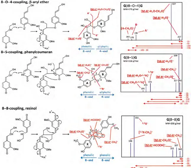
The authors compared the CID-fragmentation pattern of the deprotonated isobaric dimers G(β–5′)G and G(β–β′)G (m/z 357), and G(β–O–4′)G (m/z 375), to data obtained from the literature.[94] The CID-MS/MS of the deprotonated molecules of G(β–5′)G linked dimer afforded a product ion at m/z 221, which is character-istic of the four-aliphatic end (B). The CID-MS/MS of the isobaric
deprotonated G(β–β′)G dimer afforded the product ion of m/z 151, which is characteristic of the eight-phenolic end (A).
Obviously, the mass of the G(β–O–4′)G linked dimer increases by 18 u because the incorporation of a molecule of water and the CID-MS/MS of the deprotonated G(β–O–4′)G molecules gives two diag-nostic product ions at m/z 179 (B) for the four-aliphatic end and at m/z 195 for the eight-phenolic end (A) (Fig. 9).
In contrast, the CID-MS/MS of the S–G dimer deprotonated an-ions followed parallel gas-phase fragmentation comparable to the G–G dimers, except that we notice that the formed product ions had a difference of 30 u between S and G monomers, which of course was due to the presence of one additional methoxy group in the S monomer. Therefore, MS/MS of the deprotonated S(β–5′) G linked dimer afforded the product ion at m/z 221, which was characteristic for G residue present on the four-aliphatic end. The precursor ion scan of the S(β–O–4′)G dimer deprotonated molecule at m/z 405 afforded the product ion at m/z 225, which indicated that the S unit was on the eight-phenolic end.
It is of noteworthy to mention that the deprotonated S(β–β′)S at m/z 417 possesses an 8–8 link, whereas, the S(β–O–4′)S dimer con-tains 18 u more than the S(β–β′)S dimer due to water incorporation. Subsequently, the MS/MS of the deprotonated S(β–β′)S dimer mol-ecule at m/z 435 afforded the product ions at m/z 225 and 209, which were characteristic for this structure.
Kiyota et al. also identified the following deprotonated molecules by comparison of their MS/MS data with those obtained from the literature: G(β–O–4′)G(β–O–4′)G at m/z 571, G(β–O–4′)S(β–5′)G at m/z 583, S(β–O–4′)G(β–5′)G at m/z 583, S(β–O–4′)S(β–5′)G at m/z 613, G(β–O–4′)S(β–β′)S at m/z 613 and S(β–O–4′)S(β–β′)S at m/z 643. Consequently, the CID-MS/MS spectra of the trimers afforded product ions that were characteristic of the monomers composed of the aromatic part A, whereas the BC product ions were character-istic of the remaining dimer ions, allowing the elucidation of the complete structure. The following are examples of characterization of trimers performed using MS/MS analysis (Fig. 10).[108]
It is interesting to note that these authors chose not to use the complex CID nomenclature proposed by Morreel et al. described in section APCI-MS/MS-based Sequencing of Oligolignols,[94] but Figure 9. Dimers of G units showing the three main links. Their characteristic fragmentation patterns are indicated by red lines. (I) Theβ–O–4′, β-aryl ether link yields fragment ions A– (m/z 195) and B – (m/z 179). (II) The β–5′, phenylcoumaran link yields fragment ion B – (m/z 221). (III)The β–β′, resinol link yields fragment ion A– (m/z 151). Reproduced with permission from E. Kiyota et al. (2012). Copyright 2012 by American Chemical Society. All rights reserved.
rather, a very simple and comprehensible system. The work of Kiyota et al.[108]illustrated the simplicity of the CID-MS/MS process.
ESI-MS/MS and APCI-MS/MS characterization of lignans iso-lated from sesame seeds
During acidic extraction of glucosylated lignans (sesaminol gluco-sides), hydrolysis occurs to release the aglycones sesamin and sesamolin, which are found in either the oil fraction and/or in the defatted fraction of sesame seeds.[109–111] It has been demon-strated that the exact lignan composition of sesame seeds changes with the developmental stage of the seeds.[112–117]
Struijs has shown that the extracted sesaminol glucosides from the defatted sesame meal, when purified by chromatography, yielded a series of well-resolved peaks.[118]The identities of this se-ries of separated compounds were confirmed by ESI-MS, APCI-MS and CID-MS/MS.[119]
The ESI-MS (+ ion mode) of sesaminol produced only traces of the [M + H]+protonated molecule. However, when the ESI-MS was measured in the negative ion mode, it afforded appropriately the deprotonated molecule at m/z 369.1. However, in contrast to that shown by ESI-MS (+ ion mode), APCI-MS (+ ion mode) afforded the major protonated molecule [M + H]+at m/z 371.1 and the frag-ment ions [M + H–H2O]+at m/z 353.1.
On the other hand, it is interesting to note that when APCI-MS was used in the negative ion mode, it afforded the abundant deprotonated molecule [M–H] at m/z 369.1 identical to that ob-tained by ESI-MS. For this reason, APCI permitted the characteriza-tion of the dibenzylbutadiene lignans containing the hydroxyl groups.[110,111]Similarly, it was shown that the CID-MS/MS fragmen-tations of the precursor deprotonated molecule isolated from sesaminol were similar when using either APCI-MS or ESI-MS in the negative ion mode.
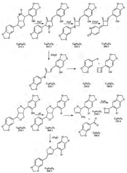
Moreover, APCI-CID-MS/MS (+ ion mode) of the protonated molecules [M + H]+at m/z 371.1 gave the product ions at m/z 353.1, 335.1, 323.1, 305.1 and 135.0. On the other hand, CID-MS/MS ( ion mode) of the deprotonated molecules at m/z
369 afforded a series of product ions at m/z, 339.1, 219.1, 191.0 and 150.0.[120] The tentative fragmentation pathways for sesaminol in the negative and positive ionization modes are pre-sented in Fig. 11.[105,121,122]
Finally, by using the same methodology, it was also established that the protonated sesamolin contained the methylenedioxy-bridged furanofuran structure and lacked phe-nolic hydroxyl groups.
APCI-MS/MS and ESI-MS/MS characterization of PML and model lignin compounds
Haupert et al. was able to identify the most suitable atmospheric pressure ionization method(s) for the tandem mass spectrometric characterization of complex mixtures related to PML degradation products. They used a set of lignin-degraded products as model compounds, which were subjected to positive and negative ion mode APCI and ESI under traditional as well as novel conditions using LQIT-MS.[123]They found that when using APCI-MS (+ ion mode) only guaiacol and eugenol formed an abundance of proton-ated molecules.
However, in contrast, it was found that vanillyl alcohol and coniferyl alcohol formed exclusively the [M + H–H2O]+ frag-ment ions. The loss of water from these protonated molecules was facilitated by virtue of their presence near to allylic and benzylic hydroxyl locations. This was also rationalized by the fact that after protonation, the precursor protonated molecule loses water to generate the resonance-stabilized carbocations (Table 1).
Obviously, guaiacol and eugenol do not contain these func-tionalities; however, the dimer, guaiacylglycerol-β-guaiacylether, contains a benzylic hydroxyl group. Consequently, this dimer easily loses water to form the [M + H–H2O]+ ion. Nevertheless, when using APCI-MS, the two most abundant fragment ions are [M + H–2H2O]+ and/or [M + H–H2O–CH2O]+ from formalde-hyde (Table 1).
When the model compounds were studied by traditional neg-ative mode APCI ionization (methanol and water as solvents), Figure 10. The general structures of the trimers identified by UPLC-MS (R1= CH3, R2= H or CH3). The (β–O–4) link present in these structures results in fragment ions A monomer plus a molecule of water and the BC dimer. The deprotonated fragment ion BC mass has the same m/z as the dimers composed of S and G. Structure with C fragmentation can only have twoβ–5 links as there is no increase in the mass of the monomers and fragment ion C is typical of the phenylcoumaran link. Reproduced with permission from E. Kiyota et al. (2012). Copyright 2012 by American Chemical Society. All rights reserved.
they gave ions with poor abundances. The lack in sensitivity and the weak observed fragmentation make this a poor ionization method for the characterization of lignin degradation products.[123]
Using the model compounds, Haupert et al. have shown that it was possible to use ESI-MS (+ion mode) doped with sodium chlo-ride, to generate abundant [M + Na]+sodiated molecules without any fragmentation. This approach allowed the direct determination of the molecular weights of the lignin degradation products in mixtures.[123]However, as expected, no structural information could be obtained by examining the CID-MS/MS of the sodiated mole-cules using either triple quadrupole or QIT tandem mass spectro-metric instruments because the sodiated molecules are known not to fragment during MS/MS analysis.
When Haupert et al. used ESI-MS ( ion mode) with the dopant sodium hydroxide, they obtained abundant deprotonated mole-cules [M-H] without any fragmentation. The ESI-MS ( ion mode) of a synthetic mixture is showed in Fig. 12.
The CID-MSn analysis of this series of precursor deprotonated molecules established the connectivities between precursor and product ions; these were obtained by measuring multiple consecu-tive ion isolation and CID-MSn(up to MS7). This methodology has since been found to improve significantly the information that can be obtained by mass spectrometric analysis for lignin degrada-tion products and lignome (Table 2).[123]
Characterization of VRL degradation products by HPLC-ESI-MSn Owen et al. developed a high-performance liquid chroma-tography/multiple-stage MS/MS method with a commercial linear QIT/Fourier-transform ion cyclotron resonance mass spectrometer (HPLC-ESI-LQIT-FT-ICR-MSn) as a tool for the analysis of complex mixtures obtained from PML degradation products.[124] In their study, the HPLC separation method of the lignin degradation prod-ucts was followed by ESI-MS analysis doped with NaOH ( ion mode). This method was established by Hauper et al. to be the most appropriate for the identification of lignin-related model compounds.[123]
Owen et al. showed that for the analysis of artificial and real mixtures, data-dependent scans allow the instrument to select automatically the most abundant three ions from the ion source.[124]This allowed the authors to acquire separate MS ac-quisitions simultaneously for the same ions with two different mass analysers. Thus, the LQIT-FT-ICR spectrometer operated in the higher duty-cycle LQIT when performing tandem mass spec-tral acquisitions for precursor ion selection, whereas the lower duty-cycle FT-ICR carried out the high-resolution measurements for elemental composition determination for the same ions.[124] Accordingly, the most abundant product ion formed in the MS2 experiments, was subjected to a further stage of ion isolation and MS3fragmentation.[124]
Figure 11. Proposed fragmentation of sesaminol in the negative ionization mode and in the positive mode. Reproduced with permission from K. Struijs (2008). Copyright 2008 by Wageningen Universiteit, The Netherlands. All rights reserved.
Ta bl e 1 . Sym b o ls ,re ten tio n tim es, chara ct er ist ic m ass fr agm ent s a n d limi ts o f d et ecti on for C u O o xi d a tio n p ro d u cts .Rep ro d uced w ith p erm iss io n fro m K. Ka ise r et al .( 2012). C opyright 2012 by Americ an Ch em-ical Soci ety. All ri ghts re ser ve d Com p ound (M W) AP CI (+ ) (m/ z)A P C I ( )( m/ z) ESI (+) (m/ z) ESI ( )( m/ z) Re la ti ve a b u n dance R el at iv e abu nda n ce Rel at iv e abun da nce R e lat iv e abu nda n ce Guaiacol (124) [M + H ] + (125) 100% Poor sign al [M + H] + (125) 100% Poor sign al [M –H] (123) 100% [M –H] (123 ) 100 % Vanillyl alcohol (154) [M + H –H 2 O] + (137) 100% Poor sign al [M + H –H2 O] + (137) 100% [M –H] (153 ) 100 % [M –H] (153) 100% Eugenol (164) [M + H ] + (125) 100% Poor sign al No Signal Poor sign a l [M –H] (163) 100% [M –H] (163 ) 100 % [M –H –CH 3 ] (148) 15% Con ifery l a lcohol (180) [M + H –H 2 O] + (163) 100% Poor sign al [M + H –H2 O] + (163) 100% [M –H] (179 ) 100 % [M –H –H2 O –H] (160) 41% [M –H –H2 O] (161) 31% [M –H] (179) 100% Guai acylgl ycerol-β -g uaiacylether (320) [M + H –H 2 O] + (303) 40% Poor sign al [2M + Na] + (663) 15% Poor sign al [M + H –2H 2 O] + (285) 95% [M –H] (319) 100% [M + N a] + (343) 100% [2M –H] (6 39 ) 2 7% [M + H –H 2 O –CH 2 O] + (273) 97% [M –H –H2 O –CH 2 O] (271) 47% [M –H] (319 ) 100 % m/ z 285-C 6 H3 OCH 3 (m/ z 179 ) 20% m/ z 285-HOC 6 H2 OCH 3 (m/ z 163 ) 25% m/ z 285-HOC 6 H4 OCH 3 (m/ z 161 ) 20% m/ z 273-HOC 6 H4 OCH 3 (m/ z 149 ) 30% m/ z 285-HOC 6 H3 (OCH 3 )CCH (m/z 137) 16%
The authors used degradation products, pure standard solutions as well as a mixture of 12 model compounds of lignin degradation products (Table 3), which were separated by reversed-phase HPLC using an acetonitrile and water gradient elution followed by high-resolution MS3analysis. The authors used three different HPLC col-umns to separate a mixture of 12 lignin-related model compounds. Using water and acetonitrile gradient buffered with ammonium for-mate, full baseline separation was attained for all components of the mixture using both the Zorbax SB-C18 and Zorbax SB-Phenyl columns. It was noted that a poorer separation was attained using Kinetex PFP column.[124]
All of the model compounds studied exclusively by ESI-MS ( ion mode) gave the abundant deprotonated ion [M–H] . Furthermore, the authors have shown that MS/MS analysis of all model com-pounds containing methoxyl groups attached to the aromatic ring were initiated via a homolytic cleavage, by the loss of a methyl rad-ical. Additionally, all ions containing allylic or benzylic hydroxyl groups (vanillyl alcohol, coniferyl alcohol, and sinapyl alcohol) fragmented primarily by loss of a water radical.[124]In addition, the authors indicated that the deprotonated guaiacylglycerol- β-guaiacyl ether was shown to mainly lose water followed by loss of formaldehyde.[96]
To explore whether additional structural information could be obtained, the authors performed CID-MS3experiments on the most abundant product ion formed in the MS2 experiment. For some ions that lack a weakly bound group, no further fragmentation were observed. However, most of the secondary product ions fragmented to yield structurally informative product ions in the MS2experiments, demonstrating that multiple-stage MS/MS is use-ful in the structural characterization of unknown lignin degradation products.
Owen et al. showed that the deprotonated sinapyl alcohol con-taining two methoxyl groups could lose sequentially one methyl radical in the MS2experiment followed by another methyl radical in the MS3experiment. These losses permitted the counting of
the methoxyl substituents in the analyte. In the MS2experiment of the deprotonated 2-methoxyl-4-propylphenol, the product ion formed by loss of the methyl radical was further subjected to MS3. Very interestingly, this product ion fragmented by eliminating an ethyl radical by homolytic cleavage of the benzylic bond, to re-veal the presence of a propyl substituent.[124]An analogous ben-zylic bond cleavage was also observed for deprotonated eugenol. The MS2of deprotonated eugenol afforded a product ion by loss of the methyl radical. MS3 of this product ion was subjected to the consecutive cleavage of the benzylic carbon–hydrogen bond and loss of a hydrogen atom. In contrast, MS2of the deprotonated isoeugenol lost a methyl radical to afford a product ion that, when subjected to an MS3experiment, did not fragment any further be-cause it lacked a weakly bound substituent. This difference in these two MS3fragmentations allowed the differentiation between these structural isomers.[124]
In addition, the authors successfully separated by HPLC, the dia-stereomeric pair (RS/SR and RR/SS) of guaiacylglycerol-β-syringyl ethers. These were shown to yield the same CID-fragmentation product ions from the deprotonated diastereomeric pairs.
To assess the ability of this method to identify unknown compo-nents, a VRL organosolv oak lignin extract was analysed by HPLC-ESI-MS. Though the organosolv lignin sample is a very complex mix-ture, the HPLC was able to separate many analytes, including iso-baric and isomeric molecules.[124]
ESI-MS study of PML Kraft black liquor
Pinto et al. attempted to characterize the lignin structural changes during Kraft pulping (PML), using1H-NMR and13C-NMR techniques and to estimate the changes in the lignin molecular weight using ESI-MS and gel permeation chromatography (GPC).[125,126]The au-thors used four Kraft cooking processes that were designated as representative of the different delignification phases as follows: ini-tial cooking (19.0% delignification), iniini-tial-to-bulk transition cooking Figure 12. Negative mode ESI mass spectrum obtained for an equimolar mixture of guaiacol (MW 124 u), 2-methoxy-4-methylphenol (MW = 138 u), vanillin (MW = 152 u), vanillyl alcohol (MW = 154 u), eugenol (MW = 164 u), isoeugenol (MW = 164 u), 2-methoxy-4-propylphenol (MW = 166 u), coniferyl alcohol (MW = 180 u), sinapyl alcohol (MW = 210 u), guaiacylglycerol-β-guaiacylether (MW = 320 u) and guaiacylglycerol-β-syringylether (MW = 350 u) dissolved in 50/50 (v/v) methanol/water and doped with sodium hydroxide. Reproduced with permission from L. J. Haupert et al. (2012). Copyright 2012 by Elsevier. All rights reserved.
Ta bl e 2 . CID-MS n a n al ysis fo r som e o f the ne gat iv e io ns list e d in F ig .25. Rep ro d u ced wit h pe rm issi on fr om L. J. Ha u p er t et al .( 2012). C opyright 20 12 by Else vier. A ll rights reserved Compound (m/ z of [M –H] )M S 2 fr agm ent ati o ns (product ions ’m/z ) re la ti ve a b u n d ance MS 3 fr agm ent ati o n s (product ions ’m/z ) re la ti ve a b u n d ance MS 4 fr agm e nt at io ns (product ions ’m/ z) rel a ti ve abundan ce MS 5 fragmentations (product ions ’m/ z) re lat iv e ab u n da nce MS 6 fr agm ent ati o n s (product ions ’m/z ) re la ti ve a b u n d ance MS 7 fragm e n tat io ns (p ro du ct io ns ’m/ z) re la ti ve abundance Vanillyl alcoh o l (153) 153 CH 3 (138) 52% 138 H2 O (120) 100% 120 + H2 O (138) 20% No further fr agm e nt at io n o b ser v e d 153 H2 O (135) 100% 135 CH 3 (120) 100% 120 CO (92) 100% 120 CO (92) 100% Eugenol (163) 163 CH 3 (148) 100% 148 H (147) 100% No further frag me ntat io n o bse rve d Isoeugenol (163) 163 CH 3 (148) 100% No further fr agm ent ati o n o bser ved Sinapyl a lc ohol (209) 209 CH 3 (194) 100% 194 CH 3 (179) 100% 179 CO (151) 100% 151 H2 O (133) 100% 209 H2 O (191) 35% 191 CH 3 (176) 100% 176 CH 3 (161) 100% 151 CH 2 O (121) 100% 121 C O (93) 100% 105 CO (77) 100% 209 H2 O –CH 3 (176) 12% 176 CH 3 (161) 100% 161 CO (133) 100% 161 CO (133) 100% 13 3 CO (105) 100% 161 –2C O (105) 75% 133 CO (105) 100% 133 2C O (77) 47% 133 2CO (77) 19% 105 CO (77) 100%
(40.0% delignification), bulk cooking (76.0% delignification) and re-sidual phases cooking (94.4% delignification). The dioxane lignin isolated from E. globulus (referred as EDL) represented the lignin in the starting wood material (0% delignification).[125,126]
Accordingly, Pinto et al. reported the ESI-MS spectrum of Kraft black liquor lignin (BL) obtained from the residual phase of delignification and showed the presence of both lower and higher m/z ions, which were attributed to lignin oligomeric substructure ions containing fragments bounded by oligosaccharides.[126]
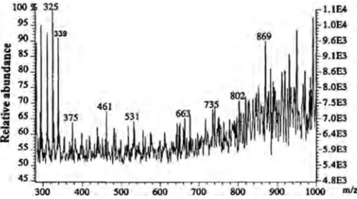
To estimate the molecular weights of components in the cooked Kraft BL, the obtained residue was purified by selective precipita-tion in dioxane to yield two fracprecipita-tions: BLp (about 70% yield, sugars content 1.5%) and BLs (about 30% yield, sugars content 18.9%). The BLp fraction showed an ESI-MS containing lower ions around m/z 800, whereas the BLs fraction showed an MS with a higher mass dis-tribution at m/z 1100. Such a difference is probably due to the BL fraction containing lignin residues attached to oligosaccharides (Fig. 13).
The expansion of the MS of BL showed series of fragment ions corresponding to LOs having m/z differences of 202–206 u, which is the approximate molecular mass of the phenylpropane unit (C-9) (Fig. 14). In addition, Pinto et al. observed that the additional ion at m/z 339 was assigned respectively to a disaccharide com-posed of 4-O-methyl-D-glucuronic acid and D-xylose residues (GlcpA→ Xylp). This disaccharide was previously detected in the composition of E. globulus xylan.[125]
Nonetheless, it is noteworthy that the authors actually deduced a low mass distribution value for such a complex ESI-MS. The absence of higher masses was a consequence of the presence of the lignin degradation products formed by this complex mixture of LOs.
Elucidation of the PML Eucalyptus globulus Labill lignins by ESI-MS
Evtuguin et al. studied the chemical structure of PML isolated from Eucalyptus globulus Labill.[106,127]
The authors found that the Eucalyptus globulus lignins fraction was mainly composed of S/G lignols with an extremely high pro-portion of syringyl (S) units (82–86 mol %) and a minor proportion of p-hydrophenyl propane (H) units (roughly 2–3 mol %). They also described the presence of unknown C-6 substituted andβ–O–5′ type syringyl substructures representing about 65% of lignin con-densed structures. ESI-MS analysis revealed a wide molecular weight distribution within the lignin with a mass distribution around 2500 u.[106]
In a different study, Evtuguin and Amado attempted to study the structures of PML E. globulus lignins and its low molecular weight fraction (EDLF), by ESI-MS and CID-MS/MS using a quadrupole time-of-flight (QTOF) MS instrument.[107]In this study each of the lignin components were isolated by means of GPC as described previously by the same group.[106]In addition, for the qualitative Table 3. Twelve model compounds of lignin degradation products. Reproduced with the permission of B. C. Owen et al. (2012) Copyright 2012 by American Chemical Society. All rights reserved
Model compound (m/z of [M–H]+; determined elemental composition) Measured exact m/z (error in ± mTh from the expected exact mass)
[A] Guaiacol (123; C7H7O2) 123.0458 (1.2) [B] 2-Methoxy-4-methylphenol (137;C8H9O2) 137.0615 (1.3) [C] Vanillin (151; C8H7O3) 151.0408 (1.3) [D] Vanillyl alcohol (153; C8H9O3) 153.0564 (1.2) [E] Eugenol (163; C10H11O2) 163.0771 (1.2) [F] Isoeugenol (163; C10H11O2) 163.0771 (1.2) [G] 2-Methoxy-4-propylphenol (165; C10H13O2) 165.0927 (1.2) [H] Coniferyl alcohol (179; C10H11O3) 179.0717 (0.9) [I] Sinapyl alcohol (209; C11H13O4) 209.0819 (0.5) [J] Guaiacylglycerol-β-guaicylether (319; C17H19O6) 319.1186 (0.4) [K + L] Guaiacylglycerol-β-syringylether stereoisomers (349; C18H21O7) 349.1292 (0.5)
Figure 13. Negative mode ESI-MS spectrum of BL (A), BLp (B) and BLs (C) isolated from black liquor after 94.4% delignification of eucalypt wood. Reproduced with the permission of P. C. Pinto et al. (2002). Copyright 2002 by Marcel Dekker, Inc. All rights reserved.
structural elucidation of their obtained ions, the authors synthe-sized standards models composed of lignin dimeric model of β-arylglycerol, pinoresinol, syringaresinol and dehydrodiconiferyl al-cohol according to established procedures.[128]
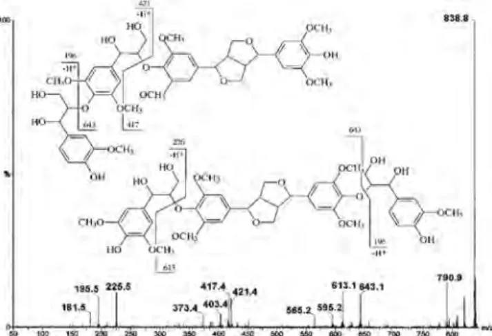
The ESI-MS of the EDLF fraction showed a complex mixture of ions, which were assigned as the deprotonated LOs [M–H] over a mass range at m/z 150–1500 (Fig. 15). The group of signals at m/z 150–210 represents monomeric degradation products ob-tained during acidolytic isolation of lignins. In addition, the authors were able to elucidate the CID-MS/MS of the major deprotonated molecules at m/z 319.4, 349.4, 357.4, 417.4, 643.1 and 838.8. (Fig. 15) They interpreted the CID-fragmentation patterns, which helped to elucidate the structures of these deprotonated molecules, which were assigned and compared with dimeric model compounds. Fi-nally, they concluded that their ESI-MS data revealed a significant abundance of LOs composed of linear fragments ofβ–O–4′ linked syringyl and guaiacyl units as well as syringaresinol.[107]
Because the average molecular weight of the monomeric guaiacyl phenyl propane unit (G) is 190 u and that of the syringyl phenyl propane unit (S) is 220 u, the series of signals centred around m/z 500–1400, 640, 870, 1070 and 1300 were assigned as lignin dimer, trimer, tetramer, pentamer and hexamer units,
respectively.[107]Another dominant ion series in the same ESI-MS was assigned as trimer at m/z 643, tetramer at m/z 868, pentamer at m/z 1065, and hexamer at m/z 1291. These ions appear to differ from each other by 226 and 196 u, which can be assigned to S and G units, respectively, in the composition of oligomers.[107]
The structure of the most abundant LO was the ion at m/z 643, which was assigned as LO643 and was studied in more detail. The comparison between the CID-MS/MS analysis of LO643 at m/z 643 with that of a synthetic dimeric lignin model precursor ion at m/z 417 showed common fragmentation product ions (Fig. 16).[107] So, CID-MS/MS of the precursor ion LO643 at m/z 643 afforded the diagnostic product ion at m/z 417 by consecutive elimination of 226 u by the loss of the terminalβ–O–4′-linked syringyl propane unit containing the benzylic hydroxyl group. Also, it was suggested that the mechanism of this CID-fragmentation was driven by the homolytic splitting of the alkyl-O-aryl bond, to afford the product ions at m/z 417 and 225. As a result, Evtuguin and Amado Figure 14. Negative mode ESI-MS spectrum (expanded mass range from
m/z 300 to 1000) of BLs isolated from black liquor after 94.4% delignification of eucalypt wood. Reproduced with the permission of P. C. Pinto et al. (2002). Copyright 2002 by Marcel Dekker, Inc. All rights reserved.
Figure 15. The ESI-MS of EDLF. Reproduced with permission from D. V. Evtuguin et al. (2003). Copyright 2003 WILEY-VCH Verlag GmbH & Co. KGaA, Weinheim. All rights reserved.
Figure 16. The product ion scan of the precursor ion extracted from the tetramer represented at m/z 643. Reproduced with permission from D. V. Evtuguin et al. (2003). Copyright 2003 WILEY-VCH Verlag GmbH & Co. KGaA, Weinheim. All rights reserved.
concluded that the structure of LO643 was a trimerβ–O–4-linked S unit with syringaresinol [S–(β–O–4′)–S–(β–β′)–S] (Fig. 16).[107]
The same approach was used to elucidate the ion at m/z 839, which was attributed to mixture of isobaric tetramer composed of G–(β–O–4′)–S–(β–O–4′)–S–(β–β′)–S and S–(β–O–4′)–S–(β–O–4′)–S– (β–β′)–G. The CID-MS/MS of the precursor deprotonated molecule at m/z 839 showed the presence of two product ions at m/z 643 and m/z 613, and the authors concluded that such an assignment confirmed both structures assigned to this isobaric ion at m/z 839 (Fig. 17).[107]
Similarly, CID-MS/MS of the ion at m/z 1065 was tentatively assigned to two alternative structures: S–(β–O–4′)–S–(β–O–4′)–S–
(β–β′)–S–(β–O–4′)–G and G–(β–O–4′)–S–(β–O–4′)–S–(β–O–4′)–S– (β–β′)–S.
In addition, the authors have shown that CID-MS/MS analysis of the ions obtained from lignin dimers (particularly 8–O–4′, 8–8′ and 8–5′ linked), formed diagnostic product ions from which the spe-cific gas-phase fragmentation patterns can be deduced.
In conclusion, the authors proposed that CID-MS/MS frag-mentation of the precursor ion of LOs occured predominantly by the cleavage of ether linkages. They also proposed that such fundamental information about lignin primary structure would form a sound basis to enable new structure elucidation studies to be made more reliably and rapidly by employing ESI-MS.
ESI-FT-ICR-MSnstructural analysis of PML thioacidolysis degradation lignin products
Onnerud et al. investigated the structures of the pinoresinol thioacidolysis degradation products by ESI-FT-ICR-MSnfrom lignin model compounds and PML wood lignin.[129]
The authors used the thioacidolytsis (BF3/C2H5SH) treatment for the degradation of pinoresinol and PML, which resulted in the con-version of uncondensed monomeric units in lignin into simple dia-stereomeric mixtures of 1,2,3-trithioethane phenylpropanoid monomers.[130]
The authors mentioned that two major peaks were detected for thioacidolysed MWL, and these were identical when compared with the thioacidolysed pinoresinol and acetylated pinoresinol. Thioacidolysis is known to produce a large fraction of the mono-meric C-6–C-3′ adduct containing three thioethyl groups in the side chain as the major component.[130]
The authors used ESI in both positive and negative ionization modes for the identification of the obtained products. In Figure 17. The product ion scan of the precursor ion extracted from the
tetramer represented at m/z 839. Reproduced with permission from D. V. Evtuguin et al. (2003). Copyright 2003 WILEY-VCH Verlag GmbH & Co. KGaA, Weinheim. All rights reserved.
Figure 18. The CID-fragmentation pattern of the thioacidolysed acetylated pinoresinol at m/z 531.19. Reproduced with permission from H. Onnerud et al. (2003). Copyright 2003 by Walter de Gruyter GmbH & Co. KG. All rights reserved.
addition, they used tandem MSn to elucidate the structure of thioacidolysed pinoresinol before and after acetylating the products.
The ESI-MS (+ ion mode) of the thioacidolysed acetylated pinoresinol and the thioacidolysed pinoresinol did not afford the expected protonated molecules but instead produced the pseudo-molecular ions obtained by elimination of the ethane thiol CH3CH2SH ( 62 u) assigned as the acetylated dithioether [MAc– SCH2CH3]+and the non-acetylated dithioether [M–SCH2CH3]+, re-spectively, at m/z 531.19 and m/z 447.17.[129]
The acetylated dithioether [MAc–SCH2CH3]+precursor ion at m/z 531.19 was analysed by CID-MS/MS, which showed two major product ions at m/z 469.17 and 427.17 that were attributed respec-tively to the loss of the second CH3CH2SH group and a molecule of ketene. Consequently, these two product ions were assigned as [MAc–CH3CH2S–CH3CH2SH]
+
at m/z 469.17 and [MAc–SCH2CH3– CH2CO]+at m/z 427.17, respectively. The latter product ion at m/z 427.16 also eliminated individually a molecule of formaldehyde and thioethanol to afford respectively the ions at m/z 385.15 and 365.14 (Fig. 18).
In contrast, MS/MS of the non-acetylated thioacidolysed precur-sor ion at m/z 447.17 afforded only two product ions [M–SCH2CH3]
+ and [M–SCH2CH3–HSCH2CH3]+, respectively, at m/z 385.15 and 323.13 (Fig. 19).[129]
On the other hand, it is important to note that the overall perfor-mance of the ESI-FT-ICR-MS of lignin did not provide unambiguous information about the detailed structure of the milled wood lignin given that many of the expected degradation products could not be identified.[129]
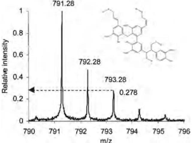
In reality, when Onnerud et al. used FT-ICR-MS (+ ion mode) to analyse the total thioacidolysis extract of PML wood lignin, they ob-tained a very heterogeneous MS composed of a multitude of lignin ions up to about 1500 u, including a high abundance ion at m/z 791.28. The authors suggested that the assignment of this series of specific ions was complex. In addition, they indicated that the ion at m/z 791.28 was the most abundant ion (base peak) and diffi-cult to assign. Indeed, CID-MS/MS analysis of this precursor ion did not yield any product ions under any CID conditions used. The au-thors indicated that this CID-MS/MS resilience of the precursor ion at m/z 791.28 was due to its very stable structure. Nevertheless, the authors indicated that the position of the thioethyl groups
found in this precursor ion was most probably different to that ob-tained from the thioacidolysed pinoresinol.[129]
From the relative abundances of the different isotope peaks, Onnerud et al. calculated that the second isotopic peak (third peak) containing34S, [M–34SCH2CH3]+, at m/z 793.28 was 0.278 compared with the base peak, the monoisotopic [M–32SCH2CH3]
+ at m/z 791.28. (Fig. 20) This value was compared with a theoretical value of the isotopic ion distribution measured using computer software, and it provided the number of sulfur atoms in the compound. For that reason, the molecular formula was calculated to be C43H51O8S3. The authors also calculated the double-bond equiva-lent (DBE) value for this molecular formula, which was found to be 18.5. This DBE value corresponded to a tetrameric structure con-taining four aromatic rings (that is 16 DBE), two double bonds (2 DBE) and the + charge (0.5 DBE). Accordingly, the authors calcu-lated that the theoretical m/z would be 791.27. Onnerud et al. con-cluded that the non-acetylated thioacidolysed PML C43H51O8S3at m/z 791.28 was a tetramer consisting of two coniferyl alcohol units, connected through aβ–O–4′ bond, with further links through a β–β′ andβ–1′ structure (Fig. 20).[129]
Figure 19. The CID-fragmentation pattern of thioacidolysed pinoresinol 1 without acetylation. Reproduced with permission from H. Onnerud et al. (2003). Copyright 2003 by Walter de Gruyter GmbH & Co. KG. All rights reserved.
Figure 20. ESI-FT-ICR-MS (+ ion mode) of the total thioacidolysis extract of MWL. Reproduced with permission from H. Onnerud et al. (2003). Copyright 2003 by Walter de Gruyter GmbH & Co. KG. All rights reserved.