Classic nuclear localization signals and a novel nuclear localization motif are required for nuclear transport of porcine parvovirus capsid proteins
Texte intégral
Figure
![Table 1 . All resulting proteins (including the wild-type [wt)]) contained the mutation to inactivate the viral PLA 2 (data not shown)](https://thumb-eu.123doks.com/thumbv2/123doknet/2954271.80657/3.877.63.431.122.258/table-resulting-proteins-including-contained-mutation-inactivate-viral.webp)
Documents relatifs
The π-turn motif is ancestral and has been lost several times independently The analysis of sponge nuclear receptor sequences show the presence of the RxxxE motif in helix H7 of
Expression of viral polymerase and phosphorylation of core protein determine core and capsid localization of the human hepatitis B virus.. Phosphorylation in the
Similar HIV-1 restriction was observed when expressing the different SAMHD1 variants in THP-1 cells that were stably silenced for expression of endogenous SAMHD1 (Figure 4D,
This result shows that the 1-galactosidase fused to MyoD deleted at NLS234 and NLS6 fails to be actively transported into the nucleus and demonstrates that the nuclear staining
Three of these (D378G, H383Q and S436P) localizing on the capsid surface was enough to abolish NADL-2 replication in primary bovine testis cells (TV) and reduce titer and cytopathic
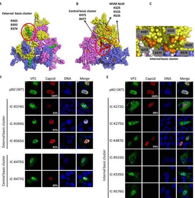
In summary, we demonstrated that capsid protein targeting to the nucleus for porcine parvovirus depended on both classic nu- clear localization signals and a novel structural
By knocking down or overexpressing MYO1B in immortalized MEFs or melanoma cells, we demonstrate that MYO1B, by interacting with PTEN, through the motor domain, prevents localization
Surprisingly, in addition to the relatively even cytoplasmic and nuclear distribution of α7 detected by indirect immunofluorescence in parental U 2 OS-tTA cells,
