HAL Id: tel-03020641
https://tel.archives-ouvertes.fr/tel-03020641
Submitted on 24 Nov 2020HAL is a multi-disciplinary open access
archive for the deposit and dissemination of sci-entific research documents, whether they are pub-lished or not. The documents may come from teaching and research institutions in France or abroad, or from public or private research centers.
L’archive ouverte pluridisciplinaire HAL, est destinée au dépôt et à la diffusion de documents scientifiques de niveau recherche, publiés ou non, émanant des établissements d’enseignement et de recherche français ou étrangers, des laboratoires publics ou privés.
Functional analysis of catalase mutants and their
application to the analysis of NADPH-linked pathways
in oxidative signaling in Arabidopsis thaliana
Zheng Yang
To cite this version:
Zheng Yang. Functional analysis of catalase mutants and their application to the analysis of NADPH-linked pathways in oxidative signaling in Arabidopsis thaliana. Botanics. Université Paris Saclay (COmUE), 2018. English. �NNT : 2018SACLS464�. �tel-03020641�
Functional analysis of catalase mutants and
their application to the analysis of
NADPH-linked pathways in oxidative signaling in
Arabidopsis thaliana
Thèse de doctorat de l'Université Paris-Saclay préparée à l'Université Paris-Sud au sein de l’Institut des Plantes des Sciences de Paris-Saclay, IPS2 École doctorale n°567 :
Sciences du végétal : du gène à l’écosystème, SDV
Spécialité de doctorat : Biologie
Thèse présentée et soutenue à Gif-sur-Yvette, le 15.11.2018, par
Zheng Yang
Composition du Jury :
Dr. Marianne Delarue
Professeur, Univ. Paris-Sud, Orsay Présidente
Dr. Yves Jolivet
Professeur, Univ. Lorraine, Nancy Rapporteur
Dr. Arnould Savouré
Professeur, Sorbonne Université, Paris Rapporteur
Dr. Graham Noctor
Professeur, Univ. Paris-Sud, Orsay Directeur de Thèse
N N T : 201 8S A C LS 46 4
ACKNOWLEDGMENTS
I would like to express my gratitude to everyone who helped me throughout the process of thesis writing. It would not have been so smoothly to finish the work without their support.
My deepest gratitude goes first to Professor Graham Noctor, my supervisor, for his patient guidance, detailed advice and continuous encouragement during my PhD study. It is my great pleasure to have a supervisor with immense knowledge and rigorous attitude to scientific work. His instruction and words helped me a lot in my research and the writing of thesis, and for sure, will influence me more in the future.
I am thankful to the jury members of my defence, Prof. Yves Jolivet (Université de Lorraine, FR), Prof. Arnould Savouré (Sorbonne Université) and Prof. Marianne Delarue (Université Paris Sud, FR) for having accepted to evaluate my PhD work and spending their valuable time to read my thesis.
I would like to thank the former and the current colleagues in our group. Amna Mhamdi, my first officemate, helped me a lot in learning different techniques and in being familiar with the working environment. The scientific discussion with her has benefited me a lot. Her enthusiasm, responsibility and preciseness has set a food example to me in my routine work. I am very grateful to Gilles Chatel-Innocenti and Hélène Vanacker for the opportunity to work on the mutants they had obtained, and for their help and discussions, enabling me to produce the results presented in Chapter 4 of this thesis. I am also thankful to Marie-Sylviane Rahantaniaina for the help in my work and life. Our communication about the culture, food and everything else is so nice and unforgettable. To Caroline Lelarge for salicylic acid analysis. To Emmanuelle Issakidis-Bourguet and Daoxiu Zhou for the valuable scientific discussion and help in life. To Yuan Shen and Xiaoyun Cui for much help in work and friendship in life.
During the PhD career, I obtained much help and support from colleagues in IPS2. It is really lucky to meet these nice people in the institute. Their passionate smile and kindly words have warmed me so much. I would like to express my gratitude to these members, Raynaud Cécile, Linda De Bont, Jingfang Hao, Shengbin Liu, Marie Dufresne, Sophie Massot, Sophie Blanchet, Severine Domenichini, Holger Ornstrup, Jean-paul Bares….
This thesis would not have been possible without the financial support of China Scholarship Council and French Agence Nationale de la Recherche grants ANR12BSV60011 (Cynthiol).
Finally, I would like to take the opportunity to thank my parents and brother for your understanding and support. It is the love of the family that provided continuous power throughout my PhD career.
在这段异国他乡的难忘的博士生涯中,来自家人的爱是支持我在科研道路上不断前进的动力和源泉。 所有的经历都因为你们而有意义。亲爱的爸爸,妈妈和哥哥,感谢你们的理解和鼓励,我爱你们!
Abbreviations
2-OG 2-Oxoglutarate
3-AT 3-Aminotriazole
3Chl Triplet state of chlorophyll 3O
2 Triplet oxygen
5-OPase 5-Oxoprolinase
6PGDH 6-Phosphogluconate dehydrogenase
ABA Abscisic acid
AO Ascorbate oxidase
APX Ascorbate peroxidase
ASC Reduced ascorbate
C Cytosine
CAT Catalase
Chl Chlorophyll
CPR5 CONSTITUTIVE EXPRESSION OF PR GENES5
cICDH Cytosolic ICDH
DCFH2-DA Dichlorodihydrofluorescein-diacetate
DDR DNA damage response
DHA Dehydroascorbate
DHAR Dehydroascorbate reductase
DTNB 5,5’-dithiobis (2-nitro-benzoic acid)
γ-ECS γ-EC synthetase
EMS Ethyl methanesulfonate
FNR Ferredoxin-NADP reductase G Guanine G6PDH Glucose-6-phosphate dehydrogenase GA Gibberellins Ga3P Glyceraldehyde-3-phosphate GGC γ-Glutamyl cyclotransferase GGT γ-Glutamyl transpeptidase
G proteins GTP-binding proteins
GPX Glutathione/thioredoxin peroxidase
GR Glutathione reductase
GRX Glutaredoxin
GS/GOGAT Glutamine synthetase/glutamate synthase
GSH Reduced glutathione
GSH-S Glutathione synthetase
GSK3 Glycogen synthase kinase 3
GSSG Glutathione disulfide
GST Glutathione S-transferase
hCO2 High CO2
H2O2 Hydrogen peroxide
HPLC High performance liquid chromatography
HR Hypersensitive response
ICDH Isocitrate dehydrogenase
ICS1 Isochorismate synthase1
JA Jasmonic acid
Ler Landsberg erecta
LD Long days (16 h photoperiod)
L-Gal L-Galactose
MAPK Mitogen-activated protein kinase
MAPKK MAPK kinase
MAPKKK MAPK kinase kinase
MDHA Monodehydroascorbate
MDHAR Monodehydroascorbate reductase
MIPS1 myo-Inositol phosphate synthase
NADHK NADH kinase
NADPH Reduced nicotinamide adenine dinucleotide phosphate
NADP-ME NADP-malic enzyme
NO Nitric oxide
npGAPDH Nonphosphorylating glyceraldehyde-3-phosphate dehydrogenase
NTR NADPH-thioredoxin reductase
OPPP Oxidative pentose phosphate pathway
OXI1 Oxidative signal inducible 1
PCS Phytochelatin synthase PEP Phosphoenolpyruvate PGA 3-Phosphoglycerate PR Pathogenesis-related PRX Peroxiredoxin PS Ι Photosystem Ι PS ΙΙ Photosystem ΙΙ
PTS1 Peroxisomal Targeting Sequence1
RBOH Respiratory burst oxidase homolog RETC Respiratory electron transport chain
ROS Reactive oxygen species
RT-PCR Reverse transcription-Polymerase chain reaction
RuBP Ribulose 1,5-bisphosphate
SA Salicylic acid
SAR Systemic acquired resistance
SD Short days (8 h photoperiod)
SOD Superoxide dismutase
Sugar-P Sugar phosphate
T Thymine
TILLING Targeting Induced Local Lesions in Genomes
TNT 2,4,6-Trinitrotoluene
TRX Thioredoxin
TRXox/TRXred Oxidized/reduced thioredoxin
VPD 2-Vinylpyridine
TABLE OF CONTENTS
CHAPTER 1 GENERAL INTRODUCTION
General overview of energy conversion in plants 1
1.1 ROS in plants 3
1.1.1 ROS: definition 3
1.1.2 ROS generation 5
1.1.2.1 ROS generation and compartmentation 5
1.1.2.2 Stress and ROS 7
1.1.3 ROS signaling 9
1.1.3.1 ROS and photosynthesis 9
1.1.3.2 ROS and redox homeostasis 11
1.1.3.3 ROS and Mitogen-activated protein kinase (MAPK) 12
1.1.3.4 ROS and photohormones 12
1.1.3.5 Other components involved in ROS signaling 14
1.2 ROS processing 15
1.2.1 Catalase 15
1.2.2 Ascorbate and glutathione in ROS metabolism 20
1.2.2.1 Glutathione in plants 20
1.2.2.2 Ascorbate in plants 21
1.2.2.3 Ascorbate-glutathione pathway 22
1.2.2.4 Other pathways of ROS processing 24
1.2.2.5 NADPH-linked reaction in plants 25
1.2.2.5.1 Glucose-6-phosphate dehydrogenase 26
1.2.2.5.2 Isocitrate dehydrogenase 28
1.2.2.5.3 Nonphosphorylating glyceraldehyde-3-phosphate dehydrogenase 28
1.2.2.5.4 NADP-malic enzyme 29
1.3 Lesion-mimic mutants in plants 30
1.4 Arabidopsis: a model to aid quick progress in understanding plant function 31
1.5 Aims of the project 32
CHAPTER 2 THE FUNCTION OF SPECIFIC CATALASES IN PLANTS
2.1 Introduction 36
2.2 Material and methods 38
2.2.1 Plant material 38
2.2.2 Growth conditions and sampling 38
2.2.3 Enzyme activities 39
2.2.4 Measurements of transcript abundance 39
2.2.5 Ascorbate, glutathione and ROS assays 39
2.3 Results 40
2.3.1 CAT expression and activity in roots 40
2.3.2 Root and seed phenotypes in CAT mutants 40
2.3.3 CAT2 and CAT3 function in leaves 45
2.4.2 The enigmatic roles of non-photorespiratory CATs in Arabidopsis 54 2.4.3 Growth day length affects oxidative signaling independent of oxidative stress duration 55
CHAPTER 3 EFFECT OF A SPECIFIC ISOFORM OF GLUCOSE-6-PHOSPHATE
DEHYDROGENASE ON H2O2-INDUCED SA SIGNALING: A GENETIC SCREEN FOR REVERTANT
LINES
3.1 Introduction 74
3.2 Results 79
3.2.1 The evaluation of mutagenesis efficiency 79
3.2.2 The revertant screen 80
3.2.3 Backcross to cat2 g6pd5 and segregation analysis 81
3.2.4 Identification of causal mutations 84
3.3 Discussion 86
CHAPTER 4 ANALYSIS OF THE ROLES OF MONODEHYDROASCORBATE REDUCTASES IN H2O2 METABOLISM USING GENE-SPECIFIC MUTANTS
4.1 Introduction 92
4.2 Results 94
4.2.1 Identification of mdhar single mutants 94
4.2.2 MDHAR transcripts in response to intracellular oxidative stress 96 4.2.3 Impact of mdhar mutations on cat2-triggered lesion formation and phytohormone signaling 100 4.2.4 Impact of the loss of MDHAR functions on leaf redox status 104
4.3 Discussion 108
CHAPTER 5 GENERAL CONCLUSION AND PERSPECTIVES
5.1 Conclusions 115
5.1.1 The function of specific CAT isoforms 115
5.1.2 Effect of G6PD5 on the H2O2-induced SA signaling pathway 116 5.1.3 Functions of specific MDHAR isoforms in response to oxidative stress 117
5.2 Perspectives 118
5.2.1 Specificity of CAT functions and interactions between oxidative signaling and day length 118 5.2.2 Further analysis of revertant mutations that allow lesion formation in cat2 g6pd5 119
5.2.3 MDHAR isoforms in responses to H2O2 120
5.2.4 A functional link between G6PD5 and MDHAR2 in H2O2 signaling? 122
CHAPTER 6 MATERIALS AND METHODS
6.1 Plant materials and growth conditions 125
6.1.1 Plant materials 125
6.1.2 Growth and sampling 125
6.2 Methods 126
6.2.1 Phenotypic analysis and lesion quantification 126
6.2.2 DNA extraction and plant genotyping 126
6.2.3 RNA extraction and transcripts analysis 126
6.2.4 Antioxidative enzyme activity measurements 129
6.2.4.1 Extraction 129
6.2.5 Metabolite analysis 131
6.2.5.1 Glutathione and ascorbate assay by plate reader 131
6.2.5.1.1 Extraction 131
6.2.5.1.2 Glutathione analysis 131
6.2.5.1.3 Ascorbate analysis 132
6.2.5.2 Total SA assay by High Performance Liquid Chromatography (HPLC) 132
6.2.6 ROS visualization in roots 133
6.2.7 EMS screen 133
6.2.7.1 EMS mutagenesis 133
6.2.7.2 Phenotype screen 134
6.2.7.3 Backcross with cat2 g6pd5 134
6.2.7.4 Sample collection for sequencing 135
6.2.8 Nuclear DNA isolation for sequencing 135
6.2.9 Statistical analysis 136
CHAPTER 1
General overview of energy conversion in plants
Plants are sessile organisms and so cannot move away from adverse conditions. Because they are constantly challenged by environmental changes such as pathogen infection, fluctuations in temperature and light intensity, water availability and chemical pollution, plants have developed extensive physiological systems to cope with a range of stimuli and to maintain normal growth and development. Photosynthesis and respiration are two fundamental physiological processes in plants because they play a critical role in balancing carbon fixation and efflux, as well as energy transfer and production. Photosynthesis captures light energy to fix CO2 into carbohydrates such as sugars. This is driven by reduced nicotinamide adenine dinucleotide phosphate (NADPH) and ATP, which are produced by the activities of photosystem II (PSII), the cytochrome b6/f complex, photosystem I (PSI) and the ATP synthase. Because photosynthesis takes place in an oxygen-rich environment and involves highly energetic reactions, it is accompanied by generation of reactive oxygen species (ROS) such as singlet oxygen in PSII and superoxide in PSI (Foyer et al., 2012). Photorespiration, which occurs alongside CO2 fixation, is initiated by the oxygenation of ribulose 1,5-bisphosphate (RuBP) in the chloroplast. The product, phosphoglycolate, is transferred to the peroxisomes and undergoes a reaction catalyzed by glycolate oxidase, in which hydrogen peroxide (H2O2) is produced. Thus, H2O2 can be produced at high rates during photosynthesis, either in the peroxisome directly from O2 or in the chloroplast following dismutation of superoxide. The rate of H2O2 production through both of these routes is influenced by environmental conditions such as temperature, water availability, and light intensity.
Mitochondrial respiration is another essential energy-transducing reaction that can also give rise to ROS formation. During this process carbohydrates and O2 are consumed to produce CO2, water and energy. Unlike photosynthesis which relies on light, respiration is not directly dependent on light and can produce energy in the dark. Like the photosynthetic electron transport chain, the respiratory electron transport chain (RETC) can also produce H2O2 via superoxide. However, respiration probably makes only a relatively minor contribution to overall cellular ROS production, at least in the light. It has been estimated that reactions associated with photosynthesis and photorespiration are the major
2
sources of ROS within plant cells in the light (Foyer and Noctor, 2003).
Increased ROS production can modify intracellular redox state. Because ROS production is influenced by external conditions, these molecules have become recruited by plants as important signals that provide information on environmental conditions relative to the internal status of plant cells. Under non-stressful physiological conditions, intracellular redox homeostasis is maintained by various systems. When the environment becomes more challenging, ROS accumulation can occur, leading to the perturbation of physiological processes that depend on cellular redox state. Depending on its intensity, context and duration, such perturbations can lead to increased resistance in plants through acclimation processes. Alternatively, they may lead to cell death. These responses are well known as outcomes of stress, although it is important to note that even cell death is not necessarily deleterious to plant function and can be used, for instance, as a strategy to resist pathogen invasion.
There is keen interest in understanding plant responses to stress, as they can often determine agricultural yields. The central role of ROS in stress responses explains the explosion of research interest in these molecules over recent decades. While earlier concepts saw ROS as toxic byproducts, they are now considered to be key signaling molecules involved in many physiological processes (Foreman et al., 2003; Wagner et al., 2004; Han et al., 2013a, b; Foyer et al., 2017). It is important to note that, in the natural and field environments, plants are often exposed to more than one type of stress condition simultaneously. For instance, a plant might have to cope with soil pollutants, excessive salt, and pathogen attack at the same time. Although it is accepted that ROS are key regulators of the outcome of such conditions, the network that controls ROS and plant responses to ROS remains to be elucidated. As explained below, one of the key gaps in our knowledge is the specific role of different enzymes that are involved in controlling accumulation of ROS and cell redox state. This is the context within which the work presented in this thesis has been conducted.
1.1 ROS in plants
1.1.1 ROS: definition
The term ROS refers to a group of derivatives of O2 that are more reactive than O2 itself. Since 2.7 billion years ago, when significant amounts of O2 began to accumulate in the earth’s atmosphere following the evolution of PSII in O2-evolving cyanobacteria, plants have had to evolve mechanisms to deal with ROS generation within their cells (Mittler et al., 2004). The production of ROS relies on the distinct molecular characteristics of O2. As a diatomic radical, O2 has two unpaired electrons with the same spin quantum number. Because of the spin restriction, O2 prefers to accept electrons one at a time, which initially produces superoxide. Photodynamic excitation of O2 can also lead, without reduction, to singlet oxygen (Krieger-Liszkay, 2005). Both of these processes lead to the generation of partially reduced or activated derivatives of O2 which are, collectively, known as ROS (Halliwell, 2006). As well as the superoxide anion and singlet oxygen, ROS include H2O2 and the very reactive hydroxyl radical (Figure 1.1).
Figure 1.1. Generation of different ROS forms.
Singlet oxygen is a highly reactive form of ROS that is formed in PSII within chloroplasts. It has a short half-time of around 200 ns in cells and can react with various biomolecules such as lipids, proteins and DNA/RNA (Gorman and Rodgers, 1992). Singlet oxygen can be generated during photoinhibition, which is caused by excess photosynthetically active radiation (Hideg et al., 1998;
3O 2 1O 2 O2•- O 2 2-H2O2 O23- O -OH• Dioxygen Singlet oxygen Superoxide radical Peroxide ion Hydrogen peroxide Hydroxyl radical H2O e- e- e -2H+ H+ 2H+
4
Krieger-Liszkay, 2005). Photosensitizers like Rose Bengal can also lead to the generation of singlet oxygen (Gutiérrez et al., 2014). Singlet oxygen production and signaling have been well studied in flu mutants in which a defect in a chlorophyll (Chl) biosynthesis regulator leads to ROS accumulation (Laloi et al., 2007; Lee et al., 2007).
As noted above, superoxide, a short-lived anion radical, is produced as a byproduct of electron transport chains during photosynthesis and respiration. Plasma membrane NADPH oxidases are another source of superoxide anion in plants. Such enzymes translocate electrons from NADPH in the cell across the membrane to O2, which is reduced at the cell surface (Foyer and Noctor, 2000; Mittler et al., 2004; Li et al., 2017). Because of its high reactivity and toxic characteristic, superoxide anion is then converted to H2O2 and O2 by chemical transformation or by dismutation catalyzed by superoxide dismutase (SOD).
H2O2 has received particular attention as an oxidant and signaling molecule because of its relative stability. As well as being produced secondarily from superoxide (Figure 1.1), H2O2 can be generated at high rates by a two-electron reduction of O2 catalyzed by enzymes such as the photorespiratory glycolate oxidase (Foyer and Noctor, 2003). H2O2 is not a free radical, and it has a relatively long lifespan compared with other ROS. This property allows it to traverse cellular membranes and migrate between different compartments to function in physiological processes. H2O2, however, is highly oxidizing, with a standard redox potential of +1.32 V at pH 7.0 (Winterbourn, 2013). Despite this potential oxidizing power, it reacts poorly with most biological molecules because of a high activation energy barrier that explains its relative stability. Therefore, the reactions of H2O2 are kinetically rather than thermodynamically driven. This means that they are largely dependent on catalysts such as enzymes. Among various types of enzymes that can metabolize H2O2, catalase (CAT) has received attention because it is highly abundant and active and because it can catalyze the dismutation of H2O2 without the requirement of any reducing co-factor other than H2O2 itself.
The hydroxyl radical is the three-electron reduction product of O2, and can notably be produced by metal-catalyzed reductive cleavage of H2O2 (Figure 1.1). As a free radical, it is short-lived, and is
considered the most reactive ROS. It is highly destructive of cellular components, and can attack lipids, proteins, DNA and RNA. Therefore, it is kept at a minimal level compared with the superoxide anion and H2O2, although it is thought to be important in the formation of cell wall polymers. The hydroxyl radical can notably be produced via the Fenton reaction involving suitable transition metals (Halliwell and Gutteridge, 2015; Li et al., 2017). Plants lack enzymatic mechanisms to scavenge the hydroxyl radical, but its formation can be prevented by metal chelators and rapid removal of H2O2 via a variety of enzyme systems that are discussed below. Together, these mechanisms act to limit Fenton-induced damage through the hydroxyl radical (Genaro-Mattos et al., 2015).
1.1.2 ROS generation
1.1.2.1 ROS generation and compartmentation
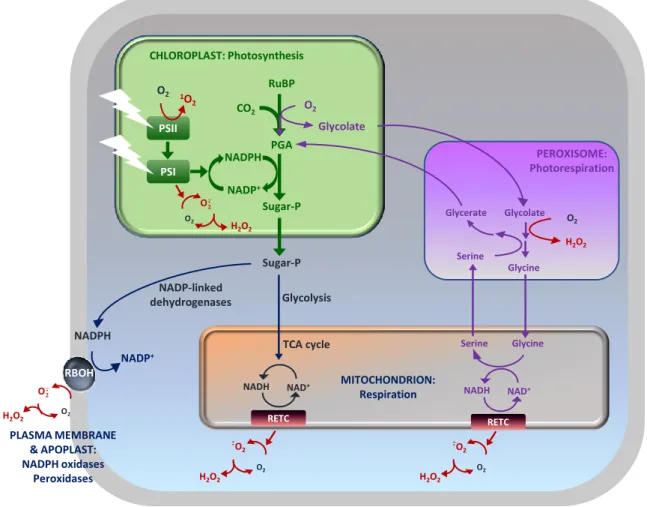
Many developmental processes involve signaling through ROS that are probably generated at lower levels than those associated with stress. It has been well established that reactions associated with photosynthesis, photorespiration and respiration are major sources of ROS. As described above, ROS can also be generated at the extracellular surface by NADPH-dependent oxidases, as well as in the apoplast by peroxidases and oxidases (Figure 1.2; Corpas et al., 2001; Foyer and Noctor, 2003; Asada, 2006; Mhamdi et al., 2012; Noctor et al, 2017) .
The chloroplast was one of the very first sources of ROS to be described in plants (Mehler, 1951; Asada et al., 1974). ROS generation occurs during photosynthesis at the sites of both PSI and PSII (Asada et al., 2006; Foyer et al., 2017). Specifically, singlet oxygen is produced by the photosensitizer Chl in the reaction center of PSII. When light absorption exceeds the capacity of photosynthetic electron transport, the lifetime of excited triplet states of Chl (3Chl) increases, favoring transfer of the absorbed light energy to ground-state triplet oxygen (3O
2) to form singlet oxygen (Hideg et al., 1998; Krieger-Liszkay, 2005). In contrast, PSI is considered to be a less important contributor to singlet oxygengeneration in photosynthetic organisms. However, it is the major site for the generation of superoxide anion and H2O2; that is, reduced forms of O2 (Mehler, 1951; Asada, 2000; Fryer et al.,
6 2002).
Figure 1.2. The basics of ROS formation in plants. PGA, 3-phosphoglycerate. PSI/II, photosystem I/II. RBOH, respiratory
burst oxidase homolog. RETC, respiratory electron transport chain. RuBP, ribulose 1,5-bisphosphate. Sugar-P, sugar phosphate. Figure taken from Noctor et al. (2017).
As well as the chloroplast, the peroxisome is an important site of ROS generation within plant cells, notably through photorespiration-related reactions (Mhamdi et al., 2012). During this pathway, which can be rapid, H2O2 is generated by the enzymatic activity of glycolate oxidase which catalyzes the oxidation of glycolate to glyoxylate. In addition, for oilseed plants such as Arabidopsis, fatty acid catabolism is a major source of ROS in peroxisomes during germination. The breakdown of lipid storage by β-oxidation leads to H2O2 accumulation as a result of acyl-CoA oxidase activity (Graham and Eastmond, 2002; Nyathi and Baker, 2006). In addition, H2O2 can also be formed by other enzyme systems such as xanthine oxidase coupled to SOD (Corpas et al., 2006).
CHLOROPLAST: Photosynthesis PSII PSI RuBP PGA Sugar-P NADPH NADP+ 1O 2 O2 O2 O· H2O2 CO2 Glycolate O2 PEROXISOME: Photorespiration Glycerate Glycolate Glycine O2 H2O2 Serine Sugar-P NADPH NADP+ RBOH O2 O· Glycolysis TCA cycle NADP-linked dehydrogenases NAD+ NADH RETC Glycine Serine NAD+ NADH RETC O2 -O 2 · O2 MITOCHONDRION: Respiration PLASMA MEMBRANE & APOPLAST: NADPH oxidases Peroxidases -O 2 ·
In animals, the mitochondrial electron transport chain is a major site of ROS generation (Liu et al., 2002). Complex I and III are the main sites of ROS production in mitochondria, whereby O2 is reduced to superoxide anion (Møller, 2001; Jezek and Hlavata, 2005). Subsequently, the superoxide anion is converted to H2O2 either by the action of MnSOD or by spontaneous dismutation (Finkel and Holbrook, 2000). Compared to chloroplasts and peroxisomes, the rate of H2O2 generation in plant mitochondria is probably lower (Foyer and Noctor, 2003; Halliwell and Gutteridge, 2015). In the light at least, the mitochondria are not regarded as a major source of ROS in leaves, although this obviously does not exclude an important function for these organelles in ROS-related signaling.
The apoplast is a major site of ROS generation. For example, during plant-pathogen interactions, ROS-producing enzymes are thought to be largely responsible for the apoplastic oxidative burst (Qi et al., 2017). This system consists chiefly of NADPH oxidases, cell wall peroxidases and amine oxidases (Petrov and Van Breusegem, 2012; Sierla et al., 2013). NADPH oxidases, also called respiratory burst oxidase homologs (RBOHs), are one of the major ROS-generating enzymes in plants (Umezawa et al., 2010; Mittler et al., 2011). As noted above, they transfer electrons from cytosolic NADPH to apoplastic oxygen, leading to the production of the superoxide anion, which is then converted to H2O2 spontaneously or by SOD. Ten NADPH oxidase genes have been identified in Arabidopsis. Among them, AtrbohD and AtrbohF play important roles in the hypersensitive response (HR; Torres et al., 2006; Chaouch et al., 2012; Kadota et al., 2015).
1.1.2.2 Stress and ROS
In optimal physiological conditions, redox homeostasis is maintained and ROS levels are relatively low. However, as sessile organisms, plants often endure environmental challenges that limit growth, reproductive success, yield, quality, or other traits desirable to humans. Collectively, these conditions are defined as stress, and are typically divided into abiotic and biotic stresses. Abiotic stress refers to the negative impact of non-living factors on organisms. Examples are excess salt, heavy metals, heat, chilling, insufficient water or high irradiance. The second is biotic stress that occurs due to damage instigated by other living organisms including bacteria, fungi, viruses, parasites, insects and other
8
native or cultivated plants. A central theme in many stress responses is the accumulation of ROS and ROS-induced changes in cellular redox state. This condition is commonly known as oxidative stress (Foyer and Noctor, 2000; Dat et al., 2001; Pastori and Foyer, 2002).
Pollution linked to heavy metals that are redox-active may result in superoxide anion formation because of autoxidation, and subsequently lead to H2O2 production via Fenton-type reactions (Li and Trush, 1993; Schutzendübel and Polle, 2002). Drought is another abiotic stress condition in which the roles of ROS have received much attention. In drought conditions, stomatal closure is induced, causing a drop in intracellular CO2 concentrations. This effect tends to favor RuBP oxygenation, thereby increasing glycolate production and accelerating H2O2 production in the peroxisomes (Cornic and Briantais, 1991; Noctor et al., 2014). Moreover, drought stress favors electron flux to O2 and restricts NADP+ regeneration. The rise in NADPH:NADP+ ratio will lead to promotion of the Mehler reaction, and increased photosynthetic control will tend to promote singlet oxygen production. Furthermore, over-reduction of the electron transport chain in chloroplasts and decreased availability of other oxidants for the chain during drought may enhance singlet oxygen, superoxide anion or H2O2 production (Asada, 2006; Fischer et al., 2013).
The above paragraph describes examples of how abiotic stress conditions can alter ROS production by their effects on basal metabolism. In addition to such effects, rapid programmed production of ROS, known as “oxidative bursts”, may also occur. Such processes have received particular attention for their importance in responses to biotic stress (Torres et al., 2006). Indeed, the first documented “oxidative burst” in plants was described following infection of potato tubers with the pathogenic oomycete, Phytophthora infestans (Doke, 1983). Since then numerous studies have shown that ROS production at the cell wall and apoplast is involved in biotic stress responses. Plasma membrane NADPH oxidases and cell wall peroxidases are thought to be the major sources of ROS production during plant-pathogen interactions, although intracellular redox reactions are also involved. NADPH oxidases, especially RBOHD and RBOHF in Arabidopsis, play a key role in apoplastic ROS generation in immune responses (Torres et al., 2002; Nühse et al., 2007; Zhang et al., 2007). In addition, it has been reported that class III apoplastic peroxidases PRX33 and PRX34 are essential for
ROS production in response to flg22 and elf18 in Arabidopsis (Daudi et al., 2012).
1.1.3 ROS signaling
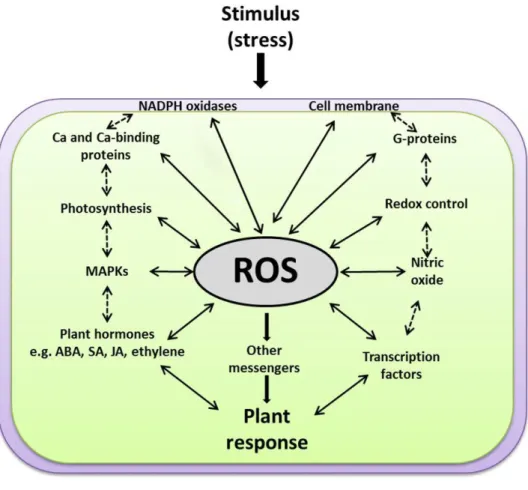
A long-held view of the biological role of ROS was that they are toxic by-products of plant metabolism through the processes described above (Asada, 1999). According to this view, they would have little or no importance in physiological processes, but rather cause damage to lipids, proteins and DNA in the stressed cells, leading to the notion that resistance to stress could be engineered by enhancing antioxidative capacity or by avoiding the production of ROS (Noctor and Foyer, 1998; Asada, 1999; Maxwell et al., 1999; Mittler, 2002). However, since the beginning of this century, substantial experimental data suggests that ROS are highly controlled signaling molecules. For ROS to be effective as signaling components, living cells have very efficient antioxidant systems to keep them under precise control (Foyer and Noctor, 2005a,b). Secondary messengers activated by ROS can regulate downstream pathways that are physically non-adjacent to the original ROS signaling pathway (Nathan, 2003). All these characteristics render ROS able to set specificity as signaling molecules. ROS function is intimately involved in the activation of gene expression, the induction of specific protein kinases and calcium signatures, the interaction with NADPH oxidases, hormone signal transduction, and the requirement of specific gene modulation for ROS-induced cell death (Figure 1.3; Kovtun et al., 2000; Desikan et al., 2001; Foreman et al., 2003; Kwak et al., 2003; Vandenabeele et al., 2003; Wagner et al., 2004; Vandenbroucke et al., 2008; Sewelam et al., 2016).
1.1.3.1 ROS and photosynthesis
Chloroplasts were one of the very earliest sources of ROS to be described in plants. As described above, ROS production in the thylakoid membrane is notably determined by the balance between light capture and the utilisation of light energy by the photosynthetic electron transport chain, which in turn depends on metabolic consumption of ATP and reductant for its continued operation. Any change or
10
Figure 1.3. A scheme explaining how ROS function at the cross-roads of various key signaling events. ROS work
upstream and downstream of the other signaling components, e.g. membranes, NADPH oxidases, G-proteins, calcium, redox homeostasis, photosynthesis, MAPKs, plant hormones [such as salicylic acid (SA), jasmonic acid (JA), abscisic acid (ABA), and ethylene] and transcription factors. Solid arrows for direct ROS interactions with other signaling components, dashed arrows for expected indirect interactions. Figure taken from Sewelam et al. (2016).
imbalance associated with photosynthesis in the chloroplast can affect, directly or indirectly, other functions in plants. It is not surprising that plants have evolved mechanisms to sense photosynthesis-associated ROS accumulation induced by environmental challenges, to enable the ensuing change of redox states in the chloroplast to influence other signaling pathways and lead to appropriate physiological responses (Huner et al., 1996). It has been shown that signal transmission from the chloroplast can change nuclear gene expression, which is proposed to act as a retrograde signaling pathway (Koussevitzky et al., 2007; Pogson et al., 2008; Pfannschmidt et al., 2009; Leister, 2012; Godoy Herz et al., 2014; Sewelam et al., 2016). For example, when pharmacological approaches were used to block carotenoid synthesis and hence increase the ability of ROS production in chloroplasts,
Arabidopsis seedlings suffered severe photo-oxidative damage. Simultaneously, ROS-responsive marker genes were rapidly up-regulated (Kim and Apel, 2013). Another approach used a genetic system of glycolate oxidase overexpression to perturb H2O2 levels in chloroplasts. The resulting ROS accumulation induced early signaling responses by regulating gene transcription and secondary signaling messengers, providing further evidence for ROS-dependent retrograde signaling pathways (Sewelam et al., 2014). The redox state of the plastoquinone pool, which plays a key role in electron transport in photosynthesis, was suggested to modulate the expression of cytosolic ascorbate peroxidase (APX) under excess light stress (Karpinski et al., 1997). Other studies also show that ROS signaling from chloroplast is involved in abiotic and biotic stress responses (Fryer et al., 2003; Agrawal et al., 2004; Joo et al., 2005; Belhaj et al., 2009; Zurbriggen et al., 2009; Serrato et al., 2013).
1.1.3.2 ROS and redox homeostasis
Redox regulation is an essential factor to keep energy conversion and consumption in plants under good control. When suffering environmental challenges, ROS perturbation is perceived and transformed into redox signals, followed by responses at various levels of regulation and in different subcellular compartments (Scheibe and Dietz, 2012). The major soluble non-protein redox couples in plants like glutathione, ascorbate, NAD and NADP, are at the hub of the redox network surrounding ROS generation and control. It was reported that photorespiration-derived H2O2 led to a photoperiodic effect on redox signaling, in which intracellular glutathione state was perturbed by the accumulation of glutathione disulfide (GSSG; Queval et al., 2007). The changes of intracellular redox state could mediate various signaling events through their interaction with many other secondary messengers such as protein kinase and phosphatases, phytohormones and calcium. Much evidence indicates that ROS-antioxidant interactions act as a metabolic interface between environmental changes and the ensuing signaling responses (Foyer and Noctor, 2005a, 2012), and this is partly accomplished through modifications of redox state (Dietz, 2008). The plant antioxidative systems that promote redox homeostasis in the face of constant ROS production are discussed further in Section 1.2.
12
1.1.3.3 ROS and Mitogen-activated protein kinase (MAPK)
MAPK signaling pathways have been well accepted as a general signal transduction mechanism in eukaryotes. These pathways consist of three functionally linked protein kinases: MAPK, MAPK kinase (MAPKK) and MAPK kinase kinase (MAPKKK). By protein phosphorylation cascades from MAPKKK to MAPK, this system links different receptors to their cellular and nuclear targets (Tena et al., 2001). MAPK signaling cascades in Arabidopsis are complex, involving multiple isoforms of each MAPK gene and more than 20 pathways (Wrzaczek and Hirt, 2001; Ichimura et al., 2002).
Many environmental constraints were found to stimulate MAPK cascades, like cold, heat, drought, wounding, and pathogens, while these cascades are also involved in phytohormone signaling (Bowler and Fluhr, 2000). In plants and eukaryotes in general, it has been demonstrated that the transmission of oxidative signals is controlled by protein phosphorylation involving MAPKs (Kyriakis and Avruch, 1996; Gustin et al., 1998; Pitzschke and Hirt, 2006; Xing et al., 2008). It was reported that H2O2 initiated MAPK signaling by activating ANP1, an isoform of MAPKKK. This effect led to the phosphorylation of MPK3 and MPK6 (Kovtun et al., 2000). Oxidative signal inducible 1 (OXI1), a protein kinase, is also involved in the activation of MPK3 and MPK6. Infection by virulent fungal pathogens caused HR in oxi1 null mutant, concomitantly the activation of MPK3 and MPK6 by oxidative stress was compromised (Rentel et al., 2004). Protein phosphorylation was also demonstrated to trigger Ca2+-dependent ROS production, which was mediated by NADPH oxidases in Arabidopsis. This study also suggested the existence of a positive feedback regulation of Ca2+ and ROS (Kimura et al., 2012).
1.1.3.4 ROS and phytohormones
Plant stress responses are highly integrated with hormone signaling to regulate biological processes. Salicylic acid (SA) signaling is associated with resistance to biotrophic pathogens as well as abiotic stress (Vlot et al., 2009). Intracellular and extracellular ROS accumulation can induce SA production, pathogenesis-related (PR) gene expression, and cell death (Chamnongpol et al., 1998; Torres et al.,
2005; Chaouch et al., 2010). All these effects can be reverted in the cat2 mutant by blocking SA synthesis, showing that H2O2-triggered cell death and related responses are not a direct consequence of damage (Chaouch et al., 2010). Intriguingly, the defence responses triggered by increased peroxisomal availability of H2O2 in cat2 occur in a photoperiod-dependent manner: the PR responses such as lesions and SA accumulation appear when plants are grown in long days (LD) but not short days (SD) (Queval et al., 2007; Chaouch et al., 2010). ROS-induced SA accumulation can have several effects, notably including enhanced resistance to pathogens through the induction of PR proteins and phytoalexins. In addition, it can also promote stomatal closure, which may also contribute to defence against pathogens that enter plants by this route (Khokon et al., 2011). The application of SA can also induce ROS production via a peroxidase-catalyzed reaction, which also favors stomatal closure (Melotto et al., 2006; Miura et al., 2013).
Unlike SA, JA and the related compound methyl jasmonate (MeJA) are involved in the response to necrotrophic pathogens and wounding (Devoto and Turner, 2005). Accumulating evidence reveals a strong relationship between ROS and JA signaling. For instance, ROS derived from NADPH oxidases are critical for induced gene expression regulated by MYC2, a transcription factor involved in JA-mediated response (Maruta et al., 2011). It was revealed that JA signaling in response to intracellular oxidative stress requires an accompanying accumulation of glutathione (Han et al., 2013b). A complex relationship between SA and JA in physiological processes has been elucidated. Many studies show the opposite effect between JA signaling and SA-dependent pathways (Dangl and Jones, 2001; Spoel et al., 2003; Takahashi et al., 2004; Koorneef et al., 2008). However, the two pathways may interact positively and can also be induced together. For example, it was demonstrated that increased H2O2 levels in cat2 induced both SA and JA signaling pathways, and that both pathways are less induced when glutathione accumulation is genetically blocked by the cad2 mutation (Han et al., 2013b). These observations point to some glutathione-dependent signaling process in the link between H2O2 and induction of phytohormone pathways, an issue that will receive attention in Chapters 4 and 5 of this thesis.
14
induced together with ROS production in different environmental stress responses. It has been reported that ABA-induced stomatal closure during stress is mediated by ROS which is derived from NADPH oxidases (Kwak et al., 2003). Besides, ABA is found to be required for H2O2 production in chloroplasts, mitochondria and peroxisomes under water stress (Hu et al., 2006). Moreover, ABA has been considered to play a negative role in biotic stress signaling orchestrated by SA, JA and ethylene (Coego et al., 2005). Ethylene is well documented to be a key player in programmed cell death during senescence, ozone stress or pathogen infection (Orzaez and Granell, 1997; Lund et al., 1998; Overmyer et al., 2000). Gibberellins (GA), a cyclic diterpene compound with multiple functions in the plant life cycle, are linked with ROS through DELLA proteins which modulate transcript levels of antioxidant enzymes (Achard et al., 2008).
1.1.3.5 Other components involved in ROS signaling
In addition to the aspects mentioned above, there are also some other second messengers such as GTP-binding proteins (G proteins) and Ca2+ which mediate the ROS signals. The role of G proteins in stress responses, especially in plant-pathogen interactions, has been extensively reported (Assmann, 2005; Trusov et al., 2009; Maruta et al., 2015). It was revealed that under ozone stress, the first biphasic oxidative burst is greatly attenuated or absent in mutants lacking Gα protein or Gβ protein (Joo et al., 2005). AtRbohD and AtRbohF were suggested to receive initial signals from G proteins to mediate ozone responses in guard cells (Suharsono et al., 2002). Calcium signaling is involved in many signal transduction pathways. Elevations in cytosolic Ca2+ represent an early response to many different biotic and abiotic stresses (McAinsh and Pittman, 2009; Dodd et al., 2010). ABA and H2O2 treatments are among those that can increase cytosolic Ca2+ concentrations. Moreover, Ca2+ has been reported to work both upstream and downstream of ROS production in signaling pathways (Bowler and Fluhr, 2000; Abuharbeid et al., 2004; Monshausen et al., 2009; Sewelam et al., 2013).
1.2 ROS processing
The above discussion underlines the close integration of ROS-dependent redox signaling in numerous physiological processes, while also revealing that our knowledge of the details of redox signaling remains incomplete. Although many downstream responses to ROS have been described, the key redox events that lie at the heart of ROS-triggered signaling remain to be elucidated. A key player in enabling and regulating ROS signals is the plant antioxidative system. This system is complex and includes efficient enzymatic and non-enzymatic mechanisms that have been developed during evolution. Enzymatic systems consist of CAT, APX, SOD, various types of peroxiredoxins (PRX), glutathione/thioredoxin peroxidases (GPX) and glutathione S-transferases (GST). Non-enzymatic components include glutathione and ascorbate, which can be considered major components of a redox hub, but also tocopherol, carotenoids and phenolic compounds. Through the network of these mechanisms, plants regulate ROS accumulation and maintain redox homeostasis (Figure 1.4;Noctor et al., 2017).
1.2.1 Catalase
CAT was the first discovered antioxidative enzyme. All known CAT forms in eukaryotes are haem-dependent. Among them two main types have been characterized, which are monofunctional CATs (also known as typical CATs) and bifunctional CAT-peroxidases (Zamocky et al., 2008). They can be distinguished by the affinity for H2O2 and the sensitivity to the inhibitor 3-amino-1,2,4-triazole (3-AT) (Margoliash and Novogrodsky, 1960; Regelsberger et al., 2002). Unlike monofunctional CATs, which exist in diverse organisms, the CAT-peroxidases are only found in some fungi and prokaryotes (Mutsuda et al., 1996; Regelsberger et al., 2002).
The typical CAT reaction is the dismutation of H2O2. The reaction is initiated by splitting the O-O bond of H2O2 to produce a molecule of H2O, as well as an oxy-ferryl enzyme intermediate (compound I) and a porphyrin cation radical. Then a second H2O2 is oxidized to O2. Simultaneously, compound I is reduced back to the initial state by releasing the bound O which is involved in the formation of the second molecule of water (Regelsberger et al., 2001; Alfonso-Prieto et al., 2009). Intriguingly,
16
monofunctional CATs can also catalyze some H2O2-dependent peroxidation of reducing substrates, making the functional division between the two types of CAT indistinct. During this process, the reduction of compound I is performed by the interaction with small compounds like ethanol instead of the second H2O2 (Zamocky et al., 2008). CAT-associated peroxidation has been reported in both mammalian and plants (Havir and McHale, 1989; Kirkman and Gaetani, 2007), but its physiological significance remains as yet unclear.
Figure 1.4. Plant antioxidative systems and their localization within the cell. APX, ascorbate peroxidase. CAT, catalase.
DHAR, dehydroascorbate reductase. GR, glutathione reductase. GRX, glutaredoxin. MDHAR, monodehydroascorbate reductase. NTR, NADPH-thioredoxin reductase. PRX, peroxiredoxin. SOD, superoxide dismutase. TRX, thioredoxin. Figure taken from Noctor et al. (2017).
CATs are notably distinguished from other H2O2-metabolizing enzymes based on two factors. Firstly, when functioning as a dismutase, CAT function does not require reductant other than H2O2. Secondly,
CHLOROPLAST 1O 2 Carotenoids Tocopherols O· CuZnSOD H2O2 FeSOD PRX Ascorbate Glutathione MDAR DHAR GR TRX NTR GRX APX
Regenerators of reduced forms
APX CuZnSOD FeSOD CAT O· H2O2 PEROXISOME
Regenerators of reduced forms Ascorbate Glutathione MDAR GR PRXII CuZnSOD APX O· H 2O2 Ascorbate Glutathione PRXII MnSOD CuZnSOD APX O· H2O2 Ascorbate Glutathione PRX H2O2 Ascorbate Glutathione NUCLEUS CYTOSOL MITOCHONDRION MDAR GR TRX NTR GRX Regenerators of reduced forms GRX GR TRX NTR GRX Regenerators of reduced forms DHAR GRX TRX NTR Regenerators of reduced forms MDAR GR
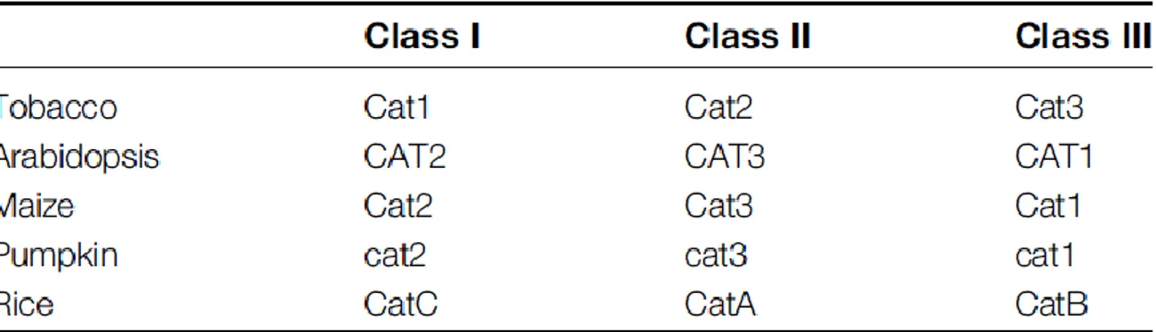
CAT has high specificity for H2O2. So far three CAT genes have been identified in angiosperm species such as Arabidopsis, rice, tobacco, maize and pumpkin (Willekens et al., 1995; Frugoli et al., 1996; Guan and Scandalios, 1996; Esaka et al., 1997; Iwamoto et al., 2000). In Arabidopsis, CAT1 and CAT3 are located contiguously on chromosome 1 while CAT2 is located on chromosome 4 (Frugoli et al., 1996). According to the classification system based on tobacco genes and first proposed by Willekens et al. (1995), Class I CATs are mainly expressed in photosynthetic tissues, Class II CATs are associated with vascular tissues, while Class III CATs are notably expressed in seeds and reproductive tissues. For Arabidopsis, CAT1, CAT2 and CAT3 correspond to Class III, Class I and Class II, respectively (Table 1.1). The CAT1 gene is mainly expressed in pollens and seeds and at very low levels in leaves, CAT2 is expressed in photosynthetic tissues but also in roots and seeds, while CAT3 is associated with vascular tissues. It has been reported that compared to CAT1, the transcript levels of CAT2 and CAT3 are much higher in mature Arabidopsis rosettes (Frugoli et al., 1996; McClung, 1997). In addition, CAT2 transcripts show a day-night rhythm with a peak expression at the night/day transition, which is opposite to that of CAT3 (Zhong et al., 1994; Mhamdi et al., 2010a). Studies on subcellular localization showed that CATs exist mainly in peroxisomes in plants. Evidence supporting this conclusion includes the high CAT activity in peroxisomes and the identification of an import mechanism due to a Peroxisomal Targeting Sequence 1 (PTS1) pathway (Mullen et al., 1997).
Table 1.1. Probable classification of the three CATs found in different plant species (taken from Mhamdi et al., 2010a)
Previous studies on gene-specific T-DNA insertion mutants showed that CAT2 encodes the major leaf CAT isoform and this enzyme makes the major contribution to leaf CAT activity (Queval et al., 2007; Mhamdi et al., 2010a). In cat2 knock-out mutants, the CAT activity in rosettes is decreased by around
18
90% compared with that in wild type, and root CAT activity is also attenuated, though less severely (Bueso et al., 2007). In cat3, there is around 20% activity lost. In contrast, the effect of the cat1 mutation on leaf CAT activity is negligible. This is consistent with its relatively low transcript levels in leaves. To date, therefore, it seems that CAT2 and CAT3 may play the major roles in Arabidopsis rosette tissue. However, the relative contribution of these CAT isoforms changes according to the developmental stage of the plants (Zimmermann et al., 2006). For instance, as a senescence-associated gene, CAT3 transcripts increase with leaf age.
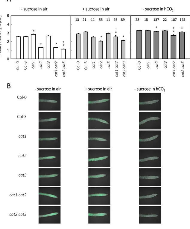
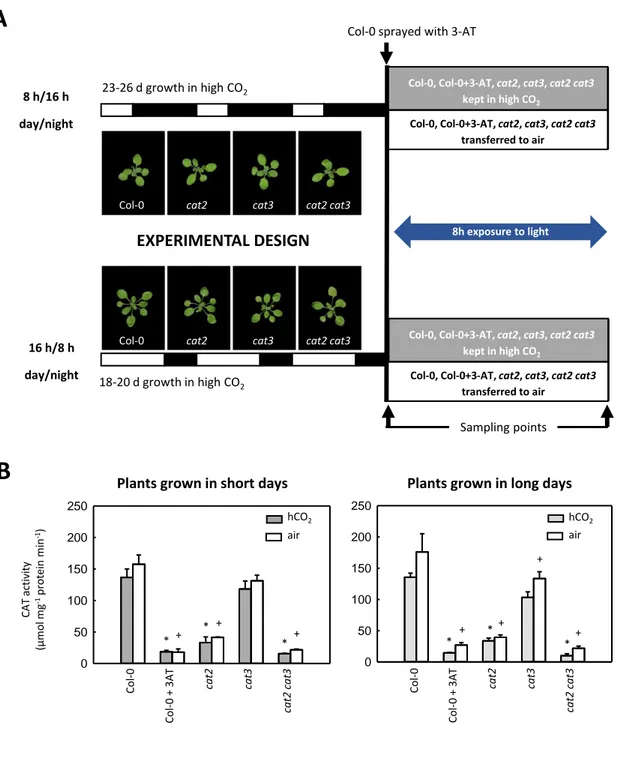
Because of the close association with photorespiration, Arabidopsis CAT2-deficient mutants (cat2) have been extensively used in various studies of plant development and physiology. Under conditions where photorespiratory H2O2 production is highly active, CAT function deficiency leads to increased availability of endogenous H2O2, with effects on cell redox state. The intracellular H2O2 signal can be modulated by changing photorespiration-related growth conditions, such as CO2 concentration and irradiance (Queval et al., 2007). When grown in air (400 µL CO2 L-1) with a moderate irradiance of 200 μmol m-2 s-1 at the surface of leaves, cat2 shows a dwarf phenotype accompanied by redox perturbation, evidenced by the decreases in the GSH:GSSG ratio and increases in total glutathione. By contrast, it has a wild-type phenotype when grown in high CO2 (hCO2; 3000 µL L-1) or if the irradiance is lower than 50 μmol m-2 s-1, both conditions in which photorespiration is rather inactive.
Among the most obvious phenotypes of cat2 grown in air at moderate light intensity are necrotic lesions. This phenotype is photoperiod-dependent. When grown in a 16 h photoperiod (LD), cat2 shows spreading necrotic lesions. By contrast, no lesions are observed in plants grown in an 8 h photoperiod (SD), even though the dwarf phenotype and redox perturbation are as marked as in LD (Queval et al., 2007, 2009). The lesion appearance in cat2 bears a striking resemblance to the HR, and includes SA accumulation, PR genes induction (e.g. PR1 and PR2), activation of camalexin and its synthesis pathway, and induced resistance to bacterial challenge. As well as necrotic lesions, all these effects are absent in SD. By the combination of pharmacological and genetic approaches, it is revealed that exogenous SA treatment could induce these responses in SD and revert it in LD when SA synthesis is genetically blocked. All these observations show that SA is involved in the peroxisomal
H2O2-triggered HR-like lesion formation in cat2 grown in LD air condition (Chaouch et al., 2010).
Because of their influence on cell redox state, peroxisomal CATs act as regulators to fine-tune redox signaling, which is also a function of the carbon flux through photorespiration. This makes cat2 mutants an interesting model system to investigate the possible relationship between photorespiration and other metabolic, transcriptional and physiological processes, especially those possessing a redox component. Reductive pathways for H2O2 processing appear to compensate quite rapidly when CAT is deficient. Even though glutathione redox status is perturbed within hours after the onset of photorespiratory H2O2 production in cat2, little or no change is found in the redox states of glutathione-associated redox compounds, like ascorbate/dehydroascorbate or NADPH/NADP+ (Queval et al., 2007; Mhamdi et al., 2010b,c). Glutathione status appears to play an active part in the H2O2 -triggered signal transduction provoked by CAT deficiency. For example, the GR knockout line gr1, which shows qualitatively similar changes in glutathione to those observed in cat2, has a wild-type phenotype, but nevertheless shows gene expression patterns that partly recapitulate those observed in cat2. Further, when the gr1 mutation is introduced into the cat2 background, H2O2-associated transcript profiles are significantly affected, suggesting that glutathione plays a role in transmitting H2O2-induced signals (Mhamdi et al., 2010b). Further evidence for this conclusion was obtained by analysis of cat2 lines in which glutathione accumulation was genetically impaired (Han et al., 2013a,b).
Extensive evidence reveals that phytohormones are important in determining cat2 phenotypes. Transcriptome analysis of cat2 roots points to the modulation of ethylene and auxin signaling (Bueso et al., 2007). Another study showed that the decreased CAT activity in cat2 induced effects on ABA signaling (Jannat et al., 2011). Glutathione was shown to be a key player in linking H2O2 to SA signaling (Han et al., 2013a). It was also reported that CAT2 participates in SA-mediated repression of auxin accumulation and JA biosynthesis during pathogen infection (Yuan et al., 2017). Another study reported that CAT2 and APX1 work in a coordinated way during the nuclear DNA damage response (DDR). Compared with the cat2 single mutant, the apx1 cat2 double mutant is more tolerant to oxidative stress imposed by high light, heat and paraquat application, and genome-wide
20
transcriptome analysis shows an activation of typical DDR hallmarks (Vanderauwera et al., 2011).
1.2.2 Ascorbate and glutathione in ROS metabolism
Many compounds function as effective antioxidants in cells by regulating ROS accumulation. Among the best studied players are the cellular redox buffers, ascorbate and glutathione. They are well accepted to be key in controlling concentrations of ROS, although both metabolites are involved in multiple physiological processes (Cobbett et al., 1998; Vernoux et al., 2000; Dowdle et al., 2007). Ascorbate and glutathione are distinguished from most other antioxidant small molecules by (1) specific enzyme systems (peroxidases) that couple them to H2O2 metabolism; (2) the relative stability of their oxidized forms; (3) recycling of oxidized forms to reduced compounds by high-capacity reductases and associated systems that depend on the key electron carrier, NAD(P)H. Based on these features, ascorbate and glutathione can effectively regulate cellular redox state by repeated redox cycling (Foyer and Noctor, 2011). The pools of these two metabolites are generally highly reduced (over 95%) within the cytosol, chloroplasts and mitochondria, while the oxidized forms accumulate in compartments lacking efficient redox-recycling mechanisms like the vacuole and the apoplast (Schwarzländer et al., 2008; Queval et al., 2011; Noctor et al., 2016).
1.2.2.1 Glutathione in plants
Glutathione (γ-glutamylcysteinylglycine) is the principal low-molecular-weight thiol in most cells. It is an essential metabolite with multiple functions in plant development, biosynthetic pathways, detoxification, antioxidant biochemistry, and redox homeostasis. The fundamental function of glutathione is in thiol-disulfide interactions, in which the interconversion of reduced glutathione (GSH) and GSSG allows an appropriate cell redox state to be achieved. The concentration of cellular glutathione is high, as is its reduction state in the absence of stress. Under optimal physiological conditions the average ratio of GSH:GSSG in tissues such as leaves is at least 20:1 (Mhamdi et al., 2010a; Han et al., 2013a). However, this value may be higher (e.g. cytosol) or lower (e.g. vacuole) in specific subcellular compartments (Meyer et al., 2007; Queval et al., 2011).
In plants glutathione is synthesized by two ATP-dependent steps which rely on the activity of γ-EC synthetase (γ-ECS) and glutathione synthetase (GSH-S) (Rennenberg, 1980; Meister, 1988; Noctor et al., 2002; Mullineaux and Rausch, 2005). Each synthetic enzyme is encoded by a single gene (May and Leaver, 1994; Ullman et al., 1996), and both are indispensable to plant growth and development. Knocking out GSH1, the gene encoding γ-ECS, leads to lethality at the embryo stage (Cairns et al., 2006) while knocking out GSH2, the gene encoding GSH-S, causes a seedling-lethal phenotype (Pasternak et al., 2008). In Arabidopsis γ-ECS is located in plastids (Wachter et al., 2005), while GSH-S is found in both chloroplasts and cytosol, with the latter compartment considered to contain the major form of GSH-S because of the higher abundance of the corresponding transcript (Wachter et al., 2005). Glutathione synthesis, which responds to factors such as oxidative stress or heavy metals, can be modulated by regulating γ-ECS expression and activity, or by changing cysteine availability (Strohm et al., 1995; Noctor et al., 1998; Creissen et al., 1999; Harms et al., 2000; Noji and Saito, 2002; Wirtz and Hell, 2007). Both GSH1 and GSH2 transcripts respond to phytohormone and heavy metals (Xiang and Oliver, 1998; Sung et al., 2009), as well as to light, drought and pathogens.
While glutathione synthesis is stress-responsive, degradation is also important to achieve appropriate levels. In Arabidopsis there are four different types of enzyme which have been implicated in glutathione degradation. These are phytochelatin synthase (PCS), glutamyl transpeptidase (GGT), γ-glutamyl cyclotransferase (GGC), and 5-oxoprolinase (5-OPase). Most attention has thus far focused on GGT, which is considered to degrade mainly GSSG located in the vacuole or the apoplast (Masi et al., 2007; Ferretti et al., 2009; Ohkama-Ohtsu et al., 2011; Su et al., 2011).
1.2.2.2 Ascorbate in plants
Ascorbate, also known as vitamin C, is a highly abundant metabolite and a water-soluble antioxidant in both animals and plants. It occurs in a reduced form (ascorbic acid) and two oxidized forms, which are the immediate product of one-electron oxidation (monodehydroascorbate, or MDHA) and dehydroascorbate (DHA), which has lost two electrons compared to ascorbate and is largely the result of MDHA dismutation. Homeostasis between reduced and oxidized ascorbate is essential for plants to
22
cope with environmental stress. Ascorbate is found in different subcellular compartments like chloroplasts, mitochondria, peroxisomes, and vacuoles (Jiménez et al., 1997, 1998; Noctor et al., 2002; Takahama, 2004; Zechmann, 2011). It has been well documented that ascorbate can be synthesized through the L-galactose (L-Gal) pathway starting from glucose (Linster and Clarke, 2008; Wheeler et al., 1998, 2015; Bulley and Laing, 2016). It was shown that vtc1 and vtc2 mutants, deficient in the enzymes associated with this pathway, contain only 25-30% and 10-20% of wild type ascorbate levels, respectively. Furthermore, the loss of ascorbate biosynthetic capacity in the vtc2 vtc5 mutant cannot be compensated by other identified ascorbate biosynthetic pathways (Dowdle et al., 2007). Hence, the L-Gal pathway is suggested to be the primary pathway that generates ascorbate in most plant organs. Nevertheless, alternative ascorbate biosynthetic pathways through galacturonate and glucuronate have also been proposed (Li et al., 2010). Ascorbate has been considered of great importance in plants with key functions in growth development, metabolism and stress responses (Noctor and Foyer, 1998; Smirnoff, 2000; Pastori et al., 2003; Anjum et al., 2014). The finding that near-total ascorbate depletion in the vtc2 vtc5 mutant is associated with seedling lethality (Dowdle et al., 2007) suggests that, like glutathione, ascorbate is an indispensable player during at least some stages of plant development.
1.2.2.3 Ascorbate-glutathione pathway
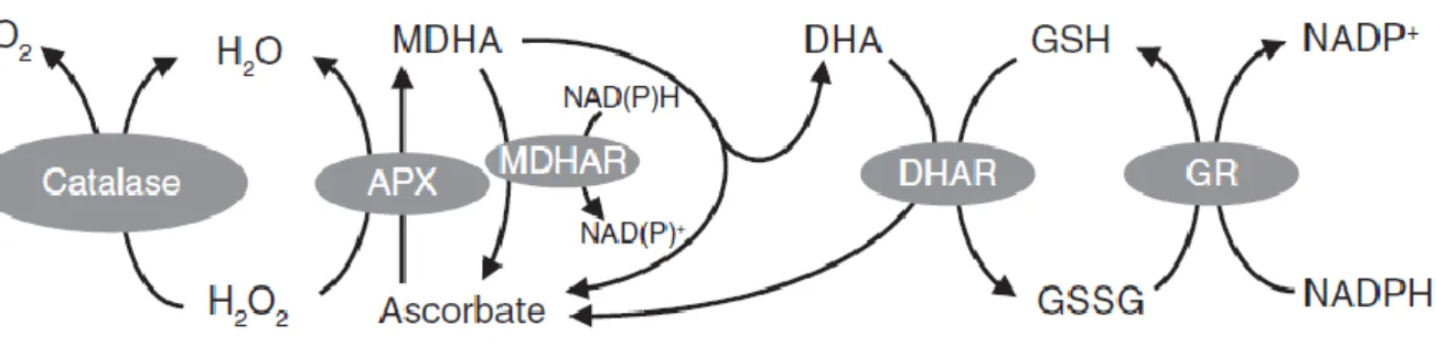
While ascorbate and glutathione can react chemically with ROS, and also have other functions as enzyme co-factors and in defense, their roles in the ascorbate-glutathione pathway continue to be a major research focus. The pathway is involved in ROS metabolism in plant cells by removal of H2O2 (Figure 1.5;Foyer and Noctor, 2009). Like CATs, APX has high specificity for H2O2. APX can reduce H2O2 to water by consuming two molecules of ascorbate, which leads to the generation of two molecules of MDHA. This primary oxidation product can be converted back to ascorbate by MDHA reductase (MDHAR) in an NAD(P)H-dependent reaction. If not reduced rapidly, MDHA will disproportionate to ascorbate and DHA because of its characteristic as a short-lived radical. GSH can reduce DHA to regenerate ascorbate, a reaction catalyzed by DHA reductases (DHAR), accompanied by the production of GSSG. In addition to MDHAR and DHAR, non-enzymatic routes for ascorbate
regeneration exist, including the reduction of DHA by GSH or of MDHA by chloroplast ferredoxin (Polle, 2001; Smirnoff, 2011; Gest et al., 2013; Johnston et al., 2015; Noctor, 2015). Consumption of GSH in this pathways leads to GSSG, which can be reduced back to GSH by the action of glutathione reductase (GR). This reaction needs electrons provided by NADPH. During this sequence of reactions, ascorbate and glutathione are not consumed: instead they are involved in a cyclic transfer of reducing equivalents by the action of four enzymes, which permits the reduction of H2O2 to H2O using electrons derived from NAD(P)H (Foyer and Halliwell, 1976; Noctor and Foyer, 1998; Foyer and Noctor, 2009).
Figure 1.5. Two of the major pathways for H2O2 metabolism in plants. APX, ascorbate peroxidase; MDHA,
monodehydroascorbate; MDHAR, monodehydroascorbate reductase; DHA, dehydroascorbate; DHAR, dehydroascorbate reductase; GSH, reduced glutathione; GSSG, glutathione disulfide; GR, glutathione reductase. Figure taken from Foyer and Noctor (2009).
Both ascorbate and glutathione are abundant and stable antioxidants which may be maintained at thermodynamic equilibrium with each other under optimal conditions. Within the highly reducing intracellular context, ROS accumulation can lead to the transient or sustained adjustment of this system, and such adjustments can be sensed by signaling pathways, leading to downstream responses. For glutathione, there is a close correlation between its status and the expected intracellular ROS availability. For example, it has been extensively reported that pharmacologically or genetically blocking CAT activities causes glutathione to become both more oxidized and more abundant (Smith et al., 1985; May and Leaver 1993., Willekens et al., 1997; Queval et al., 2009; Mhamdi et al., 2010a). Work going back over two decades has pointed to novel outcomes of modulating parts of the ascorbate-glutathione pathway, suggesting that the pathway does not function simply as an
24
antioxidative system but may also act in redox signaling. For example, cytosolic DHARs are involved in the regulation of glutathione status triggered when H2O2 metabolism shifts from CAT to reductive pathways. Genetically blocking these enzymes weakens, rather than reinforces, cell death and defense responses induced by CAT deficiency (Rahantaniaina et al., 2017). Another report showed that MDHAR can act as a pro-oxidant rather than an antioxidant by conferring sensitivity to soil-borne pollutant 2,4,6-trinitrotoluene (TNT) on Arabidopsis (Johnston et al., 2015; Noctor, 2015).
One current view is that perturbation of the status of the ascorbate-glutathione pathway may be important in signaling, i.e., the pathway has a dual antioxidant-signaling role in which some downstream responses are triggered by increased H2O2 metabolism through the pathway, rather or in addition to increased H2O2 concentrations (Foyer and Noctor, 2016). In such a scenario, glutathione status seems likely to be an influential player, based on possible interactions with protein thiol-disulfide groups. In contrast to glutathione, the ratio of ascorbate to DHA is less likely to be involved in the transmission of ROS signals. Instead, ascorbate limits the lifetime of ROS signals by scavenging ROS chemically and enzymatically. Ascorbate contents are sensitive to factors such as irradiance (Gatzek et al., 2002; Smirnoff et al., 2011), and this response may be partly related to functions in tocopherol regeneration and xanthophyll cycle.
1.2.2.4 Other pathways of ROS processing
In addition to CATs and the ascorbate-glutathione pathway, plants also contain other antioxidative systems to cope with H2O2 accumulation. Among them, PRX has been well documented to function in maintaining appropriate H2O2 levels, at least in chloroplasts (Awad et al., 2015). PRX is a thiol-dependent peroxidase existing in almost all organisms. Depending on the isoform, PRX regeneration from the oxidized form to the reduced state can be accomplished by glutaredoxin (GRX), thioredoxin (TRX), NADPH-thioredoxin reductase (NTR), glutathione or ascorbate (Dietz, 2003; Rouhier and Jacquot, 2005; Meyer, 2008; Pérez and Cejudo, 2009; Liebthal et al., 2018). GPX is also able to remove H2O2 but these enzymes are likely to function with TRX instead of GSH (Herbette et al., 2002; Iqbal et al., 2006). GST could also play a role as GSH-dependent peroxidases to remove H2O2 (Dixon
et al., 2009), although their exact importance in this respect is still not clear.
All the enzymes discussed above are found in the aqueous cell phases. Within the membrane phase, carotenoids and tocopherols are well known as important compounds to control the accumulation of ROS and derived molecules. Carotenoids can act either to avoid ROS production (e.g. by quenching excited Chl states) or react with existing ROS by quenching singlet oxygen (Fischer et al., 2013). Most evidence available so far suggests that the functions of tocopherols (vitamin E) are more essential in seeds than in leaves. Tocopherol-deficient mutants of Arabidopsis show clear effects associated with enhanced lipid peroxidation and the activation of defense pathways during germination (Sattler et al., 2006).
1.2.2.5 NADPH-linked reaction in plants
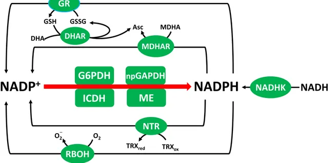
NADPH is a key redox carrier in the aqueous phase of many cells and plays a crucial role in physiological processes like cell growth, proliferation, detoxification, redox homeostasis and biosynthesis. It is the reductant required for sugar-phosphate production in the photosynthetic Calvin-Benson cycle and is also required for other key anabolic pathways such as lipid and amino acid synthesis. Alongside its roles in biosynthetic pathways, NADPH functions in defence reactions and the regulation of cellular redox status. For example, in the ascorbate-glutathione pathway, NADPH is required for the reactions catalyzed by GR and MDHAR (Figure 1.5; Noctor, 2006) while TRX needs NADPH to protect against oxidative damage (Nordman et al., 2003). The functions of NADPH are not limited to providing electrons for biosynthetic reactions and antioxidative enzymes. Indeed, NADPH may have a pro-oxidant function, for example, in providing reductant for plasma membrane NADPH oxidase that produce suyperoxide and, subsequently, H2O2 (Foreman et al., 2003; Torres et al., 2006). NADPH is also a cofactor for the generation of nitric oxide (NO) from arginine, catalyzed by animal-type NO synthases (Corpas et al., 2009), although the existence of such enzymes in plants is controversial.