ACADEMIE UNIVERSITAIRE WALLONIE-EUROPE UNIVERSITE DE LIEGE
FACULTE DE MEDECINE VETERINAIRE
DEPARTEMENT GIGA-INFLAMMATION, INFECTION AND IMMUNITY LABORATOIRE D'IMMUNOLOGIE CELLULAIRE ET MOLÉCULAIRE
Développement d’outils moléculaires de détection des moisissures présentes dans
l’air intérieur afin de déterminer leur impact sur la santé publique
Development of molecular tools for rapid detection and quantification of indoor airborne molds
to assess their impact on public health
Libert Xavier
THESE PRESENTEE EN VUE DE L’OBTENTION DU GRADE DE
DOCTEUR EN SCIENCES VETERINAIRE
ACADEMIE UNIVERSITAIRE WALLONIE-EUROPE UNIVERSITE DE LIEGE
FACULTE DE MEDECINE VETERINAIRE
DEPARTEMENT GIGA-INFLAMMATION, INFECTION AND IMMUNITY LABORATOIRE D'IMMUNOLOGIE CELLULAIRE ET MOLÉCULAIRE
Développement d’outils moléculaires de détection des moisissures présentes
dans l’air intérieur afin de déterminer leur impact sur la santé publique
Development of molecular tools for rapid detection and quantification of
indoor airborne molds to assess their impact on public health
Libert Xavier
THESE PRESENTEE EN VUE DE L’OBTENTION DU GRADE DE
DOCTEUR EN SCIENCES VETERINAIRE
Supervisors
Prof. F. Bureau, ULg
Dr. Ir. S. De Keersmaecker, WIV-ISP
Dr. Ir. A. Packeu, WIV-ISP
Dr. Ir N. Roosens, WIV-ISP
Members of the Examination Committee
Prof. B. Dewals, ULg
Prof. P. Le Cann, EHESP, France
Prof. J.-B. Watelet, UGent
Prof. C. Charlier, ULg
Prof. D. Cataldo, ULg
Prof. B. Mignon, ULg
Prof. D. Peeters, ULg
Dissertation presented for the degree of
Doctor in Veterinary Sciences (ULg)
i
Résumé
Aujourd’hui, la contamination de l'environnement intérieur par des moisissures aéroportées est considérée comme un problème de santé publique. Les méthodes analytiques classiques de surveillances, basées sur la culture et l'identification microscopique, présentent des limitations liées à la dépendance vis-à-vis de la culture et du temps requis pour les analyses.
Par conséquent des biais peuvent être introduits, notamment concernant la fraction morte (champignons non-cultivables ou morts), pouvant avoir potentiellement un impact sur la santé humaine.
Dans ce contexte, les outils moléculaires semblent être d’excellentes alternatives pour la surveillance des contaminations fongiques aéroportées d’intérieurs. Ainsi, différents outils moléculaires ont été développés lors de cette thèse pour détecter et identifier les champignons dans l'air intérieur (qPCR SYBR®green, High Resolution Melting (HRM), Luminex xMAP® et NGS). L'objectif étant d'améliorer la détection des contaminants fongiques, la fraction morte inclue, par rapport aux méthodes de surveillance classiquement utilisées, mais également d’améliorer les connaissances actuelles sur la contamination fongique aéroportée d’intérieur.
Ainsi, un test PCR en temps réel (qPCR) a été développé pour la détection d’Aspergillus versicolor, une moisissure pathogène de l’air intérieur,et d’Exophiala jeanselmei, une moisissure pathogène suspectée de faire partie de la "fraction morte". Bien que validée selon des critères stricts prouvant la qualité des outils développés, des limites, notamment concernant la discrimination des espèces génétiquement proches et le multiplexage ont été observées.
Dans cette thèse, la première question a été résolue par l'utilisation d’une analyse post-PCR High Resolution Melting (HRM). Utilisé tel un proof of concept, le HRM a été testé sur 3 Aspergillus génétiquement proches (A. versicolor, Aspergillus creber et Aspergillus sydowii) démontrant que son utilisation peut améliorer le suivi de ce type de contaminants.
La problématique du multiplexage a été résolue grâce à la technologie Luminex xMAP®. Développé pour la détection simultanée de 10 moisissures fréquemment observées dans l'air intérieur. Ainsi, il a été démontré que l’introduction de cette technique dans un protocole de monitoring permettrait de réduire le temps d’analyse et, in fine, le temps de transmission des résultats à l’équipe médicale en charge du patient. Cependant, puisqu’une sélection des espèces à identifier est nécessaire avant l’analyse, cette technologie ne convient pas pour l'étude de la diversité fongique.
Dans ce contexte, la technologie next generation sequencing (NGS) semble offrir une alternative valable en tant qu’outil universel d’identification des moisissures intérieures. Dans cette étude, la fraction « morte » a été investiguée grâce à une analyse NGS métagénomique, permettant par la
ii même occasion de détecter pour la première fois E. jeanselmei dans l'air intérieur. De plus, une analyse de la diversité fongique de l’air intérieur provenant de résidences contaminées a été réalisée démontrant qu’une analyse NGS métagénomique peut contribuer à l'amélioration des données sur la diversité fongique aéroportée d’intérieur, nécessaire au développement de méthodes de détections, ainsi que de tests immunologiques représentatifs de cette diversité.
Les méthodes développées ainsi que les résultats obtenus lors de ce doctorat sont une première étape pour une meilleure compréhension du lien existant entre les champignons aéroportés présents dans l’intérieur de l’habitat et la santé publique.
iii
Summary
Currently, contamination of the indoor environment by fungi is suggested to be a public health problem, although scientific evidence on the causal link is still limited. The monitoring of indoor airborne fungal contamination is a common tool to help understanding the link between fungi in houses and respiratory problems. Classical monitoring methods, based on cultivation and microscopic identification, have some limitations. For example, uncultivable or dead fungi (“unknown” fraction) cannot be identified, although they could have an impact on human health.
In this context, molecular tools seem to be a valuable alternative. In this PhD work, different molecular tools were developed, from simplex to multiplex, to detect and identify indoor airborne fungi. The goal was to improve the detection of fungal contaminants, including the “unknown” fraction, as compared to the currently used classical monitoring methods. The necessary air sampling and DNA extraction protocols, adapted to the downstream molecular monitoring methods have also been developed. Through the application of the developed tools to specific case studies, we aimed to improve the current knowledge on fungal contamination.
At first, we developed a specific ITS-based SYBR®green real-time PCR (qPCR) assay for Aspergillus versicolor, a species frequently observed in indoor air and known to be allergenic. Additionally, an ITS-based qPCR assay was developed for the specific detection of Exophiala jeanselmei, a pathogenic yeast suspected to be a part of the “unknown fraction”. The performance of these qPCR methods was assessed. This comparison demonstrated that SYBR®green qPCR assays can be used as a molecular alternative for monitoring of contaminated samples while eliminating the need for culturing and thereby considerably decreasing the required analysis time.
However, qPCR has some limitations especially concerning the discrimination of genetically close species and multiplexing. The first issue was addressed through the use of post-qPCR high resolution melting (HRM) analysis, providing a proof-of-concept for this approach, using 3 closely related Aspergillus, i.e., A. versicolor, Aspergillus creber and Aspergillus sydowii. This HRM tool will allow a more accurate monitoring of these closely related indoor air contaminants, thereby contributing to an improved insight in the causal link between the specific presence of these species and health issues.
The multiplexing issue was overcome through a Luminex xMAP® assay, developed for the simultaneous detection of the 10 most frequently in indoor air found fungi. All the species identified with the classical method were also detected with the xMAP® assay, however in a shorter time frame, and using less sample material. This assay will improve the communication with the involved medical team and the patient.
iv problems, the full diversity needs however to be identified. This cannot be achieved by using a targeted assay. Therefore, next generation sequencing (NGS) could offer a valuable alternative as an open approach multiplex monitoring method. An NGS-based metagenomics approach was used to investigate the “unknown” agents in air samples of offices in contact with air-conditioning reservoirs and showed the first detection of E. jeanselmei in indoor air. Finally, a metagenomics analysis was performed to investigate the indoor airborne fungal diversity in contaminated residences in Brussels where people with health problems were living. This demonstrated that NGS could contribute to improved data concerning the indoor airborne fungal diversity, as compared to the currently used classical methods.
The methods developed in this PhD work and the insights obtained are a first step for a better understanding of the causal link between indoor airborne fungi and public health.
v
Abbreviations
µl microliter
∆ Cq quantitative cycle difference
∆G Gibb’s energy
Ac Avogadro’s constant
Aversi_ITS assay qPCR SYBR®green assay developed for the detection of Aspergillus versicolor
BCCM/IHEM Biomedical Fungi and Yeasts Collection BLAST basic local alignment search tool BLAST standard nucleotide BLAST
bp base pair
CFU colony forming unit
CFU/m³ colony forming unit per cubic meter CFU/m³ colony forming unit per cubic meter CFU/ml colony forming unit per milliliter Cn genomic copy number
Cq quantitative cycle
CRIPI Cellule Régionale d’Intervention en Pollution Intérieure from Brussels Environment
CTAB hexodecyltrimethylammonium bromide
ddNTP 2’,3’-deoxythimidine triphosphate DNA desoxyribonucleic acid
dNTP deoxynucleotide triphosphate
dsDNA double stranded desoxyribonucleic acid
E efficiency
EDTA ethylenediaminetetraacetic acid
Ejeanselmei_ITS assay qPCR SYBR® green assay developed for the detection of Exophiala jeanselmei
ELISA enzyme-linked immunosorbent assay ENGL European Network of GMO Laboratories
EPA Environmental Protection Agency of the Unites States of America ERMI environmental relative moldiness index
FNR false negative ratio FPR false positive ratio
FRET fluorescence resonance energy transfer
g unit of speed
gDNA genomic desoxyribonucleic acid
GIGA Groupe Interdisciplinaire de Génoprotéomique Appliquée GMO genetically modified organism
Gs genome size
HPLC high performance liquid chromatography HRM analysis high resolution melting analysis
IgE immunoglobuline E IgG immunoglobuline G ITS internal transcribed spacer
vi ITS-1 internal transcribed spacer 1
ITS-2 internal transcribed spacer 2
l/min liter per minutes
LDA ligation dependent amplification LNA locked nucleic acids
LOD limit of detection
LSU large subunit of ribosomal RNA
m amount of DNA
m³ cubic meter
Mb megabase
MEA malt extract agar
MFI median fluorescence intensity MFI median fluorescence intensity value
mg milligrams
min minutes
ml milliliters
MSQPCR mold-specific qPCR
MUSCLE multiple sequence comparison by log-expactation Mw base pair mean molecular weight
NCBI National Center of Biotechnology Information ND not determined
ng nanograms
NGS next generation sequencing NTC no template control
NRL National Reference Laboratory
OTU operational taxonomic unit
PacBio Pacific Biosciences PC positive control
PCR polymerase chain reaction
PG pulse group of the air-conditioning system PNA peptide nucleotide acids
qPCR real-time polymerase chain reaction
r repeatability
R² coefficient of determination RAST radioallergosorbent test
RCS Reuter Centrifugal Sampler® plus – air sampler rDNA ribosomal DNA
RFU relative fluorescence unit RNA ribonucleic acid
RSDr relative standard deviation of the repeatability
s seconds
SAPE streptavidin-R-phycoerythrin SBS sick building syndrome SD standard deviation
SMRT sequencing single-molecule real-time sequencing SMRT single molecule real-time sequencing
spores/m³ spores per cubic meter
vii SN signal-to-noise ratio
Tm melting temperature
Taq Thermus aquaticus (DNA polymerase) TMAC tetramethylammonium chloride
ULg University of Liège
VFMs volatile fungal metabolites VOCs volatile organic compounds
WHO World Health Organisation
WIV-ISP Wetenschappelijk Instituut Volksgezondheid – Institut Scientific de Santé Public
viii
Contents
Résumé ... i
Summary ... iii
Abbreviations ... v
Contents ... viii
List of Figures ... xiv
List of Tables ... xv
Chapter 1
General introduction, rationale and outline of the PhD thesis ... 1
1.1.
Indoor air pollution and public health ... 1
1.2.
Fungi: definition, reproduction, classification and ecology ... 2
1.2.1.
The kingdom of Fungi ... 2
1.2.2.
Morphological characteristics of the vegetative growth ... 3
1.2.3.
Reproduction ... 4
1.2.4.
Classification ... 6
1.2.5.
Ecology ... 8
1.3.
Fungal contamination of indoor environment ... 9
1.3.1.
Indoor airborne fungi and public health ... 11
1.3.2.
Fungal immunological testing ... 15
1.3.3.
Some important fungal species found indoor and
their clinical relevance ... 17
1.4.
Fungal monitoring ... 22
ix
1.4.2.
Classical method for fungal identification: culture and microscopic
determination ... 26
1.4.3.
Molecular tools for fungal detection ... 27
1.4.4.
Quantitation of the indoor air fungal contamination ... 37
Rationale and outline of the thesis ... 38
Chapter 2
Development and performance assessment of a qualitative SYBR
®green real-time
PCR assay for the detection of Aspergillus versicolor in indoor air... 41
2.1
Introduction ... 43
2.2
Materials and methods ... 45
2.2.1
Fungal strains ... 45
2.2.2
Culture conditions and DNA extraction ... 45
2.2.3
Design of primers ... 47
2.2.4
Qualitative SYBR
®green qPCR assay ... 48
2.2.5
Strain confirmation: Sequencing and theoretical T
mcalculation ... 49
2.2.6
Aversi_ITS assay: Performance assessment ... 50
2.2.7
Environmental testing: Inhibition test ... 51
2.2.8
Environmental testing: Proof of concept ... 52
2.3
Results ... 53
2.3.1
Design and selection of A. versicolor qPCR primer pair ... 53
2.3.2
Aversi_ITS assay: Performance assessment ... 53
2.3.3
Proof of concept: Environmental testing ... 59
2.4
Discussion ... 63
Chapter 3
A molecular approach for the rapid, selective and sensitive detection of Exophiala
jeanselmei in environmental samples: Development and performance assessment
of a real-time PCR assay ... 69
3.1
Introduction ... 71
3.2
Materials and methods ... 73
x
3.2.2
Culture conditions ... 73
3.2.3
DNA extraction ... 73
3.2.4.
Primer design ... 75
3.2.5
Qualitative SYBR
®green qPCR assay ... 75
3.2.6
Inhibition test (pure cultures) ... 76
3.2.7
Sequencing ... 77
3.2.8
Theoretical T
mcalculation ... 77
3.2.9
Ejeanselmei_ITS assay performance assessment ... 77
3.2.10
Proof-of-concept: Environmental testing ... 79
3.3
Results ... 80
3.3.1
Design and selection of qPCR primer pair ... 80
3.3.2
Ejeanselmei_ITS assay performance assessment ... 83
3.3.3
Environmental testing ... 89
3.4
Discussion ... 90
Chapter 4
Discrimination of three genetically close Aspergillus species by using high
resolution melting analysis applied to indoor air as case study ... 95
4.1
Background ... 97
4.2
Results ... 98
4.2.1
HRM assay development ... 98
4.2.2
Symmetric and asymmetric DNA concentration test ... 104
4.2.3
Specificity assessment... 106
4.2.4.
Proof-of-concept ... 106
4.3
Discussion ... 108
4.4
Conclusions ... 111
4.5
Material and methods ... 111
4.5.1
Strains, culturing and DNA isolation ... 111
4.5.2
QPCR and high resolution melting (HRM) conditions ... 111
4.5.3
HRM data analysis ... 112
4.5.3
Sensitivity test: Limit of detection ... 113
4.5.4.
Symmetric and asymmetric DNA concentration test ... 113
xi
4.5.6.
Proof-of-concept using environmental air samples ... 114
Chapter 5
Development and performance assessment of a Luminex xMAP
®Direct
hybridization assay for the detection and identification of indoor air fungal
contamination ... 116
5.1
Introduction ... 118
5.2
Materials and methods ... 120
5.2.1
Fungal strains and DNA isolation ... 120
5.2.2
PCR amplification ... 122
5.2.3
Probe selection ... 122
5.2.4
Probe coupling to xMAP beads ... 122
5.2.5
Coupled beads hybridization and MagPix analysis ... 123
5.2.6
Data analysis and interpretation ... 123
5.2.7
Specificity test ... 124
5.2.8
Proof of concept with real-life environmental samples ... 125
5.3
Results ... 126
5.3.1
Probe selection ... 126
5.3.2
Specificity test ... 127
5.3.3
Proof of concept using environmental samples ... 133
5.4
Discussion ... 136
Chapter 6
Exploiting the advantages of molecular tools for the monitoring of fungal indoor
air contamination: First detection of Exophiala jeanselmei in indoor air ... 141
6.1
Introduction ... 143
6.2
Material and methods ... 145
6.2.1
Sampling ... 145
6.2.2
Classical analysis ... 147
6.2.3
DNA extraction ... 147
6.2.4
qPCR screening ... 147
6.2.5
Massive parallel sequencing ... 148
xii
6.3
Results ... 149
6.3.1
Classical analysis by culturing ... 149
6.3.2
qPCR detection ... 153
6.3.3
Massive parallel sequencing ... 154
6.4
Discussion and Conclusion ... 158
Chapter 7
Exploring the indoor airborne fungal community with a metagenomic approach
and next generation sequencing ... 162
7.1
Introduction ... 163
7.2
Material and methods ... 165
7.2.1
Sample collection ... 165
7.2.2
Classical analysis ... 165
7.2.3
DNA extraction ... 165
7.2.4
Massive parallel sequencing ... 165
7.2.5
Bio-informatics analysis ... 166
7.2.6
Statistical analysis ... 167
7.3
Results ... 168
7.3.1
Description of sample collection ... 168
7.3.2
Classical analysis: relative abundance and species richness ... 171
7.3.3
Massive parallel sequencing data analysis ... 179
7.3.4
Clustering: ITSone database ... 179
7.3.5
Clustering: UNITE database ... 180
7.3.6
ANOVA analysis ... 181
7.4
Discussion ... 181
Chapter 8
General conclusions and future perspectives ... 187
Appendix A ... 196
xiii
Appendix B ... 201
Supporting information for Chapter 7... 201
Bibliography ... 210
List of publications ... 230
Curriculum vitae ... 232
xiv
List of Figures
1.1.Worldwide distribution of fungal allergy 2
1.2. Relathionship between organisms referred to fungi 3
1.3.Fungal life cycle: the example of Ascomycota 5
1.4.Phylogeny and classification of Fungi 8
1.5.Schematic reprensentation of the allergenic reaction types I, II, III and IV 12
1.6. RAST test - schematic principle 16
1.7. Figure 1.4: ELISA test – schematic principle 16
1.8. Stages Andersen cascade impactor mimicking the deposition in the respiratory tract 24
1.9.(a) RCS air sampler (b) Hycon Agar strips 25
1.10. The SartoriusTM Airport MD8 25
1.11. (a) Coriolis® µ sampler (b) concentration of particles into the liquid sample 26
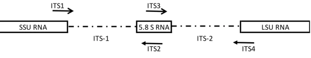
1.12. Internal transcribed spacer region and ITS primers 28
1.13. SYBR®green and TaqMan® assays 31
1.14. (a) Bead-suspension array detection by CCD camera imager (b) read-out of liquid bead suspension array with a laser read-ou 32
1.15. Sanger sequencing – schematic principle 34
1.16. Illumina® sequencing technology – schematic principle 35
1.17. Pacific Biosciences® technology- schematic principle 36
1.18. Schematic outline of the thesis 40
2.1. Alignement of selected forward and reverse Aversi_ITS primers on ITS1 region sequences of A. versicolor, A. creber, and A.sydowii 55
2.2. Melting curves obtained with Aversi_ITS qPCR assay for the A. versicolor pure strains 56
2.3. R² and PCR efficiency of the Aversi_ITS qPCR assay 59
3.1. Alignement of ITS sequences of E. jeanselmei, E. lecanii-corni, E. spinifera and A. fumigatus strains, and selected primers 82
3.2. Calibration curve (inhibition test) 83
3.3. Melting curves obtained with E.jeanselmei_ITS qPCR assay for the E.jeanselmei pure strains 84
3.4. Melting curves obtained with E.jeanselmei_ITS qPCR assay for the E.jeanselmei and the E. spinifera pure strains 86
xv
3.5. Coefficient of determination and PCR efficiency of Ejeanselmei_ITS qPCR assay 88
4.1. High resolution melting analysis plots (a) the normalized melt curves (b) the difference temperature plot 100
6.1. Neighbor Joining tree obtained with the water samples 154
6.2. Neighbor Joining tree obtained with the air samples 155
7.1. Classical analysis: Relative abundance per species 171
7.2. ITSone clustering: Relative abundance per OTU 179
7.3.UNITE clustering: Relative abundance per OTU 180
8.1 Schematic representation of the main results of the thesis and their implication for public healh 196
A1 Alignment of the 9 Versicolores species and the Aversi_ITS primers 199
B1 General phylogenetic tree obtained during the clustering analysis of all air samples using the ITSone database as reference 208
B2 General phylogenetic tree obtained during the clustering analysis of all air samples using the UNITE database as reference 208
xvi
List of Tables
1.1.Frequently found indoor fungal species and their mycotoxins 14
2.1. Selectivity evaluation of SYBR®green qPCR Aversi_ITS assay 46
2.2. Primer sequences developed in silico 48
2.3. Cq values obtained during the six runs of the limit of detection estimation for the Aversi_ITS SYBR®green qPCRassay 57
2.4. Limit of detections results (Cq mean, SD and % positive) for Aversi_ITS SYBR®Green qPCR assay 58
2.5. Environmental testing, comparison of classical analysis methods with the SYBR®green Aversi_ITS qPCR assay 61
3.1. Selectivity evaluation of SYBR®green qPCR Ejeanselmei_ITS assay 74
3.2. Primer sequences developed in silico 82
3.3. Cq values obtained during the six runs of the limit of detection estimation for the Ejeanselmei_ITS qPCR SYBR®green assay 87
3.4. Limit of detection results for Ejeanselmei_ITS qPCR SYBR®green assay 88
3.5. Environmental testing on water from air-conditioning reservoirs 90
4.1. Species discrimination by HRM analysis 101
4.2. Limit of detection of HRM assay 103
4.3. Symmetric, asymmetric and specificity assessment of different mixes 105
4.4. Environmental test 107
5.1. Fungal species and probes used in this study 121
5.2. Composition of DNA Mixes analysis 125
5.3. Simplex xMAP® analysis 128
5.4. Multiplex xMAP® analysis to test the bead- probe specificity 129
5.5. DNA Mixes analysis 131
5.6. Limit of detection a of the fungal Luminex® assay 132
5.7.a. Proof of concept with environmental samples: Culture, microscopic determination and quantification 134
5.7.b. Proof of concept with environmental samples: Luminex xMAP® analysis 135
xvii 6.1. Detection of E. jeanselmei in indoor air and water in air-conditioning systems:
Classical analysis and qPCR 146 6.2. Fungal contamination in water samples, other than E. jeanselmei: classical analysis
and sequencing analysis comparison 150 6.3. Fungal contamination in air samples: Classical analysis and sequencing analysis
comparison 151
6.4. Detection of E. jeanselmei in indoor air and water in air-conditioning systems:
sequencing data and clustering results 156
7.1. Overview of air samples taken, sample and house characteristics and global fungal
diversity 169 7.2. Classical and metagenomics analysis: Species determination and
abundance 172
A1 Spike test results 197
A2 Melting temperature obtained for the species from the Versicolores group 200
1
Chapter 1
General introduction, rationale and outline of the PhD thesis
1.1
Indoor air pollution and public health
Currently, poor indoor air quality is considered as a public health issue, this is important as people spend increasing times in indoor environment such as at home, in residential buildings or professional buildings (Asikainen et al. 2016; Bernstein et al. 2008; Bruce et al. 2000; European Environment Agency 2013; United States Environmental Protection Agency 2016; World Health Organization 2009). Three groups of airborne compounds are indexed as major indoor air pollutants i.e., smoke, volatile organic compounds (VOCs) and biological agents (European Environment Agency 2013; Revah and Morgan-Sagastume 2005). ’Smoke’ groups all the products from the outdoor or indoor obtained by fuel combustion, especially from traffic, heating or cooking and cigarette smoking. The second group of indoor air pollutants includes all VOCs released in gaze by some liquids and solids such as sprays, paints, solvents, cleaners, air fresheners, plastic components, etc. VOCs are defined as all the volatile compounds with a photochemical reactivity in atmospheres which contain carbon molecules except carbon monoxide, carbon dioxide, carbonic acid, carbonates and ammonium carbonates (Revah and Morgan-Sagastume 2005). Finally, the biological agent air pollutants refer to all the compounds produced or emitted by biological organisms which could have an impact on health. So, this group includes for instance algae, bacteria, dust mites, plants, parasites, pollen, viruses but also fungi and fungal compounds such as spores, cell wall fragments, mycelium and mycotoxins.
Among those biological contaminants, indoor fungal contamination is increasingly studied in order to define the possible impact on health, especially the implication in respiratory diseases (Anses 2016; Bellanger et al. 2009; Packeu et al. 2012; Reboux et al. 2010). Indeed, a large number of studies in many geographical regions has found a consistent association between evident indoor dampness, fungal contamination and health effect in infants, children as well as adults (Fig 1.1) (Anses 2016; Bornehag et al. 2004; Douwes 2005; Douwes et al. 2003; Mendell et al. 2011). According to the World Health Organization (WHO) and the scientific literature, indoor air pollution could be implicated in some diseases including allergies, asthma exacerbation, cancer, cystic fibrosis, allergies and in some cases strokes, particularly in immune-deficient people (Amegah and Jaakkola 2016; Anses 2016; Asikainen et al. 2016; European Environment Agency 2013; Ponsoni and Gonçalves Raddi 2010; World Health Organization 2009). Poor air quality is also associated to the sick building syndrome (SBS) characterized by mucous irritation (eyes and throat especially), neurotoxic effect as fatigue and loss of concentration, chemosensory
2 disturbance, skin symptoms and respiratory problems such as wheezing or shortness of breath (Anses 2016; European Environment Agency 2013; United States Environmental Protection Agency 2016; World Health Organization 2009). Nevertheless, even if the impact of the air pollution on health is recognized, some scientific evidence, especially for the causal link between fungal airborne contamination and health, is still not well elaborated.
Figure 1.1: Worldwide distribution of fungal allergy (adapted from Twaroch et al. 2015). In yellow, countries in which sensitization to fungi has been described.
1.1.
Fungi: definition, reproduction, classification and ecology
1.1.1. The kingdom of Fungi
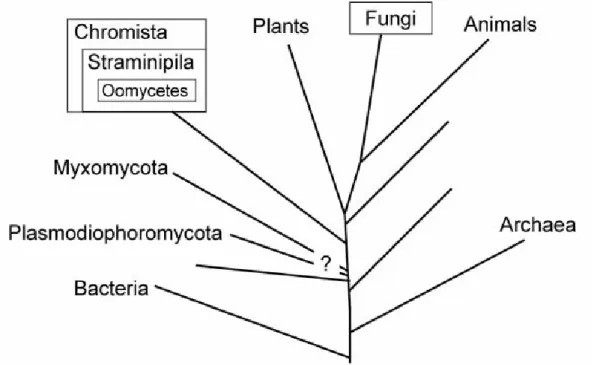
The Mycota, or Fungi kingdom brings together different groups of micro- and macroscopic multicellular and heterotrophic eukaryotes showing various forms and cellular organizations and have great economic importance (Scientific Institute for Public Health 2015; Tom and Aime 2012). They live everywhere in air, in water, on land, in soil, and on or in plants and animals. The organisms in kingdom fungi include mushrooms, yeasts, molds, rusts, smuts, puffballs, truffles, morels, and molds. The abence of cellulose as well as the wall cell composition (formed by a network of microfibrils containing chitine, β-glucans and soluble polysaccharides) distinguish the true fungi, or Eumycota (e.g. Ascomycota and Basidiomycota), from the filamentous false fungi (i.e. the Chromista group (which groups the Hyphochytriomycota, the Labyrinthulomycota, the Oomycota, diatoms or brown algae) and non-filamentous false fungi from the Protista group (i.e., the Plasmodiophoromycoyta, the Dictyosteliomycota, the Myxomycota and the Acrasiomycota) (Scientific Institute for Public Health 2015) (Fig. 1.2).
3 The diversity of the Mycota kingdom is still being investigated and might contain at least 1.5 106 species (Hawksworth 2001), but according to new estimations obtained with high-troughput sequencing methods, this diversity might reach 5.1 106 species (Blackwell 2011). Despite these estimations, until today only 99,000 species have been described (Carris et al. 2012). All of them are characterized by a cell wall containing chitin and are able to carry out a zygotic meiosis (Tom and Aime 2012).
Figure 1.2 Relationship between organisms referred to as fungi (adapted from Rossman and Palm 2017). Fungi correspond to the Eumycota or true fungi and include Ascomycota and Basidiomycota. The Chromista, Myxomycota and Plasmodiophormycota correspond to false fungi.
1.1.2. Morphological characteristics of the vegetative growth
Fungi present different kinds of morphological structures i.e., vegetative structures and reproductive structures. Vegetative structures such as the thallus or rhizoids are developed for the fungal growth and nutrition.
The fungal thallus could be unicellular (i.e., yeast such as Saccharomyctes cerevisae, Candida sp., etc) or pluricellular (mushrooms, Aspergillus sp., Alternaria sp., Cladosporium sp., etc) (Scientific Institute for Public Health 2015). The thallus is composed of a network of hyphae containing protoplasm which moves from the oldest parts to the new ones. A mass of hyphae is called mycelium. The mycelium grows through nucleic divisions and could be divided by septa in regular segments having one or more nuclei. Each septum is pierced in the middle by a pore
4 allowing the moving of the organelles, inclusions and nucleus from a segment to another (Scientific Institute for Public Health 2015). In some cases, hyphae are aggregated forming a false tissue (sort of stacking up of cells) called pseudoparenchyma or could form a solid tissue rich in nutrients called sclerotium (Scientific Institute for Public Health 2015). The mycelium could present also some particular structures such as rhizoids, or appressoria which are branched organs developed for the fixing of the organisms on substrates. In parasite fungal species, the nutrients are taken directly in the host through a special structure called haustorium (Scientific Institute for Public Health 2015).
1.1.3. Reproduction
Fungal reproduction could be sexual or asexual (Fig. 1.3) and most of the true fungi are pleomorphic (or polymorphic), i.e., able to produce different forms of spores (i.e., sexual or asexual) having a reproductive function. The sexual reproductive stage is called teleomorph or perfect stage, while the asexual reproductive stage is called anamorph or imperfect stage. The term holomorph is used to appoint the whole fungus (i.e., including the anamorph and the teleomorph) (Scientific Institute for Public Health 2015).
Asexual reproduction
The asexual reproduction produces identical individuals through the production of asexual “spores”, called conidia. Conidia are produced either through the budding of the mycelium (or a cell for unicellular thallus) or through particular cells (i.e., conidiogenous cell) set into the mycelium or on specific hyphae called conidiophores (Cyr 2009; Scientific Institute for Public Health 2015).
Sexual reproduction
The sexual spores are diploid cells (true spore) obtained through the fusion of 2 particular cells (i.e., gametangium) from a same mycelium or from 2 compatible mycelia. The gametangium is a structure which contains gametes (haploids), which could be morphologically identical (i.e., isogamy) or different (heterogamy). In the case of heterogamy, the gametangium containing the male gametes is called the antheridium (Fig. 1.3). The one containing the female gametes is called ascogonium (Fig. 1.3). In some cases, the vegetative thallus is totally converted in gametangium (or in reproductive structure). The thallus is then considered as holocarpic (Cyr 2009; Scientific Institute for Public Health 2015). In other cases, vegetative and reproductive structures coexist forming a eucarpic thallus.
5 conidium, giving a haploid mycelium (Cyr 2009; Scientific Institute for Public Health 2015). Two compatible mycelia or spores form a diploid cytoplasm (fusion of cytoplasm without fusion of nuclei) (Fig. 1.3). Then, after caryogamy, the meiose occurs and the zygote is transformed in sexual spores (4, 8 or more) (Cyr 2009; Scientific Institute for Public Health 2015). Finally, after dispersion, spores grow (germination) and give rise to new mycelia (Fig. 1.3).
Figure 1.3 Fungal life cycle: the example of Ascomycota (Cyr 2009). Sexual part of Ascomycota life cycle:
(1) Formation of the ascogonium and antheridium from haploid hyphae type “+” and type “-“. (2) Nuclei from antherium moved into the ascognium.
(3) Ascocarp formation with dikaryotic hyphae.
(4) Formation of ascus (dikaryotic) into an ascocarp at the tips of dikaryotic mycelium. (5) Karyogamy and formation of diploid nucleus (diploid ascus).
(6) Meiosis of the diploid ascus, giving new genetically distinct nuclei into the ascus. (7) Mitosis and formation of new haploid nuclei into the ascus.
(8) Formation of ascopores containing haploid nucleus (one nucleus per ascospore). (9) Ascospore release in the environment.
(10) Development of haploid mycelia from the released ascopores. Asexual part of Ascomycota life cycle:
(11)Segmentation of the hyphae and formation of haploid conidia (asexual spores). Conidia dispersion and germination giving a new haploid mycelium identical with the original hyphae.
6 For most of the fungi, spore maturation, as well as sporulation (i.e., spores dropping into the environment), is light and circadian cycle dependent (Scientific Institute for Public Health 2015). Most of the time, the sporulation is due to an increase of pression inside the ascus which causes a membrane breakdown and then, the drop of spores into the environment. In some cases, the sporulation is caused by humidity variation which causes structural changes (turgescence, torsion…) and finally the propelling of the spores (Scientific Institute for Public Health 2015). Other factors could also induce sporulation such as rain or thermic changes as well as physical interactions (Scientific Institute for Public Health 2015).
Spore forms and sizes are variable. They could be small, large, curved, ovoid, spheric, ramified or colored. They could also present ornamentation such as wings or hooks to favour the dispersion into the air or by animals (Scientific Institute for Public Health 2015). Most of the time, spores are required to perform the species identification (Scientific Institute for Public Health 2015).
Spore viability is variable according to the taxa. Some of them are resistant to extreme conditions (desiccation, insolation,…) such as the spores of rusts, other ones have a short life time (e.g., 10 hours for the Cronartium ribicola spores) (Scientific Institute for Public Health 2015). The germination is also variable. Parasitic species require hosts to germinate; other ones require a specific substrate. In some cases spores could result in an infertile mycelium (i.e., a mycelium which is not able to sporulate) which do not show the morphological structures required for their identification (Scientific Institute for Public Health 2015).
1.1.4. Classification
In mycology, the classical classification is still based on the microscopic visualisation and the identification of reproductive structures from teleomorphs (e.g., spore form and size, presence of special structure on the spores, form of the reproductive structures…). However, while in environmental conditions, anamorphs and teleomorphs stages coexist, in culture, anamorphic colonies are often observed due to the culture conditions (especially due to the selected medium), the absence of a compatible partner or because the strains are not able to sporulate. In addition, some species, such as Penicillium chrysogenum, are also only known in the anamorphic stage (Scientific Institute for Public Health 2015). Therefore, before the use of molecular tools, the anamorphic forms were included with a specific nomenclature into a group called Fungi imperfecti, till the observation of the teleomorphs. But this classification system is complex, because a double nomenclature occurs (i.e., one name for the anamorph, one for the teleomorph), e.g. Aspergillus glaucus (anamorphe) and Eurotium herbariorum (teleomorphe) (Scientific Institute for Public Health 2015). Moreover, in some cases the classical identification does not
7 give the same species for the 2 stages due to the absence of the anamorphic stage on plate, or because the strain cannot produce conidia and asexual spores (Scientific Institute for Public Health 2015). In this case, the closely related species are grouped into a complex of species. However, since 2011, new fungal taxonomy rules were defined and it was decided to use only one name to identify the anamorph and the corresponding teleomorph fungus. This decision has had an impact on the whole nomenclature and induced some bias or loss of information. Indeed, most of the species were determined according to their physiology and some species are known only under their anamorphic stage. For example Penicillium marneffei (anamorph) is now named Talaromyces marneffei (because the genus Talaromyces groups together the sexual stage of Penicillium) (Scientific Institute for Public Health 2015; Yilmaz et al 2014).
The more recent classification is based on the use of molecular tools and the DNA sequence of the fungi. Because the use of new molecular tools improves the knowledge on fungal phylogeny, the fungal classification is continuously adapted. However, it is now considered that Fungi are divided in 7 phyla, two of which, the Ascomycota and the Basidiomycota, are contained within a branch representing subkingdom Dikarya (Fig. 1.4) (Hibbett et al. 2007; Scientific Institute for Public Health 2015). The Ascomycota and the Basidiomycota phyla are characterized by the production of dicaryons (i.e., cells containing two nuclei during the dicryotic stage of the sexual reproduction) and non-mobile spores. The Basidiomycota phylum contains the most familiar fungi such as mushrooms, toadstools, stink-horns, puffballs, shelf fungi and plant pathogens such as rust or smuts (Scientific Institute for Public Health 2015). The Ascomycota contains taxa such as yeasts, Aspergillus, Penicillium, Cladosporium, but also edible morels or truffles (Scientific Institute for Public Health 2015). In scientific literature, the term “molds” is often used to show anamorphic fungi, as well as some microscopic teleomorphic fungi from the Ascomycota or Basidimycota phyla (Scientific Institute for Public Health 2015).
8 Figure 1.4 Phylogeny and classification of Fungi (Hibbett et al. 2007).
Branch lengths and genetic distances are not proportional (Hibbett et al. 2007).
1.1.5. Ecology
Adapted to various ecological niches, fungi are found in several habitats from the sea to the air as well as on soil, living organism as hosts (plant or animals) or rocks. Most of the fungi grow between 15°C and 30 °C, but some could grow in extreme environments (below 10 °C or up to 50 °C) ( Scientific Institute for Public Health 2015). Based on this, fungi could be classified as - thermophilic if the organism could growth till 50 °C, but not below 20 °C
- thermotolerant if the organism has a minimum temperature of growth at or above 20 °C, and a maximum temperature of growth extending up to 60 to 62 °C
9 - mesophilic if the organism has an optimal temperature of growth around 25 °C
- psychrophilic if the organism has an optimal temperature of growth lower than 10 °C.
The humidity level is also an important factor for the fungal growth. Actually, two groups of fungi are observed (Scientific Institute for Public Health 2015) i.e., the osmophilic species which require a water saturation of the substrate lower than 80 % and the hygrophilic species which require a water saturation of the substrate higher than 90 %.
Fungi are generally aerobic taxa. Some species, such as Mucor species, consume a high level of oxygen and so, could only grow in upper area. A contrario, Stachybotrys is able to grow at depths where the level of oxygen is very low (Scientific Institute for Public Health 2015).
Fungi are key species for the ecosystem. These heterotrophic organisms (no chlorophyll) are able to digest the biologic matter externally and absorbing the nutrient through their cell wall. Some of them are biotrophs (i.e., obtain nutrients from a living host), other are saprotrophs (i.e., nutrients are taken from dead organisms) or necrotrophs (i.e., species growing on living host and causing the death of the cell) (Carris et al. 2012).
In the ecosystem, fungi play the role of decomposer and so, are important for the recycling of nutrients. The source of carbon used by fungi comes from glucose and fructose especially. But because only a low number of fungi can produce the enzyme required for the hydrolysis of starch and cellulose, only a few species are able to grow in indoor environment (Scientific Institute for Public Health 2015). However, even if the number of fungal species able to grow in indoor environment is limited in comparison to the diversity observed in outdoor environment, their impact on health could be important due to their ability to produce toxins and allergens, but also due to their strong association to non-healthy environments (water-damadged and damp habitats).
1.2
Fungal contamination of indoor environment
Contamination of indoor environment by fungi is commonly observed in industrialized countries (European Environment Agency 2013; United States Environmental Protection Agency 2016; World Health Organization 2009). For example, in 1994 the prevalence of fungal contamination inside houses was estimated at 50 % in the United States of America (USA) (Mudarri and Fisk 2007). In 2013, Moularat and colleagues sampled 94 houses located in the urban and rural Auvergne region (France), and they showed that 59 % of these houses were contaminated by fungi and 19 % by visible moisture (Moularat et al. 2011). In Brussels (Belgium), 42.2 % of houses sampled between 2000 and 2006 in the framework of the regional intervention unit for indoor pollution, called Cellule Régionale d’Intervention en Pollution Intérieure or CRIPI (Brussels
10 Environment, Belgium), showed fungal contamination (visible or not) (Brussels Environment 2007). In 2011, 20 % of by the CRIPI investigated housings in Brussels showed more than 3 m² of visible fungal colonies in a single room (Brussels Environment 2015).
Fungal contamination could consist of active compounds (spores or mycelium) or of spores in dormancy. They penetrate into buildings via different ways of contamination. Most of them come from the direct outdoor environment and are transported in buildings via aeration and ventilation systems (i.e., windows, air conditioning, etc). Others are carried on build materials or on cloths (Nevalainen et al. 2015). Because they are ubiquitous, fungi grow on various substrates such as building materials (e.g., gypsum, paste, wood) or finishing materials (e.g., paints, linoleum, wall paper), mattress, pipes, and even on fabric or on compost of indoor plants (Andersen et al. 2011; Kelley and Gilbert 2013; Horner et al. 2004; Nevalainen et al. 2015; Nishimura et al. 1987; World Health Organization 2009; World Health Organization Regional Office for Europe 2004). Additionally, energy saving measures not correctly implemented could also favor fungal development (World Health Organization Regional Office for Europe 2004). Indeed, moisture, poor ventilation and darkness could stimulate the development of fungal contamination (World Health Organization Regional Office for Europe 2004). According to the WHO, in 2009, among 10 to 50 % of buildings around the world were affected by water-damage and 15 to 40 % of them showed fungal contamination (World Health Organization 2009). In Brussels, in 1994, it was estimated that 90 % of water-damaged dwellings showed also fungal contamination (Beguin and Nolard 1994).
Even if numerous data on indoor fungal diversity are available in scientific literature, the community of indoor fungi is complex to study due to the variability of the indoor environment and the fungal ecology (Nevalainen et al. 2015). Furthermore, climate changes and urbanization could also affect the diversity of the fungal community observed in indoor environment with the detection of species not yet observed in our industrialized countries (Vardoulakis et al. 2015; World Health Organization Regional Office for Europe 2004). However, in our industrialized countries, it is commonly accepted that indoor fungal communities are dominated by 6 genera i.e., Alternaria, Aspergillus, Cladiosporium, Penicillium, Stachybotrys and Ulocladium (Andersen et al. 2011; Beguin and Nolard 1994; Chew et al. 2003; Gots et al. 2003; Jones et al. 2011; Shelton et al. 2002). More specifically among these genera, the most common species are Alternaria alternata, Aspergillus fumigatus, Aspergillus versicolor, Cladosporium cladosporioides, Cladosporium herbarum, Cladosporium sphaerospermum, Penicillium chrysogenum, Stachybotrys chartarum and Ulocladium botritys (Andersen et al. 2011; Beguin and Nolard 1994; Chew et al. 2003; Jones et al. 2011; Shelton et al. 2002). A. versicolor and P. chrysogenum are considered as the major species in terms of prevalence and public health effects among the species
11 in indoor environment especially in water-damaged buildings (Andersen et al. 2011; Andersson et al. 1997; Beguin and Nolard 1994) and to a lesser extent, S. chartarum which is particularly observed on humid substrata containing cellulose such as wood or wall paper (Andersen et al. 2011). As for A. versicolor, U. botrytis, S. chartarum is commonly observed on gypsum and other mural coatings (Scientific Institute for Public Health 2015). These species are typically considered as “real indoor fungi” because their occurrence in indoor environments is higher than that observed outdoors (Fradkin et al. 1987). Conversely, some species are observed with a lower occurrence in indoor area than in outdoor, such as A. alternata, A. fumigatus and the 3 Cladosporium species. Their levels of detection are closely linked to the level of their contamination observed in outdoor air (Beguin and Nolard 1994). Present in the air from May to October, these species present a seasonality with an important peak of sporulation during summer. During this period, their spores could reach numbers of more than 10.000 spores/m³ (Sautour et al. 2009; Scientific Institute for Public Health 2015) and could correspond to more than 50 % of the total diversity of fungal spores observed in the outdoor air (Sautour et al. 2009; Scientific Institute for Public Health 2015). In Belgium, it was estimated that the contamination of indoor air by Alternaria sp. spores correspond to 1.5 % of the total amount of spores yearly observed in outdoor air (i.e., minimum 500 spores/m³) and that around 50 % of dust samples contain A. alternata (Scientific Institute for Public Health 2015).
1.2.1 Indoor airborne fungi and public health
Because fungi are ubiquitous, but also because many of them are found all around the world, fungal contamination of indoor environment, by spores, mycelia or other fungal particles, is increasingly assimilated into a public health question.
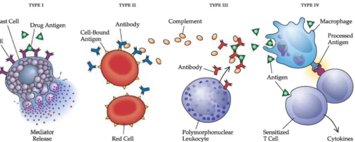
Fungi cause adverse human health effects through 3 specific mechanisms: direct infection by the organism, generation of a harmful immune response (e.g., allergy or hypersensitivity pneumonitis) and toxic-irritant effects from fungi by-products (Bush et al. 2006). The world-wide prevalence of fungal allergy among atopic subjects is estimated to be from 3 to 10 % depending on the allergic population studied, the test system and extract used and fungi species tested (Horner et al. 1995). Additionally, it was reported that 80 % of the asthmatic patients are sensitized to fungi (Simon-Nobbe et al. 2007). Indeed, today, the exposure to fungal components is considered as a potential source for allergic diseases like allergic asthma, allergic rhinitis, allergic sinusitis and hypersensitivity (Pieckova and Wilkins 2004). Allergens could be found in food and feed but also in air. The fungal allergens group mycotoxins (< 1kDa), volatile fungal metabolites (VFMs), spores (viable or not), hyphae and fungal fragments containing especially (1-3)-β-D glucans, intracellular proteins, secreted proteins and many glycopeptides which have enzymatic activities (Douwes 2005; Khan and Karuppayil 2012; Horner et al. 1995; Portnoy et al. 2008; World Health
12 Organization 2009). Most of the these allergens are type I allergens (Fig. 1.5) (Crameri et al. 2014; Green et al. 2006; Khan and Karuppayil 2012). The fungal allergens’ aerosolization could occur by differents mechanisms i.e., the sporulation and associated mechanisms of spore dispersion, fungal fragment dispersion by air, physical disturbances and suspension of dust containing fungal allergens (Gorny et al. 2002). However, the exact mechanism of aerosolization and inhalation is still not well understood (World Health Organization 2009).
It is considered that the airborne fungal genera mostly implicated in allergies are Alternaria, Aspergillus, Cladosporium and Penicilium which are all common in indoor environments and which are characterized by their capacity to excrete allergens.
These allergens could induce IgE-mediated hypersensitivity (Portnoy et al. 2008) which could cause systemic anaphylaxis and localized anaphylaxis such as asthma, eczema and wheeze (Janeway et al. 2001). But, fungi are also regarded as type III (IgG-inducing) allergens producers, especially species from the Aspergillus and Penicillium genera (World Health Organization Regional Office for Europe 2004, Khan and Karuppayil 2012). In some case, fungal contamination could also be implicated in the combination of type III and IV allergic reactions (Fig. 1.5), such as is the case for hypersensitivity pneumonitis (World Health Organization Regional Office for Europe 2004).
Figure 1.5: Schematic representation of the allergenic reaction types I, II, III and IV (
http://what-when-how.com)
Indeed, as mentioned above, fungi are able to produce compounds such as spores, proteins, mycotoxins, (1-3)-β-D glucan and volatile fungal metabolites (VFMs) which could induce illness by inhalation or ingestion. Some scientific studies showed a correlation between some diseases such as allergies, asthma exacerbation, SBS or rhinitis and fungal contamination (de Ana et al. 2006; Gots et al. 2003; Horner et al. 1995; Jarvis and Miller 2005; Jones et al. 2011; Meheust et al. 2014; Mendell et al. 2011; Packeu et al. 2012a,b; Piecková and Wilkins 2004; Portnoy and Jara 2015; Rosenbaum et al. 2010; United States Environmental Protection Agency 2016; Vardoulakis
13 et al. 2015; Verhoeff and Burge 1997; Vesper et al. 2013; World Health Organization 2009). The following paragraphs discuss the most important fungal compounds which could be implicated in health problems.
Proteins
Most of the time fungal allergens are proteins from the cell wall of airborne spores or inserted in the mycelium membrane (Horner et al. 1995; Kurup 2003). Produced by several species, these proteins are frequently observed in indoor environments. Indeed allergenic proteins are observed in several genera and species commonly observed in indoor environment as for example, in Alternaria such as the Alt a1 from A. alternata; in Aspergillus such as Asp f1 and Asp f2 from A. fumigatus, in Cladosporium such as Cla c9 form C. cladosporioides or Cla h1 and Cla h2 from C. herbarum; in Penicillium such as Pen ch13 from P. chrysogenum; and in Ulocladium such as Ulo C1 (Horner et al. 1995; Fukutomi and Taniguchi 2015).
Among all proteins currently found, Alt a1 from A. alternata (Kustrzeba-Wójcika et al. 2014; Gabriel et al. 2016), Asp f2 from A. fumigatus (Banerjee et al. 1998) and Cla c9 from C. cladosporioides (Chou et al. 2008) are considered as major allergens, involved in IgE mediated allergy.
Mycotoxins
Mycotoxins are toxic secondary metabolites produced by several fungal species and which could contaminate food and feed. Generally not volatile, these toxics compounds could be found in air because of their attachment to some small biological particles (Portnoy et al. 2008). Mycotoxin inhalation could have an impact on health inducing mucous irritations, skin rash, immune system suppression and immunotoxic effects, liver damage, damage of the central nervous system and the endocrine system (Fromme et al. 2016; Jarvis and Miller 2005; World Health Organization 2009). Some mycotoxins are also known to be cytotoxic and carcinogenic (De Ruyck et al. 2015; Jarvis and Miller 2005).
Today, around 400 different mycotoxins have been identified and they are produced especially by Alternaria spp., Aspergillus spp., Fusarium spp., Penicillium spp. and Stachybotrys spp. (Fromme et al. 2016; World Health Organization 2009). These toxins are found in the indoor air of water-damaged, air-conditioned or ventilated buildings and agricultural buildings such as cereals depots, stables, compost or manure depots (Fromme et al. 2016; World Health Organization Regional Office for Europe 2004). In indoor environment, the most often detected mycotoxins are the aflavotoxins produced especially by Aspergillus species; the ochratoxins produced for the most part by Aspergillus and Penicillium; cladosporin from Cladosporium species; and alternariol and alternariol methyl esther from Alternaria species (Fromme et al. 2016). As shown in Table 1.1,
14 more than one mycotoxin could be observed simultaneously in indoor environments. Polizzi and colleagues investigated 99 samples from 7 Belgian houses and showed that 62 samples contained at least one mycotoxin. Using liquid chromatography-tandem massive spectrometry (LC-MS/MS), they have identified 7 major mycotoxins i.e., roquefortine C, chaetoglobosin A, sterigmatocystin, roridin E, ochadrine A, aflavotoxin B(1) and aflavotoxin B(2) (Polizzi et al. 2009). The mycotoxin production depends largely on the growth conditions both in terms of quantity and of number of toxins produced by species (Jarvis and Miller 2005; World Health Organization Regional Office for Europe 2004). For example, A. versicolor, one of the most important indoor fungal contaminants, produces sterigmatocystin only under a high level of humidity (Jarvis and Miller 2005; World Health Organization Regional Office for Europe 2004).
It should be noted that chromatographic techniques, such as LC-MS/MS, are not the only ones used to detect these toxins, but MALDI-TOF (matrix assisted laser desorption/ionization - time of flight massive spectrometry) and immunochemical-based methods such as ELISA (enzyme linked immunosorbent assay) are also largely exploited and have contributed to improve mycotoxins monitoring (Anfossi et al. 2015). However, today the causal link between exposure to indoor mycotoxins and specific diseases is still poorly documented (Khan and Karuppayil 2012).
Table 1.1: Frequently found indoor fungal species and their mycotoxins
Species Mycotoxins Reference
Alternaria alternata Alternariol, alternariol monomethyl esther, Fumonisin B1
Ren et al. 1998; Jarvis and Miller 2005 Aspegillus creber Sterigmatocystins Jurjevic et al. 2013 Aspergillus flavus Aflavotoxin B1, Aspergillic acid, Kojic
acid, cyclopiazonic acids, 3-nitropropionic acid
Nielsen 2003
Aspergillus fumigatus Gliotoxins, fumigaclavines, fumitoxins, fumitremorgens, tryptoquivalins, verruculogen
Nieminen et al. 2002; Nielsen 2003
Aspergillus niger Ochratoxin A, naphtho-c-pyrones,
tetracyclic compounds, nigragillin, kotanin, orlandin, malformin A, B, and C
Nielsen 2003
Aspergillus sydowii Aspermutarubrol, sydowinins, sydowic acid Jarvis and Miller 2005 Aspergillus versicolor versicolorins, Sterigmatocystins,
5-methoxysterigmatocystin
Jarvis and Miller 2005
Cladosporium cladosporioides
Asperentin, cladosporic acids Jarvis and Miller 2005
Penicillium chrysogenum Roquefortine C, meleagrin, chrysogine, x-hydroxyemodine, pyrovoylamin-obenzamides, xanthocillin X
Jarvis and Miller 2005; Nielsen 2003
Stachybotrys chartarum Macrocyclic trichothecenes, atranones, dolabellanes stachybotrylactones and lactams, stachybotrydials
15 (1-3)-β-D glucans
(1-3)-β-D glucan is a polymer of glucose, found in most of the fungal species, and it has a structural function in the cell. Inside the cell wall, this polymer is bound to carbohydrates, lipids, proteins and other polymers such as (1-6)-β-D glucans (World Health Organization Regional Office for Europe 2004). According to the scientific literature, bioaerosols containing (1-3)-β-D glucans could be implicated in respiratory problems and non-specific inflammation (Douwes et al. 2003; Douwes 2005).
Volatile Fungal metabolites (VFMs)
Besides glucans and mycotoxins, indoor air could also contain volatile fungal metabolites (VFMs). VFMs are secondary metabolites such as alcohols, aldehydes or ketones produced during the exponential growth of several fungal species (Wilkins et al. 2003). The impact of VFMs on health is poorly documented, but some studies consider that VMFs inhalation could have some respiratory irritation (Weinhold 2007).
1.2.2 Fungal immunological testing
Today, fungal sensitivity is detected through cutaneous tests. Briefly, these tests consist of putting some drops of standardized allergens from different fungal species, on the skin of the patient. After incubation, a patient is considered as sensible to the allergen when immunological signs such as red patches, inflammation or rashes are visible.
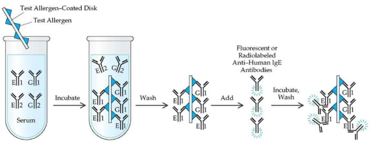
A second approach is based on allergen-specific serum IgE antibodies such as in the radioallergosorbent test (RAST) and enzyme-linked immunosorbent assay (ELISA) (Horner et al. 1995; Kurup et al. 2003). The RAST diagnosis consists of an in-vitro detection of IgE antibodies in blood, through the use of allergens coated to an insoluble substrate and a radiolabeled anti-human IgE (Fig. 1.6). Briefly, some specific antigens coated to an insoluble substrate are dropped into the patient serum. If the patient is sensitive to the allergens, specific IgE will bind the allergens. After a wash step, radiolabeled anti-human IgE are added into the solution and bind the antigens-antibodies complexes. The presence of IgE antibodies are detected by the radioactivity measurement. The radioactivity level is proportional to the level of IgE anti-allergens binding in the serum (Negrini et al. 1985).
16 Figure 1.6: RAST test - schematic principle (http://what-when-how.com)
With the ELISA test (Fig. 1.7), antigens of interest are coated to the bottom of a polystyrene plate with proteins blocking the free plastic sites. When the serum of patients is added, if they are present in the serum, specific IgE (primary antibodies) bind the antigens. After a wash of the non-binding IgE, enzyme labeled antibodies (secondary antibodies) bind primary antibodies. Finally, a specific substrate is added and consumed by the enzyme which changes the color of the liquid.
Figure 1.7: ELISA test – schematic principle (Adapted from Day 2015) A: Positive test. B. corresponding to the negative control
[1] If they are present in the serum, specific IgE (primary antibodies) recognize the coated antigens.
[2] Addition of secondary antibodies labeled with an enzyme (E) which bind primary antibodies. [3] After a wash, free secondary antibodies are removed.
17 Even though the allergenicity of several fungal species is well known, the health impacts of a large part of the diversity are unknown. Indeed, today, numerous fungal monitoring protocols, still culture-based, are not able to detect dead or uncultivable species (also called dead fraction or unknown agent). But, because these unknown agents could contain allergenic compounds, they could be allergenic and could have an impact on health.
Moreover, the lack of data concerning the unknown agents as well as the difficulties to produce and to standardize allergens required for clinical tests has an impact on the representativeness of the fungal species tested during immunological tests (Portnoy et al. 2008; World Health Organization 2009). Thus, today, representative fungal extracts available for immunological tests are focused on allergens from fungi only frequently found indoor. However, to understand the causal link between fungal contamination and health problems such as respiratory diseases and allergies, the representativeness of immunological tests is required to demonstrate that a direct exposure of an affected person to the detected fungal species occurred.
1.2.3 Some important fungal species found indoor and their clinical relevance
This paragrah describes the fungal genera and species targeted in this doctoral research and their clinical relevance.
Alternaria
The genus Alternaria is found all around the world and contains about fifty species. Alternaria species are plant pathogens, but are also decomposers of organic matter from vegetables, food, and soil.
At the clinical level, Alternaria is associated with allergies. In outdoor air, it is considered that a concentration of 100 spores/m³ is a clinical threshold. In Europe, between June and October, this concentration is commonly reached (Scientific Institute for Public Health 2015). In addition, a concentration of 500 spores/m³ is considered as a threshold for asthmatic reactions. Also this level of outdoor air contamination is often reached during the year.
In indoor environment, Alternaria species are also frequently observed, but most of their spores found inside are coming from outdoor air. The total of indoor Alternaria spores corresponds to 1.5 % of the Alternaria spores observed outdoor (Scientific Institute for Public Health 2015). However, Alternaria colonies are also observed inside buildings on walls or mattresses. In Belgium, 50 % of dust samples contain Alternaria species (Scientific Institute for Public Health 2015).