R E S E A R C H A R T I C L E
Open Access
Effects of light deprivation on visual evoked
potentials in migraine without aura
Gianluca Coppola
1*, Julien Crémers
2, Pascale Gérard
2, Francesco Pierelli
3,4and Jean Schoenen
2,5Abstract
Background: The mechanisms underlying the interictal habituation deficit of cortical visual evoked potentials (VEP) in migraine are not well understood. Abnormal long-term functional plasticity of the visual cortex may play a role and it can be assessed experimentally by light deprivation (LD).
Methods: We have compared the effects of LD on VEP in migraine patients without aura between attacks (MO, n = 17) and in healthy volunteers (HV, n = 17). Six sequential blocks of 100 averaged VEP at 3.1 Hz were recorded before and after 1 hour of LD. We measured VEP P100 amplitude of the 1st block of 100 sweeps and its change over 5 sequential blocks of 100 responses.
Results: In HV, the consequence of LD was a reduction of 1st block VEP amplitude and of the normal habituation pattern. By contrast, in MO patients, the interictal habituation deficit was not significantly modified, although 1st block VEP amplitude, already lower than in HV before LD, further decreased after LD.
Conclusions: Light deprivation is thought to decrease both excitatory and subsequent inhibitory processes in visual cortex, which is in line with our findings in healthy volunteers. The VEP results in migraine patients suggest that early excitation was adequately suppressed, but not the inhibitory mechanisms occurring during long term stimulation and habituation. Accordingly, deficient intracortical inhibition is unlikely to be a primary factor in migraine pathophysiology and the habituation deficit.
Background
Deficient habituation of pattern reversal visual evoked potentials (VEPs) during long lasting stimulus repetition characterizes migraine patients during the pain-free interval [1-3].
This abnormal information processing could be attribu-ted to hypofunctioning intracortical inhibitory circuits [4,5] or to a reduced preactivation level of the visual cor-tex, which may be due to hypoactive aminergic projections from the brainstem [1,2].
Immediate and longer-lasting cortical changes, such as those induced by repetitive sensory stimuli, are thought to reflect central nervous system (CNS) plasticity and modifications of synaptic effectiveness in the stimulated cortex through short- and long- term depression (LTD) [6,7]. We suggested therefore that altered functional
plasticity of sensory cortices could be responsible for the electrophysiological abnormalities found in migraine [8].
Light deprivation (LD) is a well-known, validated in vivo model for the study of functional plasticity in the visual system [9,10]. LD reduces time dependently peripheral [11,12], and central [13,14] neural activity in animal mod-els, which is accompanied by behavioural changes [14]. In humans, LD slows the electroencephalogram (EEG), by increasing low-band power and decreasing alpha activity [15,16].
In studies of visually deprived animals [17-19] and humans [20,21], smaller VEP amplitudes have been observed. Conversely, phosphene thresholds after tran-scranial magnetic stimulation (TMS) were decreased after 1 hour of light deprivation [22,23] which was attributed to decreased excitability of cortical inhibitory interneurons [22].
We decided therefore to use light deprivation in order to investigate more precisely long-term functional plasticity of the visual cortex and the role of cortical inhibition in migraine patients between attacks in whom habituation is
* Correspondence: gianluca.coppola@gmail.com 1
G.B. Bietti Eye Foundation-IRCCS, Dept of Neurophysiology of Vision and Neuroophtalmology, Rome, Italy
Full list of author information is available at the end of the article
© 2011 Coppola et al; licensee BioMed Central Ltd. This is an Open Access article distributed under the terms of the Creative Commons Attribution License (http://creativecommons.org/licenses/by/2.0), which permits unrestricted use, distribution, and reproduction in any medium, provided the original work is properly cited.
absent and in healthy subjects who have a normal habitua-tion. We measured VEP amplitudes to low numbers of stimuli and VEP habituation over sequential blocks during uninterrupted durable visual stimulation.
Methods
Subjects - We enrolled 17 consecutive migraine patients without aura (MO, ICHD-II code 1.1) (14 women and 3 men, mean age 28.9 ± 12.1 years) from our headache clinic. They underwent VEP recordings during the interic-tal period, i.e. at least three days before and after the recordings. No preventive anti-migraine drugs were allowed during the preceding 3 months. For comparison, 17 healthy subjects of comparable age and gender distribu-tion (14 women and 3 men, mean age 28.8 ± 11.4 years) were recruited among medical school students and health-care professionals. They had to be devoid of any overt medical condition, personal or family history of migraine or epilepsy, and regular drug intake. To minimize variabil-ity due to hormonal influences on cortical excitabilvariabil-ity, female subjects were always recorded at mid-cycle.
Participants taking medications on a regular basis and subjects who failed to reach a best corrected visual acuity of > 8/10 were excluded. None of the enrolled subjects had sleep deprivation or alcohol ingestion the day pre-ceding the recordings. Caffeinated beverages were not allowed on the day of recordings. All participants received a complete description of the study and granted written informed consent. The project was approved by the ethical review board of the Faculty of Medicine, University of Liège, Belgium.
Visual evoked potential recordings - Subjects were seated in an acoustically isolated room with dimmed light in front of a TV monitor surrounded by a uniform luminance field of 5 cd/m2. To obtain a stable pupillary diameter, each subject adapted to the ambient room light for 10 min before the VEP recording. VEPs were elicited by monocular stimulation. Visual stimuli consisted of full-field checkerboard patterns (contrast 80%, mean luminance 250 cd/m2) generated on a TV monitor and reversed in contrast at a rate of 3.1/s. At the viewing dis-tance of 80 cm, the single check edges subtended a visual angle of 15 minutes. Subjects were instructed to fixate a red dot in the middle of the screen with the left eye cov-ered by a patch to maintain stable fixation. VEPs were recorded from the scalp through pin electrodes posi-tioned at Oz (active electrode) and Fz (reference elec-trode, 10/20 system). A ground electrode was placed on the right forearm. The evoked potential signals were amplified by CED™ 1902 preamplifiers (band-pass 0.05-2000 Hz, Gain 1000) and recorded with a CED™ 1401 device (Cambridge Electronic Design Ltd, Cambridge, UK). A total of 600 consecutive sweeps each lasting 200 ms were collected and sampled at 4000 Hz.
After applying off-line a 45 Hz low-pass digital filter, cortical responses were partitioned in 6 sequential blocks of 100, consisting of at least 95 artefact-free sweeps. Responses in each block were averaged off-line (“block averages”) using the Signal™ software package version 3.10 (CED Ltd).
VEP were off-line analysed by one investigator (J.C.) not blinded for subjects’ diagnosis. VEP components were identified according to their latencies: N1 was defined as the most negative peak between 60 and 90 ms, P1 as the most positive peak following N1 between 80 and 120 ms, and N2 as the most negative peak following P1 at between 125 and 150 ms (Figure 1). We measured peak-to-peak amplitude of the N1-P1 com-plex. Habituation was defined both as the change in N1-P1 amplitude between the 1st and the 6th block of averages and the slope of the linear regression line over the 6 blocks. VEP habituation was evaluated before and immediately after light deprivation. All recordings were collected in the morning (between 09.00 and 11.00 a.m.) by the same investigator.
Light deprivation (LD) - Light deprivation was obtained by having subjects wear opaque goggles covered by a mask. They had to report complete absence of light per-ception and freedom of eyelid movement. Subjects were instructed to relax, keep their eyes open and blink as usual. VEPs were recorded before and immediately after 1 hour of LD.
Statistical analyses - We used the Statistical Package for the Social Sciences (SPSS) for Windows, version 15.0
Figure 1 Representative recordings of visual evoked potential (VEP): habituation at baseline (left) and after 1 h light deprivation (right) in a healthy subject [HS] and a migraine without aura patient [MO].
for all analyses. We constructed a multivariate analysis of variance (ANOVA) taking as a within-subject factor “block” and as between-subject factors “Group” (HV, MO) and“session” (before and after LD). A regression analysis was used to disclose linear trends in VEP ampli-tude across blocks (slope) in each condition and group. The paired Student’s t-test was used to compare VEP amplitude in block 1 before and after LD in both groups of subjects. Fisher’s least significant difference (LSD) test was used for post hoc analysis. Pearson’s correlation test was used to search for correlations among the VEP amplitude slopes and clinical variables. P values smaller than 0.05 were considered to indicate statistical significance.
Results
VEP recordings from all participants yielded analysable data. Examples of VEP recordings before and after 1 h of light deprivation, obtained from a healthy subject and from a MO patient, are shown in Figure 1.
ANOVA for amplitude in averaged VEP blocks disclosed a significant two-way interaction of group by block (F(5,320)= 3.72, p = 0.002), and time by block (F(5,320)=
3.57, p = 0.003), but not of group by block and time (F(5,320)= 1.13, p = 0.340). Linear regression analysis of
VEP amplitudes recorded over all 6 blocks differed between sessions in healthy subjects (F(1,32)= 10.04, p =
0.003) but not in patients (F(1,32)= 0.52, p = 0.47). Post
hoc analysis showed that before LD the slope of VEP amplitudes from block 1 to block 6 was negative (-0.22 ± 0.16) in healthy subjects whereas in patients it was positive (+0.04 ± 0.39, Figure 2). Conversely, after 1 hour of LD, the slope of VEP amplitudes from block 1 to block 6 became positive in healthy volunteers (+0.03 ± 0.07), while there was little change in patients (+0.12 ± 0.22).
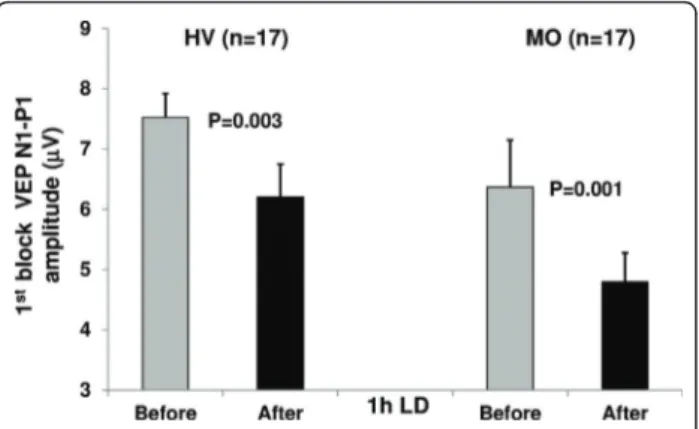
VEP block 1 amplitude decreased significantly after LD in both healthy subjects (t(1,17) = 3.47, p = 0.003)
and patients (t(1,17)= 4.25, p = 0.001; Figure 3).
With Pearson’s test there was no significant correla-tion between clinical characteristics and VEP amplitude slopes (before and after LD) in migraine patients.
Discussion
Light deprivation produced differential changes of pat-tern reversal VEPs in healthy subjects and migraine with-out aura patients recorded between attacks. Whereas in healthy subjects LD decreased VEP amplitude in block 1 and abolished the normal VEP habituation, in patients it also further decreased the already low VEP amplitude in block 1, but did not significantly change the abnormal VEP habituation pattern.
The reduction in 1stblock VEP amplitude after LD is in line with current neurobiological knowledge on LD-induced changes in the visual system. Chronic light
deprivation reduces retinal ganglion cell responses [11], induces plastic changes in lateral geniculate nucleus [13], alters neural activity in monoamine brain systems, especially in locus coeruleus, and induces loss of cortical noradrenergic fibres to the visual cortex [14]. These LD-induced changes produce behavioural deficits in decision making and attention tasks in animal models [14]. In humans, prolonged [15] as well as brief visual depriva-tion for 1 or 2 hours slows the electroencephalogram (EEG) and decreases alpha frequency power [15,16,24]. The latter observation is of particular interest for the LD-induced reduction of VEP amplitudes in the 1st block. It is well known indeed that EEG alpha activity predominates in occipital areas and that it is directly correlated with VEP amplitude in healthy subjects [25]. The changes in alpha power may reflect a change in general arousal [26] and are probably induced by a dif-fuse activation from the brainstem reticular formation [27,28]. The latter is thought to modulate visual evoked responses [29-31] and, consequently, the mechanisms of LD-induced cortical plasticity [14]. In line with this con-cept, VEP amplitudes were found reduced in animals during and immediately after LD [17,19,21], which was attributed to an increase of synchronic thalamic inhibi-tory influences on the cortex, resulting, as in our study, in a decrease of excitatory drive and thus of VEP ampli-tude [19,21].
The second distinctive finding in our study is that in healthy subjects experimental LD attenuates VEP habitua-tion. In animals it was shown previously that LD reduces excitation at early, but also inhibitory processes at later time points, which results in a loss of VEP habituation during continuous stimulus repetition [19,21]. The dynamics of the excitatory-inhibitory balance that finally results in a lack of long-term depression, i.e. lack of habi-tuation, after 1 h of LD, is likely to depend on glutamater-gic and GABAerglutamater-gic mechanisms. In animal models, LD alters NMDA-receptor-dependent synaptic plasticity [13,32,33] and impairs both short- and long- term depres-sion in visual cortex [34]. In animals reared in the dark from birth the effectiveness of inhibitory synapses on the soma of pyramidal neurons in the superficial layers of the visual cortex is markedly reduced [35]. Altered GABA neurotransmission in the visual cortex by LD was also considered to be the mechanism underlying the decrease in magnetophosphene thresholds after LD in healthy humans [22,23]. However, the magnetophosphene and VEP studies are not comparable without reservation, as the habituation phenomenon is only studied in the latter. VEP amplitudes tend already to habituate between the 4th and 6thblock of averagings, i.e. during the 4thminute of stimulation (-3.0%, Figure 2), while the decrease of magne-tophosphene thresholds is still observed 180 minutes after the end of LD. As suggested by pharmacological studies
[36], besides cortical GABAergic inhibition, it is thus likely that excitatory neurotransmitters like acetylcholine and glutamate also play a role in LD-induced visual cortex plasticity.
Another finding in our study is that 1 h LD has no effect on the interictal habituation deficit in migraineurs. This contrasts with a“normal” behavior of the 1stblock VEP which, as in healthy volunteers, decreases after LD despite its already lowered amplitude. In migraineurs, unlike healthy subjects, VEP amplitude does not habituate between the 4thand 6thblocks, but on the contrary con-tinues to increase (+6.7%) confirming once more that the brain mechanisms responsible for long-term habituation are malfunctioning in migraine [1-3]. We have shown
previously that thalamo-cortical activation is reduced in migraine between attacks [37,38] which might be attribu-ted to functional disconnection of the thalamus from its control by aminergic brainstem nuclei [39]. Light depriva-tion precisely has an effect on brainstem noradrenergic and serotoninergic neurons in rat [14]. The transient dys-function of these neurons might worsen the already reduced thalamo-cortical activity, producing an additional decrease in cortical excitation and further reduction of VEP amplitude in the 1stblock of averaged responses. In the thalamo-cortical dysrhythmia [40,41] model of migraine, the deficient habituation in later blocks of VEP averagings can be attributed to inefficient lateral inhibition in the visual cortex [37]. It was hypothesized that the
Figure 2 VEP N1-P1 block amplitudes (mean + SEM) before and after 1 h of light deprivation (LD) in healthy volunteers [HV] and migraine patients without aura [MO].
deficient habituation in migraineurs is due to a hypofunc-tion of inhibitory cortical interneurons [4,5]. In this case, however, one would expect that after LD that inhibits inhi-bitory interneurons (see above), the habituation deficit worsens. The fact that this was not our finding does not favor this hypothesis, unless one assumes that the habitua-tion deficit is already maximal before LD in migraine patients and cannot be further increased. It is known that the effect of LD on the visual cortex depends on the underlying level of cortical activation that is not fixed [42] and might be genetically determined [34,42,43]. Taken together, these data suggest that the migraine disease-related decrease in thalamocortical preactivation prevents the occurrence of the normal plastic changes induced by 1 h LD.
Conclusions
In conclusion, the reduction in VEP 1stblock amplitude after LD in healthy subjects and migraineurs might reflect a transient thalamo-cortical dysfunction due to interference with neural activity in brainstem modula-tory aminergic centres [14,19,24,44]. This initial inhibi-tory effect on VEP amplitude is worsened in migraine, probably because of further impairment of an already reduced thalamo-cortical drive. This reduction results in abnormal modulation of the visual cortex and failure of long term VEP inhibitory mechanisms, which prevents further changes in the pre-existing habituation deficit. Consequently, reduced cortical preactivation, and not primary dysfunction of intracortical inhibition, is likely to be the main cause of the habituation deficit found in migraine patients between attacks.
Author details
1G.B. Bietti Eye Foundation-IRCCS, Dept of Neurophysiology of Vision and Neuroophtalmology, Rome, Italy.2Headache Research Unit. University Dept. of Neurology, Liège University, Belgium.3Dept of Medico-Surgical Sciences
and Biotechnologies,“Sapienza” University of Rome Polo Pontino, Latina, Italy.4IRCCS-Neuromed, Pozzilli (IS), Italy.5GIGA-Neurosciences, Liège University, Liège, Belgium.
Authors’ contributions
GC made substantial contributions to analysis and interpretation of data as well as in drafting the manuscript. JC, PG & FP were implied in recording data and analysis. JS was implied in the interpretation of data as well as in drafting the manuscript; gave critical revision of the manuscript for important intellectual content. All authors read and approved the final manuscript.
Competing interests
The authors declare that they have no competing interests.
Received: 26 April 2011 Accepted: 27 July 2011 Published: 27 July 2011 References
1. Schoenen J, Wang W, Albert A, Delwaide P: Potentiation instead of habituation characterizes visual evoked potentials in migraine patients between attacks. Eur J Neurol 1995, 2:115-122.
2. Schoenen J: Deficient habituation of evoked cortical potentials in migraine: a link between brain biology, behavior and trigeminovascular activation? Biomed Pharmacother 1996, 50(2):71-78.
3. Coppola G, Pierelli F, Schoenen J: Habituation and migraine. Neurobiol Learn Mem 2009, 92(2):249-259.
4. Mulleners WM, Chronicle EP, Palmer JE, Koehler PJ, Vredeveld JW: Visual cortex excitability in migraine with and without aura. Headache 2001, 41(6):565-572.
5. Brighina F, Palermo A, Fierro B: Cortical inhibition and habituation to evoked potentials: relevance for pathophysiology of migraine. J Headache Pain 2009, 10(2):77-84.
6. Thompson RF, Spencer WA: Habituation: a model phenomenon for the study of neuronal substrates of behavior. Psychol Rev 1966, 73(1):16-43. 7. Rankin CH, Abrams T, Barry RJ, Bhatnagar S, Clayton DF, Colombo J,
Coppola G, Geyer MA, Glanzman DL, Marsland S, McSweeney FK, Wilson DA, Wu CF, Thompson RF: Habituation revisited: an updated and revised description of the behavioral characteristics of habituation. Neurobiol Learn Mem 2009, 92(2):135-138.
8. Coppola G, Currà A, Serrao M, Di Lorenzo C, Gorini M, Porretta E, Alibardi A, Parisi V, Pierelli F: Lack of cold pressor test-induced effect on visual-evoked potentials in migraine. J Headache Pain 2010, 11(2):115-121. 9. Smith GB, Heynen AJ, Bear MF: Bidirectional synaptic mechanisms of
ocular dominance plasticity in visual cortex. Philos Trans R Soc Lond B Biol Sci 2009, 364(1515):357-367.
10. Rittenhouse CD, Shouval HZ, Paradiso MA, Bear MF: Monocular deprivation induces homosynaptic long-term depression in visual cortex. Nature 1999, 397(6717):347-350.
11. Tian N, Copenhagen DR: Visual deprivation alters development of synaptic function in inner retina after eye opening. Neuron 2001, 32(3):439-449.
12. Giovannelli A, Di Marco S, Maccarone R, Bisti S: Long-term dark rearing induces permanent reorganization in retinal circuitry. Biochem Biophys Res Commun 2008, 365(2):349-354.
13. Duffy KR, Slusar JE: Monocular deprivation provokes alteration of the neuronal cytoskeleton in developing cat lateral geniculate nucleus. Vis Neurosci 2009, 26(3):319-328.
14. Gonzalez MMC, Aston-Jones G: Light deprivation damages monoamine neurons and produces a depressive behavioral phenotype in rats. Proc Natl Acad Sci USA 2008, 105(12):4898-4903.
15. Zubek JP, Welch G: Electroencephalographic changes after prolonged sensory and perceptual deprivation. Science 1963, 139:1209-1210. 16. Marjerrison G, Keogh RP: Electroencephalographic changes during brief
periods of perceptual deprivation. Percept Mot Skills 1967, 24(2):611-615.
17. Snyder A, Shapley R: Deficits in the visual evoked potentials of cats as a result of visual deprivation. Exp Brain Res 1979, 37(1):73-86.
18. Postnikova NN, Nachkebiia AI, Lordkipanidze SO: [Evoked potentials in cortical and subcortical structures of the visual system to photic and acoustic stimuli under conditions of early light deprivation]. Zh Vyssh Nerv Deiat Im I P Pavlova 1977, 27(1):169-176.
Figure 3 VEP N1-P1 amplitude (mean + SEM) in the 1stblock of averaged responses before and after 1 h of light
deprivation (LD) in healthy volunteers [HV] and migraine without aura patients [MO].
19. Zislina NN: [Effect of early deprivation of visual evoked potentials of rabbit cerebral cortex under conditions of dark and light adaptation]. Zh Vyssh Nerv Deiat Im I P Pavlova 1977, 27(1):161-168.
20. Glass JD, Crowder JV, Kennerdell JS, Merikangas JR: Visually evoked potentials from occipital and precentral cortex in visually deprived humans. Electroencephalogr Clin Neurophysiol 1977, 43(2):207-217. 21. Grigor’eva LP, Zislina NN, Tolstova VA: [Plasticity of the visual system and
learning]. Fiziol Cheloveka 1996, 22(1):55-62.
22. Boroojerdi B, Bushara KO, Corwell B, Immisch I, Battaglia F, Muellbacher W, Cohen LG: Enhanced excitability of the human visual cortex induced by short-term light deprivation. Cereb Cortex 2000, 10(5):529-534.
23. Fierro B, Brighina F, Vitello G, Piazza A, Scalia S, Giglia G, Daniele O, Pascual-Leone A: Modulatory effects of low- and high-frequency repetitive transcranial magnetic stimulation on visual cortex of healthy subjects undergoing light deprivation. J Physiol 2005, 565(Pt 2):659-665. 24. Marjerrison G, Keogh RP: The neurophysiology of schizophrenia. Field
dependency and electroencephalogram (EEG) responses to perceptual deprivation. J Nerv Ment Dis 1971, 152(6):390-395.
25. Koch SP, Koendgen S, Bourayou R, Steinbrink J, Obrig H: Individual alpha-frequency correlates with amplitude of visual evoked potential and hemodynamic response. Neuroimage 2008, 41(2):233-242.
26. Hughes SW, Crunelli V: Thalamic mechanisms of EEG alpha rhythms and their pathological implications. Neuroscientist 2005, 11(4):357-372. 27. Steriade M, Llinás RR: The functional states of the thalamus and the
associated neuronal interplay. Physiol Rev 1988, 68(3):649-742. 28. Lopes da Silva F: Neural mechanisms underlying brain waves: from
neural membranes to networks. Electroencephalogr Clin Neurophysiol 1991, 79(2):81-93.
29. Bremer F, Stoupel N: [Facilitation and inhibition of evoked cortical potentials during cerebral arousal]. Arch Int Physiol Biochim 1959, 67(2):240-275.
30. Armengol V, Lifschitz W, Palestini M: Inhibitory influences on primary and secondary cortical photic potentials originating in the lower brain stem. J Physiol 1961, 159:451-460.
31. Fuster JM, Docter RF: Variations of optic evoked potentials as a function of reticular activity in rabbits with chronically implanted electrodes. J Neurophysiol 1962, 25:324-336.
32. Yashiro K, Corlew R, Philpot BD: Visual deprivation modifies both presynaptic glutamate release and the composition of perisynaptic/ extrasynaptic NMDA receptors in adult visual cortex. J Neurosci 2005, 25(50):11684-11692.
33. Goel A, Lee HK: Persistence of experience-induced homeostatic synaptic plasticity through adulthood in superficial layers of mouse visual cortex. J Neurosci 2007, 27(25):6692-6700.
34. Kirkwood A, Rioult MC, Bear MF: Experience-dependent modification of synaptic plasticity in visual cortex. Nature 1996, 381(6582):526-528. 35. Kreczko A, Goel A, Song L, Lee HK: Visual deprivation decreases somatic
GAD65 puncta number on layer 2/3 pyramidal neurons in mouse visual cortex. Neural Plast 2009, 2009:415135.
36. Boroojerdi B, Battaglia F, Muellbacher W, Cohen LG: Mechanisms underlying rapid experience-dependent plasticity in the human visual cortex. Proc Natl Acad Sci USA 2001, 98(25):14698-14701.
37. Coppola G, Ambrosini A, Di Clemente L, Magis D, Fumal A, Gérard P, Pierelli F, Schoenen J: Interictal abnormalities of gamma band activity in visual evoked responses in migraine: an indication of thalamocortical dysrhythmia? Cephalalgia 2007, 27(12):1360-1367.
38. Coppola G, Vandenheede M, Di Clemente L, Ambrosini A, Fumal A, De Pasqua V, Schoenen J: Somatosensory evoked high-frequency oscillations reflecting thalamo-cortical activity are decreased in migraine patients between attacks. Brain 2005, 128(Pt 1):98-103.
39. Panconesi A: Serotonin and migraine: a reconsideration of the central theory. J Headache Pain 2008, 9(5):267-276.
40. Llinás RR, Ribary U, Jeanmonod D, Kronberg E, Mitra PP: Thalamocortical dysrhythmia: A neurological and neuropsychiatric syndrome
characterized by magnetoencephalography. Proc Natl Acad Sci USA 1999, 96(26):15222-15227.
41. Walton KD, Dubois M, Llinás RR: Abnormal thalamocortical activity in patients with Complex Regional Pain Syndrome (CRPS) Type I. Pain 2010. 42. Bear MF, Cooper LN, Ebner FF: A physiological basis for a theory of
synapse modification. Science 1987, 237(4810):42-48.
43. Bienenstock E, Cooper L, Munro P: Theory for the development of neuron selectivity: orientation specificity and binocular interaction in visual cortex. J Neurosci 1982, 2(1):32-48.
44. Montero V: Amblyopia decreases activation of the corticogeniculate pathway and visual thalamic reticularis in attentive rats: a‘focal attention’ hypothesis. Neuroscience 1999, 91(3):805-817. Pre-publication history
The pre-publication history for this paper can be accessed here: http://www.biomedcentral.com/1471-2377/11/91/prepub
doi:10.1186/1471-2377-11-91
Cite this article as: Coppola et al.: Effects of light deprivation on visual evoked potentials in migraine without aura. BMC Neurology 2011 11:91.
Submit your next manuscript to BioMed Central and take full advantage of:
• Convenient online submission
• Thorough peer review
• No space constraints or color figure charges
• Immediate publication on acceptance
• Inclusion in PubMed, CAS, Scopus and Google Scholar
• Research which is freely available for redistribution
Submit your manuscript at www.biomedcentral.com/submit
![Figure 1 Representative recordings of visual evoked potential (VEP): habituation at baseline (left) and after 1 h light deprivation (right) in a healthy subject [HS] and a migraine without aura patient [MO].](https://thumb-eu.123doks.com/thumbv2/123doknet/6872070.192566/2.892.457.806.697.1024/figure-representative-recordings-potential-habituation-baseline-deprivation-migraine.webp)
![Figure 2 VEP N1-P1 block amplitudes (mean + SEM) before and after 1 h of light deprivation (LD) in healthy volunteers [HV] and migraine patients without aura [MO].](https://thumb-eu.123doks.com/thumbv2/123doknet/6872070.192566/4.892.86.810.130.765/figure-block-amplitudes-deprivation-healthy-volunteers-migraine-patients.webp)