Intraosseous Schwannoma of the Jaws: An Updated Review of the Literature and Report of 2 New Cases Affecting the Mandible
Dru Perkins, BSc, Tudor I. Stiharu, DMD, James Q. Swift, DDS, Dipl ABOMS, FACS, Tran Volong Dao, DMD, FRCD(C), Dipl ABOMS, Gisele N. Mainville, DMD, MS, FRCD(C), Dipl ABOMP
PII: S0278-2391(17)31542-2
DOI: 10.1016/j.joms.2017.12.017
Reference: YJOMS 58099
To appear in: Journal of Oral and Maxillofacial Surgery
Received Date: 12 August 2017 Revised Date: 19 December 2017 Accepted Date: 19 December 2017
Please cite this article as: Perkins D, Stiharu TI, Swift JQ, Dao TV, Mainville GN, Intraosseous Schwannoma of the Jaws: An Updated Review of the Literature and Report of 2 New Cases Affecting the Mandible, Journal of Oral and Maxillofacial Surgery (2018), doi: 10.1016/j.joms.2017.12.017. This is a PDF file of an unedited manuscript that has been accepted for publication. As a service to our customers we are providing this early version of the manuscript. The manuscript will undergo copyediting, typesetting, and review of the resulting proof before it is published in its final form. Please note that during the production process errors may be discovered which could affect the content, and all legal disclaimers that apply to the journal pertain.
M
AN
US
CR
IP
T
AC
CE
PT
ED
Title:Intraosseous Schwannoma of the Jaws: An Updated Review of the Literature and Report of 2 New Cases Affecting the Mandible
Authors:
Dru Perkins1, BSc. Tudor I. Stiharu2, DMD
James Q. Swift3, DDS, Dipl ABOMS, FACS Tran Volong Dao4, DMD, FRCD(C), Dipl ABOMS
Gisele N. Mainville5, DMD, MS, FRCD(C), Dipl ABOMP (corresponding author)
Affiliations:
1
Dental Student, Faculty of Dentistry, Université de Montréal, Montréal, QC, Canada
2
Chief Resident, Division of Oral and Maxillofacial Surgery, School of Dentistry, University of Minnesota, Minneapolis, MN, United States
3
Professor, Director of Advanced Education Program, Division of Oral and Maxillofacial Surgery, School of Dentistry, University of Minnesota, Minneapolis, MN, United States
4
Assistant Professor, Oral and Maxillofacial Surgery Chief of Service, Department of Stomatology, Faculty of Dentistry, Université de Montréal, Montréal, QC, Canada
5
Assistant Professor, Oral and Maxillofacial Pathologist, Department of Stomatology, Faculty of Dentistry, Université de Montreal, Montréal, QC, Canada
Correspondence: Gisele N. Mainville
gisele.mainville.1@umontreal.ca
2900 Edouard-Montpetit, room A-218, Montreal, QC, H3T 1J4 Telephone: 514 343-6111 ext. 37381
M
AN
US
CR
IP
T
AC
CE
PT
ED
AbstractSchwannomas are benign nerve sheath neoplasms composed almost entirely of Schwann cells. These tumors most often arise in the soft tissues of the head and neck. However, seldom do they occur within bone. This article presents a rare case of a recurrent intraosseous schwannoma of the anterior mandible and another case of a posterior intraosseous mandibular schwannoma accessed via a sagittal split ramus osteotomy. Furthermore, we provide an updated review of the literature on intraosseous schwannomas affecting the mandible and maxilla.
M
AN
US
CR
IP
T
AC
CE
PT
ED
IntroductionSchwannomas are benign peripheral nerve sheath neoplasms originating from Schwann cells, the glial cells that wrap around axons to form the myelin sheath. These slow-growing tumors are often solitary and usually arise in the soft tissues of the head and neck region and the flexor surfaces of the upper and lower extremities[1]. However, oral lesions are seldom encountered, the tongue representing the most common site[2]. Very rarely, schwannomas may arise centrally within bone[3]. These examples are frequently confined to the posterior mandible, a phenomenon presumably explained by the long course of the inferior alveolar nerve inside the mandibular canal[3].
In this article, we review the literature on intraosseous schwannomas of the jaws and describe two additional cases involving the mandible.
Case Report Case 1
An otherwise healthy 22-year-old man was referred to the University of Minnesota’s oral surgery department for the evaluation and treatment of an anterior mandibular lesion causing extraoral swelling. His medical history included a functional heart murmur as a child, tonsillectomy, adenoidectomy and myringotomy tubes. He was involved in a motorcycle accident two years prior to presentation, sustaining trauma to the right mandible. No specific treatment was rendered. A year later, the patient developed pain on mastication in his anterior mandibular teeth and felt like his teeth and jaw were shifting. He first noticed the swelling at that time, but only sought out care five months later. A CT scan of facial bones was obtained at a local hospital emergency
M
AN
US
CR
IP
T
AC
CE
PT
ED
department in which the patient was noted to have a lesion in the anterior mandible. With concern for malignancy, no further workup was done at the hospital and the patient was immediately referred to the University for evaluation and treatment.
Clinical examination revealed a marked expansion of the anterior mandible spanning both mental foramina, obliterating the labiomental fold and deviating the chin point to the right in the setting of a skeletal class III occlusion (Figures 1A, B). There was no evidence of paresthesia or pulp necrosis and the anterior mandibular teeth presented grade 2 mobility despite a stable and reproducible occlusion. No bruits were audible upon auscultation of the area.
Imaging revealed an expansile well-circumscribed, non-corticated, multilocular anterior mandibular radiolucency, crossing the midline and extending laterally from tooth 20 to 30. External root resorption was noted from the first left premolar to the mesial root of the right first molar. The lesion had scalloping borders, especially between the teeth and along the anterior mandibular cortex. These initial findings from the thick-cut maxillofacial CT scan without contrast were confirmed by additional panoramic and occlusal radiographs. (Figures 2A, B, C)
The patient was first seen in the Otolaryngology Department. Based on clinical presentation and imaging findings, a decision was made to perform a fine needle aspiration biopsy of the mandibular swelling. The sample revealed proteinaceous fluid with occasional macrophages, consistent with a benign lesion and cystic fluid. The patient was then referred to the Oral and Maxillofacial Surgery Department for further evaluation and treatment.
M
AN
US
CR
IP
T
AC
CE
PT
ED
histopathologic evaluation revealed organizing granulation tissue and connective tissue with numerous, irregular, variably sized pseudocystic spaces that sometimes contained unclotted blood, hemosiderin deposits and siderophages. No fibrous capsule, odontogenic epithelium or giant cells were noted. The biopsy findings prompted a preliminary diagnosis of aneurysmal bone cyst by the oral and maxillofacial pathologist (OMP) at the time.
A conservative definitive surgical plan was devised in light of these results. Through a mental nerve-sparing intraoral approach, curettage of the lesion was performed under general anesthesia. Intraoperative findings revealed an expansile cystic lesion composed of hemosiderin-laden soft tissue along with a small amount of straw-colored fluid with mild bleeding. (Figure 3) Support of the dentition was maintained with a lingual splint for 8 weeks.
The histopathological examination of the surgical specimens showed a fragmented soft tissue tumor composed predominantly of spindle cells arranged around pseudocystic spaces. Also present were abundant fibrosis, inflammatory cells and hemorrhage consistent with degenerative changes, hence the initial diagnostic challenge following the incisional biopsy. The spindle cells stained strongly and diffusely for S-100 protein, indicating their Schwann cell origin. A final diagnosis of schwannoma with cystic degeneration – termed “old” or “ancient” schwannoma – was rendered. (Figures 4A, B)
Immediate and delayed post-operative follow up showed a stable occlusion, intact dentition, markedly decreased swelling of the anterior mandibular region and gradual filling of the bony defect with fibro-osseous tissue. Two years later, when the patient
M
AN
US
CR
IP
T
AC
CE
PT
ED
presented for the removal of his third molars, he continued to be faring well and had no complaints regarding his prior intervention, namely paresthesia or tooth mobility. All teeth overlying the previously affected region remained responsive to electric and cold pulp testing.
The patient was lost to follow-up thereafter, but presented six years post- operatively after a local dentist noticed mandibular asymmetry and radiographic anomalies. The patient remained asymptomatic and the dentition in the affected area remained intact (Figure 5A). The only clinical finding was a slight expansion of the inferior border of the anterior mandible immediately to the right of the midline. A panoramic radiograph and a CT scan revealed extensive post-operative changes in the form of bone scarring with sclerosis, as well a radiolucent, scalloped and corticated lesion causing expansion and cortical thinning at the inferior border of the lingual surface of the mandibular symphysis. (Figures 5B)
Due to concern for local recurrence, enucleation and curettage of the tissue was performed through an anterior mandibular gingival sulcus nerve-sparing incision with vertical releases distal to the canines, under general anesthesia. Intraoperative examination showed good bone fill of the prior defect with a small remaining defect at the inferior border of the anterior mandible, immediately to the right of the midline. (Figure 5C) The excised soft tissue from the bony defect demonstrated diffuse positive staining for S-100 protein within the spindle cells, indicating local recurrence of the previous schwannoma (Figure 6A, B). The patient recovered well from this minor procedure and remained disease-free six years after the second intervention (Figure 7).
M
AN
US
CR
IP
T
AC
CE
PT
ED
Case 2An otherwise healthy 39-year-old male presented to his dentist complaining of an occasional dull pain of the right mandible. Upon clinical examination, all teeth were vital and there was no sign of bony expansion or V3 paresthesia. A panoramic radiograph revealed an elongated and scalloped, well-defined but non-corticated radiolucency centered on the right mandibular canal (Figure 8). The patient was referred to a local oral and maxillofacial surgeon in Montreal, QC for further evaluation.
An incisional biopsy showed nodules and whorls of spindle cells organized in Antoni A and Antoni B tissue. Lymphocytes and mast cells were sparsely scattered throughout the lesion. Verocay bodies were absent and mitotic activity was not seen (Figure 9A). Immunohistochemical staining was diffusely positive for S-100 protein and negative for EMA, SMA and CD68 (Figure 9B). These findings lead to the diagnosis of an intraosseous schwannoma by an OMP.
Surgical enucleation was performed under general anaesthesia. A subperiosteal flap exposing the lateral border of the mandible and the anterior ramus was elevated and the temporalis muscle was stripped from the coronoid process to access the medial ramus above the lingula. A standard osteotomy for a sagittal split was then performed, exposing the inferior alveolar nerve and the attached mass (Figure 10A). The tumor was carefully peeled away from the nerve and the osteotomy was rigidly fixated (Figures 10B, C).
Yearly clinical and radiographic follow-up for 3 years showed no tumor recurrence and full nerve function.
M
AN
US
CR
IP
T
AC
CE
PT
ED
Review of the literature
A review of the English-language literature was performed in order to update Chi and colleagues’ 2003 review of 44 intraosseous schwannomas[3]. The key words “schwannoma” OR “neurilemmoma” AND “gnathic” OR “jaws” were entered in the search fields of Pubmed, Scopus, and Google Scholar, and reference lists were searched for any previously undetected reports. Forty-two additional acceptable cases were identified[4-42]. Excluded were cases that were not truly intraosseous in nature, reports lacking histopathological features of schwannomas, and cases of malignant schwannomas. The principal features of the additional cases and those of the two currently reported cases are summarized in Table 1. Data pertaining to all 88 cases are compiled in Tables 2, 3, and 4.
Age data were available for all but 3 of the 75 cases with mandibular schwannomas [3]. The average age was 36.9 years (range, 8-77 years), with peak prevalence in the third and fourth decades of life. A slight female predilection for mandibular schwannomas was noted. Of the 71 cases disclosing gender, a female-to-male ratio of 1.5:1 was evident [3, 17, 18-20, 22-32, 34, 36, 37, 39, 41-42]. Mandibular tumors most often involved posterior locations with 56 cases (78%) affecting the posterior body/ascending ramus region[3, 5, 7-17, 21-32, 34, 36, 39, 42] versus 16 cases (22%) involving the anterior mandible [3, 6, 41, 20, 19]. Specific location was not disclosed in 3 cases [3].
Patient age and sex information was available in all 13 maxillary cases [3, 18, 33, 35, 37, 38, 40]. Average patient age was 29.7 years (range, 9-64 years) and a female predilection was noted, with a female-to-male ratio of 2.25:1. In contrast to mandibular
M
AN
US
CR
IP
T
AC
CE
PT
ED
tumors, maxillary cases were evenly distributed between anterior and posterior segments. Clinical features were not described in 5 mandibular reports [3, 42]. The most common clinical finding associated to maxillary and mandibular schwannomas was swelling or expansion and was present in 59 cases (71%) [3, 5, 6, 11, 12, 14-18, 19-23, 25-28, 30, 31, 33, 35, 38-41]. Twenty-four patients (29%) reported pain or tenderness [3, 11, 12, 14, 21, 26, 31, 32, 35] and ten mandibular cases (14%) presented paresthesia [3, 7, 14, 27, 29, 30, 34]. Tooth mobility and displacement was found in 17 instances (20%) [3, 6, 10, 16, 20, 21, 26, 28], while only one case presented rapid growth, surface ulceration and infection [3]. Thirteen cases (16%) presented with no clinical signs or symptoms [3, 8, 9, 13, 24, 36].
Radiographic features were not described in 6 mandibular reports [3, 42]. The typical radiographic presentation, found in 76% of all cases, was that of a well-defined, unilocular radiolucency with a thin sclerotic border [3-11, 13-16, 18-24, 26, 28, 29, 31, 34, 36-41]. Rarely were the radiolucent lesions multilocular (13 cases, 16%) [3, 17, 25-27, 30-32, 35] or diffuse (3 cases, 4%)[3, 12, 33]. Accompanying features included external root resorption (21 cases, 26%) [3, 6, 9, 10, 16, 21, 24, 26, 31, 33, 37, 41], cortical thinning/erosion (22 cases, 27%) [3, 9-11, 14, 17, 20, 22, 25-28, 31, 40], cortical expansion (13 cases, 16%) [3, 16, 17, 25, 27, 32, 39, 41], tooth displacement/impaction (6 cases, 7%) [3, 6, 16, 28], and spotty calcifications or focal radiopacities (4 cases, 5%) [3, 25]. In 13 instances, mandibular canal distension was suggestive of a neural lesion [3, 9, 10, 13, 20, 23, 24, 32, 34, 36]. Otherwise, radiographic presentations were non-specific, yet consistent with benign processes. Direct association with the inferior alveolar neurovascular bundle was intraoperatively noted in 43 mandibular cases (57%) [3, 5-7,
9-M
AN
US
CR
IP
T
AC
CE
PT
ED
11, 13-17, 20-27, 29-32, 34, 36, 39]. Furthermore, associations with the nasopalatine and superior alveolar neurovascular bundles were separately noted in two maxillary examples (15%) [3, 33].
Microscopically, intraosseous and soft tissue schwannomas are identical. They are encapsulated tumors composed of eosinophilic, spindle-shaped cells with oval or comma-shaped nuclei arranged in alternating areas of Antoni A and Antoni B tissue[2]. Compact and cellular, Antoni A areas often exhibit nuclear palisading around eosinophilic acellular areas known as Verocay bodies[1, 3]. In contrast, Antoni B areas are hypocellular and present haphazardly arranged spindle-shaped cells in a loose stroma with microcystic and cystic areas[1, 3]. Stromal features such as hyalinized blood vessels, lipid-laden macrophages, hemosiderin deposits, delicate collagen fibers and lymphoid aggregates are seen in schwannomas. Importantly, strong diffuse cytoplasmic and nuclear immunoreactivity for S-100 protein is nearly always observed, but staining is often diminished in Antoni B areas[3].
In addition to the classic schwannoma, several histopathological subtypes exist. Ancient schwannomas are usually large tumors of long duration having undergone degenerative changes such as cyst formation, calcification, hemorrhage, and hyalinization[1, 3]. Mitotic figures are generally absent, and nuclei are often large, hyperchromatic and multilobated. Cellular schwannomas are predominantly composed of Antoni A tissue and lack Verocay bodies[1, 43]. Similarly, plexiform schwannomas, named for their plexiform, multinodular pattern, lack Antoni B tissue and are associated with NF2 and schwannomatosis syndrome[1, 2, 33]. The epithelioid variant consists of clusters and cords of small, round epithelioid cells that stain strongly for S-100 protein[1,
M
AN
US
CR
IP
T
AC
CE
PT
ED
3]. Finally, melanotic schwannomas are grossly pigmented, as the Schwann cells contain melanosomes and are immunoreactive with melanotic markers. These tumors often lack a capsule, Verocay bodies, and Antoni A and B tissues[1, 44]. Although most of these subtypes are of no clinical significance, it must be noted that the cellular variant is associated with an increased rate of recurrence and melanotic schwannomas present a higher risk of malignant transformation[1].
The clear majority of intraosseous gnathic schwannomas described in the literature were of the classic type. However, cases of each histopathological subtype, except the epithelioid type, have been reported. The ancient schwannoma was the most common variant. Including our first case, a definitive diagnosis of ancient schwannoma was described in seven instances, six involving the mandible[14, 22, 24, 25, 34] and one involving the maxilla[40]. Our review also revealed examples of mandibular[17] and maxillary[33] plexiform schwannomas, two mandibular cellular schwannomas[43, 45], and one mandibular melanotic schwannoma[44]. Recently, a very rare variant termed microcystic/reticular schwannoma has been reported in the mandible[39]. These tumors often lack a capsule, show focal signs of infiltration, and are composed of spindle-shaped cells arranged in a prominent microcystic pattern with evidence of reticular growth[39].
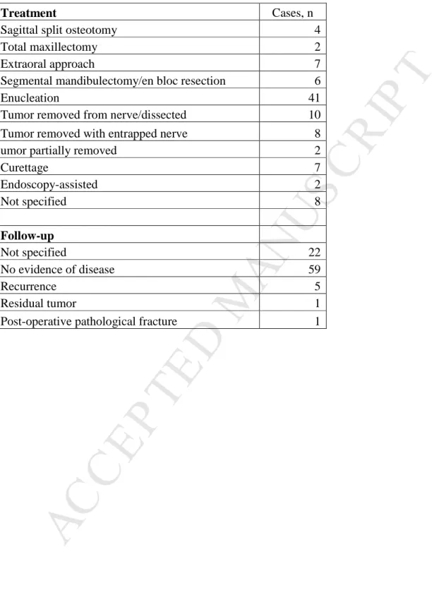
Conservative enucleation following the reflection of a mucoperiosteal flap and creation of a bony window was the treatment of choice for most mandibular and maxillary schwannomas [3, 5-8, 10, 12, 13, 18-21, 27, 28, 31, 33, 37-41]. Complete removal was possible in all but two cases where the lesion was only partially removed [3]. An extraoral approach was favored in seven instances [3, 5, 22, 31], while four authors accessed mandibular tumors via sagittal split ramus osteotomies (SSRO) [11, 34,
M
AN
US
CR
IP
T
AC
CE
PT
ED
42]. Schwannoma dissection from the affected nerve was possible in 10 cases [3, 23, 25, 31, 32, 36], but eight examples required the entrapped nerve be removed as well [3, 9, 11, 14, 17, 22, 34, 42]. In one instance, a sural nerve graft was performed after sacrificing the inferior alveolar nerve and partial regain in sensation was achieved[42]. Larger resections were deemed necessary for six cases treated by segmental mandibulectomy or en bloc resection [3, 16, 26, 30, 36] and two cases treated by total maxillectomy [35]. Recently, two minimally invasive, endoscopy-assisted procedures were performed to remove two mandibular lesions located in the ascending ramus [23, 31].
Follow-up data was available for 66 of the 88 cases [3, 4, 6-14, 17, 19, 21-23, 28, 29, 31, 32, 34-38, 41]. No evidence of disease was noted in 59 cases (89%) [3, 4, 6-12, 14, 17, 19, 21-23, 28, 29, 31, 32, 34-38, 41], recurrence was detected in five cases (8%) [3], a residual tumor was noted once (2%) [3], five patients experienced post-operative paresthesia (8%) [21, 23, 29, 32, 36], and one pathological fracture (2%) following the surgical removal of a mandibular lesion was described [13]. Unfortunately, information pertaining to the improvement of pre-operative paresthesia is lacking in eight of the 10 reports[3, 7, 14, 27, 30]. One patient regained most sensation following surgery [29], while the entrapped nerve was removed with the tumor in the other case [34].
Discussion
In theory, schwannomas may affect any bone, but this is exceedingly rare. In the Mayo Clinic series of 11,087 primary bone tumors, 14 cases of intraosseous schwannomas were identified, accounting for less than 1% of these benign primary bone tumors[46]. Three mechanisms by which bony involvement may occur have been
M
AN
US
CR
IP
T
AC
CE
PT
ED
proposed: (1) a tumor may arise centrally within bone, (2) a tumor may arise within the nutrient canal leading to canal enlargement, (3) a soft tissue or periosteal tumor may cause secondary erosion and penetrate into bone[3]. The first two mechanisms describe lesions that are truly intraosseous in nature. However, only schwannomas derived from the first mechanism are considered to be genuine primary bone tumors. Indeed, primary schwannomas of the bone are limited to the substance of bone, most likely arising from nutrient artery-associated vasomotor nerves, and are often centered on the medullary cavity[47]. As such, most of the lesions described herein are not primary in nature, since their associated nerves do not directly innervate bone. This difference is often visible histologically, as most primary tumors are not encapsulated[47].
Schwannomas may occur sporadically, in most cases, or in the context of familial tumor syndromes[1]. Neurofibromatosis type 2 (NF2) is a dominantly inherited syndrome characterized by mutations in the tumor suppressor-encoding gene NF2. Loss of function of this gene is also noted in most sporadic cases. NF2 patients are predisposed to schwannomas and often develop bilateral vestibular nerve tumors leading to hearing loss, peripheral schwannomas and other glial neoplasms like neurofibromas, meningiomas and gliomas[1]. Schwannomatosis is a distinct form of neurofibromatosis. Individuals develop multiple schwannomas, sometimes localized to a specific body segment, but lack other hallmark traits of NF2[1]. Finally, Carney complex patients are prone to developing melanotic schwannomas[1]. Fawcett and Dahlin describe a mandibular lesion showing histological signs of both a schwannoma and neurofibroma in the context of von Recklinghausen’s disease (Neurofibromatosis type 1)[48]. However, our review of the literature did not uncover any intraosseous cases associated to the above-mentioned
M
AN
US
CR
IP
T
AC
CE
PT
ED
conditions.Schwannomas are not considered an aggressive neoplasm. Surgical excision with preservation of the neighboring structures and periodic follow-up is considered the first-line treatment for these lesions. Furthermore, radiotherapy should be avoided, as the tumor is radio-resistant[3]. Including our first case, recurrence was reported in only five of the mandibular cases and one maxillary case. Recurrence is likely due to incomplete removal or tumor seeding. Post-operative paresthesia is possible, but rare. Malignant transformation of schwannomas is highly unlikely and has not been documented in any intraosseous cases of the jaws, though cases have been reported in other parts of the body.
Conclusion
Intraosseous schwannomas of the jaws are an exceedingly rare entity. We have presented two new mandibular examples and our review of the literature revealed 86 previously reported cases. To our knowledge, this is the largest literature review to date. These tumors most commonly involve the posterior mandible and usually present as an asymptomatic swelling, although pain or paresthesia are not uncommon. Radiographically, these tumors typically present a well-defined unilocular radiolucency, consistent with a benign process. Although the differential diagnosis for a radiolucent lesion of the jaws is wide, including odontogenic, fibro-osseous, vascular and reactive lesions to name a few, the identification of a lesion clearly centered on the mandibular canal should bring to mind the possibility of a peripheral nerve tumor such as a schwannoma. The final diagnosis requires histopathologic examination. Conservative
M
AN
US
CR
IP
T
AC
CE
PT
ED
surgical enucleation of a schwannoma is generally curative.
For mandibular lesions, SSRO is a tissue-sparing way of accessing mandibular schwannomas clearly centered on the mandibular canal. Classically, lesions in this area were approached by aggressive buccal window osteotomies. Segmental resection and reconstruction with autogenous iliac bone graft has also been reported [16]. SSRO affords adequate access to the inferior alveolar neurovascular bundle and its attached tumor, while minimizing bone loss and preserving nerve function. For anterior mandibular lesions, a conservative genioplasty-like approach, such as the one presented in our first case, can permit access to the surgical site, complete tumor excision and minimize post- operative morbidity.
References
1. Hilton DA, Hanemann CO: Schwannomas and their pathogenesis. Brain Pathol 24:205, 2014
2. Neville BW, Damm DD, Allen CM, Chi AC: Oral and Maxillofacial Pathology. 4th ed. St. Louis, MO, Elsevier, 2016, p 492
3. Chi AC, Carey J, Muller S: Intraosseous schwannoma of the mandible: a case report and review of the literature. Oral Surg Oral Med Oral Pathol Oral Radiol Endod 96:54, 2003
4. Minic A: Central schwannoma of the mandible. J Oral Maxillofac Surg 21:297, 1992
5. Kodani I, Ueyama Y, Mori T, et al: Intraosseous schwannoma of the mandible. Asian J of Oral Maxillofac Surg 15:64, 2003
M
AN
US
CR
IP
T
AC
CE
PT
ED
6. de Lacerda SA, Brentegani LG, Rosa AL, et al: Intraosseous schwannoma of mandibular symphysis: case report. Braz Dent J 17:255, 2006
7. Pimkhaokham A, Piriyasupong P, Swasdison S, et al: Central neurilemmoma of the jaw in concurrence with radicular cyst: a case report. Oral Surg Oral Med Oral Pathol Oral Radiol Endod 102:e34, 2006
8. Martins MD, Taghloubi SA, Bussadori SK, et al: Intraosseous schwannoma mimicking a periapical lesion on the adjacent tooth: case report. Int Endod J 40:72, 2007
9. Minowa K, Sakakibara N, Yoshikawa K, et al: CT and MRI findings of intraosseous schwannoma of the mandible: a case report. Dentomaxillofac Radiol 36:113, 2007
10. Hseih MY, Chen CM, Lin LM, et al: Intraossesous neurilemmoma (schwannoma) of the mandible - report of a case. Cases Journal TAOMFR 1:1, 2009
11. Ito S, Mandai T, Ishida K, et al: Intraosseous schwannoma of the mandible: a case report. Kawasaki Med J 35:249, 2009
12. Buric N, Jovanovic G, Pesic Z, et al: Mandible schwannoma (neurilemmoma) presenting as periapical lesion. Dentomaxillofac Radiol 38:178, 2009
13. Gallego L, Junquera L, Rodriguez-Recio C, et al: Intraosseous mandibular schwannoma mimicking an odontogenic keratocyst, with a postsurgical pathological fracture. J Laryngol Otol 123:560, 2009
14. Jang KY, Moon WS, Park HS: Intraosseous neurilemmoma of the mandible - a case report. Korean J Pathol 43:88, 2009
M
AN
US
CR
IP
T
AC
CE
PT
ED
15. Manor E, Tetro S, Noyhous M, et al: Translocation (2;13) and other chromosome abnormalities in intraosseous schwannoma of the mandible. Cancer Genet Cytogenet 193:116, 2009
16. Patil K, Mahima VG, Srikanth HS, et al: Central schwannoma of mandible. J Oral Maxillofac Pathol 13:23, 2009
17. Vera-Sempere F, Vera-Sirera B: Intraosseus plexiform schwannoma of the mandible: immunohistochemical differential diagnosis. J Craniofac Surg 21:1820, 2010
18. Parikh NR, Desai N: Intraoral schwannoma (neurilemmoma): an unusual anterior palatal swelling - a case report. J Int Oral Health 2:87, 2010
19. Kawasaki G, Yanamoto S, Yoshida H, et al: Intraosseous schwannoma of the mandibular symphysis: report of a case. Oral Sci Int 7:76, 2010
20. Metwaly H, Maruyama S, Cheng J, et al: Central schwannoma of the mandible: report of a case and review of the literature. Oral Med Pathol 12:29, 2010
21. Cristofaro MG, Giudice A, Donato G, et al: An unusual localization of intraosseus schwannoma: mandibular localization and new pathogenetic prospectives. Ann Ital Chir 82:205, 2011
22. Jahanshahi G, Haghighat A, Azmoodeh F: Intraosseous neurilemmoma of the mandible: report of a rare ancient type. Dent Res J (Isfahan) 8:150, 2011
23. Jiang WH, Brillo GV, Cheng AH, et al: Endoscope-assisted removal of intraosseous schwannoma with preservation of inferior alveolar nerve. J Craniofac Surg 22:617, 2011
M
AN
US
CR
IP
T
AC
CE
PT
ED
24. Kim NR, Chung DH, Park D-S, et al: Ancient schwannoma in oral cavity: a report of two cases. J Korean Assoc Oral Maxillofac Surg 37:530, 2011
25. Saghafi S, Salehinejad J, Rahpeyma A, et al: Intraosseous ancient schwannoma of the mandible: a case report. Iranian J Pathol 6:101, 2011
26. Sun Z, Sun L, Li T, et al: Intraosseous trigeminal schwannoma of mandible with intracranial extension. J Laryngol Otol 125:418, 2011
27. Agarwal K, Umargi HR, Tupkari JV, et al: Slowly growing swelling on body of the mandible with paresthesia on lower lip. Oral Surg Oral Med Oral Pathol Oral Radiol 114:677, 2012
28. Kargahi N, Razavi SM, Hasheminia D, et al: Mandibular intraosseous schwannoma in a child: report of a rare case. Dent Res J (Isfahan) 9(Suppl 1):S119, 2012
29. Simsek HO, Aksoy MC, Can C, et al: Intraosseous schwannoma of the mandible. Int J Exper Dent Sci 1:48, 2012
30. Thakur N, Bagewadi A, Keluskar V, et al: Intraosseous schwannoma of mandible. Int J Dent Clin 4:74, 2012
31. Zhang L, Xia B, Sun H, et al: Intraosseous schwannomas of the jaws: 2 case reports and review of the literature. Oral Surg Oral Med Oral Pathol Oral Radiol 114:e13, 2012
32. Suga K, Ogane S, Muramatsu K, et al: Intraosseous schwannoma originating in inferior alveolar nerve: a case report. Bull Tokyo Dent Coll 54:19, 2013
33. Lambade PN, Lambade D, Saha TK, et al: Unusual intramaxillary plexiform schwannoma. Oral Maxillofac Surg 17:137, 2013
M
AN
US
CR
IP
T
AC
CE
PT
ED
34. Mahmood L, Demian N, Weinstock YE, et al: Mandibular nerve schwannoma resection using sagittal split ramus osteotomy. J Oral Maxillofac Surg 71:1861, 2013
35. Verma A, Banerjee K, Verma A, et al: Maxillary neurilemmoma - rarest of the rare tumour: report of 2 cases. Int J Surg Case Rep 4:1044, 2013
36. Abouchadi A, Guerrouani A, Ribag Y, et al: Intrabony schwannoma of the mandible: case report and review of literature. Open J Stomatol 4:233, 2014 37. Garg B, Batra J, Chavda R, et al: Intraosseous schwannoma (neurilemmoma): an
unusual anterior maxillary swelling: a case report and review of literature. J Otol Rhinol 4:5, 2015
38. Meundi MA, Anekar J, Raj AC, et al: Intraosseous schwannoma of the maxilla mimicking a periapical lesion: a diagnostic challenge. J Clin Diagn Res 9:ZD01, 2015
39. Yin Y, Wang T, Cai Y-P, et al: Microcystic/reticular schwannoma of the mandible first case report and review of the literature. Medicine (Baltimore) 94:e1974, 2015
40. Avinash T, Sandhya T, Dodal S, et al: Recurrent ancient intraosseous neurilemmoma of maxilla: a rare case report. Iran J Pathol 11:176, 2016
41. Kawasaki Y, Kobashi H, Ishii S, et al: Intraosseous schwannoma localized in the anterior mandible with multilocular radiolucency: a case report. Oral Science Int 13:37, 2016
42. DeLeonibus A, Bassiri Gharb B, Papay F, et al: Surgical management of mandibular intraosseous schwannomas. J Craniofac Surg 28:e307, 2017
M
AN
US
CR
IP
T
AC
CE
PT
ED
43. Ogutcen-Toller M, Metin M, Karagoz F: Cellular schwannoma of the mandible: a case report. J Oral Maxillofac Surg 59:826, 2001
44. Hodson JJ: An intra-osseous tumour combination of biological importance-invasion of a melanotic schwannoma by an adamantinoma. J Pathol Bacteriol 82:257, 1961
45. Redman RS, Guccion JG, Spector CJ, et al: Cellular schwannoma of the mandible: a case report with ultrastructural and immunohistochemical observations. J Oral Maxillofac Surg 54:339, 1996
46. Unni KK, Dahlin DC: Dahlin's Bone Tumors. General Aspects and Data on 11,087 Cases. 5th ed. Philadelphia, PA, Lippincott-Raven, 1996
47. Ida CM, Scheithauer BW, Yapicier O, et al: Primary schwannoma of the bone: a clinicopathologic and radiologic study of 17 cases. Am J Surg Pathol 35:989, 2011
M
AN
US
CR
IP
T
AC
CE
PT
ED
Figure legendsFigure 1. A. Clinical photograph of patient at presentation demonstrating anterior mandibular swelling. B. Intraoral photograph of patient at presentation demonstrating swelling and hypervascularity localized to the anterior mandibular vestibule.
Figure 2. A. Representative axial cut of the initial maxillofacial CT scan without contrast demonstrating an anterior mandibular multilocular radiopaque lesion with expansile borders. B. Occlusal radiograph at presentation demonstrating finely defined expanded mandibular borders on the buccal and lingual aspects with a mixed radiopaque aspect of the symphysis further denoting erosion of cortical borders. C. Panoramic radiograph at presentation demonstrating a multi-loculated anterior mandibular radiolucency eroding the roots of mandibular teeth from the first left premolar to the medial root of the right first molar and scalloping the inferior mandibular border.
Figure 3. Intraoperatively, vestibular incision with buccal corticotomy to expose the anterio- superior aspect of the lesion, which was comprised of thick, hemosiderin-laden soft tissue fragments. Photograph taken after complete curettage of the lesion.
Figure 4. A. Low power magnification of the final excisional biopsy specimen demonstrating multiple areas of cystic degeneration (asterisks) with prominent fibrosis and inflammatory cell infiltrate. B. High power magnification of the final excisional biopsy specimen demonstrating whorls of spindle cells staining strongly and diffusely for S-100 protein.
M
AN
US
CR
IP
T
AC
CE
PT
ED
Figure 5. A. Six-year post-op intraoral photograph demonstrating absence of hypervascularity of the soft tissues, minimal scarring of vestibule and intact appearing dentition. B. Six-year post-op maxillofacial CT scan without contrast. Slice reveals a cyst-like lesion affecting the inferior border of the symphysis, in addition to extensive bone scarring with bony thickening and sclerosis. C. Exposure of the mandibular symphysis to the inferior aspect reveals the reparative sclerotic bone formation, and the defect at the inferior border noted on imaging.
Figure 6. A. High power magnification demonstrating spindle cells arranged predominantly in Antoni B pattern. B. Immunohistochemical staining with S-100 protein showing diffuse and strong immunoreactivity.
Figure 7. Panoramic radiographic taken 12 years after the initial diagnosis and revealing continued enlargement of the left mental foramen, bony sclerosis of the symphysis area and preservation of dentition.
Figure 8. A cropped panoramic radiograph revealing a well-defined unilocular radiolucency centered on the right mandibular canal.
Figure 9. A. Spindle cells arranged in areas of Antoni A (bottom left corner) and Antoni B (top right corner) tissue (H&E stain, original magnification 10x). B.
Immunohistochemical staining with S-100 protein showing diffuse and strong
immunoreactivity (original magnification 10x) Note the reduction in S-100 staining in Antoni B tissue (top right corner).
M
AN
US
CR
IP
T
AC
CE
PT
ED
Figure 10. A. Right inferior alveolar nerve (white arrows) and attached tumor (black arrows) accessed via sagittal split ramus osteotomy. B. Tumor (black arrow) peeled off of inferior alveolar nerve (white arrow) with forceps. C. Inferior alveolar nerve (white arrows) replaced following tumor removal.
M
AN
US
CR
IP
T
AC
CE
PT
ED
Table 1. Cases of intraosseous schwannomas involving the mandible and maxilla.
Authors Study year
Patient age
Gender Site Clinical findings Associated with a nerve? Radiographic features
Treatment Follow-up Histology Comments
Minic et al. [4] 1992 9 M Maxilla (A) Asymptomatic swelling of the anterior maxilla of 10 mo. duration. Expansion of the alveolar ridge. Intact mucosa. Yes (nasopalatine nerve) Ill-defined radiolucency involving the alveolar bone between the roots of the maxillary central incisors.
Surgical excision
NED after 6 y Antoni A. IHC: S100 strongly expressed by most of the tumor cells.
M
AN
US
CR
IP
T
AC
CE
PT
ED
Kodani et al. [5] 2003 45 F Mandible (R) Slight diffuse swelling of the right cheek. Yes Well-demarcated radiolucent lesion at the right ramus detected 3 mo previously. CT: clear borders. MRI: high signal on T2-wighed images. Complete enucleation via an extraoral incision at the inferior border of the mandible NS Antoni A and B. IHC: strong S100+, Desmin-, SMA-. GFAP+. de Lacerda et al. [6] 2006 11 M Mandible (A) Slow-growing asymptomatic swelling over 3 y, buccal and lingual expansion, displaced incisors. Yes (peripheral nervous plexus of the anterior mandible) Well-demarcated radiolucent unilocular intraosseous lesion, precluding the eruption of the permanent mandibular incisors, canines and premolars, discrete root resorption, agenesis of the central incisors. Surgical enucleationNED after 5 y Antoni A predominated. Occasional Antoni B areas. Nuclear atypia, but no other signs of ancient change.
M
AN
US
CR
IP
T
AC
CE
PT
ED
Pimkhaokham et al. [7] 2006 29 F Mandible (P) Numbness of the right side of the lower lip for 3 mo. Yes Bilocular homogenous radiolucent lesion 10x15mm, with well-defined margins, extending from the right lower second premolar to the distal root of the 1st molar. Mucoperiosteal flap; bony window; resection of the tumor mass, preservation of the nerve. Enucleation of the adjacent cystic lesion.NED after 1 y Antoni A and B. IHC: NS Radicular cyst directly adjacent to intraosseous schwannoma Martins et al. [8] 2007 34 F Mandible (P) Painful carious, but vital teeth. No bony expansion. No Radiolucency measuring 15x10mm between the roots of teeth 29 and 30. Intact periodontal ligament and lamina dura on both teeth. Surgical excision. Easy separation from surrounding tissue.
NED after 3 y Antoni B predominated. S-100+.
M
AN
US
CR
IP
T
AC
CE
PT
ED
Minowa et al. [9] 2007 67 F Mandible (P) No clinical signs. Lesion identified radiologically during examination after facial trauma.Yes Unilocular, well-defined, radiolucent lesion. Slight molar root resorption. CT: buccolingual erosion of the mandibular cortex, destructive changes in the cortical wall of the mandibular canal by the tumor. MRI: canal encased by the solid tumor mass.
Complete removal of the tumor and entrapped inferior alveolar nerve NED after 14 mo. Antoni B predominantly. Foci of Antoni A without Verocay bodies.
M
AN
US
CR
IP
T
AC
CE
PT
ED
Hsieh et al. [10] 2009 54 M Mandible (P) Grade 2 mobility of the mandibular right first and second molars of a few months duration. Teeth tested vital. Yes Well-defined, unilocular, oval-shaped, radiolucent lesion with peripheral cortication, involving the inferior mandibular canal and extending from teeth 30 to 32. External root resorption. CT: lingual cortical plate perforation. Surgical excision.NED after 3 y. Alternating Antoni A and B. IHC: diffuse S100+, Desmin-, SMA-.
M
AN
US
CR
IP
T
AC
CE
PT
ED
Ito et al. [11] 2009 70 M Mandible (P)
Swelling and pain.
Yes Large, unilocular, radiolucent lesion with distinct borders, extending from the second molar to the upper portion of the ramus. CT: cortical resorption. Extraction of the third molar; mucoperiosteal flap; bony window; enucleation of the tumor and
en bloc resection of the inferior alveolar neurovascular bundle. NED after 16 mo. Antoni A predominated. Occasional Antoni B areas. IHC: diffuse S100 positivity. Buric et al. [12] 2009 23 F Mandible (P) Painful swelling persisting after endodontic treatment on a molar. Slightly expanded buccal cortical plate. No Unilocular periapical radiolucency with an ill-defined border at the mesial root of endodontically-treated tooth 30. Surgical enucleation and apicoectomy. 10 x 5 x 5 mm.
NED after 1 y. Fascicles of spindle-shaped cells arranged in a palisades (Antoni A) with a rich vascular component. IHC: S100 stongly +, desmin-, SMA-. Mistaken for an angioleiomyoma histologically.
M
AN
US
CR
IP
T
AC
CE
PT
ED
Gallego et al. [13] 2009 60 M Mandible (R)None Yes Well-circumscribed, unilocular, radiolucent lesion in the left mandibular angle along the inferior alveolar nerve.
Surgical enucleation from the bony cavity. The course of the nerve was not clearly identified. Pathological fracture of the left mandibular angle 45 d post-op. NED after 6 mo. Antoni A predominated. Occasional Antoni B areas. IHC: diffuse S100 positivity. Jang et al. [14] 2009 77 F Mandible (P) Painful swelling of the right chin, evolving over 3 y. Paresthesia of the right lower lip and chin.
Yes 3x2cm, well-demarcated radiolucent lesion in the anterior mandibular body. CT: thinning of the buccal and lingual cortical plates. Mucoperiosteal flap; bony window; resection of the tumor mass with the right mental nerve and overlying thinned cortical bone. NED after 2 mo. Alternating Antoni A and B. Ancient change. IHC: S100 +, SMA and CD34 negative. Ancient change.
M
AN
US
CR
IP
T
AC
CE
PT
ED
Manor et al. [15] 2009 57 F Mandible (P) Asymptomatic, slight buccal and lingual expansion of the mandible. Yes Radiolucent, unilocular, well-circumscribed lesion with densely sclerotic borders, situated between the canine and second molar.Enucleation NS Spindle shaped cells with palisading nuclei and prominent Verocay bodies. Alternating Antoni A and B. IHC: uniformly S100+. t(2;13)(p13;q34) Patil et al. [16] 2009 23 F Mandible (P) Swelling for 6 mo. Gradual increase in size. Tooth mobility for 15 d.
Yes Unilocular, well-defined, radiolucent lesion. Slight root resorption and tooth displacement. Buccolingual expansion. Segmental mandibular resection; reconstruction with iliac crest bone graft.
NS Antoni A and B. IHC: diffuse S100 positivity .
M
AN
US
CR
IP
T
AC
CE
PT
ED
Vera-Sempere et al. [17] 2010 46 F Mandible (R) Buccolingual expansion for 6 mo.Yes Large, well-defined, radiolucency involving the angle and ramus. Internal multilocular appearance. Cortical expansion and thinning. Surgical enucleation including a portion of the inferior alveolar nerve.
NED after 2 y Multiple, well-demarcated tumor nodules; encapsulation; Antoni A predominated. IHC: S100 stongly +, Vimentin+, CD57+ in tumor cells. 1st case of plexiform intraosseous schwannoma. No evidence of a syndromic association Parikh et al. [18] 2010 64 F Maxilla (A) History of asymptomatic gradual swelling on palate over 3 y. Smooth surfaced, 2x2cm, oval swelling in anterior hard palate region No Well-defined radiolucency with a sclerotic lining in anterior hard palate in relation with maxillary anterior teeth. Surgical enucleation under local anesthesia. NS Antoni A, Antoni B, areas of necrosis, hyalinization and myxoid degeneration.
M
AN
US
CR
IP
T
AC
CE
PT
ED
Kawasaki et al. [19] 2010 27 M Mandible (A) Asymptomatic, slight anterior mandibular buccal expansion. All teeth were vital. NS Well-circumscribed, unilocular, radiolucent lesion 2 cm in diameter, inferior to the roots of mandibular incisors. CT:18 × 20 × 22 mm mass between the mental foramina. Surgical enucleation under general anesthesia. NED after 22 mo. encapsulated, well-demarcated tumor lobules, Antoni A. IHC: S100+.M
AN
US
CR
IP
T
AC
CE
PT
ED
Metwaly et al. [20] 2010 34 M Mandible (A) Slight swelling, hard and painless on palpation, in right incisor apical area of mandible. Slight mobility of incisors. No paresthesiaYes Well defined, corticated radiolucency extending from the apices of 22 to 26 down to the inferior mandibular cortex CT: thinning and partial perforation of buccal and lingual cortices. Associated with mandibular canal. Surgical enucleation. NS Antoni A, capsulated, hemorrhage with associated hemosiderosis. IHC: S100+. Nerve bundle attached to fibrous capsule Cristofaro et al. [21] 2011 40 NS Mandible (P) Pain and mobility of 29 anf 31 for 3 mo. Mandibular deformation. Yes Ground-glass radiolucency in right mandibular premolar-molar region. Root resorption of 29 and 31. Extraction of 29 and 31 and lesion resected under local anesthesia. Sporadic paresthesia after 3 mo. NED after 1 y Predominantly Antoni A with Antoni B. IHC: S100+, Vimentin+, Osteopontin+,
M
AN
US
CR
IP
T
AC
CE
PT
ED
Jahanshahi et al. [22] 2011 11 F Mandible (P) Swelling for 2 mo. Yes Well-circumscribed, unilocular radiolucent lesion with thin sclerotic borders. CT: erosion of the lingual cortex. Enucleation via extraoral approach; en bloc resection of the inferior alveolar nerve. NED after 3 mo. Antoni A and B. Hyalinization, calcification, hemorrhage and mild pleomorphism. Ancient schwannoma Jiang et al. [23] 2011 39 F Mandible (R) Asymptomatic extra-oral swelling of right mandibular angle. Yes Unilocular, radiolucency 3x4cm, well defined corticated margins. Affecting mandibular ramus. Clearly associated to IAN. CT: confirmed IAN involvement. Assisted by endoscopy, lesion was deroofed and enucleated with preservation of IAN. Intermittent paresthesia after 6 mo NSM
AN
US
CR
IP
T
AC
CE
PT
ED
Kim et al. [24] 2011 35 F Mandible (P)Normal Yes Unilocular well defined radiolucency (cystic appearance), 3x1.5x2cm, left mandibular body, external root resorption of teeth 18 and 19 IAN included in lesion. Incisional biopsy, patient deceased before complete enucleation. Patient deceased Paresthesia after biopsy Cellular spindle cells in fascicles, showing occasional bizarre shaped enlarged cells. IHC: S100+, pancytokeratin-, desmin-, SMA- Diagnosis: schwannoma with focal ancient changes
M
AN
US
CR
IP
T
AC
CE
PT
ED
Saghafi et al. [25] 2011 27 F Mandible (R) Asymptomatic swelling of left mandibular angle, 2 y. Yes Multilocular radiolucency in left mandibular angle and ramus, floating teeth 17 and 18 without root resorption. Fine septae present within the lesion. CT: Cortical thinning and expansion, fine calcification visible within the lesionRetromolar incision, removal of buccal bone and tumor dissection from IAN. NS Benign proliferation of Schwann cells in fibrous stroma. Calcified foci were visible IHC: S100+
M
AN
US
CR
IP
T
AC
CE
PT
ED
Sun et al. [26] 2011 22 M Mandible (R) Pain and loosening of the left mandibular molars over 6 mo. Facial swelling over 2 mo. Yes Radiolucent lesions with clear margins and internal septation, within the left mandibular body and ramus; thinned cortex; "floating" teeth; root resorption. CT: mass extending into the crania via enlarged foramen ovale. Segmental resection of the mandible and reconstruction with a vascularised fibular flap. NED at 2 y Antoni A predominated. Occasional Antoni B areas. Nuclear atypia, large blood vessels, hyalinised vessel walls, thrombus formation, hemorrhage, hemosiderin, focal necrosis and foamy macrophage infiltraiton. IHC: S100 and NSE diffusely +, SMA-.
M
AN
US
CR
IP
T
AC
CE
PT
ED
65 F Mandible (R) Facial swelling, intermittent toothache of the right mandibular molars over 2 y; diffuse swelling of the mandibular bucco-lingual plates. Yes (superior alveolar nerve) Multilocular, radiolucent lesion with clearly defined margins extending from the molar region to the sigmoid notch. CT: lesion extending into the middle cranial fossa Segmental resection of the mandible and reconstruction with a vascularized fibular flap. NS Antoni A predominated. Occasional Antoni B areas. Nuclear atypia, but no other signs of ancient change. IHC: S100 and NSE diffusely +, SMA-M
AN
US
CR
IP
T
AC
CE
PT
ED
Agarwal et al. [27] 2012 23 F Mandible (P) Gradual swelling of the left posterior mandible, from the second premolar to the second molar. Paresthesia of the left lower lip, left buccal mucosa and left tongue. 6 mo. duration. Yes Well-defined multilocular radiolucency in the left body of tht mandible. Displacement of the second premolar. Scalloped margins. Slight expansion and thinning of the inferior border of the mandible. CT: perforation of the buccal cortical plate, residual bony septa within the lesion. MRI: isointense on T1, slightly heterogeneously hyperintense on T2, a few hypointense areas of necrosis, level Ia, Ib and IV cervical lymphadenopathy. Surgical excision NS Antoni B predominantly. Antoni A also present. IHC: S100 strongly positive. Cervical lymphadenopathy and necrosis left unexplained .M
AN
US
CR
IP
T
AC
CE
PT
ED
Kargahi et al. [28] 2012 9 M Mandible (P) Asymptomatic, swelling of lower right mandibular border, mobility and displacement of anterior teeth NS Radiolucency with sclerotic borders extending from M to 30 Transposition of impacted 28 and 29. CT: unilocular lesion 2.5 x 3 cm, well defined, expansion and thinning of buccal and lingual cortices. Excisional biopsy. NED after 4 mo. Antoni A and Antoni B, no evident capsule, slight pleomorphism and hyperchromatism. IHC: S100+, Ki67 (less than 10%) Simsek et al. [29] 2012 47 F Mandible (P) Paresthesia left mandible 1 y.Yes Well defined radiolucency. Curettage under local anesthesia and endodontic therapy on tooth 20. Most sensation regained with intermittent paresthesia after 20 mo. Verocay bodies surrounded by spindle shaped cells. IHC: S100+.
M
AN
US
CR
IP
T
AC
CE
PT
ED
Thakur et al. [30] 2012 24 M Mandible (P) Asymptomatic slow growing swelling, 1 y. 62x50mm swelling of left mandible, with expansion of buccal and lingual cortices, facial asymmetry. Palpation revealed paresthesia of left lower lip and chin.Yes Multilocular radiolucency involving entire left body and ramus of mandible, with involvement of left condyle and coronoid process. Hemi-mandibulectomy with IAN resection. NS Antoni A and Antoni B.
Similar lesion was operated on 6 y prior. 18, 19, 20 were extracted. Histology consistent with schwannoma.
M
AN
US
CR
IP
T
AC
CE
PT
ED
Zhang et al. [31] 2012 35 M Mandible (R) Slow-growing swelling over 3 y, buccal and lingual expansion, slight pain on palpation. No 2 well-circumscribed, bilocular, radiolucent, 3cm lesions, from distal of tooth 30 to the right sigmoid notch. Root resorption of tooth 32. CT: thinning cortex Mass removed via extraoral approach; thinned cortex was removed and the mass was enucleatedNED after 3 y Antoni A predominated. Occasional Antoni B areas. Hemorrhage, but no other signs of ancient change. IHC: diffuse S100 and vimentin positivity. 39 F Mandible (R) Painless extraoral swelling. No paresthesia. Slight mandibular buccal expansion. Yes 3-4cm well defined radiolucent unilocular lesion with sclerotic borders, extending from the apical region of the second molar to the ramus. Mass removed via endoscopic guidance; buccal mucoperiosteal flap; bony window; mass carefully dissected from the nerve and removed in toto.
NED after 3 y Encapsulated, well-demarcated tumor lobules composed of spindle-shaped cells with aligned long nuclei.
M
AN
US
CR
IP
T
AC
CE
PT
ED
Suga et al. [32] 2013 33 M Mandible (P) Throbbing pain involving the first molar region, persisting after pulpectomy. Pain on percussion of the first and second molars.Yes CT: vertical and lateral expansion of the mandibular canal. Fusiform radiolucent lesion. MRI: hypointense in TI, hyperintense on T2. Mass occupying the mandibular canal. Sagittal split osteotomy via an intraoral approach; most of the lesion peeled away from the nerve; partial neurectomy posteriorly NED after 7 y and 4 mo. Temporary paresthesia. Antoni B predominated. Occasional Antoni A areas. Many cells with large, atypical, hyperchromatic nuclei. IHC: diffuse S100 positivity, weak NSE positivity. Lambade et al. [33] 2013 33 M Maxilla (P) Swelling for 8 mo. Yes (superior alveolar nerve) Diffuse radiolucency distal to tooth 1, causing root resorption Surgical excision via intraoral approach NS Multiple nodules composed of alternating Antoni A and B tissue; IHC: S100+ Plexiform schwannoma of the maxillary division of the trigeminal nerve.
M
AN
US
CR
IP
T
AC
CE
PT
ED
Mahmood et al. [34] 2013 23 F Mandible (R) 5 mo. history of paresthesia along the distribution of the left V3 nerve branch. Yes Well-circumscribed radiolucent lesion at the left angle of the mandible within the inferior alveolar nerve canal. CT: second lesion detected proximally to the first. MRI: both lesions were T2-enhansing. Tumor excision and en bloc resection of the inferior alveolar nerve; sagittal split osteotomy via intraoral approach NED after 5 mo. Spindle cell neoplasm with rudimentary Verocay bodies, random nuclear pleomorphism, rare hyalinized vascular channels. IHC: strong S100+ Ancient changeM
AN
US
CR
IP
T
AC
CE
PT
ED
Verma et al. [35]2013 9 F Maxilla Swelling of the right maxilla and temporal region for 6 mo. Gradual increase in size. Slight pain. Intraoral bulging into the right buccal vestibule. NS CT: Large multilocular radiolucencies involving the maxillary antrum and orbital floor. Two simultaneous tumors. Total maxillectomy; excision of the temporal mass; reconstruction with temporalis muscle flap
NED after 1 y Antoni A and B. IHC: diffuse S100 positivity Questionable case - lack of documentation 27 F Maxilla Gradually increasing swelling of the right maxilla for 1.5 y. Intraoral bulge. NS NS Total maxillectomy
NED after 1 y Antoni A and B. IHC: diffuse S100 positivity
Questionable case - lack of
M
AN
US
CR
IP
T
AC
CE
PT
ED
Abouchadi et al. [36] 2014 39 M Mandible (P)Normal Yes Unique multilocular radiolucency sitting in left mandibular corpus between teeth 24 and 18. A concomitant radiolucency appears as a cyst of the left mandibular first premolar root without root erosion. CT: well defined lesion measuring 36 × 18 mm continuous to the path of IAN. Pre-op endodontic therapy on teeth 24 to 20. Surgical enucleation, due to difficult dissection IAN was only partially respected Immediate paresthesia of left lower lip. 1 y post-op, partial resolution of paresthesia. NED after 2 y. Encapsulated tumor composed of connective tissue arranged in crossed fascicles and fusiform cells with aligned long nuclei. Presence of Verocay bodies. IHC: S100+
M
AN
US
CR
IP
T
AC
CE
PT
ED
Garg et al. [37] 2015 58 F Maxilla (A) Asymptomatic progressive swelling of upper jaw, obliterating right nasolabial fold 1 y. Intraorally: 2 x 2 cm firm to hard swelling from apex of tooth 6 to apex of tooth 10 with obliteration of labial vestibule. NS Unilocular well defined radiolucency, 2 cm in diameter, with sclerotic border. Extending from tooth 6 to 10. External root resorption of teeth 6 and 7, all teeth were vital.Surgical excision under local anesthesia
NED after 1 y Predominantly Antoni A with scattered Antoni B. Fibrous capsule. No IHC.
M
AN
US
CR
IP
T
AC
CE
PT
ED
Meundi et al. [38] 2015 20 F Maxilla (A) Asymptomatic, no facial swelling, 1 x 3 cm swelling in left anterior hard palate, in relation with teeth 10 and 11 (vital). Non-tender, bony hard mass. No Unilocular, radiolucent lesion 2 x 3 cm, overlapping teeth 10, 11 and 12 Margins were clear defined and irregularly corticated.Surgical excision under local anesthesia
NED after 2 y Antoni A and Antoni B. No IHC.
M
AN
US
CR
IP
T
AC
CE
PT
ED
Yin et al. [39] 2015 61 F Mandible (P) Right facial asymmetry for 1 y. Yes (inferior alveolar nerve) Unilocular, radiolucent lesion 3.9 x 3.4 cm, with clear defined margins. Cortical expansion. Resection. NS Eosinophilic spindle- shaped cells arranged in microcystic pattern with reticular growth. Myxoid stroma with hyalinized collagen infiltration. Thin fibrous capsule extending into the tumor to form lobules. Nervous tissue adjacent to capsule. IHC: S100 +, CD34+, CD99+, NSE+, MIB1-, CK-, EMA-, CK5/6-, p63-, Calponin-, CD10-, SMA-, Desmin-, GFAP-, NF-, Syn-, CgA-. First microcystic/reticular schwannoma described in the mandible
M
AN
US
CR
IP
T
AC
CE
PT
ED
Avinash et al. [40] 2016 38 M Maxilla (P) Asymptomatic, soft, 3x2.5x0.5 cm swelling over left maxillary area below the infraorbital region extending from the ala of the nose to malar process antero-posteriorly and from infra orbital margin to 1 cm above the corner of mouth superior-inferiorly. 1x1cm swelling of alveolar mucosa and buccal expansion from teeth 11 to 16. NS Radiolucency extending in left maxillary premolar-molar region. Partly well-defined corticated margins and discontinuous maxillary sinus floor CT: lesion extending from teeth 11 to 16, maxillary sinus and buccal cortical plate perforation. Surgical excision under general anesthesia. NS Antoni A and Antoni B, regions of cystic degeneration IHC: S100+ Carious 16 was extracted one y prior, with post-operative gradual swelling. Excisional biopsy showed a non-specific infection.Only one section of the biopsy showed histological signs of Schwannoma.
M
AN
US
CR
IP
T
AC
CE
PT
ED
Kawasaki et al. [41] 2016 69 F Mandible (A) Asymptomatic, well demarcated, non-tender swelling on labial gingiva of anterior mandible. Slight mobility of mandibular canines (vital), moderate mobility of mandibular incisors (non-vital) No Radiolucency extending from teeth 22 to 27. External root resorption of all mandibular incisors. No evidence of interaction with IAN. CT: Multilocular expansile lesion with well-defined margins, 25x22x10 mm Surgical enucleation under general anesthesia.NED after 1 y Predominantly Antoni A with scattered Antoni B. IHC: S100+. DeLeonibus et al. [42] 2017 72 F Mandible (P)
NS Yes Suspected tumor following panoramic radiograph examination. Sagittal split osteotomy; complete excision of tumor and IAN; sural nerve graft to repair IAN. NED after 1 y and partial resolution of post-operative paresthesia. NS
M
AN
US
CR
IP
T
AC
CE
PT
ED
Case report 1 22 M Mandible (A) Buccolingual expansion spanning both mental foramina, labiomental fold obliteration, chin point deviation, tooth mobility, no pulp necrosis. No Expansile, well-circumscribed, multilocular radiolucency extending from teeth 20 to 29, external root resorption. Buccal and lingual cortex erosion and perforation. Curettage under general anesthesia. Tumor recurrence after 7 y. NED for 2 y following retreatment. Antoni A and Antoni B tissue, inflammatory cells and signs of cystic
degeneration and fibrosis. IHC: S100+.
Case report 2 39 M Mandible (P)
Occasional dull pain, right mandible. Yes Well-defined radiolucency centered on the right mandibular canal. Sagittal split osteotomy; tumor pealed from IAN.
NED after 3y Antoni A and Antoni B tissue. Verocay bodies were absent and no mitotic activity. IHC: S100+, EMA-, SMA-, CD68-.
M
AN
US
CR
IP
T
AC
CE
PT
ED
Abbreviations: M: male, F: female, A: anterior, P: posterior, R: ramus, mo: month(s), y: year(s), d: day(s), NED: no evidence of
M
AN
US
CR
IP
T
AC
CE
PT
ED
Table 2: Epidemiologic data of 88 reported intraosseous schwannomas of the jaws Cases Mean age
(range) Male: Female ratio Posterior: Anterior ratio Mandible 75 36.9 (8-77) 1 : 1.5 3.6 : 1 Maxilla 13 29.7 (9-64) 1 : 2.25 1 : 1
Table 3: Clinical and radiological features of 88 intraosseous schwannomas of the jaws.
Cases, n (%) Clinical examination Total* 83 No symptoms 13 (16%) Swelling or expansion 59 (71%) Pain or tenderness 24 (29%) Tooth mobility/displacement 17 (20%) Mandibular paresthesia 10 (14%) Surface ulceration 1 (1%)
Infection and rapid growth 1 (1%)
Associated with nerve?
Yes 45 (63%)
No 27 (37%)
M
AN
US
CR
IP
T
AC
CE
PT
ED
Radiographic presentation Total* 82Well-defined unilocular radiolucency 62 (76%)
Multilocularity 13 (16%)
Root resorption 21 (26%)
Tooth displacement/impaction 6 (7%)
Cortical thinning/erosion 22 (27%)
Spotty calcification/focal radiopacity 4 (5%)
Peripheral scalloping 5 (6%)
Cortical expansion 13 (16%)
Periosteal reaction 1 (1%)
Diffuse radiolucency 3 (4%)
adjacent to a cyst/tumor 3 (4%)
M
AN
US
CR
IP
T
AC
CE
PT
ED
Table 4: Treatment and outcome data.
Treatment Cases, n
Sagittal split osteotomy 4
Total maxillectomy 2
Extraoral approach 7
Segmental mandibulectomy/en bloc resection 6
Enucleation 41
Tumor removed from nerve/dissected 10
Tumor removed with entrapped nerve 8
umor partially removed 2
Curettage 7 Endoscopy-assisted 2 Not specified 8 Follow-up Not specified 22 No evidence of disease 59 Recurrence 5 Residual tumor 1