HAL Id: dumas-01652019
https://dumas.ccsd.cnrs.fr/dumas-01652019
Submitted on 29 Nov 2017
HAL is a multi-disciplinary open access
archive for the deposit and dissemination of sci-entific research documents, whether they are pub-lished or not. The documents may come from teaching and research institutions in France or abroad, or from public or private research centers.
L’archive ouverte pluridisciplinaire HAL, est destinée au dépôt et à la diffusion de documents scientifiques de niveau recherche, publiés ou non, émanant des établissements d’enseignement et de recherche français ou étrangers, des laboratoires publics ou privés.
Prévalence des manifestations rhumatologiques apparues
sous immunothérapie anticancéreuse et corrélation à la
réponse tumorale : une étude prospective monocentrique
Léa Rouxel
To cite this version:
Léa Rouxel. Prévalence des manifestations rhumatologiques apparues sous immunothérapie anti-cancéreuse et corrélation à la réponse tumorale : une étude prospective monocentrique. Médecine humaine et pathologie. 2017. �dumas-01652019�
1
Université Bordeaux
UFR MEDICALES
Année 2017 N° 3109
Thèse pour l’obtention du
DIPLOME d’ETAT de DOCTEUR EN MEDECINE
Présentée et soutenue publiquementle 22/09/2017
par Léa ROUXEL
Interne des Hôpitaux de Bordeaux Née le 05/08/1988 à Bordeaux.
Prévalence des manifestations rhumatologiques apparues sous
immunothérapie anticancéreuse et corrélation à la réponse tumorale :
une étude prospective monocentrique.
Directeur de thèseMonsieur le Pr Thierry Schaeverbeke Jury
Monsieur le Professeur Christophe Richez, Président Monsieur le Professeur Thierry Schaeverbeke, Directeur
Madame le Docteur Nadia Mehsen-Cetre, Juge Monsieur le Docteur Rémi Veillon, Juge Monsieur le Professeur Jean-Luc Pellegrin, Juge
Madame le Professeur Estibaliz Lazaro, Juge
Rapporteur
2
REMERCIEMENTS
A mon Président du jury,
Monsieur le Professeur Christophe Richez,
Merci d’avoir accepté de présider ma thèse, mais surtout merci d’avoir été un coordonnateur de DES présent, efficace, et à notre écoute. Tu répondais toujours présent lorsque j’avais besoin d’un avis en consultation (sauf quand t’avais piscine mais c’était rare), et tes blagues détendaient un peu la visite du vendredi. Tu m’as beaucoup appris et guidé au cours de mon internat et je suis ravie de continuer d’apprendre à tes côtés pendant les 2 prochaines années.
A mon directeur de thèse,
Monsieur le Professeur Thierry Schaeverbeke,
Vous m’avez énormément aidée pour ce travail, et consacré de votre temps et de votre énergie pour qu’il soit le plus accompli possible. Vous m’avez également donné le goût de la
rhumatologie, déjà sur les bancs de la faculté lors de vos cours, n’ayant jamais eu le privilège d’effectuer un stage d’externat dans votre service, et ensuite lors de mes stages d’internat. Je vous remercie de m’accepter pour 2 ans de plus (si si vous avez signé !) pour mon assistanat dans le service de rhumatologie, j’en suis honorée et ravie.
A mon rapporteur,
Madame le Professeur Estibaliz Lazaro,
Tu m’as beaucoup appris lors de mon passage au G2, sur le plan des connaissances médicales mais aussi par ta rigueur professionnelle impressionnante. Tu trouveras ici la marque de ma plus profonde admiration.
Aux membres du jury :
Madame le Docteur Nadia Mehsen-Cetre,
C’est toi qui m’as confié ce travail, m’as aidée à le démarrer, c’est donc toi qui es à l’initiative de ce bel article !! Tu as toujours été présente lorsque j’en avais besoin, pour m’aider à me
3 sortir de situations délicates au cours du poste de consultations. Merci pour ta bonne humeur qui fait que l’HDJ est encore plus « bisounours » que le reste du service, et ta façon de pratiquer la médecine qui fait de toi un de mes modèles.
Monsieur le Docteur Rémi Veillon,
Vous avez suivi ce travail depuis le début lorsque j’apprenais tout juste ce qu’étaient les anti-PD1. Votre expertise dans le domaine de l’oncologie broncho-pulmonaire offre une vision supplémentaire au jugement de ce travail. Je vous remercie pour votre collaboration des 2 dernières années et d’avoir accepté d’être membre du jury de ma thèse.
Monsieur le Professeur Jean-Luc Pellegrin,
J’ai énormément appris lors de mon passage dans votre service. Je vous remercie d’avoir accepté de faire partie de mon jury de thèse ; votre expertise de l’immunologie rend votre jugement précieux. Je suis honorée de faire partie des personnes qui sont passées au G2.
Au Docteur Marie Kostine,
Qui a réalisé ce travail avec moi, et qui a mis énormément de temps et d’énergie pour qu’il soit le plus abouti possible. Tu m’as guidée, corrigée, aidée, rassurée, motivée, tout cela en remplissant tes fonctions de chef de clinique, et avec brio en plus ! (tout le monde est
d’accord). J’ai adoré travailler avec toi. En plus de ça, j’ai découvert une personne pétillante, drôle, toujours prête pour l’aventure (y compris partir manger des racines à l’autre bout du monde). Je te souhaite plein de bonheur pour cette toute nouvelle aventure !
A Thomas Barnetche,
statisticien émérite qui a travaillé avec nous sur cet article. Je te remercie de toujours
t’arranger pour être disponible lorsqu’on a besoin de tes lumières en matière de statistiques. Aux autres médecins de rhumatologie qui m’ont marqué pendant mon parcours : à Nico, pour ta présence, ton aide infaillible, tes blagues, l’épaule de Yann, les parties de baby foot, les petits dej d’astreinte. A M. Bannwarth, pour vos cours improvisés sur les AINS devant la chambre « Tagada », vos visites de fin d’après midi en hôpital de jour (aussi appelées mission compote sans sucre). A Marie Elise, pour ta patience et tes cours sur la sclérodermie (mais pas
4 seulement !). A Claire, à Marion, super chefs de clinique. A Jean-Philippe, alias Dr V., et Severin, pour votre accueil dans le monde de la rhumatologie.
A l’équipe de rhumatologie du 12ème, les IDE, secrétaires, AS et ASH qui font de ce service un endroit que j’aime, surtout grâce à la bienveillance de notre maman à tous, Patricia, cadre tant convoitée par les autres services mais fidèle à la rhumatologie jusqu’au bout je l’espère ! À toutes les personnes rencontrées au cours de mon parcours, les équipes de neurologie et de rhumatologie de Libourne, l’équipe de dermatologie de Saint André, l’équipe du G2, du l’Hôpital Suburbain du Bouscat, et merci au Pr Houssiau de m’avoir accueillie à la Clinique Saint Luc pendant 6 mois très riches sur le plan professionnel et personnel.
A mes co-internes de rhumatologie : Alice et Fanny, Laeti, Pauline, Alexia, Lorraine, Lisou, Adeline, Vincent, François, Marc.
A mes proches,
Morganne Skreck, Paoletta et Aseel, les Desperate housewives de Libourne et la fashion police. Helene et Manon les Parisiennes au grand coeur. Aux co-internes du G2, qui étaient pour moi une petite famille pendant ces 6 mois. Ma Clotinete/ Clotasse/ Boloss, je ne pourrais pas mettre tous nos moments cultes sur papier (vive l’accent belge, et les prognathes). A Jojo et Ludo, des colocs en or.
A mon groupe de sous-colle, Juju Wouilles et Popote la barjotte, car vous avez fait de ces moments de boulot des moments mythiques (Cacastel, ses concours).
A la FF, pour tous ces bons moments souvent orchestrés par papa Aldi. Titou le kangourou, JAT notre dexter, Caro ma colloc chérie, Marie pour ta force, Cam pour ta douceur, Jibouze pour tes yeux bridés, Charly pour ton rire contagieux, nos 3 pruneaux adorés, Geogeo et notre tchexplosion, Benouze ma future collègue, Carole pour ta générosité, Jé pour la chanson du shark gay, Claire pour le souvenir du bikini/boots au ski, Ben pour tes accords de guitare, Diegui pour ton humour et tes mimiques, Clem pour la baignoire ;) Quentin pour ta bonne humeur, Sophie l’espagnole, Laurette mon Hally baba, Brice, Horty, Manon des sources, Rafiki le chef cuistot et Segogo.
A ma Syb, vivement que tu quittes les poitevins et que tu viennes nous retrouver avec Q et le baby, Mathou la coquillette ma partenaire de course, ma Kelly la force tranquille du groupe et
5 les moments formidables que tu m’as fait vivre, Loulou et ta joie infaillible (et les séances gainage chez les Viet), Marinou kktou la gogol parce que même en habitant loin, tu es toujours la quand ça va, et quand ça va pas. Pensée pour Alice, partenaire de
pala/squash/footing, et Paulinou la néo-nantaise.
Au groupe fidèle de Roses, Miss Bonob, Miss Blonde, Palisso, Gogo, Pedro, Bedaine, Coma, Sancho, Paulinou. A Emeline et cette rencontre à l’aire de jeux du Domac en CE1.
A Pierre pour m’avoir supportée tous les jours ces derniers mois de préparation de thèse, pour entre autres me faire rire et me laisser gagner aux jeux de temps en temps. Pour tous ces beaux moments passés et à venir.
A ma famille, qui est ce que j’ai de plus cher, pour m’avoir soutenue depuis toujours. Merci à mes grand parents et ma grand mère, pour vous être si bien occupés de nous. Merci à mes parents, de rendre notre famille si soudée, et de nous aimer de manière inconditionnelle. A mon frérot : à part avancer plus vite que didoute et moi, j’ai rien à te reprocher, t’es un frère hors pair. A ma sœurette : pour être mon modèle depuis toujours. Je vous aime fort. A Gege et Paulo, merci de les rendre heureux.
6
TABLE DES MATIERES
I/ INTRODUCTION 10
A. L’activation lymphocytaire T et la costimulation 10
B. Inhibiteurs de checkpoint immunitaire utilisés en oncologie 11
C. Les Immune-Related Adverse Effects (irAEs) 12
D. Les irAEs rhumatologiques 12
E. Objectif de l’étude 13
II/ ARTICLE : Rheumatic disorders associated with immune checkpoint inhibitors in cancer patients: clinical aspects and relationship with tumour
response. A single-center prospective cohort study. 14
A. INTRODUCTION 15
B. PATIENTS AND METHODS 17
1. Study design 17
2. Rheumatologic evaluation 17
3. Evaluation of other irAEs and tumour response 18
4. Statistics 18
C. RESULTS 19
1. Patient characteristics 19
2. Description of musculoskeletal irAEs 19
3. Inflammatory arthritis 24
7
5. Other irAEs 25
6. Tumour response according to irAEs 26
D. DISCUSSION 27
E. CONCLUSION 29
F. REFERENCES 30
III/ DISCUSSION 32
8
TABLE DES ILLUSTRATIONS
Figures:
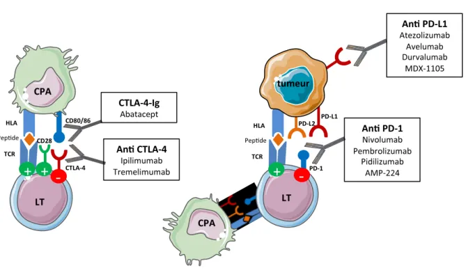
Figure I: Principales thérapeutiques ciblant la costimulation 11
Figure 1: Flow diagram for description of musculoskeletal irAEs 23
Tableaux:
Table 1: Demographic data, type of cancer and ICI molecules 20
Table 2: Demographic data, type of cancer, ICI molecules, and tumour response
correlated with irAEs 20
Supplementary Table 1: Characteristics of patients experiencing rheumatic irAEs 21 Supplementary Table 2: Description of rheumatic irAE and management 22
9
ABBREVIATIONS
irAEs : immune-Related Adverse Effects ICI : immune checkpoint inhibitors
CTLA-4 : cytotoxic T-lymphocyte-associated protein 4 PD-1 : programmed cell death 1
LT : T lymphocyte TCR : T cell receptor
CMH : complexe majeur d’histocompatibilité CPA : cellule présentatrice d’antigène
PD-L1 : programmed cell death 1 ligand IA : inflammatory arthritis
RA : rheumatoid arthritis PMR : polymyalgia rheumatica PsA : psoriatic arthritis
NSCLC : non–small cell lung cancer
RECIST : Response Evaluation Criteria In Solid Tumors RF : rheumatoid factor
CCP : cyclic citrullinated peptide AAN : antinuclear autoantibodies
10
I/ INTRODUCTION
A. L’activation lymphocytaire T et la costimulation
L’immunothérapie constitue actuellement une révolution thérapeutique en oncologie, avec l’arrivée des inhibiteurs de checkpoint immunitaire (ICI), sous la forme d’anticorps monoclonaux, notamment les anti-CTLA-4 (cytotoxic T-lymphocyte-associated protein 4) et les anti PD-1 (programmed cell death 1). Leur mécanisme d’action original cible le co-signal inhibiteur des cellules tumorales/cellules présentatrices d’antigènes sur les lymphocytes T, activant ainsi une immunité anti-tumorale.
Pour devenir pleinement activé, le lymphocyte T (LT) a besoin de deux signaux d’activation (Figure I). Le premier est celui transmis par le TCR, récepteur T spécifique de l’antigène lorsque celui-ci est présenté par la cellule présentatrice d’antigènes via le complexe majeur d’histocompatibilité (CMH, ou HLA). Le deuxième signal est un signal non spécifique de co-stimulation transmis par la liaison du CD28 exprimé à la surface du lymphocyte T, avec un de ses ligands CD80/86 à la surface de la cellule présentatrice d’antigène (CPA). Si le lymphocyte T reçoit ces deux signaux alors il sera activé, pourra proliférer et déclencher une réponse immunitaire spécifique.
Pour éviter que le système ne s’emballe, il existe des molécules notamment le CTLA-4, de structure similaire au CD28, se fixant aux mêmes ligands CD80/86 mais avec une plus grande affinité que le CD28 et transmettant un signal inhibiteur sur le lymphocyte T. Ce signal de co-stimulation négative provoque alors l’anergie ou la mort par apoptose du lymphocyte T, supprimant de potentiels lymphocytes autoréactifs et prévenant ainsi le développement de maladies auto-immunes. Cette voie est bien connue des rhumatologues car
11 un analogue du CTLA-4 est utilisé comme traitement de la polyarthrite rhumatoïde (Abatacept).
B. Inhibiteurs de checkpoint immunitaire utilisés en oncologie
Les oncologues vont chercher à moduler positivement cette co-stimulation afin d’activer une réponse immunitaire T anti-tumorale. A côté de la voie du CTLA-4, un autre checkpoint immunitaire majeur et exploité par les cellules tumorales pour échapper au système immunitaire est le récepteur 1 (à la surface du lymphocyte T) et son ligand PD-L1 (à la surface de la cellule tumorale et CPA). En exprimant PD-PD-L1, les cellules tumorales utilisent en quelque sorte le frein pour bloquer l’activation lymphocytaire T et empêcher leur destruction par le système immunitaire.
12 En stimulant la réponse cellulaire T anti tumorale, des réponses tumorales au long cours ont été observées chez des patients avec un cancer non opérable ou métastatique. La remarquable efficacité des inhibiteurs de checkpoint immunitaire a rapidement conduit à des autorisations de mise sur le marché dans plusieurs cancers (mélanome, cancer bronchique non à petites cellules, cancer rénal, lymphome de Hodgkin), et de nombreux protocoles sont en cours pour d’autres cancers.
C. Les Immune-Related Adverse Effects (irAEs)
Cependant, en inhibant les freins naturels des voies du CTLA-4 et du PD-1, on a favorisé l’émergence de manifestations auto-immunes par perte de la tolérance périphérique. Dans la littérature, ces manifestations auto-immunes sont identifiées sous le terme d’Immune-Related Adverse Effects (irAEs).
Dans les essais cliniques, les principaux irAEs rapportés concernent le tractus gastro-intestinal, la peau, les glandes endocrines, le foie et les poumons, mais quasiment tous les organes peuvent être atteints, comme illustré avec le nombre croissant de case reports. Le spectre de l’auto-immunité induite par ces traitements ne cesse de s’élargir, d’autant plus qu’ils sont maintenant utilisés à grande échelle.
D. Les irAEs rhumatologiques
Une récente analyse systématique de la littérature s’est concentrée sur les effets secondaires rhumatologiques. Les arthralgies et myalgies ont été les effets secondaires rhumatologiques les plus rapportés dans les essais cliniques, avec une prévalence de 1 à 43% et de 2 à 21%, respectivement (ref. 10 article). Plusieurs séries de cas et case reports ont décrit
13 de manière plus détaillée des cas d’arthrite inflammatoire, de pseudo-polyarthrite rhizomélique, de vasculite, de polymyosite ou de réaction sclérodermique secondaires aux ICI.
E. Objectif de l’étude
Dans le but d’évaluer la prévalence et le type d’effets secondaires rhumatologiques apparaissant sous ICI dans notre hôpital, ainsi que leur corrélation avec la réponse tumorale, nous avons mis en place un recrutement systématique des patients déclarant des symptômes rhumatologiques sous ICI, de septembre 2015 à mai 2017, et réalisé une étude prospective monocentrique observationnelle.
14
II/ ARTICLE
Soumis à Annals of the Rheumatic Diseases
Rheumatic disorders associated with immune checkpoint inhibitors in
cancer patients: clinical aspects and relationship with tumour response.
A single-center prospective cohort study.
M. Kostine (1)*, L. Rouxel (1)*, T. Barnetche (1), R. Veillon (2), F. Martin (2), C. Dutriaux (3), L. Dousset (3), A. Pham-Ledard (3), S. Prey (3), M. Beylot-Barry (3), A. Daste (4), M. Gross-Goupil (4), J. Lallier (4), A. Ravaud (4), N. Milpied (5), B. Bannwarth (1), M.E. Truchetet (1), C. Richez (1), N. Mehsen-Cetre (1), T. Schaeverbeke (1)
* These authors contributed equally to this work
(1) Rheumatology Department, CHU Bordeaux, Bordeaux, France (2) Pneumology Department, CHU Bordeaux, Pessac, France (3) Dermatology Department, CHU Bordeaux, Bordeaux, France (4) Oncology Department, CHU Bordeaux, Bordeaux, France (5) Hematology department, CHU Bordeaux, Pessac, France.
15
INTRODUCTION
Immune checkpoint inhibitors (ICIs) targeting the cytotoxic T-lymphocyte-associated protein 4 (CTLA-4) or programmed cell death protein 1 (PD-1) axes have accelerated the immunotherapy revolution in oncology. Since the approval of ipilimumab (anti CTLA-4) for the treatment of metastatic melanoma in 2011 (1), pembrolizumab and nivolumab (anti PD-1) have also demonstrated a survival benefit for patients with advanced melanoma, non–small cell lung cancer (NSCLC) and renal cell carcinoma and are now being tested in many other cancers (2)(3)(4)(5). Recently, agents targeting PD-1 ligand 1 (PD-L1), as well as other immune checkpoint molecules and combination strategies have been developed(6)(7).
In healthy individuals, the role of these immune checkpoints is to promote tolerance to self-antigens, therefore preventing autoimmunity and damage to normal tissues. Many tumour types appropriate these T-cell inhibitory pathways as immune escape mechanisms in order to evade activated cytotoxic T-cells(8)(9). ICIs currently used in the clinical setting target CTLA-4 or PD-1 receptors expressed on T-cells, or PD-L1 expressed on both antigen-presenting cells (APCs) and tumour cells, allowing T-cell activation and tumour destruction. By enhancing anti-tumour T-cell activity, these treatments have resulted in some unprecedented long-lasting tumour responses in patients with unresectable or metastatic disease. However, releasing the natural brakes of CTLA-4 and PD-1 signalling on T-cells may affect self-tolerance to healthy tissues, leading to immune side effects known as immune-related adverse effects (irAEs).
In clinical trials, reported irAEs mainly involved the gastrointestinal tract, skin, endocrine glands, liver and lung, but they can affect nearly all organs, as illustrated by the increasing number of case reports. Rheumatic and musculoskeletal irAEs observed in patients receiving ICIs have been the focus of a recent systematic review of the literature(10).
16 Arthralgia and myalgia were by far the most commonly reported rheumatic irAEs in clinical trials, with a prevalence ranging from 1 to 43% and from 2 to 21% respectively. However, prevalence may have been underestimated as only high-grade irAEs were reported in some trials. In addition, case series and reports provide more details on patients with inflammatory arthritis or disease patterns suggesting polymyalgia rheumatica, vasculitis, polymyositis or sclerodermoid reaction induced by ICIs (11)(12)(13). To date, the largest series of patients with rheumatic irAEs included 13 and 15 patients from the United States of America, highlighting differences compared to classical rheumatic diseases, and cases of seropositive RA were recently reported in a French retrospective study (14)(15)(16).
To evaluate the prevalence and type of rheumatic and musculoskeletal irAEs in patients receiving ICI treatment at a single institution (University Hospital of Bordeaux, France), we implemented systematic referral to the rheumatology department for evaluation of any rheumatic symptoms occurring with ICIs from September 2015 to May 2017.
17
PATIENTS AND METHODS
Study design
All patients aged 18 years and older treated with anti CTLA-4 (ipilimumab, tremelimumab), anti PD-1 (nivolumab, pembrolizumab) or anti PD-L1 (atezolizumab, avelumab, durvalumab, MSB0011359C) agents alone or in combination, either in trials or in routine clinical practice from September 2015 to May 2017 were included in this prospective observational study. Safety data were recorded at each follow-up visit before ICI administration.
To evaluate the prevalence and type of rheumatic events related to ICIs, rheumatologists contacted oncology, dermatology and pulmonology departments from our institution for referral of any musculoskeletal symptom occurring during or after ICI treatment, up to May 2017. Patients with a known rheumatic or autoimmune disease were also included if they had a flare of the underlying disease or new musculoskeletal symptoms.
Rheumatologic evaluation
For each referred patient, a rheumatologist (M.K. or L.R.) performed the history of musculoskeletal symptoms and the clinical evaluation. Demographic data (age, gender), personal or familial history of autoimmune disease, type of cancer, ICIs received, the starting date, number of cycles and days before the occurrence of rheumatic irAE, previous cancer therapy and other associated irAEs were recorded by the examining rheumatologist. After the
18 physical examination, blood tests including inflammation markers and autoantibodies were obtained based on the clinical findings. HLA-DR phenotyping was also performed for some patients to search for shared epitopes. X-ray imaging and ultrasounds of swollen and/or painful joints, performed by an ultrasound-certified rheumatologist or radiologist, were also obtained when indicated. Finally, a treatment decision was made after discussion with the referring oncologist.
Evaluation of other irAEs and tumour response
Demographic data, type of cancer, the occurrence of other irAEs and tumour response according to ICI agent were also abstracted from medical records for patients without rheumatic manifestations, using local software DxCare and CHIMIO. For each patient, the best tumour response was collected and defined by the Response Evaluation Criteria In Solid Tumors (RECIST) 1.1 criteria as read by a radiologist on serial CT imaging(17).
Statistics
Data analyses were performed using Stata/SE software version 13.1 (College Station, TX: StataCorp LP). Associations of the occurrence of rheumatic or non-rheumatic irAEs and tumour response in relation to ICI treatment were analysed using Pearson’s chi-squared test or Fisher’s exact test as needed. Subgroup analyses were also completed when appropriate, according to tumour type or ICI type. Odds ratios were estimated with their 95% confidence intervals. P-values less than 0.05 were considered as statistically significant.
19
RESULTS
Patient characteristics
From September 2015 to May 2017, 524 patients received ICIs at our institution and were included in the study. Patients were treated either with anti CTLA-4 (n=5) or anti PD-1/anti PD-L1 (n=407) agents alone, or received a combination or sequential administration of both molecules (n=112). Demographic data, type of cancer and ICI molecules are listed in
Table 1.
Description of musculoskeletal irAEs
35 patients developed musculoskeletal symptoms after initiation of immunotherapy and were referred to the rheumatology department (6.6%). The median exposure time to ICIs was 70 days (range: 1 day - 650 days) and for 80% of patients, the exposure time was less than 8 months. The mean age was 64 years (+/- standard deviation 12 years) and 65.7% of patients were male. Cancer types included melanoma (n=16), Merkel carcinoma (n=1), non-small cell lung cancer (n=13) and renal cancer (n=5)(Table 2). Patients were treated with anti-PD-1 or anti-PD-L1 alone (n=30), or received anti-CTLA-4 and PD-1 in combination or as sequential treatment (n=5), as described in Supplementary Table 1.
20
Table 1 : Demographic data, type of cancer and ICI molecules
Caracteristics at Baseline n (%)
Age (mean, St. Dev.) 64.5 (12.8) Gender Male 360 (68.7) Female 164 (31.3) Tumour type Melanoma 239 (45.6) Merkel 5 (1.0) NSCLC 129 (24.6) Renal 71 (13.5) Urothelial 14 (2.7) Head and neck 29 (5.5) Blood cancer 15 (2.9) Gastrointestinal/liver 18 (3.4) Lung+head and neck 3 (0.6) Glioblastoma 1 (0.2) Cancer therapy anti CTLA-4 5 (1.0) ipilimumab 3 (0.6) tremelimumab 2 (0.3) anti PD-1 337 (64.3) nivolumab 225 (42.9) pembrolizumab 112 (21.4) anti PD-L1 70 (13.3) avelumab 25 (4.8) atezolimumab 23 (4.4) durvalumab 18 (3.4) MSB0011359C 4 (0.7) sequential or combined 112 (21.4) anti CTLA-4 + anti PD-1/PL-L1
Table 2 : Demographic data, type of cancer, ICI molecules, and tumour response correlated with irAEs
rheumatic irAEs (n=35) non rheumatic irAEs (n=137) no irAEs (n=368) n (%) n (%) n (%)
Age (mean, Std. Dev.) 63.8 (11.9) 65.5 (12.8) 64.0 (12.9)
Gender Male 23 (65.7) 95 (69.3) 253 (68.7) Female 12 (34.3) 42 (30.6) 115 (31.2) Tumour type Melanoma 16 (45.7) 83 (62.0) 150 (40.7) Merkel 1 (2.8) 0 (0) 4 (1.0) NSCLC 12 (34.2) 17 (12.4) 103 (27.9) Renal 6 (17.1) 13 (9.4) 55 (14.9) Urothelial 0 (0) 2 (1.4) 12 (3.2) Head and neck 11 (8.0) 18 (4.8) Blood cancer 0 (0) 3 (2.1) 12 (3.2) Gastrointestinal/liver 0 (0) 7 (5.1) 11 (2.9) Lung+head and neck 0 (0) 1 (0.7) 2 (0.5) Glioblastoma 0 (0) 0 (0) 1 (0.2)
Cancer therapy
anti CTLA-4 0 (0) 3 (2.1) 2 (0.5) anti PD-1/PD-L1 30 (85.7) 82 (59.8) 305 (82.8) sequential or combined 5 (14.2) 52 (37.9) 61 (16.5)
Best overall tumour response
Responders 30 (85.7) 103 (75.1) 130 (35.3) Complete response 3 (8.5) 14 (10.2) 15 (4.0) partial response 11 (31.4) 33 (24.0) 45 (12.2) stable disease 16 (45.7) 56 (40.8) 70 (19.0) Non responders 5 (14.2) 28 (20.4) 192 (52.1) Unknown 0 (0) 6 (4.3) 46 (12.5)
21 Pa ti en ts G en de r A ge Ty pe o f m ali gn an cy IC I th er ap y Pr ev io u s ca n ce r th er ap y Ty pe o f i rA E A sso ci ate d iR A es (Ma le/F ema le) (y ea rs) 1 M 60 NS C LC ni vol umab ch em oth er ap y 60 PM R none co m pl ete re spo n se 2 F 54 NS C LC ni vol umab ch em oth er ap y 30 PM R none sta bl e di se ase 3 M 65 mel anoma pe m br oli zu m ab none 10 PM R h ype rth yr oidi sm , v iti ligo pa rti al re spo n se 4 M 69 NS C LC ni vol umab ch em oth er ap y 56 PM R none sta bl e di se ase 5 M 66 re n al ca rci n om a ni vol umab ch em oth er ap y 120 PM R pn eu m on iti s sta bl e di se ase 6 M 61 NS C LC ate zo liz u m ab ca rbo pl ati n e/ ta xo l/be va ci zu m ab asso ci ate d 350 PM R ra sh pa rti al re spo n se 7 M 79 NS C LC ni vol umab ch em oth er ap y 120 PM R none pa rti al re spo n se 8 F 67 mel anoma ni vol umab ch em oth er ap y, Ipi lim u m ab 365 PM R h ypo ph ysi ti s pa rti al re spo n se 9 F 81 mel anoma pe m br oli zu m ab none 210 PM R none pa rti al re spo n se 10 M 75 m er ke l ca rci n om a av elu m ab ch em oth er ap y 20 PM R /S A none pr ogr essi ve di se ase 11 M 83 mel anoma ni vol umab ch em oth er ap y, Ipi lim u m ab 21 PM R none pa rti al re spo n se 12 F 67 mel anoma pe m br oli zu m ab none 240 RA ke ra ti ti s sta bl e di se ase 13 F 53 mel anoma ipi lim u m ab ch em oth er ap y 56 RA h ypo ph ysi ti s pa rti al re spo n se 14 M 48 mel anoma ni vol umab none 60 RA di ar rh ea pa rti al re spo n se 15 F 71 mel anoma pe m br oli zu m ab ipi lim u m ab 90 RA none pr ogr essi ve di se ase 16 M 60 NS C LC av elu m ab ch em oth er ap y 77 RA none sta bl e di se ase 17 M 68 mel anoma pe m br oli zu m ab ch em oth er ap y 7 RA viti ligo pa rti al re spo n se 18 M 79 NS C LC ni vol umab ch em oth er ap y 42 RA pn eu m on iti s sta bl e di se ase 19 M 50 re n al ca rci n om a ni vol umab su n iti n ib , r adi ati on th er ap y 210 PsA th yr oidi ti s, pso ria si s co m pl ete re spo n se 20 M 68 mel anoma pe m br oli zu m ab none 14 PsA pso ria si s pr ogr essi ve di se ase 21 F 73 mel anoma pe m br oli zu m ab ipi lim u m ab 150 m ech an ic none co m pl ete re spo n se 22 M 70 mel anoma ni vol umab none 180 m ech an ic none pr ogr essi ve di se ase 23 F 59 NS C LC ni vol umab ch em oth er ap y 105 m ech an ic none pr ogr essi ve di se ase 24 M 61 NS C LC ni vol umab ch em oth er ap y 450 m ech an ic none pr ogr essi ve di se ase 25 F 65 NS C LC ni vol umab ch em oth er ap y 1 m ech an ic none pr ogr essi ve di se ase 26 M 63 mel anoma pe m br oli zu m ab none 10 m ech an ic none pr ogr essi ve di se ase 27 F 52 mel anoma ni vol umab ch em oth er ap y 240 m ech an ic h epa ti ti s pr ogr essi ve di se ase 28 M 43 mel anoma pe m br oli zu m ab ch em oth er ap y, ipi lim u m ab , r adi ati on th er ap y 650 m ech an ic th yr oidi ti s sta bl e di se ase 29 M 61 NS C LC ni vol umab ch em oth er ap y 70 m ech an ic none pa rti al re spo n se 30 F 36 re n al ca rci n om a ni vol umab ch em oth er ap y 56 m ech an ic none pa rti al re spo n se 31 M 59 re n al ca rci n om a ate zo liz u m ab none 180 m ech an ic none sta bl e di se ase 32 M 54 NS C LC n iv olu m ab+ ipi lim u m ab none 63 m ech an ic viti ligo , h epa ti ti s pa rti al re spo n se 33 M 36 mel anoma pe m br oli zu m ab ch em oth er ap y 28 m ech an ic th yr oidi ti s, co liti s pa rti al re spo n se 34 F 60 re n al ca rci n om a ni vol umab ch em oth er ap y 360 m ech an ic pn eu m on iti s sta bl e di se ase 35 M 62 NS C LC ni vol umab ch em oth er ap y 70 m ech an ic none sta bl e di se ase E xpo su re ti m e to IC I be fo re rh eu m ati c irA E B est ov er all re spo n se (RECIS T 1.1) S u p p le m e n tar y T ab le 1 . C h ar ac te ris tic s o f p at ie n ts e x p e rie n ci n g r h e u m at ic ir A E s NS C LC : n on sm all ce ll lu n g ca n ce r; IC I: im m u n e ch eck po in t in h ibi to rs; ir A E s: im m u n e‐ re la te d adv er se e ffe cts; PM R : po ly m ya lgi a rh eu m ati ca ; PsA : pso ria ti c ar th riti sR A : r h u m ato id ar th riti s; R E C IS T: R espo n se E va lu ati on C rite ria in S oli d Tu m ou rs
22 Pa ti en ts R h eu m ati c co m pl ain s Jo in t sw ell in g C R P (m g/ L) C K ( m g/ L) A u to an ti bo di es H LA D R ph en ot ypi n g X ra y U ltr aso u n d Ty pe o f i rA E irA E tr ea tm en t E vo lu ti on 1 sh ou lde rs+ th igh s none 7 -A A N 1 /3 2 0 HL A -D RB 1 * 0 4 * 1 5 :0 1 normal normal PM R Pr edn iso n e 3 0 m g im pr ov em en t 2 sh ou lde rs+ th igh s+ ce rv ica l spi n e none 64 32 A A N 1 /8 0 -PM R Pr edn iso n e 1 5 m g th en ta ppe re d of f im pr ov em en t 3 sh ou lde rs+ h an ds+ fe et none 0,6 329 A A N 1 /8 0 HL A -D RB 1 * 0 1 :0 3 * 0 7 normal normal PM R Pr edn iso lo n e 1 2 ,5 m g re so lu ti on 4 sh ou lde rs+ th igh s none 27 40 -PM R NS A ID s im pr ov em en t 5 sh ou lde rs+ h an ds h an ds 3,5 51 A A N 1 /8 0 -normal m ild su ba cr om ia l bu rsi ti s PM R Pr edn iso n e 1 0 m g im pr ov em en t 6 sh ou lde rs none 3,6 -n ega ti ve HL A -D RB 1 * 0 1 :0 1 (S E) normal m ild su ba cr om ia l bu rsi ti s PM R NS A ID s re so lu ti on 7 sh ou lde rs none 2,5 -HL A -D RB 1 * 0 1 :0 1 (S E) normal m ild su ba cr om ia l bu rsi ti s PM R Pr edn iso n e 1 5 m g im pr ov em en t 8 sh ou lde rs+ th igh s none 64 44 A A N 1 /8 0 -normal m ild su ba cr om ia l bu rsi ti s PM R Pr edn iso n e 1 5 m g th en ta ppe re d of f re so lu ti on 9 sh ou lde rs none 14 34 -normal m ild su ba cr om ia l bu rsi ti s PM R Pr edn iso n e 1 0 m g re so lu ti on 10 sh ou lde rs+ do rsa l spi n e none 48 -A A N 1 /8 0 -normal normal PM R /S A Pr edn iso n e 7 m g im pr ov em en t 11 sh ou lde rs+ th igh s h an ds 234 -normal sy n ov iti s PM R Pr edn iso n e 1 5 m g re so lu ti on 12 h an ds+ sh ou lde rs w rists 6 -A A N 1 /6 4 0 -normal w rists+ h an gs te n osy n ov iti s RA Pr edn iso n e 1 0 m g im pr ov em en t 13 fe et+ w rists+ lu m ba r spi n e w rists 4,6 -A A N 1 /8 0 -normal w rists sy n ov iti s RA Pr edn iso n e 1 0 m g an d M eth otr ex ate im pr ov em en t 14 sh ou lde rs+ h an ds+ fe et+ spi n e none 5 -an ti -C C P+ , A A N 1 /8 0 HL A -D RB 1 * 0 1 :0 1 (S E) normal sy n ov iti s RA Pr edn iso n e 5 m g re so lu ti on 15 h an ds h an ds 2,8 -A A N 1 /8 0 HL A -D RB 1 * 0 8 * 1 5 Jo in t spa ce n ar ro w in g sy n ov iti s RA Pr edn iso n e 7 m g th en ta pe re d of f a n d sto ppe d re so lu ti on 16 h an ds+ w rists+ kn ee s+ sh ou lde rs none 8 -A A N 1 /8 0 HL A -D RB 1 * 0 3 * 1 5 normal normal RA Pr edn iso n e 1 5 m g im pr ov em en t 17 sh ou lde rs+ h an ds h an ds 25 -A A N 1 /8 0 H LA -D R B 1 0 1 :0 1 e t 0 1 :0 2 ( S E ) normal sy n ov iti s RA Pr edn iso n e 1 5 m g im pr ov em en t 18 h an ds none 46 -A A N 1 /1 6 0 HL A -D RB 1 * 0 3 * 1 2 normal normal RA Pr edn iso lo n e 3 0 m g th en ta pe re d of f a n d sto ppe d re so lu ti on 19 sh ou lde rs+ fe et none 7,7 113 A A N 1 /1 6 0 HL A -D RB 1 * 0 3 * 1 1 normal -PsA NS A ID s im pr ov em en t 20 w rists+ sh ou lde rs+ kn ee s+ ce rv ica l spi n e h an ds+ w rists+ kn ee s 150 61 -Ps A Pr edn iso n e 1 5 m g th en NS A ID s th en M eth otr ex ate im pr ov em en t 21 sh ou lde rs+ th igh s+ h an ds+ ce rv ica l spi n e h an ds 4,3 -n ega ti ve -E ro si ve O A ( PI P, D IP) normal m ech an ic Pr edn iso n e 1 5 m g th en 7 m g im pr ov em en t 22 sh ou lde rs none 12,7 -m ech an ic Ph ysi oth er ap y re so lu ti on 23 lu m ba r a n d ce rv ica l spi n e+ w rists+ elbo w s+ th igh s+ kn ee s none -m ech an ic M or ph in e pa tch es re so lu ti on 24 h an ds none 49 -A A N 1 /1 6 0 -m ech an ic Pr edn iso lo n e 3 0 m g fo r pr ogr essi ve di se ase re so lu ti on 25 lu m ba r a n d do rsa l spi n e+ sh ou lde rs none 58 -normal -m ech an ic Tr am ado l im pr ov em en t 26 sh ou lde rs+ h an ds none 4 -Jo in t spa ce n ar ro w in g normal m ech an ic Ph ysi oth er ap y im pr ov em en t 27 le ft elbo w + righ t fo ot none 5 -n ega ti ve -normal normal m ech an ic S te ro id pe ri ar ti cu la r i n je cti on o f e pi co n di ly ti s im pr ov em en t 28 righ t elbo w + ri gh t w rist none 3,4 -A A N 1 /1 6 0 -normal te n di n iti s m ech an ic NS A ID s im pr ov em en t 29 righ t sh ou lde r none 3 -normal m ild su ba cr om ia l bu rsi ti s m ech an ic A n alge si cs an d ph ysi oth er ap y im pr ov em en t 30 kn ee s+ th igh ts+ an kle s+ h an ds+ w rists+ sh ou lde rs none 1,2 102 -normal -m ech an ic A n alge si cs im pr ov em en t 31 le ft sh ou lde r none -normal normal m ech an ic Ph ysi oth er ap y im pr ov em en t 32 spi n e+ th igh s+ h an ds+ sh ou lde rs none -m ech an ic N one re so lu ti on 33 knees none 1,7 -normal -m ech an ic NS A ID s re so lu ti on 34 w rists+ an kle s+ sh ou lde rs none 0 -normal normal m ech an ic Pr edn iso n e 6 0 m g (in te rsti ti al lu n g di se ase ) im pr ov em en t 35 sh ou lde rs+ h an ds+ kn ee s+ an kle s+ ce rv ica l spi n e none 7 97 -m ech an ic A n alge si cs re so lu ti on A NA : a n ti n u cl ea r a n ti bo di es; C C P: C ycl ic ci tr u llin ate d pe pti de ; C K : C re ati n e kin ase ; C R P: C -r ea cti ve pr ote in ; D IP: di sta l in te rph ala n ge al; NS A ID s: n on ste ro ida l a n ti -in fla m m ato ry dr u gs; PI P: pr ox im al in te rph ala n ge al, PM R : po ly m ya lgi a rh eu m ati ca ; PsA : pso ria ti c ar th riti s; R A : r h eu m ato id ar th riti s; R F: rh eu m ato id fa cto r; S E : sh ar ed epi to pe S u p p le m e n tar y T ab le 2 . D e sc rip tio n o f r h e u m at ic ir A E an d m an ag e m e n t
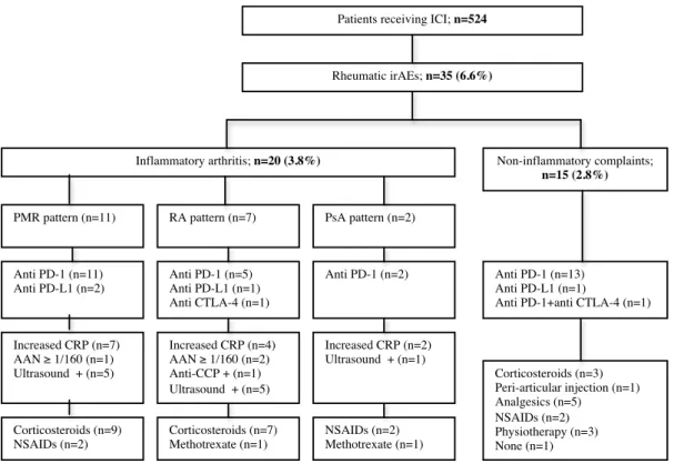
23 Reported musculoskeletal symptoms were varied, as detailed in Supplementary
Table 2. Both large and small joints in the upper and lower extremities were involved. The
most common symptom was arthralgia (n=35), with joint swelling being observed in only 8 patients and myalgia in 5 patients. Ten patients complained of rachialgia. Overall, two distinct clinical presentations were observed: 1) inflammatory arthritis (IA) mimicking either rheumatoid arthritis (RA) (n=7), polymyalgia rheumatica (PMR) (n=11) or psoriatic arthritis (n=2), and 2) non-inflammatory musculoskeletal conditions (n=15) (see Figure 1).
Figure 1 : Flow diagram for description of musculoskeletal irAEs
ICI: immune checkpoint inhibitors; irAEs: immune related adverse effects; PMR: polymyalgia rheumatica; RA: rheumatoid arthritis; PsA: psoriatic arthritis; PD-1: programmed cell death 1; PD-L1: programmed cell death 1 ligand; CTLA-4: cytotoxic T-lymphoc:te-associated protein 4; CRP: C-reactive protein; AAN: antinuclear autoantibodies; CCP: cyclic citrullinated peptide; NSAIDs: non steroidal anti-inflammatory drugs.
Patients receiving ICI; n=524
Rheumatic irAEs; n=35 (6.6%)
Inflammatory arthritis; n=20 (3.8%)
PMR pattern (n=11) RA pattern (n=7) PsA pattern (n=2)
Non-inflammatory complaints; n=15 (2.8%) Anti PD-1 (n=11) Anti PD-L1 (n=2) Anti PD-1 (n=5) Anti PD-L1 (n=1) Anti CTLA-4 (n=1) Anti PD-1 (n=2) Increased CRP (n=7) AAN ≥ 1/160 (n=1) Ultrasound + (n=5) Increased CRP (n=4) AAN ≥ 1/160 (n=2) Anti-CCP + (n=1) Ultrasound + (n=5) Increased CRP (n=2) Ultrasound + (n=1) Corticosteroids (n=9) NSAIDs (n=2) Corticosteroids (n=7) Methotrexate (n=1) NSAIDs (n=2) Methotrexate (n=1) Anti PD-1 (n=13) Anti PD-L1 (n=1)
Anti PD-1+anti CTLA-4 (n=1)
Corticosteroids (n=3) Peri-articular injection (n=1) Analgesics (n=5) NSAIDs (n=2) Physiotherapy (n=3) None (n=1)
24
Inflammatory arthritis
Inflammatory arthritis occurred in 20 patients (3.8%). PMR was diagnosed in 11 patients, with a clinical presentation of predominant bilateral shoulder pain and stiffness. One patient had a pre-existing stable rheumatic disease (axial spondyloarthritis) and developed a PMR-like condition twenty days after ICI initiation. Increased levels of C-reactive protein, defined as >5 mg/L, were found in 7 patients and ultrasound analysis detected subdeltoid bursitis in 5 patients. Nine patients were treated with oral corticosteroids (mean dosage 15 mg/day) and clinical improvement was achieved in all of them. For two patients, proximal pain and stiffness completely resolved with NSAIDs.
Bilateral and symmetric hand pain and stiffness were observed in 7 patients who developed rheumatic irAEs mimicking rheumatoid arthritis (RA), mainly within the first 3 months after ICI initiation. Two patients developed psoriatic arthritis, including one with pre-existing psoriasis who experienced severe arthritis associated with psoriasis exacerbation. Clinically significant joint swelling and/or ultrasound-confirmed synovitis were present in 5 patients. Elevated inflammatory markers were noted in 4 patients. Of note, one patient tested positive for anti-cyclic citrullinated peptide antibodies (anti-CCP) but negative for rheumatoid factor (RF). Mild biological immune dysregulation with anti-nuclear autoantibodies (ANA) (positive threshold 1/160) was observed in 2 patients and a shared HLA-DRB1 *01:01 epitope was present in two patients. X-ray evaluation did not detect any structural joint damage. All patients required prednisone or prednisolone at low-to-moderate doses, with a maximum of 30 mg/day, leading to clinical improvement or remission. Disease-modifying antirheumatic drugs (methotrexate) were required for one RA patient and for one psoriatic arthritis patient. When remission was achieved, a progressively tapered corticosteroid regimen was initiated, allowing complete steroid withdrawal in only 2 patients at 6 months of follow-up.
25 ICI therapy was pursued for all but one patient and was temporarily discontinued in accordance with the clinical trial protocol.
Non-inflammatory musculoskeletal conditions
15 patients presented with non-inflammatory rheumatic and musculoskeletal disorders (2.8%), characterized by pain worsened by physical activity and relieved with rest, and the absence of joint stiffness. All suffered from arthralgia of proximal or distal joints (mainly shoulders and hands), with associated axial symptoms in 5 patients. Only one patient presented with swelling of interphalangeal joints associated with hand, shoulder and cervical pain, associated with osteoarthritis based on the clinical examination, imaging and blood tests. Increased levels of C-reactive protein were observed in 4 patients but were probably related to their cancer, as they were present before the development of rheumatic symptoms. One patient benefited from a peri-articular steroid injection for left epicondylitis, five patients received analgesics, two patients were treated with NSAIDs, three patients recovered after physiotherapy, and one patient did not need any treatment. Importantly, no modification of immunotherapy was necessary. Finally, two patients received prednisone (15 and 30 mg/day) due to cancer progression with development of renal metastasis, and one was treated with steroids for concomitant interstitial lung disease, leading to complete resolution of rheumatic symptoms. Importantly, no modification of immunotherapy was necessary.
Other irAEs
Overall, non-musculoskeletal irAEs were observed in 137 patients (26.1%) according to medical records (Table 2) and were associated with rheumatic irAEs in 16 patients.
26 Reported irAEs were mainly cutaneous (rash, vitiligo, psoriasis, bullous pemphigoid), endocrine (thyroiditis, dysthyroidism) and digestive (colitis). Some cases of pneumonitis and hepatitis were also observed, as well as rarer irAEs such as pancreatitis, sarcoidosis, myasthenia, nephritis or uveitis.
Tumour response according to irAEs
Tumour response to ICI treatment was abstracted from medical records for all patients, with data available for 472 patients. Overall, there was a tumour response in 248 patients (52.5%), including complete response (n=31; 6.6%), partial response (n=84; 17.8%) or stable disease (n=133; 28.2%), while progressive disease was observed in 224 patients (47.5%). Rheumatic irAEs were observed in 12.1% of responders and in 2.2% of non-responders. For patients with rheumatic irAEs, the tumour response rate was higher compared to patients without rheumatic irAEs (85.7% versus 35.3%; OR=8.8 (95%CI: 3.2, 29.8), p<0.0001), as detailed in Table 2. The number of responders to ICI treatment was also higher in patients presenting with other irAEs compared to patients without irAEs (75.1% versus 35.3%; OR=5.4 (95%IC: 3.3, 9), p<0.0001). Subgroup analyses according to tumour type confirmed this association between rheumatic or other irAEs and tumour response across different cancers.
27
DISCUSSION
Arthralgia and myalgia were by far the most commonly reported rheumatic irAEs in clinical trials while inflammatory arthritis was less commonly described(10). Our prospective series estimates the prevalence of rheumatic manifestations in patients treated with ICI at 6.6%, and illustrates the wide range of clinical presentations, with some patients presenting with inflammatory arthritis mimicking PMR, RA or psoriatic arthritis (3.8%), while others developed non-inflammatory disorders (2.8%). Moreover, our data clearly emphasize the strong association of these manifestations with tumour response.
In accordance with the current literature describing rheumatic irAEs, PMR and RA were the two major clinical entities observed, with mainly seronegative RA, and only one patient being anti-CCP-positive in our series (14)(15)(16). However, the systematic referral of any musculoskeletal symptoms revealed new phenotypes of rheumatic irAEs. First, we observed some cases of PMR with a typical clinical presentation (acute onset, bilateral scapular and/or pelvic pain and stiffness) with no increase in inflammation markers or only mild elevation. Second, non-inflammatory disorders were also noted in several patients, sometimes occurring a few days after ICI initiation and therefore suspected to be attributable to the treatment. When using the Naranjo algorithm, the adverse drug reaction probability score was rated as possible or doubtful for such non-inflammatory manifestations due to improvement despite ICI continuation, with alternative causes being possible, and because of the lack of similar events reported in the literature (18). Future clinical reports and a better understanding of the pathogenesis of these disorders will help to answer this question (7).
The distinction between inflammatory versus non-inflammatory irAEs is worthwhile for treatment decisions, as patients might not require systemic corticosteroids. For non-inflammatory conditions, symptomatic treatment with analgesics, NSAIDs, local injection or
28 physiotherapy were often effective. For inflammatory irAEs, low-to-moderate doses of corticosteroids were generally sufficient to achieve improvement or remission, without ICI discontinuation. When needed, methotrexate was added, which was the case for two patients. In our experience, as in the series reported by Belkhir et al. (16) classical anti-inflammatory treatment was able to control rheumatic disorders without any modification of the ICI regimen, conversely to the experience of American colleagues, who reported a requirement for higher steroid dosage (often >40 mg/day), the use of TNF inhibitors for some patients, and usually ICI discontinuation (14)(15). Furthermore, the increasing number of cases reported in the literature illustrates these differences regarding rheumatic irAE management, highlighting the need for developing either national or international recommendations.
The higher prevalence of rheumatic irAEs with anti PD-1/anti PD-L1 exposure, either as a single agent or in combination with anti CTLA-4 (19)(20)(21), is a common observation from case reports and case series and was also recently reported from a single-centre pharmacosurveillance database (22). All patients referred to our department except one were receiving anti PD-1 or anti PD-L1 treatment. Even if ipilimumab is less commonly used currently, it is clear that anti PD-1 and anti CTLA-4 therapies have a distinct spectrum of irAEs (23)(24). Of note, rheumatic irAEs were rarely reported with CTLA-4 blockade when ipilimumab was the major ICI used in melanoma patients, raising the question of whether the PD-1 pathway is the one mainly involved in ICI-related rheumatic manifestations (25). Animal models support this hypothesis, with arthritis being observed in PD-1 knockout mice but not in CTLA-4 knockout mice (26)(27). Moreover, heterozygous CTLA-4 mutations in humans are associated with severe immune disorders such as thrombocytopenia, hypogammaglobinaemia, and lung, brain and gastrointestinal lymphocytic infiltrates but no clear rheumatic manifestations (28). Interestingly, some CTLA-4 and PD-1 polymorphisms are associated with RA (29)(30), and recently, a breakdown of PD-1/PD-L1 checkpoints has
29 been reported in giant-cell arteritis (31). Unifying research efforts in our classical inflammatory/auto-immune diseases and in the ones induced by ICIs would result in a better understanding of co-stimulation pathways and would benefit the management of both diseases.
This study is the first to demonstrate that patients experiencing rheumatic irAEs had a higher rate of response to ICI treatment compared to those without irAEs. Given the poor prognosis of advanced cancers, this clinical observation is of major interest and might be related, in part, to the fact that ICI treatment was continued in all but one patient. This strong association with tumour response was also observed with non-rheumatic irAEs. However, a longer follow-up is needed to assess whether the occurrence of irAEs affected patient survival. The correlation between irAE occurrence and patient outcomes has been actively investigated but not fully elucidated. Among two retrospective analyses, one reported a better overall survival for nivolumab-treated melanoma patients experiencing irAEs compared to those without irAEs, while the other study, which involved ipilimumab-treated patients, concluded that tumour response or survival were not affected by the occurrence of irAEs (32)(33). In fact, the majority of case reports associated irAEs with tumour response but this association has yet to be demonstrated in larger prospective studies.
CONCLUSION
Since ICIs are used with increasing frequency and represent a new standard of care in some advanced cancers, knowledge of irAEs and their management is of major interest. Rheumatic irAEs occurred in 6.6% of treated patients and were mainly associated with anti PD-1 or anti-PD-L1 therapies; they were also associated with better tumour response (85% of ICI responders). Importantly, these irAEs were easily manageable, either with a
low-to-
30 moderate dose of prednisone for inflammatory irAEs or various symptomatic therapies for non-inflammatory irAEs, and did not require ICI discontinuation. Collaboration between oncologists and rheumatologists is worthwhile to achieve a better understanding of these rheumatic irAEs and to define appropriate treatment algorithms.
REFERENCES
1. The Department of Health and Human Services. FDA approves new treatment for a type of late-stage skin cancer. FDA [online] http://www.fda.gov/NewsEvents/ Newsroom/PressAnnouncements/ucm1193237.htm (2011)
2. Chan MM, Kefford RF, Carlino M, et al. Arthritis and tenosynovitis associated with the anti-PD1 antibody pembrolizumab in metastatic melanoma. J Immunother. 2015 Jan;38(1):37-9
3. Patnaik A, Kang SP, Rasco D, et al. Phase I study of MK-3475 (anti-PD-1 monoclonal antibody) in patients with advanced solid tumors. Clin Cancer Res. 2015 Oct 1;21(19):4286-93
4. Topalian SL, Hodi FS, Brahmer JR, et al. Safety, activity, and immune correlates of anti-PD-1 antibody in cancer. N. Engl. J. Med. 2012 Jun 28;366(26):2443-54
5. Westin JR, Chu F, Zhang M, et al. Safety and activity of PD1 blockade by pidilizumab in combination with rituximab in patients with relapsed follicular lymphoma: a single group, open-label, phase 2 trial. Lancet
Oncol. 2014 Jan;15(1):69-77
6. Topalian SL, Drake CG, Pardoll DM. Immune checkpoint blockade: a common denominator approach to cancer therapy. Cancer Cell 2015;27:450–61
7. Van der Vlist M, Kuball J, Radstake TR, et al. Immune checkpoints and rheumatic diseases: what can cancer immunotherapy teach us?, Nat Rev Rheumatol. 2016 Oct;12(10):593-604
8. Chen DS, Irving BA, Hodi FS, et al. Molecular Pathways: Next-Generation Immunotherapy— Inhibiting Programmed Death-Ligand 1 and Programmed Death-1. Clin Cancer Res; 18(24) 2012 Dec 15;18(24):6580-7
9. Granier C, Karaki S, Roussel H, et al. [Cancer immunotherapy: Rational and recent breakthroughs]. Rev
Med Interne. 2016 Jun 28. pii: S0248-8663(16)30413-1.
10. Cappelli LC, Gutierrez AK, Bingham CO, et al. Rheumatic and musculoskeletal immune-related adverse events due to immune checkpoint inhibitors: A systematic review of the literature. Arthritis Care Res (Hoboken). 2016 Dec 20
11. Bilen MA, Subudhi SK, Gao J, et al. Acute rhabdomyolysis with severe polymyositis following ipilimumab-nivolumab treatment in a cancer patient with elevated anti-striated muscle antibody, J Immunother
Cancer. 2016 Jun 21;4:36
12. Salmon J-H, Lambrecht I, Brochot P, et al. A case of arthritis under pembrolizumab. Joint Bone Spine. 2017 Mar;84(2):243-244
13. Kimura T, Fukushima S, Miyashita A, et al. Myasthenic crisis and polymyositis induced by one dose of nivolumab, Cancer Sci. 2016 Jul;107(7):1055-8
14. Cappelli LC, Gutierrez AK, Baer AN, et al. Inflammatory arthritis and sicca syndrome induced by nivolumab and ipilimumab. Ann Rheum Dis. 2017 Jan;76(1):43-50
15. Calabrese C, Kirchner E, Kontzias K, et al. Rheumatic immune-related adverse events of checkpoint therapy for cancer: case series of a new nosological entity. RMD Open. 2017 Mar 20;3(1):e000412
31
after immune checkpoint inhibitor treatment. Ann Rheum Dis. 2017 Jun 9
17. Eisenhauer EA, Therasse P, Bogaerts J, et al. New response evaluation criteria in solid tumours: revised RECIST guideline (version 1.1). Eur J Cancer 2009;45:228–47
18. Naranjo CA, Busto U, Sellers EM, et al. A method for estimating the probability of adverse drug reactions. Clin Pharmacol Ther. 1981 Aug;30(2):239-45
19. Bonigen J, Raynaud-Donzel C, Hureaux J, et al. Anti-PD1-induced psoriasis. A study of 21 patients. J
Eur Acad Dermatol Venereol. 2016 Oct 14
20. Zimmer L, Goldinger SM, Hofmann L, et al. Neurological, respiratory, musculoskeletal, cardiac and ocular side-effects of anti-PD-1 therapy. Eur J Cancer. 2016 Jun;60:210-25
21. Garel B, Kramkimel N, Trouvin AP, et al. Pembrolizumab-induced polymyalgia rheumatica in two patients with metastatic melanoma. Joint Bone Spine. 2017 Mar;84(2):233-234
22. Le Burel S, Champiat S, Mateus C, et al. Prevalence of immune-related systemic adverse events in patients treated with anti-Programmed cell Death 1/anti-Programmed cell Death-Ligand 1 agents: A single-centre pharmacovigilance database analysis. Eur J Cancer. 2017 Jun 21;82:34-44
23. Spain L, Diem S, Larkin J. Management of toxicities of immune checkpoint inhibitors. Cancer Treat
Rev. 2016 Mar;44:51-60
24. Boutros C, Tarhini A, Routier E. Safety profiles of anti-CTLA-4 and anti-PD-1 antibodies alone and in combination. Nat Rev Clin Oncol. 2016 Aug;13(8):473-86
25. Bertrand A, Kostine M, Barnetche T, et al. Immune related adverse events associated with anti-CTLA-4 antibodies: systematic review and meta-analysis. BMC Med. 2015 Sep 4;13:211
26. Nishimura H, Nose M, Hiai H, et al. Development of lupus-like autoimmune diseases by disruption of the PD-1 gene encoding an ITIM motif-carrying immunoreceptor. Immunity. 1999 Aug;11(2):141-51
27. Tivol EA, Borriello F, Schweitzer AN, et al. Loss of CTLA-4 leads to massive lymphoproliferation and fatal multiorgan tissue destruction, revealing a critical negative regulatory role of CTLA-4. Immunity. 1995 Nov;3(5):541-7
28. Kuehn HS, Ouyang W, Lo B, et al. Immune dysregulation in human subjects with heterozygous germline mutations in CTLA4. Science. 2014 Sep 26;345(6204):1623-1627
29. Vaidya B, Pearce SH, Charlton S, et al. An association between the CTLA4 exon 1 polymorphism and early rheumatoid arthritis with autoimmune endocrinopathies. Rheumatology (Oxford). 2002 Feb;41(2):180-3 30. Lee YH, Bae SC, Kim JH, et al. Meta-analysis of genetic polymorphisms in programmed cell death 1. Associations with rheumatoid arthritis, ankylosing spondylitis, and type 1 diabetes susceptibility. Z Rheumatol. 2015 Apr;74(3):230-9
31. Zhang H, Watanabe R, Berry GJ, et al. Immunoinhibitory checkpoint deficiency in medium and large vessel vasculitis. Proc Natl Acad Sci U S A. 2017 Feb 7;114(6)
32. Freeman-Keller M, Kim Y, Cronin H, et al. Nivolumab in Resected and Unresectable Metastatic Melanoma: Characteristics of Immune-Related Adverse Events and Association with Outcomes. Clin Cancer
Res. 2016 Feb 15;22(4):886-94
33. Horvat TZ, Adel NG, Dang TO, et al. Immune-Related Adverse Events, Need for Systemic
Immunosuppression, and Effects on Survival and Time to Treatment Failure in Patients With Melanoma Treated With Ipilimumab at Memorial Sloan Kettering Cancer Center. J Clin Oncol. 2015 Oct 1;33(28):3193-8
32
III/ DISCUSSION
Ce travail a été réalisé en collaboration avec les services de dermatologie, de pneumologie, d’oncologie et d’hématologie du CHU de Bordeaux. Il a consisté à la mise en place d’un recrutement systématique, dans le service de rhumatologie, des patients développant des manifestations musculosquelettiques sous inhibiteurs de checkpoint immunitaire, qui se poursuit depuis la fin de notre étude. Cette collaboration entre la rhumatologie et les différents services pré cités a permis de gagner en expérience des deux côtés afin d’améliorer la prise en charge de nos patients communs. En ce sens, les choix thérapeutiques étaient discutés avec le médecin référent dans le but de soulager le patient tout en poursuivant l’immunothérapie.
Devant l’expansion considérable de l’utilisation des ICI (multiplication des molécules, des indications), il est primordial de former des groupes d’étude pour recenser les Immune-Related Adverse Effect (irAES), mieux les comprendre et les traiter, et décider d’arbres décisionnels et thérapeutiques pour que la prise en charge des irAEs interfère le moins possible sur l’efficacité et la poursuite du traitement anti tumoral.
Récemment, un groupe bordelais regroupant les différents spécialistes d’organes impliqués dans la gestion de ces immunothérapies et de leur toxicité s’est constitué. A l’image de nos collègues parisiens de Gustave Roussy qui ont crée un véritable centre expert autour de l’immunothérapie, l’organisation de ce groupe local a plusieurs objectifs :
- l’identification d’un réseau d'experts d'organes (local puis régional) pour la prise en charge des effets secondaires de ces ICI ;
- le développement d’algorithmes de prise en charge pour chaque toxicité, ou du moins les plus communes ;
33 - une réflexion autour de la mise en place de projets de recherche translationnelle autour des irAEs.
Concernant la prise en charge spécifique des irAEs rhumatologiques, un groupe de travail regroupant des médecins rhumatologues, internistes et oncologues, s’est également constitué sous l’égide du CRI (Club Rhumatismes et Inflammation), dans l’idée de développer des recommandations nationales basées sur une revue de la littérature existante et l’avis d’experts. Ces recommandations auraient un double intérêt : 1) servir de référence pour les rhumatologues qui seront de plus en plus confrontés à la prise en charge de ce type de manifestations induites par les ICI, et 2) de simplifier et accélérer la gestion des irAEs par les médecins prescripteurs d’immunothérapie.
Enfin, à l’image des registres mis en place avec succès par la communauté rhumatologique pour le suivi des patients sous biothérapie (ORA, REGATE, AIR-PR..), il serait intéressant de mettre en place de tels registres pour suivre les patients avec irAEs rhumatologiques. Ceux-ci permettraient, grâce à un suivi à long terme, d’apporter des réponses à des questions cliniques telles que l’évolution à long terme des irAEs, l’impact de la corticothérapie et d’autres traitements immunosuppresseurs sur la réponse tumorale, qui restent sans réponse actuellement. Ces registres permettraient aussi de suivre nos patients atteints de maladie auto immune pré existante et mis secondairement sous immunothérapie anti cancéreuse.
En conclusion, ces irAEs rhumatologiques constituent une nouvelle entité de notre spécialité, ouvrant la porte à de nombreux projets centrés autour de la multidisciplinarité.
34
IV/ SERMENT D’HIPPOCRATE
Au moment d’être admise à exercer la médecine, je promets et je jure d’être fidèle aux lois de l’honneur et de la probité.
Mon premier souci sera de rétablir, de préserver ou de promouvoir la santé dans tous ses éléments, physiques et mentaux, individuels et sociaux.
Je respecterai toutes les personnes, leur autonomie et leur volonté, sans aucune discrimination selon leur état ou leurs convictions. J’interviendrai pour les protéger si elles sont affaiblies, vulnérables ou menacées dans leur intégrité ou leur dignité. Même sous la contrainte, je ne ferai pas usage de mes connaissances contre les lois de l’humanité.
J’informerai les patients des décisions envisagées, de leurs raisons et de leurs conséquences. Je ne tromperai jamais leur confiance et n’exploiterai pas le pouvoir hérité des circonstances pour forcer les consciences.
Je donnerai mes soins à l’indigent et à quiconque me les demandera. Je ne me laisserai pas influencer par la soif du gain ou la recherche de la gloire.
Admise dans l’intimité des personnes, je tairai les secrets qui me seront confiés. Reçue à l’intérieur des maisons, je respecterai les secrets des foyers et ma conduite ne servira pas à corrompre les moeurs.
Je ferai tout pour soulager les souffrances. Je ne prolongerai pas abusivement les agonies. Je ne provoquerai jamais la mort délibérément.
Je préserverai l’indépendance nécessaire à l’accomplissement de ma mission. Je
n’entreprendrai rien qui dépasse mes compétences. Je les entretiendrai et les perfectionnerai pour assurer au mieux les services qui me seront demandés.
J’apporterai mon aide à mes confrères ainsi qu’à leurs familles dans l’adversité.
Que les hommes et mes confrères m’accordent leur estime si je suis fidèle à mes promesses ; que je sois déshonorée et méprisée si j’y manque.
35
Prévalence des manifestations rhumatologiques apparues sous immunothérapie anticancéreuse et corrélation à la réponse tumorale : une étude prospective monocentrique.
Objectif: Déterminer la prévalence et le type d’immune-related adverse events (irAEs) rhumatologiques apparus
sous inhibiteurs de checkpoint immunitaire (ICIs), ainsi que la corrélation avec la réponde tumorale.
Méthodes: Nous avons réalisé une étude monocentrique prospective observationnelle incluant tous les patients
atteints de cancer traités par ICIs. L’apparition des irAEs et la réponse tumorale étaient rapportées. Les patients développant des atteintes rhumatologiques étaient adressés au service de rhumatologie pour évaluation et prise en charge.
Résultats: De septembre 2015 à mai 2017, 524 patients ont reçu des ICIs et 35 patients ont été adressés en
rhumatologie (6.6%). Tous les irAEs rhumatologiques sont survenus au cours d’un traitement par anti PD-1/PD-L1, à l’exception d’un patient, avec un temps d’exposition moyen de 70 jours. Il existait deux présentations cliniques distinctes: 1) arthrite inflammatoire à type de polyarthrite rhumatoïde (PR)(n=7), de pseudo polyarthrite rhizomélique (n=11) ou de rhumatisme psoriasique (n=2), et 2) manifestations rhumatologiques non inflammatoires (n=15). Un seul patient avec PR avait des Ac anti CCP. Dix-neuf patients ont été traités par corticoïdes avec une dose moyenne de 17 mg/j et deux patients ont reçu du methotrexate. Les manifestations non inflammatoires ont été traitées avec des AINS, des antalgiques simples ou bien par kinésithérapie. L’immunothérapie a pu être poursuivie chez tous les patients sauf un. Les patients avec irAEs rhumatologiques avaient une meilleure réponse tumorale par rapport à ceux sans irAEs rhumatologiques (85.7% versus 35.3%; p<0.0001).
Conclusions: Devant l’augmentation croissante de l’utilisation des ICIs, la compréhension des irAEs et de leur
prise en charge est primordiale. Tous les patients ont répondu à des doses modérées de corticoïdes ou à des traitement symptomatiques, et n’ont pas nécessité d’arrêt de l’immunothérapie, d’autant plus que le taux de réponse tumorale était significativement plus important chez les patients développant des irAEs rhumatologiques.
Mots clés : inhibiteurs de checkpoint immunitaire, réponse tumorale, effets secondaires rhumatologiques, polyarthrite rhumatoïde, pseudo-polyarthrite rhizomélique, rhumatisme psoriasique
--- Rheumatic disorders associated with immune checkpoint inhibitors in cancer patients: clinical aspects and relationship with tumour response. A single-center prospective cohort study.
Objectives: To evaluate the prevalence and type of rheumatic immune-related adverse events (irAEs) in patients
receiving immune checkpoint inhibitors (ICIs), as well as the correlation with tumour response.
Methods: This was a single-centre prospective observational study including all cancer patients receiving ICIs.
The occurrence of irAEs and tumour response was assessed on a regular basis. Patients who experienced musculoskeletal symptoms were referred to the rheumatology department for clinical evaluation and management.
Results: From September 2015 to May 2017, 524 patients received ICIs and 35 were referred to the
rheumatology department (6.6%). All but one of the rheumatic irAEs occurred with anti PD-1/PL-L1 antibodies, with a median exposure time of 70 days. There were two distinct clinical presentations: 1) inflammatory arthritis (IA) mimicking either rheumatoid arthritis (n=7), polymyalgia rheumatica (n=11), or psoriatic arthritis (n=2), and 2) non-inflammatory musculoskeletal conditions (n=15). Of note, one RA patient was anti-CCP positive. Nineteen patients required corticosteroids with a median dose of 17 mg/day and methotrexate was started in two patients. Non-inflammatory disorders were managed with NSAIDs, analgesics and/or physiotherapy. ICI treatment was pursued in all but one patient. Patients with rheumatic irAEs had a higher response rate compared to patients without rheumatic irAEs (85.7% versus 35.3%; p<0.0001).
Conclusions: Since ICIs are used with increasing frequency, knowledge of rheumatic irAEs and their
management is of major interest. All patients were responsive either to low-to-moderate doses of prednisone or symptomatic therapies and did not require ICI discontinuation especially as tumour response was significantly higher in patients who experienced rheumatic irAEs.
Key words : immune checkpoint inhibitors, immune related adverse events, tumour response, rhumatoid arthritis, polymyalgia rheumatica, psoriastic arthritis
Thèse de doctorat en médecine : Spécialité : Rhumatologie