Université de Montréal
Caractérisation bioinformatique des nouvelles protéines
mitochondriales chez les moules d’eau douce (Bivalvia :
Unionoida).
par Alyssa Mitchell
Département de sciences biologiques Faculté des arts et des sciences
Mémoire présenté à la Faculté des arts et des sciences en vue de l’obtention du grade de M.Sc.
en sciences biologiques
Décembre, 2015
Résumé
Malgré que le contenu des génomes mitochondriaux animaux soit dit bien conservé, des nouveaux gènes mitochondriaux ont été identifiés chez plusieurs espèces, surtout des invertébrés. Par exemple, les bivalves exhibant la double transmission uniparentale de leurs génomes mitochondriaux possèdent des nouveaux gènes spécifiques au sexe (M-ORF dans l’ADN de type M, F-ORF dans l’ADN de type F) qui ont été caractérisés in silico chez trois espèces de l’ordre Mytiloida, une espèce de Veneroida et une espèce de Unionoida par une précédente étude. Même si les séquences varient beaucoup entre ces trois ordres, cette étude à montré que des hélices transmembranaires ainsi que des peptides signaux sont conservés pour toutes les séquences. L’étude a aussi montré que les nouveaux gènes pourraient avoir des rôles dans la signalisation cellulaire, le cycle cellulaire et la réponse immunitaire et qu’ils pourraient être le résultat de l’endogénisation de l’ADN viral. Le projet présenté ici a pour but de mieux caractériser ces nouveaux gènes et leur origine potentielle, en plus d’étudier le H-ORF particulier aux hermaphrodites, en ciblant les espèces des unionidés. Les résultats montrent que les hélices transmembranaires et peptides signaux sont conservés chez les unionidés, les protéines semblent être associées à la membrane et être capables de lier des acides nucléiques et protéines, et les fonctions potentielles sont conservées. Les M-ORFs semblent avoir un rôle dans le transport et des processus cellulaires tels que la signalisation, le cycle cellulaire et la division, et l’organisation du cytosquelette. Les F-ORFs semblent être impliqués dans le trafic et transport cellulaire et la réponse immunitaire. Finalement, les H-ORFs semblent être des glycoprotéines structurales avec des rôles dans la signalisation, le transport et la transcription. Les résultats de ce projet pourraient supporter une origine virale ou mitochondriale pour ces gènes.
Abstract
Although animal mitochondrial gene content is generally considered to be well-conserved, new genes have been identified in a variety of species, particularly invertebrates. For example, bivalves with doubly uniparental inheritance (DUI) of the mitochondrial genome have novel, sex-specific genes (M-ORF in M-type DNA, F-ORF in F-type DNA) which have been characterized in silico in three species of the order Mytiloida, one Veneroida and one Unionoida in a previous study. Although they are highly variable across these three orders, this study found conserved N-terminal signal peptides and transmembrane helices across all species. The study also showed that the new genes may and have roles in cell signaling, cell cycle, and immune response, and that they may be the result of endogenization of viral DNA. This project aimed to better characterize these novel genes and their potential origin as well as the H-ORF specific to hermaphrodites by focusing on the Unionoida. The pattern of conserved transmembrane helices and signal peptides is present across the species studied, all proteins seem to be membrane associated and able to bind nucleic acids and proteins, and potential functions are conserved as well. M-ORFs seem to have a role in transport and cellular processes such as signalling, cell cycle and division, and cytoskeleton organisation. F-ORFs are predicted to be involved in cellular traffic and transport and immune response. Finally,
H-ORFs appear to be structural glycoproteins which may be involved in signalling, transport and
transcription. The results of this project support either a viral or a mitochondrial origin for these genes.
Table des matières
Résumé ... 1
Abstract ... 2
Table des matières ... 3
Liste des tableaux ... 5
Liste des figures ... 8
Liste des abréviations et sigles ... 9
Remerciements ... 11
Chapitre 1 : Introduction générale ... 13
Section 1.1 : La mitochondrie et son génome ... 13
Section 1.2 : La double transmission uniparentale ... 14
Section 1.3 : Les moules d’eau douce (Unionoida) ... 16
Section 1.4 : Nouveautés et nouveaux gènes ... 18
Section 1.5 : Objectifs et hypothèses ... 22
Chapitre 2 : In silico analyses of mitochondrial ORFans in freshwater mussels (Bivalvia: Unionoida) provide framework for future studies of their origin and function. ... 23
Section 2.1: Introduction ... 23
Section 2.2: Materials and methods ... 26
Section 2.2.1: Sequences used in the analyses ... 26
Section 2.2.2: Analyses of ORFan sequences and protein secondary structures ... 30
Section 2.2.3: Functional analyses of ORFan proteins ... 30
Section 2.3: Results ... 31
Section 2.3.1: Rate of evolution of ORFan genes and proteins ... 31
Section 2.3.3: Motif and functional domain scans: frequently recurring HHpred hits and
potential ligand-binding sites ... 44
Section 2.3.4: Prediction of molecular function: hits to viral proteins ... 47
Section 2.3.5: Prediction of molecular function: hits to mitochondrial proteins ... 51
Section 2.3.6: Profile HMM – sequence comparisons for F-ORFs and M-ORFs ... 53
Section 2.3.7: Prediction of molecular function (all sequences, all programs except hmmsearch ) ... 54
Section 2.4: Discussion and conclusion ... 57
Section 2.4.1: Evolution of freshwater mussel ORFan sequences and protein structures ... 57
Section 2.4.2: Conserved motifs and domains: mitochondrial export of ORFan proteins ... 59
Section 2.4.3: Putative origin for freshwater mussel mitochondrial ORFans ... 61
Section 2.4.4: Predicted functions for freshwater mussel mitochondrial ORFans ... 64
Section 2.4.5: Conclusions and future directions ... 67
Chapitre 3 : Discussion, perspectives et conclusion ... 69
Section 3.1 : Conservation des séquences et structures des ORFans ... 69
Section 3.2 : Exportation des ORFans de la mitochondrie ... 70
Section 3.3 : Fonctions potentielles ... 72
Section 3.4 : DUI et conflit génomique ... 73
Section 3.5 : Unionoida ... 74
Bibliographie ... 76
Appendice 1 : L’approche bioinformatique ... 84
Appendice 2 : Figures supplémentaires ... 87
Liste des tableaux
Table I. Sequences analyzed in the present study ... 27
Table II. p-distances (p-D) and standard error (SE) values for mitochondrial M-orfs, F-orfs, and cox1 in freshwater mussel subfamilies ... 37
Table III. p-distances (p-D) and standard error (SE) values of mitochondrial H-orfs and cox1 in hermaphroditic freshwater mussels ... 38
Table IV. p-distances (p-D) and standard error (SE) values of mitochondrial F-orfs vs H-orfs and Fcox1 vs Hcox1 in comparisons between gonochoric vs. closely related hermaphroditic freshwater mussel species ... 39
Table V. Summary of hits to ligand-binding sites in M-ORFs, F-ORFs and H-ORFs ... 46
Table VI. Hits to viral proteins from structural prediction analyses ... 48
Table VII. List of BLAST hits for mitochondrial ORFans in freshwater mussels searched against NCBI NRDB mitochondrial proteins ... 52
Supplementary Table I. Predicted transmembrane (TM) helices in M-ORFs and F-ORFs. . 95
Supplementary Table II. Predicted signal peptides in M-ORFs and F-ORFs. ... 97
Supplementary Table III. Predicted transmembrane (TM) helices in H-ORFs. ... 98
Supplementary Table IV. Predicted signal peptides in H-ORFs. ... 99
Supplementary Table V. Frequently recurring HHpred hits in F-ORFs and M-ORFs ... 100
Supplementary Table VI. Frequently recurring HHpred hits in H-ORFs ... 103
Supplementary Table VII. Hits to other motifs and domains in M-ORFs and F-ORFs ... 105
Supplementary Table VIII. Hits to other motifs and domains in H-ORFs ... 107
Supplementary Table IX. Filtered hmmsearch output for the M-ORF and F-ORF HMM profiles built using default parameters with hmmbuild. ... 108
Supplementary Table X. Filtered hmmsearch output for the M-ORF and F-ORF HMM profiles built using custom parameters with hmmbuild. ... 123
Supplementary Table XI. Venustaconcha ellipsiformis M-ORF function predictions ... 132
Supplementary Table XII. Quadrula quadrula M-ORF function predictions ... 136
Supplementary Table XIII. Pyganodon grandis M-ORF function predictions ... 139
Supplementary Table XIV. Inversidens japanensis M-ORF function predictions ... 143
Supplementary Table XVI. Solenaia carinatus M-ORF function predictions ... 150
Supplementary Table XVII. Cumberlandia monodonta M-ORF function predictions ... 154
Supplementary Table XVIII. Hyridella menziesii M-ORF function predictions ... 157
Supplementary Table XIX. Anodonta anatina M-ORF function predictions ... 161
Supplementary Table XX. Venustaconcha ellipsiformis F-ORF function predictions ... 164
Supplementary Table XXI. Quadrula quadrula F-ORF function predictions ... 168
Supplementary Table XXII. Pyganodon grandis F-ORF function predictions ... 172
Supplementary Table XXIII. Inversidens japanensis F-ORF function predictions ... 175
Supplementary Table XXIV. Utterbackia peninsularis F-ORF function predictions ... 178
Supplementary Table XXV. Solenaia carinatus F-ORF function predictions ... 181
Supplementary Table XXVI. Cumberlandia monodonta F-ORF function predictions ... 185
Supplementary Table XXVII. Hyridella menziesii F-ORF function predictions ... 188
Supplementary Table XXVIII. Lasmigona complanata F-ORF function predictions ... 191
Supplementary Table XXIX. Toxolasma lividus F-ORF function predictions ... 196
Supplementary Table XXX. Margaritifera margaritifera F-ORF function predictions ... 199
Supplementary Table XXXI. Anodonta anatina F-ORF function predictions ... 203
Supplementary Table XXXII. Utterbackia imbecillis H-ORF sequence 1 function predictions ... 206
Supplementary Table XXXIII. Utterbackia imbecillis H-ORF sequence 2 function predictions ... 213
Supplementary Table XXXIV. Utterbackia imbecillis H-ORF sequence 3 function predictions ... 219
Supplementary Table XXXV. Utterbackia imbecillis H-ORF sequence 4 function predictions ... 225
Supplementary Table XXXVI. Utterbackia imbecillis H-ORF sequences 5 & 6 function predictions ... 231
Supplementary Table XXXVII. Utterbackia imbecillis H-ORF sequences 7 function predictions ... 236
Supplementary Table XXXVIII. Margaritifera margaritifera H-ORF sequence 1 function predictions ... 243
Supplementary Table XXXIX. Margaritifera margaritifera H-ORF sequences 2 & 4
function predictions ... 246
Supplementary Table XL. Margaritifera margaritifera H-ORF sequence 3 function
predictions ... 250
Supplementary Table XLI. Toxolasma lividus H-ORF function predictions ... 253 Supplementary Table XLII. Lasmigona compressa H-ORF sequence 1 function predictions
... 257
Supplementary Table XLIII. Lasmigona compressa H-ORF sequence 2 function predictions
... 260
Supplementary Table XLIV. Lasmigona subviridis H-ORF sequence 1 function predictions
... 264
Supplementary Table XLV. Lasmigona subviridis H-ORF sequence 2 function predictions
Liste des figures
Figure 1. La double transmission uniparentale. ... 16 Figure 2. Phylogénie simplifiée d’une collection d’espèces à sexes séparés et hermaphrodites (familles Unionoida et Margaritifera). ... 18 Figure 3. Cartes des génomes de types F, H et M. ... 20 Figure 4. Alignment of M-ORF and F-ORF protein sequences. ... 36 Figure 5. Hydrophobicity profiles of M-ORFs (a), F-ORFs (b) and H-ORFs vs. F-ORFs (c).. ... 43 Figure 6. Position of motifs frequently recurring in HHpred hits. ... 45 Figure 7. Most common categories of hits for (a) M-ORFs, (b) F-ORFs, and (c) H-ORFs..
... 56
Supplementary Figure 1. Alignments of F-ORFs and H-ORFs of closely related species.
... 88
Supplementary Figure 2. Alignments of complete mitochondrial genomes of freshwater mussels with DUI. ... 90 Supplementary Figure 3. Position of frequently recurring functions in HHpred and BLAST hits for (a) M-ORFs, (b) F-ORFs, and (c) and (d) H-ORFs. ... 94
Liste des abréviations et sigles
ABC : transporteur ABC / ATP-binding cassette transporter ADN : acide désoxyribonucléique / deoxyribonucleic acid ADNmt : ADN mitochondriale / mitochondrial DNA ARN : acide ribonucléique / ribonucleic acid
ATP : adénosine triphosphate / adenosine triphosphate
CMS : stérilité cytoplasmique mâle / cytoplasmic male sterility CTERM : C-terminale / C-terminal
DUI : double transmission uniparentale / doubly uniparental inheritance e.g. : exempli gratia
Et al. : et alii
HMM : modèle de Markov caché / Hidden Markov Model i.e. : id est
MY : million d’années / million years
NADH : nicotinamide adénine dinucléotide / nicotinamide adenine dinucleotide ORF : cadre de lecture ouvert / open reading frame
ORFan : cadre de lecture ouvert sans homologie à une protéine connue / open reading frame without homology to a known protein
PPR : protéines avec répétitions de type pentatricopeptide / pentatricopeptide repeat proteins SMI : transmission strictement maternelle / strict maternal inheritance
SP : peptide signal / signal peptide Spp. : espèces / species
TMH : hélice transmembranaire / transmembrane helix
UPRmt : réponse au stress lié à l’accumulation de protéines mal repliées dans les
For all those who part ways by quietly chanting the name of a professor who looks like Santa. Hockey hamster.
Remerciements
• Je commence par Sophie, bien sûr! Surtout pour la décision aventureuse de m’accepter comme première étudiante graduée dans ton labo pas-tout-à-fait-existant, mais aussi pour les cent mille lettres de référence, les soupers, les daiquiris sans alcool et les cent mille rires.
• Don, I think you’re the hardest to fit into a bullet point. You took me on as an honours student at the absolute last minute when no one knew much of anything about me (or even who I was). The four years you’ve known me have been a bit chaotic to say the least, but you’ve been patient and supportive through it all (I deleted “very” for you). Coincidentally, your name means “gift” in French. If I ever master my great-grandmother’s molasses cookies, you’re getting a great big batch!
• Of course I have to thank my family, aka the reason I exist. I think I’ve planned the perfect celebratory feast: cheeseball, lobster dip, almond roca, gingerbread men from Yarmouth, and braid. Nobody needs a main course. Make it a kitchen party, and get ready to atone for your sins at the lobster dip (forgiiiive me
Father, I know not what I do!).
• Next is my nerd crew (plus that one “normal” guy with the tin foil hat and hatchet). Every now and then I get a bit stunned that we’re all still in touch and see each other anywhere near as often as we do. I think that’s a pretty good reflection of how highly these friendships are prioritized, and it’s nice to know that we’re pretty ride or die (or maybe hike or die). I’m thrilled to have found my people so early in life, and when I’m home for Christmas I want to celebrate this like it’s The Night Pat Murphy Died.
• Je tiens aussi à remercier mon département, surtout ceux et celles que j’ai connu le plus. D’abord, ce « grand lab avec beaucoup d’étudiants » à Bernard Angers, qui m’ont adoptée ma première année. Trouvez-vous que mes présentations ont évolué au cours de ma maitrise? Ensuite, mon labo, qui a
commencé à se peupler tout tranquillement – c’est plus fun en groupe qu’à 1 ou 2. Il faut que je rémercie les étudiants au bac avec qui j’ai travaillé et parmi lesquels je compte plusieurs amis - vous avez mis beaucoup de joie dans les TPs et séances de tutorat! Finalement, les démos et tuteurs qui ont travaillé à côté de moi, et tout le gang qui se réunit au CI. C’est grâce à vous que j’ai eu l’expérience d’immersion totale que j’ai voulue, et c’est aussi grâce à vous que j’ai pu rire au moins une fois par jour, même quand ça n’allait pas bien.
• Gli italiani are next, aka the Passamonti lab. Grazie di your contributions to my work, the article that guided mine, your contributions to our lab (Davide and Stefano), and the entertaining and delicious visits from Dr. Over-the-hill. Molte
grazie!
• For beach days, Nordic spas, masala dosa, all of the most useful Hindi vocabulary (food), and sternly telling me that I need to meditate, शुिक्रया राजेश. • I’m going to close this section by thanking all of the artists who made the most
miserable tasks a bit more fun. For all the hours of spent sorting hits, scoring my beastly matrix, and moving pieces of figures one pixel at a time, thank you Prince Royce, Taylor Swift, Lea Michele, Tim Chaisson, and Great Big Sea. For the writing process, thank you Mozart, Beethoven, and Bach.
Chapitre 1 : Introduction générale
Section 1.1 : La mitochondrie et son génome
Les mitochondries sont des organelles à double-membrane retrouvées dans le cytoplasme chez les cellules eucaryotes et responsables de la production d’énergie. Bien que ça soit leur fonction principale, elles sont également impliquées dans d’autres processus cellulaires, tels que la signalisation cellulaire, la régulation métabolique, le contrôle du cycle cellulaire, le développement, la réponse antivirale et l’apoptose [1].
Antérieurement, on croyait que les génomes mitochondriaux (ADN mitochondrial ou ADNmt) chez les espèces animales étaient tous très similaires – une molécule circulaire d’environ 15 000 à 20 000 paires de bases encodant 2 ARN ribosomaux, 22 ARN de transfert, et 13 protéines impliquées dans la synthèse de l'ATP à l'intérieur des mitochondries [7 sous-unités du complexe de la NADH-ubiquinone oxydoréductase (gènes nad1-6, nad4L), une du complexe de l’ubiquinol-cytochrome c oxydoréductase (gène cytb), 3 du complexe de la cytochrome c oxydase (gènes cox1-3), et 2 du complexe de l’ATP synthase (gènes atp6 et
atp8)] [2, 3]. Aujourd’hui on connaît plusieurs cas de réarrangements structuraux du génome
mitochondrial ou encore la présence de gènes mitochondriaux supplémentaires (Voir [4] pour une revue). Par exemple, les Medusozoa ont un génome mitochondrial linéaire [5], et certains crustacés terrestres ont une portion circulaire et une portion linéaire [6]. Au niveau des gènes, on connait plusieurs cas de duplications de gènes existants ou encore de découverte de nouveaux gènes, surtout chez les invertébrés [4]. Chez les bivalves en particulier, deux exemples de duplication de gènes codant pour des protéines sont bien connus : la duplication de cox2 chez Musculista senhousia [7], et la duplication de nad2 chez le genre Crassostrea [8]. Le gène codant l’ARN ribosomal rrnS est aussi dupliqué chez Crassostrea gigas [3, 9]. Chez Aurelia aurita, et le genre Pocillopora (Cnidaria), un nouveau cadre de lecture (« open
reading frame » ou ORF) de fonction inconnue a été retrouvé dans le génome mitochondrial
[10–12] et deux ORFs de fonctions inconnues existent également chez Iphitheon panicea (Porifera) [13].
De plus, la transmission strictement maternelle (« Strict maternal inheritance » ou SMI), qui est la norme dans le règne animal, était considérée le seul système de transmission mitochondriale [14]. On connait maintenant une exception à cette règle aussi – la double transmission uniparentale, qui sera décrite dans la prochaine section.
Section 1.2 : La double transmission uniparentale
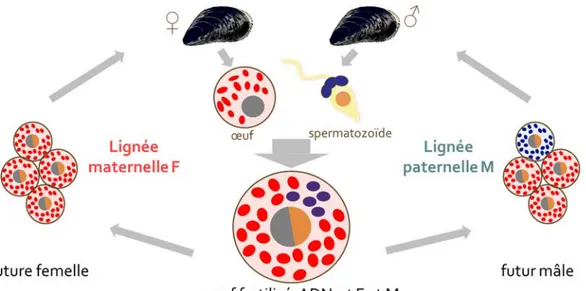
Il existe un cas exceptionnel à la « règle » de la transmission strictement maternelle de l’ADNmt chez les animaux. Plusieurs espèces de bivalves (Ordres Mytiloida, Nuculanoida, Unionoida et Veneroida) ont un mode de transmission fondamentalement différent connu sous le nom de « double transmission uniparentale » ou DUI (« Doubly uniparental inheritance ») [15–22]. Ces espèces sont caractérisées par la présence de deux ADNmt distincts : un génome M transmis par les mâles, et un génome F transmis par les femelles. Normalement, un œuf haploïde contient seulement des mitochondries de type F (voir [19, 20] pour des exceptions), et les spermatozoïdes contiennent seulement des mitochondries de type M, qui vont entrer dans l’œuf lors de la fécondation. Chez les embryons destinés à devenir femelles, les mitochondries paternelles sont dispersées dans toutes les cellules de l’embryon, et sont détruites pour permettre le développement d’une femelle homoplasmique (l’homoplasmie – où toutes les copies d’ADNmt sont identiques – est la norme sous SMI). Chez les embryons destinés à devenir mâles, par contre, les mitochondries paternelles sont regroupées dans les cellules destinées à devenir la gonade (Figure 1). Un mâle mature est hétéroplasmique, avec l’ADNmt de type F dans ses tissus somatiques, et l’ADNmt de type M dans sa gonade [21–23] (cet ADNmt M est principalement actif dans les spermatozoïdes [24, 25]).
Le taux de divergence entre les deux génomes au niveau des nucléotides peut varier d’environ 10% chez les moules marines à plus de 50% chez les moules d’eau douce, et une évolution plus rapide du génome mt de type M a été notée chez la plupart des espèces [21, 29-33]. Une explication possible pour cette différence est que le génome mt de type M serait un élément égoïste (ou « presque égoïse ») puisqu’il est fonctionnel seulement dans les cellules spermatogéniques [28]. D’autres études ont également proposé que les deux génomes évolueraient de façon neutre, mais que le type M accumulerait des mutations plus rapidement
dû à (i) sa population effective plus petite, (ii) son taux de réplication et donc de mutations élévé (on observe un total de sept divisions pendant la gamètogénèse chez le mâle versus 4 chez la femelle), ou (iii) aux dommages oxidatifs plus importants que les mitochondries subissent chez les spermatozoïdes [e.g. 21].
Deux hypothèses non-exclusives ont été proposées pour expliquer l’origine et le maintien du système atypiqueDUI chez les bivalves [29, 30] : (i) l’ADNmt mâle est impliqué dans des fonctions spécifiques et nécessaires aux spermatozoïdes et/ou (ii) ces deux génomes associés aux sexes sont impliqués dans la détermination du sexe. La détermination du sexe est méconnue chez les bivalves, mais il y a une particularité connue chez les Mytiloida et Veneroida – un effet maternel sur la sexe-ratio [36-38]. Chaque femelle produit des descendants majoritairement femelles, majoritairement mâles, ou environ 50% femelles et 50% mâles, peu importe avec quel mâle elle a été croisée. De plus, la majorité des filles présentent le même biais que la mère, mais des changements de biais qui suivent un rapport Mendelien ont également été observés. Il a été suggéré qu’un facteur nucléaire maternel clé ainsi que des facteurs secondaires nucléaires et/ou mitochondriaux serait impliqués dans le maintient de ces sexe-ratios biaisées [31].
Figure 1. La double transmission uniparentale. Mitochondries avec génome de type M en
bleu; mitochondries avec génome de type F en rouge.
Section 1.3 : Les moules d’eau douce (Unionoida)
Les moules d’eau douce (Unionoida) sont un groupe relativement ancien, avec une origine durant le Triassique il y a plus de 200 millions d’années [32, 33]. Les espèces sont généralement gonochoriques (à sexes séparés), avec un cycle de vie fondamentalement différent de celui des moules marines Mytiloida et palourdes marines Veneroida qui relâchent leurs gamètes dans la colonne d’eau, où a lieu la fécondation et le développement larvaire. Chez les Unionoida, seuls les mâles relachent leurs gamètes dans l’eau, les spermatozoïdes sont captés par les femelles et les premiers stades de développement ont lieu dans des compartiments spécialisés appelés marsupium dans les branchies des femelles [34]. Quand les embryons atteignent un certain stade de développement, la femelle les relâche, et ils doivent s’encyster sur les branchies d’un poisson hôte pour vivre une métamorphose parasitique. Une fois transformés en juvéniles, ils se détachent et tombent au fond du cours d’eau (pour plus de détails, voir [34])
Un petit nombre d’espèces hermaphrodites a été reporté chez les Unionoida (e.g., [32, 35, 36]). En Amérique du Nord, par exemple, seulement 7 espèces sont hermaphrodites sur plus de 300 espèces repertoriées [37]. De rares individus hermaphrodites sont également périodiquement trouvés chez les espèces à sexes séparés [37]. Plusieurs hypothèses ont été proposées pour expliquer l’émergence de l’hermaphrodisme chez les Unionoida, comme par exemple des facteurs environnementaux tels que la force du courant, la position d’un individu dans la population pour donner ou capter des spermatozoïdes et la densité de la population, e.g. [38]) mais cela demeure encore nébuleux [39]. Aussi, il n’est pas connu si des différences génétiques existent entre les hermaphrodites obligatoires et accidentels, cependant il y a des différences anatomiques bien documentées – tous les hermaphrodites ont un ovotestis (une gonade contenant à la fois des cellules spermatogéniques et des cellules ovogéniques), mais la distribution des cellules spermatogéniques et ovogéniques diffère entre les deux types d’hermaphrodites. Chez les hermaphrodites obligatoires on observe des acini discrèts qui produisent un type de gamète, mais chez les hermaphrodites accidentels on observe une distribution aléatoire de ces cellules [37]. Des analyses phylogénétiques démontrent que les espèces hermaphrodites en Amérique de Nord sont relativement jeunes [32, 37], et que l’hermaphrodisme est un caractère dérivé qui évolue à partir des femelles ([40, 41], voir Figure 2).
Figure 2. Phylogénie simplifiée d’une collection d’espèces à sexes séparés et hermaphrodites (familles Unionoida et Margaritifera). Les espèces marquées par une
étoile (*) sont des hermaphrodites qui ont perdu le génome mitochondrial de type M (donc possédant une transmission mitochondriale strictement maternelle). Figure tirée de Stewart et al. [41] avec permission.
Section 1.4 : Nouveautés et nouveaux gènes
Récemment, Breton et al. [40, 42] ont identifié des nouveaux gènes codant pour des protéines dans les génomes mitochondriaux des moules d’eau douce. Tel que mentionné précédemment, chez les unionidés, la grande majorité des espèces ont des sexes séparés mais il existe aussi des rares cas d’espèces hermaphrodites [32, 39]. Breton et al. [40] ont séquencé les génomes mitochondriaux des mâles et femelles pour plusieurs espèces gonochoriques, ainsi que le génome mitochondrial présent chez 5 espèces hermaphrodites proches parentes des espèces gonochoriques, mais qui ont toutes évolué de façon indépendante. Dans chacun des génomes, un quatorzième ORF a donc été découvert (F-ORF dans les génomes F, M-ORF dans les génomes M et H-ORF dans les génomes des espèces hermaphrodites) [40, 42]. La
technique Western Blot a été utilisée pour démontrer que les F-ORF et M-ORF sont exprimés, par contre, cela reste encore à être démontré pour le H-ORF. Les études de Breton et al. [40, 42] ont présenté six points importants :
i. tous les génomes F étudiés contiennent un F-ORF codant pour une protéine qui est conservée entre les espèces, mais qui ne présente aucune homologie évidente aux autres protéines connues (selon les résultats BLAST);
ii. tous les génomes M étudiés contiennent un M-ORF codant pour une protéine qui est conservée entre les espèces, mais qui n'est pas homologue au F-ORF, ni à aucune protéine connue;
iii. les hermaphrodites n’ont pas de génome M et leur ADNmt contient un F-ORF hautement modifié (H-ORF);
iv. les H-ORFs divergent des séquences F-ORF de leurs espèces proches parentes - leurs séquences sont plus longues (environ 80 acides aminés pour les F-ORFs et 150 acides aminés pour les H-ORFs) et elles possèdent plusieurs sous-unités répétitrices et plusieurs portions transmembranaires prédites versus une seule chez les F-ORFs et M-ORFs;
v. le gonochorisme est toujours accompagné de la DUI et la présence des F-ORFs et M-ORFs tandis que l’hermaphrodisme est accompagné de la SMI et la présence d’un F-ORF hautement modifié (H-ORF), ce qui mène à l’hypothèse que la DUI et les nouveaux gènes auraient un rôle dans le maintien des sexes séparés chez les unionidés;
vi. l’analyse immunohistochimique indique que la protéine encodée par le F-ORF chez l’espèce Venustaconcha ellipsiformis est non seulement présente dans la mitochondrie, mais transportée hors de l’organelle et retrouvée dans la membrane nucléaire et le nucléoplasme des œufs;
Cette dernière observation indique un rôle autre que la phosphorylation oxydative, et des études subséquentes sont venues appuyer l'hypothèse que ce produit de gène pourrait jouer un rôle dans la détermination du sexe [40, 42–45]. Présentement, il n’y a aucun cas connu dans le règne animal où les mitochondries sont directement impliquées dans la détermination
du sexe. Les fonctions des nouveaux gènes mitochondriaux découverts chez les bivalves avec la DUI demeurent pour le moment obscures (voir ci-dessous). Les modifications importantes observées dans le gène H-ORF chez les espèces hermaphrodites suggèrent une fonction différente pour ce gène ou encore une perte de fonction, mais cela reste à être étudié. Le lien entre la DUI et la détermination du sexe reste aussi à être élucidé, et la raison pour la déviation de la SMI chez les bivalves gonochoriques – et un retour vers la SMI chez les hermaphrodites – demeurent des questions ouvertes.
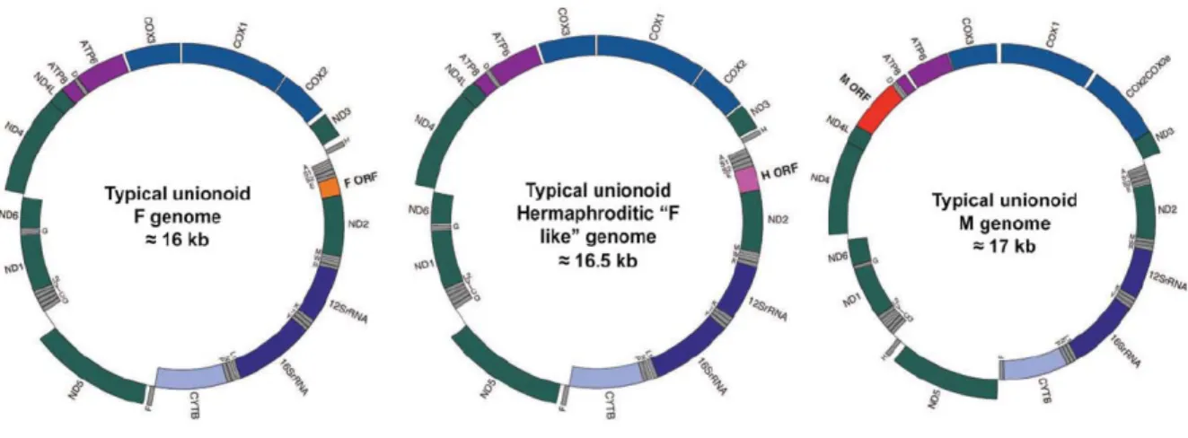
Figure 3. Cartes des génomes de types F, H et M. Identités des gènes : complexe I en vert;
complexe III en bleu pâle, complexe IV en bleu; complexe V en violet; ARN ribosomaux en bleu foncé. Les ARNs de transfert sont indiqués par leur lettre d’acide aminée. F-ORF, orange; H-ORF, rose; M-ORF, rouge. Les gènes à l’intérieure du cercle sont encodés sur le brin léger, ceux à l’extérieure du cercle sont encodés sur le brin lourd. Figure et légende tirées de Breton et al. 2011 [40] avec permission.
Milani et al. [44] ont publié les premières analyses in silico des structures et fonctions potentielles des F-ORFs et M-ORFs chez les bivalves avec la DUI (les moules marines
Musculista senhousia and Mytilus spp. (Mytiloida), la palourde marine Ruditapes philippinarum (Veneroida), et l’unionidé Venustaconcha ellipsiformis (Unionoida)). Leurs
des nucléotides et des acides aminés, et que ces gènes évoluent plus vite que tout autre gène mitochondrial chez les bivalves étudiés [40].
Les prédictions structurales et fonctionnelles indiquaient des similarités parmi toutes les espèces. D’abord, des hélices transmembranaires et peptides signaux sont conservés entre les espèces. Ensuite, les fonctions prédites pour les F-ORFs incluent la liaison avec les acides nucléiques, l’association aux membranes pour la signalisation ou l’adhésion cellulaire, ou un rôle dans la réponse immunitaire, tandis que les fonctions prédites pour les M-ORFs incluent l’association aux membranes, des interactions avec les acides nucléiques (surtout pour la signalisation cellulaire et la différentiation et développement), des interactions avec le cytosquelette, l’ubiquitination, l’apoptose et la réponse immunitaire [44].
En plus de prédire la structure et la fonction de ces protéines, cette étude a émis une hypothèse sur leur origine : l’endogénisation d’un ADN viral [44, 46]. Toutefois, en raison des taxons étudiés (5 mytilidés, un veneridé, et un unionidé) qui sont évolutivement très distants, et en raison des problèmes pour l’obtention de bons alignements des séquences, les auteurs ont également émis l'hypothèse que les ORFs chez les bivalves avec la DUI pourraient provenir d’événements d’endogénisation indépendants [17, 18]. Une augmentation du nombre d'espèces proches parentes et de séquences à l’étude pourrait aider à avoir une meilleure idée de l’origine de ces nouveaux gènes mitochondriaux. Par exemple, au moins quatre autres origines peuvent être proposées : (i) un gène homologue à un ancien gène bactérien, (ii) une duplication et néofonctionalization d’un gène mitochondrial, (iii) une origine à partir de séquences mitochondriales non-codantes, et (iv) un transfert du noyau vers la mitochondrie.
Les unionidés représentent un excellent modèle pour mieux comprendre l’origine et les fonctions de ces nouveaux gènes. Ils ont une position basale dans les Bivalvia, et les séquences complètes des M-ORF, F-ORF et H-ORF sont disponibles pour plusieurs espèces gonochoriques et des espèces hermaphrodites proche-parentes. La gamme de séquences disponibles représentent au moins 200 MY d’évolution pour ces gènes ([40]; Guerra et al., en prep), mais les séquences demeurent relativement similaires et donc plus facilement comparables.
Section 1.5 : Objectifs et hypothèses
L'objectif principal du projet est de caractériser l’évolution, la structure et la fonction des nouveaux gènes mitochondriaux F-ORF, H-ORF et M-ORF chez les moules d’eau douce, à partir de leur séquences nucléotidiques et/ou protéiques. Notre hypothèse générale est que ces gènes sont impliqués dans la détermination du sexe et donc subissent de fortes pressions sélectives. Plus précisément, on prévoit que (i) ces gènes évoluent rapidement et présentent plus de mutations non-synonymes que synonymes chez toutes les espèces (une signature répandue chez les gènes impliqués dans la détermination du sexe), (ii) malgré cette évolution rapide, les structures secondaires devraient être conservées pour chacun des nouveaux gènes : c’est-à-dire entre les espèces pour les F-ORFs et M-ORFs, et intra-espèce pour les H-ORFs, et (iii) la fonction prédite pour chacun des nouveaux gènes devrait être la même pour toutes les espèces pour un même genre (ex. les séquences F-ORF donnant des résultats similaires chez toutes les espèces).
Chapitre 2 : In silico analyses of mitochondrial ORFans in
freshwater mussels (Bivalvia: Unionoida) provide
framework for future studies of their origin and function.
Article in preparation for BMC Genomics.
Alyssa Mitchella, Davide Guerraa, Donald Stewartb, Sophie Breton a
aDepartment of Biological Sciences, Université de Montréal, Montréal, QC H3C 3J7 Canada bDepartment of Biology, Acadia University, Wolfville, NS B4P 2R6 Canada
Section 2.1: Introduction
Metazoan mitochondrial genomes (mtDNAs) are typically small, circular genomes without introns that encode 2 ribosomal RNAs, 22 transfer RNAs, and 13 proteins involved in ATP production [2, 3]. Strict maternal inheritance (SMI) of mtDNA is predominant among animals with limited or no paternal contribution [14]. There are, however, many exceptions to these characteristics. For example, linearized mitochondrial genomes have been reported in the Medusozoa [5] and some terrestrial isopod crustaceans [6]. Differences in gene content have also been found among metazoan mtDNAs, particularly in invertebrates (see [4] for a review). For example, duplications of typical protein-coding genes have been reported in several mollusc species, including cephalopods, aplacophorans, and bivalves; additional ‘atypical’ protein-coding genes with non-OXPHOS functions have been reported in cnidarians, sponges, and placozoans (e.g., atp9, dnaB, tatC); and mitochondrial ORFans, i.e., ‘atypical’ genes with unknown function, have been identified in cnidarians and in bivalves with doubly uniparental inheritance of mtDNA (DUI), which is the only known exception to SMI in the animal kingdom [4].
DUI has been reported in marine and freshwater bivalves (Orders Mytiloida, Nuculanoida, Unionoida, and Veneroida) ([26]; [47]; [27]; [21]). Species with DUI possess mitochondrial genomes that are transmitted in a sex-specific manner (known as female F-type
and male M-type mtDNAs, respectively). Haploid eggs typically contain mitochondria with only F-type mtDNA (but see [19] and [20]), while sperm mitochondria, which enter the egg when fertilization occurs, only contain the M-type. If the embryo develops as a female, sperm mitochondria are dispersed and/or destroyed, leading to homoplasmic females (similar to what happens under SMI). If the embryo develops as a male, sperm mitochondria remain grouped together, and are eventually sequestered in the germ line, which becomes homoplasmic for the M mtDNA [24, 25]. Males are therefore heteroplasmic individuals, with mitochondria inherited from their mother containing the F-type mtDNA throughout their soma, and mitochondria inherited from their father containing the M-type mtDNA in germ line cells (in males M mtDNA can be found in variable proportions also in somatic tissues [9]). DNA divergence between conspecific M- vs. F-type mitochondrial genomes over 50% has been found in Unionoida ([21]).
With their unique DUI system, bivalves not only challenge our traditional view of the SMI of mtDNA, their mitochondrial genomes also contain additional, sex-specific protein-coding genes, i.e., the mitochondrial ORFans - F-orfs and M-orfs in the F- and M-type mtDNAs, respectively - whose products are exported from the organelle and may be involved in functions other than energy production [40, 42–46]. In freshwater mussels, for example, species typically have separate sexes (gonochorism or dioecy), but hermaphroditism also occurs rarely [32, 39]. An absolute correlation has been observed between gonochorism and the presence of DUI and novel sex-specific proteins encoded by the F- and M-type mtDNAs (F-ORF and M-ORF), whereas hermaphroditic species lack the M-type altogether [43]. Hermaphroditic species appear to follow the SMI rule of mitochondrial transmission and individual mussels have only one type of mtDNA, called H-type [16]. The H-type is remarkably similar to the F-type mtDNA of closely-related gonochoric species except for the novel ORFan gene (named H-orf in these species), which is a highly mutated version of the
F-orf in their sister taxa [43]. For these reasons, Breton et al. [43] proposed a connection
between DUI and the maintenance of separate sexes in freshwater mussels. However, the link between DUI and sex determination, and the cause of deviation from the "SMI rule" in bivalves are still open questions.
The first in-depth bioinformatic analysis of the structures and potential functions of
F-ORF and M-F-ORF proteins was performed by Milani et al. [44] on the following DUI bivalve
species: the marine mussels Musculista senhousia, Mytilus edulis, Mytilus galloprovincialis,
Mytilus trossulus and Mytilus californianus (Mytiloida), the marine clam Ruditapes philippinarum (Veneroida), and the freshwater mussel Venustaconcha ellipsiformis
(Unionoida). M-orf and F-orf nucleotide sequences were found to be highly variable, with mostly non-synonymous mutations, indicating rapid evolution and supporting previous claims that these protein-coding genes are the fastest-evolving mitochondrial genes in bivalves with DUI [40, 44]. Despite this fast rate of evolution, structural similarities in their translated amino acid sequences were observed among species, and ORFan proteins were predicted to share similar functions. For example, F-ORFs were largely predicted to bind and interact with nucleic acids, associate with membranes for cell adhesion and/or signalling, or play a role in immune response. M-ORFs were also predicted to be membrane-associated and interact with nucleic acids, primarily for signalling, cell differentiation and development, and also for cytoskeleton formation and dynamics, ubiquitination, apoptosis, and immune response [44]. Even if hit probabilities were sometimes low and the regions of similarity were of short lengths, several clues suggested that these novel ORFans originated from endogenization of viral DNA [44, 46]. However, the impossibility of obtaining good alignments including
F-ORFs and M-F-ORFs from all species, due to the highly divergent nature of the ORFans,
indicated that either their fast evolution wiped out sequence similarities among species or that they originated from independent virus endogenization events [44]. It is also conceivable that the ORFans originated from different sources/processes but evolved similar function(s) in these distantly related DUI species, particularly if DUI evolved independently more than once [44]. Other than a viral origin, there are at least four other possibilities for the source of these mitochondrial ORFans; they may have originated from (i) a gene homologous to ancestral bacterial protein-coding genes, (ii) a duplicated and diverged mitochondrial gene, (iii) a gene composed from previously non-coding mitochondrial sequences, or (iv) a gene transferred from the nucleus to the mitochondrion (e.g., [40]).
The reality is that it is unfortunately not currently possible to confirm that mitochondrial ORFans in these distantly related DUI species are homologous because of their
high divergence and incomplete knowledge regarding their phylogenetic distribution. One option to better understand the origin(s) and function(s) of these ORFans is to compare more closely related sequences at a lower taxonomic level. Freshwater mussels (Unionoida) offer an excellent opportunity for this for at least two reasons: (1) the basal nature of the Unionoida within the Bivalvia, according to mtDNA-based phylogenies, suggests that their ORFans have a very ancient origin and that DUI in this group might be one of the first examples of this phenomenon in bivalves [33], and (2) complete F and M genomes or F-orf, M-orf and H-orf sequences are available for several gonochoric species and five independently evolved hermaphroditic species (e.g., [33, 43, 48]). All of them belong to the family Unionidae, but recently we have sequenced the F and M mtDNAs from Cumberlandia monodonta (Margaritiferidae) and Hyridella menziesii (Hyriidae) (Guerra et al., in prep), and these genomes possess an F-orf and an M-orf, suggesting that these unique genes have been present and functioning continuously for >200 million years in this group ([43]; Guerra et al., in prep).
The present study thus aims to predict the origin, structure, and function of the F-ORF and M-ORF protein sequences in Unionoida, and analyze the H-ORFs for the first time. Our results confirm that they are the fastest evolving genes in unionoid mitochondrial genomes, that they share structural and functional similarities, and that they may have a viral or a mitochondrial origin, bringing back on the table the evolutionary scenario of multiple origins of DUI, with the possibility of DUI systems with elements of different sources/origins and different mechanisms of action in the distantly-related DUI taxa [44, 46].
Section 2.2: Materials and methods
Section 2.2.1: Sequences used in the analyses
ORFan and cox1 nucleotide sequences of unionoid bivalve species were either obtained from the National Center for Biotechnology Information (NCBI) or from newly sequenced mitochondrial genomes (i.e., H. menziesii and C. monodonta; Guerra et al., in prep). Newly sequenced genomes were sequenced at the sequencing platform of McGill University [Montreal, Canada] using the genome sequencer FLX sequencing service, and all others were obtained by Sanger sequencing [40, 42]. All species and GenBank entries used in
this study are listed in Table I (Note: M-orf sequences for Lasmigona complanata,
Margaritifera margaritifera and Toxolasma lividus have not been obtained). The sequences
were translated with ORF Finder (http://www.ncbi.nlm.nih.gov/projects/gorf/; [49]) using the invertebrate mitochondrial genetic code, and analyzed at the nucleotide and/or amino acid level (see below). Because M-ORF and F-ORF protein sequences vary little within a species, only one sequence was used for each gonochoric species. H-ORF sequences are highly variable within species [42], and so multiple sequences were analyzed per species to provide a more complete picture of intraspecific H-ORF evolution and potential functionality.
Table I. Sequences analyzed in the present study
Species mtDNA type Accession number ORF name
Subfamiliy Ambleminae Quadrula quadrula M M F F FJ809751.1 FJ809751.1 FJ809750.1 FJ809750.1 Qqu-Morf Qqu-Mcox1 Qqu-Forf Qqu-Fcox1
Toxolasma lividus F HM849457.1 Tli-Forf
Toxolasma parvum H To come Tpa-Horf
Venustaconcha ellipsiformis M M F F FJ809752.1 FJ809752.1 FJ809753.1 FJ809753.1 Vel-Morf Vel-Mcox1 Vel-Forf Vel-Fcox1 Subfamiliy Anodontinae Anodonta anatina M F KF030962.1 KF030964.1 Aan-Morf Aan-Forf
Subfamiliy Gonideinae Inversidens japanensis M M F F AB055624.1 AB055624.1 AB055625.1 AB055625.1 Ija-Morf Ija-Mcox1 Ija-Forf Ija-Fcox1 Solenaia carinatus M M F F KC848655.1 KC848655.1 KC848654.1 KC848654.1 Sca-Morf Sca-Mcox1 Sca-Forf Sca-Fcox1 Subfamiliy Hyriidae Hyridella menziesii M M F F
Guerra et al. in prep To come To come AY785394.1 Hme-Morf Hme-Mcox1 Hme-Forf Hme-Fcox1 Subfamiliy Margaritiferinae Cumberlandia monodonta M M F F
Guerra et al. in prep To come HM849375.1 KF647374.1 Cmo-Morf Cmo-Mcox1 Cmo-Forf Cmo-Fcox1 Margaritifera falcata H H H H H H HM849545.1 HM856634.1 HM849547.1 HM849548.1 HM856634.1 NC_015476.1 Mfa-Horf (1-4) Mfa-Hcox1(1-2) Margaritifera margaritifera F F HM849399.1 HM849095.1 Mma-Forf Mma-Fcox1
Subfamiliy Unioninae
Lasmigona complanata F HM849393.1 Lco-Forf
Lasmigona compressa H H H H HM849534.1 HM849535.1 HM856638.1 NC_015481.1 Lco-Horf (1-2) Lco-Hcox1(1-2) Lasmigona subviridis H H HM849542.1 HM849543.1 Lsu-Horf (1-2) Pyganodon grandis M M F F FJ809755.1 FJ809755.1 FJ809754.1 FJ809754.1 Pgr-Morf Pgr-Mcox1 Pgr-Forf Pgr-Fcox1 Utterbackia imbecillis H H H H H H H HM849591.1 HM849595.1 HM849594.1 HM849601.1 HM849606.1 HM849597.1 HM849584.1 Uim-Horf (1-7) H H NC_015479 HM856637.1 Uim-Hcox1(1-2) Utterbackia peninsularis M M F F HM856635.1 HM856635.1 HM856636.1 HM856636.1 Upe-Morf Upe-Mcox1 Upe-Forf Upe-Fcox1
NOTE – M = M mtDNA in a DUI gonochoric breeding system, F = F mtDNA in a DUI gonochoric breeding system, H = H mtDNA in a non-DUI hermaphroditic breeding system.
Section 2.2.2: Analyses of ORFan sequences and protein secondary
structures
Alignments of ORFan and COX1 sequences were performed with M-COFFEE (DNA) and PSI-COFFEE (proteins) (http://tcoffee.crg.cat/apps/tcoffee/index.html; [50]). Nucleotide and amino acid p-distances, as well as a codon-based test of positive selection using the Nei-Gojobori method [51] were calculated using MEGA6 [52] with variance estimated using 500 bootstrap repetitions. The program VISTA [53] was also used to display the level of sequence conservation between M vs. M, F vs. F, and F vs. H complete mitochondrial genomes. M- and F-type mtDNAs were not compared due to their high divergence and previous characterization [40]. Hydropathy profiles of each amino acid sequence were calculated with the ProtScale tool at ExPASy (http://ca.expasy.org/tools/; [54]) using the method of Kyte and Doolittle [55]. Putative transmembrane (TM) helices were identified using a variety of protein signature recognition methods implemented by the following programs: Phobius
(http://phobius.sbc.su.se/; [56]), InterProScan (TMHMM)
(http://www.ebi.ac.uk/Tools/pfa/iprscan5/; [57]), TMPred (http://www.ch.embnet.org/software/TMPRED_form.html; [58]), TOPCONS
(http://topcons.cbr.su.se/; [59]), and Predict Protein (http://www.predictprotein.org; [60]).
Section 2.2.3: Functional analyses of ORFan proteins
Signal peptides (SPs) were sought using Phobius, InterProScan, PrediSi (http://www.predisi.de/; [61]), and SignalP (http://www.cbs.dtu.dk/services/SignalP/; [62]). Motif Scan (http://myhits.isb-sib.ch/cgi-bin/motif_scan; [63]) and HHpred (http://toolkit.tuebingen.mpg.de/hhpred; [64]) were used to search for motifs and functional domains. TPRpred (http://toolkit.tuebingen.mpg.de/tprpred; [65]) was used to search for potential tetratricopeptide repeat (TPR) or pentatricopeptide repeat (PPR) motifs. The following procedure was used to predict the function of ORFan proteins: (1) we performed BLASTp, tBLASTx, and PSI-BLAST searches against NCBI’s entire non-redundant protein
database (NRDB) and against mitochondrial proteins only (last accessed July, 2015) with default parameters (http://blast.ncbi.nlm.nih.gov/; [66]); (2) we used hmmbuild (v3.1b2; downloaded from http://hmmer.janelia.org) [67] to generate two HMM profiles from both the F-ORF and M-ORF protein alignments (H-ORFs were not considered given their scattered phylogenetic distribution and independent evolutionary histories) using default and custom parameters (four profiles in total), and performed profile HMM – sequence comparisons against UniProtKB, Swissprot, PDB, QfO, and Pfamseq databases using HMMER hmmsearch (http://www.ebi.ac.uk/Tools/hmmer/; [67]) with default parameters (E-value cutoff = 0.001); (3) for profile HMM – profile HMM comparisons, we used HHpred, which compares HMM profiles with databases of HMMs representing proteins with known structure (e.g. PDB, SCOP) or annotated protein families (e.g. PFAM, SMART, CDD, COGs, KOGs); and (4) the following programs were also used to predict the function of ORFan proteins: @tome2 (http://atome.cbs.cnrs.fr/; [68]), I-TASSER (http://zhanglab.ccmb.med.umich.edu/I-TASSER/; [69]) and PredictProtein, which also returned the predicted subcellular localization, binding sites, and ligands (with no accompanying measure of significance). For BLASTp and PredictProtein all matches with E-values <1.0 were kept, while for position-specific iterative or PSI-BLAST all matches with E-values <0.01 were kept as recommended by the program (except for PSI-BLAST analyses against NCBI mitochondrial genes only, where E-values <1.0 were kept, see below). For I-TASSER, top templates with Z scores over 1.0 and structural analogs with TM scores over 0.5 were recorded. All @tome2 results were kept. Motif Scan results not marked as “questionable” or “weak” were kept. Hits described as “uncharacterized,” “putative,” “unknown,” or “predicted” were not kept. Hits from all programs were scored in 122 categories covering putative ligands, subcellular localization, and protein functions, and analyzed.
Section 2.3: Results
Section 2.3.1: Rate of evolution of ORFan genes and proteins
The amino acid sequences of ORFans were generally not well conserved among unionoid species. As seen in Figure 3, a good comprehensive alignment including all M-ORF sequences was not possible due to their high divergence, however, sequences from the same
subfamily produced good alignments: P. grandis and U. peninsularis (Unionidae: Unioninae);
I. japanensis and S. carinatus (Unionidae: Gonideinae); Q. quadrula and V. ellipsiformis
(Unionidae: Ambleminae) (Figure 3b-d). A common feature of M-ORFs is that they are all lysine-rich proteins with poly-K strings found in many of them, a characteristic that is apparently absent in F-ORF and H-ORF amino acid sequences. Similar to M-ORF sequences, F-ORF sequences from the same subfamily or family, produced better alignments than for all species: L. complanata, P. grandis and U. peninsularis (Unionidae: Unioninae); I. japanensis and S. carinatus (Unionidae: Gonideinae); Q. quadrula, T. lividus and V. ellipsiformis (Unionidae: Ambleminae); C. monodonta and M. margaritifera, (Margaritiferidae: Margaritiferinae) (Figures 3e-i). Finally, because the H-ORFs were likely formed by 5 independent evolutionary events [42], these gene sequences are only conserved within a species; interspecific alignment is therefore not possible for hermaphrodites, and alignments between hermaphrodite H-ORFs and closely-related gonochoric species F-ORFs were mainly of low quality (Supplementary Figure 2).
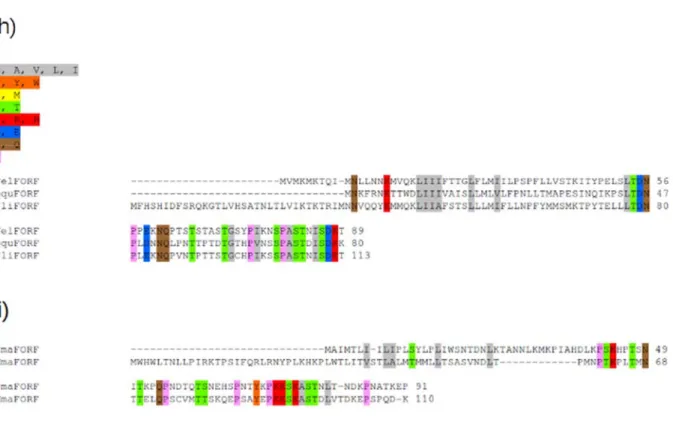
Figure 4. Alignment of M-ORF and F-ORF protein sequences. Global alignments and
alignments for each subfamily are shown. a) All M-ORF sequences, b) M-ORFs from the subfamily Unioninae, c) M-ORFs from the subfamily Gonideinae, d) M-ORFs from the subfamily Ambleminae, e) all F-ORF sequences, f) F-ORF sequences from the subfamily Unioninae, g) F-ORF sequences from the subfamily Gonideinae, h) F-ORF sequences from the subfamily Ambleminae i) F-ORF sequences from the subfamily Margaritiferidae. Colour coding is applied to amino acid groups conserved in ≥70% of sequences. Grey, aliphatic amino acids; orange, aromatic amino acids; yellow, sulfur amino acids; green, amino acids bearing a hydroxyl group; red, basic amino acids; blue, acidic amino acids; brown, amino acids with an amide group; pink, cyclic amino acids. VelMORF, V. ellipsiformis M-ORF; QquMORF, Q. quadrula ORF; PgrMORF, P. grandis ORF; IjaMORF, I. japanensis
M-ORF; UpeMORF, U. peninsularis M-M-ORF; ScaMORF, S. carinatus M-M-ORF; CmoMORF, C. monodonta M-ORF; HmeMORF, H. menziesii M-ORF; AanMORF, A. anatina M-ORF;
VelFORF, V. ellipsiformis ORF; QquFORF, Q. quadrula ORF; PgrFORF, P. grandis
F-ORF; IjaFORF, I. japanensis F-F-ORF; UpeFORF, U. peninsularis F-F-ORF; ScaFORF, S. carinatus F-ORF; CmoFORF, C. monodonta F-ORF; HmeFORF, H. menziesii F-ORF;
MmaFORF, M. margaritifera F-ORF; LcoFORF, L. complanata F-ORF; TliFORF, T. lividus F-ORF AanFORF, A. anatina F-ORF.
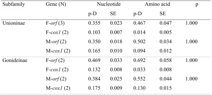
The p-distances for nucleotide and amino acid ORFan sequences as well as the outcome of the test of positive selection are reported in Table II (M-ORFs and F-ORFs) and Table III (H-ORFs), along with the values for cox1 sequences taken from the same sex-specific mtDNAs. Table IV shows the p-distances for within-genus comparisons of F-ORFs versus H-ORFs. In all cases, the novel ORFs have interspecific p-distances several times higher than cox1, which is typically the slowest-evolving protein-coding gene in animal mtDNAs [70]. All groups of sequences also returned a 100% chance of rejecting the null hypothesis of neutral selection in favor of the alternative hypothesis of positive selection. The level of sequence conservation between M vs. M, F vs. F, and F vs. H complete mitochondrial genomes also confirmed that mitochondrial ORFans are the fastest evolving genes in the mtDNA of freshwater mussels with DUI (Supplementary Figure 2).
Table II. p-distances (p-D) and standard error (SE) values for mitochondrial M-orfs, F-orfs,
and cox1 in freshwater mussel subfamilies
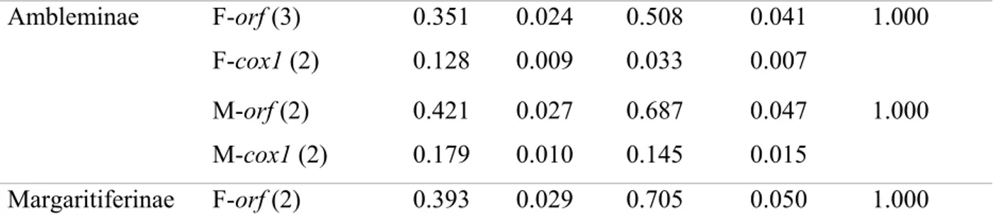
Subfamily Gene (N) Nucleotide Amino acid p
p-D SE p-D SE Unioninae F-orf (3) 0.355 0.023 0.467 0.047 1.000 F-cox1 (2) 0.103 0.007 0.014 0.005 M-orf (2) 0.350 0.018 0.502 0.034 1.000 M-cox1 (2) 0.165 0.010 0.094 0.012 Gonideinae F-orf (2) 0.469 0.033 0.692 0.058 1.000 F-cox1 (2) 0.132 0.008 0.033 0.008 M-orf (2) 0.384 0.025 0.552 0.044 1.000 M-cox1 (2) 0.175 0.009 0.130 0.015
Ambleminae F-orf (3) 0.351 0.024 0.508 0.041 1.000 F-cox1 (2) 0.128 0.009 0.033 0.007
M-orf (2) 0.421 0.027 0.687 0.047 1.000 M-cox1 (2) 0.179 0.010 0.145 0.015
Margaritiferinae F-orf (2) 0.393 0.029 0.705 0.050 1.000 NOTE – N = number of sequences used. The probability of rejecting the null hypothesis of strict-neutrality (dN = dS) in favor of the alternative hypothesis (dN > dS) (in the p column) is
shown. dS and dN are the numbers of synonymous and nonsynonymous substitutions per site,
respectively.
Table III. p-distances (p-D) and standard error (SE) values of mitochondrial H-orfs and cox1
in hermaphroditic freshwater mussels
Species Gene (N) Nucleotide Amino acid p
p-D SE p-D SE
Utterbackia imbecillis H-orf (7) 0.070 0.008 0.181 0.022 1.000
cox1 (2) 0.000 0.000 0.000 0.000
Margaritifera falcata H-orf (4) 0.003 0.002 0.004 0.004 1.000
cox1 (2) 0.000 0.000 0.000 0.000
Lasmigona compressa
H-orf (2) 0.029 0.007 0.065 0.017 1.000
cox1 (2) 0.000 0.000 0.000 0.000
Lasmigona subviridis H-orf (2) 0.016 0.005 0.021 0.010 1.000
NOTE – N = number of sequences used. The probability of rejecting the null hypothesis of strict-neutrality (dN = dS) in favor of the alternative hypothesis (dN > dS) (in the p column) is
shown. dS and dN are the numbers of synonymous and nonsynonymous substitutions per site,
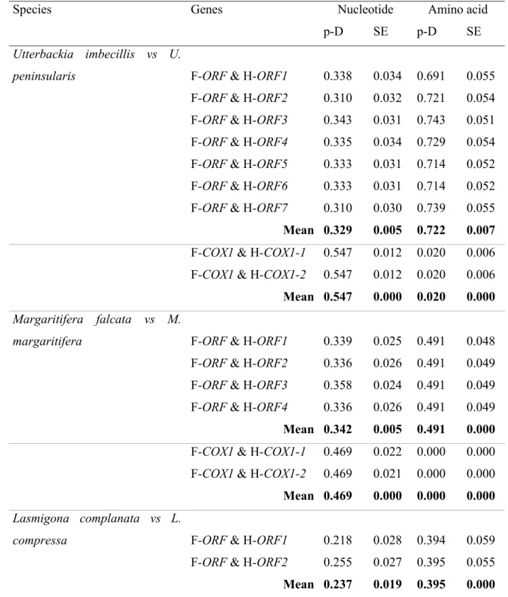
Table IV. p-distances (p-D) and standard error (SE) values of mitochondrial F-orfs vs H-orfs
and Fcox1 vs Hcox1 in comparisons between gonochoric vs. closely related hermaphroditic freshwater mussel species
Species Genes Nucleotide Amino acid
p-D SE p-D SE
Utterbackia imbecillis vs U.
peninsularis F-ORF & H-ORF1 0.338 0.034 0.691 0.055
F-ORF & H-ORF2 0.310 0.032 0.721 0.054 F-ORF & H-ORF3 0.343 0.031 0.743 0.051 F-ORF & H-ORF4 0.335 0.034 0.729 0.054 F-ORF & H-ORF5 0.333 0.031 0.714 0.052 F-ORF & H-ORF6 0.333 0.031 0.714 0.052 F-ORF & H-ORF7 0.310 0.030 0.739 0.055
Mean 0.329 0.005 0.722 0.007
F-COX1 & H-COX1-1 0.547 0.012 0.020 0.006 F-COX1 & H-COX1-2 0.547 0.012 0.020 0.006
Mean 0.547 0.000 0.020 0.000
Margaritifera falcata vs M.
margaritifera F-ORF & H-ORF1 0.339 0.025 0.491 0.048
F-ORF & H-ORF2 0.336 0.026 0.491 0.049 F-ORF & H-ORF3 0.358 0.024 0.491 0.049 F-ORF & H-ORF4 0.336 0.026 0.491 0.049
Mean 0.342 0.005 0.491 0.000
F-COX1 & H-COX1-1 0.469 0.022 0.000 0.000 F-COX1 & H-COX1-2 0.469 0.021 0.000 0.000
Mean 0.469 0.000 0.000 0.000
Lasmigona complanata vs L.
compressa F-ORF & H-ORF1 0.218 0.028 0.394 0.059
F-ORF & H-ORF2 0.255 0.027 0.395 0.055
Lasmigona complanata vs L.
subviridis F-ORF & H-ORF1 0.269 0.029 0.429 0.054
F-ORF & H-ORF2 0.295 0.029 0.442 0.055
Mean 0.282 0.013 0.436 0.007
Toxolasma parvum vs T. lividus F-ORF & H-ORF 0.443 0.027 0.736 0.044
Section 2.3.2: Conserved structures in ORFan protein sequences
One TM helix was predicted near the N-terminus of all M-ORFs (Figure 4 and Supplementary Table I), except for H. menziesii M-ORF sequence, for which one N-terminal and two additional TM helices were predicted. PrediSi and SignalP both returned predicted SPs for all M-ORF sequences, however, the programs rarely agreed about the length of the predicted signal peptide (Supplementary Table II). One TM helix was also predicted in all
F-ORF sequences, with a SP predicted to overlap with this TM structure, except in the case of T. lividus F-ORF where the location of the SP was uncertain (Figure 4 and Supplementary Tables
I and II). All H-ORFs contained one predicted TM helix near the N-terminus as well, except for U. imbecillis H-ORFs that contained multiple predicted TM helices, only the first of which had a confident location (Figure 4 and Supplementary Table III). U. imbecillis H-ORFs also returned variable SP predictions, whereas all other H-ORF sequences contain one predicted SP overlapping with the N-terminal TM helix (Supplementary Table IV). Although they could not be confidently aligned (see Supplementary Figure 2), F-ORFs and H-ORFs of closely related species showed some structural similarities in the localization of the TM helices and SPs (Figure 4). Importantly, all H-ORFs contain tandem repeats (L. compressa possesses between 3 to 7 tandemly repeated sequence motifs of 20 or 21aa; L. subviridis 7 to 9 repeats of 17aa; T.
parvum 2 to 3 repeats of 47aa; M. falcata 2 to 3 repeats of 12aa; and U. imbecillis 2 to 4
repeats of 11 or 21aa), which are not found in F-ORFs and account for most of the difference in length between F-ORFs and H-ORFs of closely related species (Supplementary Figure 2).
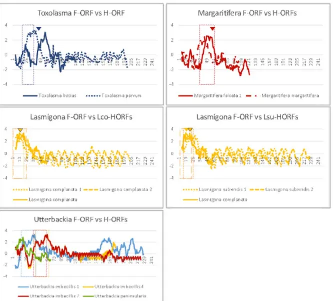
Figure 5. Hydrophobicity profiles of M-ORFs (a), F-ORFs (b) and H-ORFs vs. F-ORFs (c). Boxes indicate predicted TM helices, arrowheads indicate the end of predicted SPs. X-axis
is amino acid position, Y-axis is hydrophobicity. Margaritifera H-ORFs: Mfa1 and Mfa2&4 have nearly identical profiles. Lasmigona H-ORFs: arrowheads outlined in black indicate the end of the SP in sequence 1, arrowhead without outline is for Lco-HORF2; boxes with long dashes are for sequence #2. Utterbackia H-ORFs: Sequences 2-6 have profiles similar to that of sequence 7.
Section 2.3.3: Motif and functional domain scans: frequently recurring
HHpred hits and potential ligand-binding sites
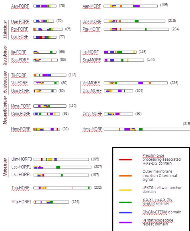
Six HHpred hits consistently appeared highly ranked in the results of M-ORFs,
F-ORFs and H-F-ORFs: (1) prepilin-type processing-associated H-X9-DG domain, (2) outer
membrane insertion C-terminal signal, (3) LPXTG cell wall anchor domain, (4) X-X-X-Leu-X-X-Gly heptad repeats, (5) GlyGly-CTERM domain and (6) a pentatricopeptide repeat (PPR) domain. Probabilities were all >92% (which the developers state can be interpreted literally [64]), and ranks were typically 1-6 in variable order, with very few of these hits falling outside of the top 10 (Supplementary Tables V and VI). Figure 5 shows the position of these six hits in the protein sequences analyzed. Other but less recurring motifs and domains are presented in detail in Supplementary Tables VII and VIII. No TPR or PPR motifs were found.
Figure 6. Position of motifs frequently recurring in HHpred hits. Protein length in amino
acids is indicated in parentheses. One representative sequence was chosen for each hermaphroditic species.
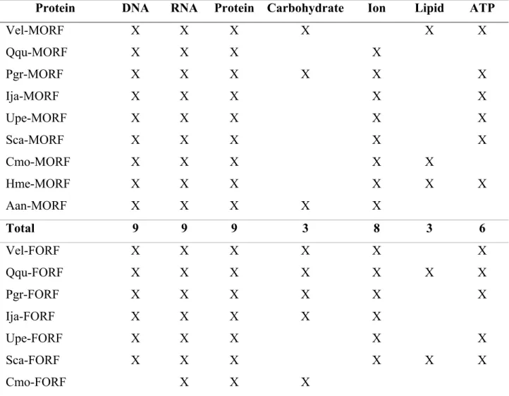
Inferred homologies and prediction of binding sites both indicated that ORFan proteins may bind several ligands (Table V). All M-ORFs returned hits to protein-binding, DNA-binding and RNA-DNA-binding proteins, while many returned hits to proteins DNA-binding ions (8 species), ATP (6 species), carbohydrates (3 species), and lipids (3 species). All F-ORFs returned hits to protein-binding and RNA-binding proteins, while many returned hits to proteins binding DNA (11 species), ions (10 species), ATP (8 species), carbohydrates (7 species), and lipids (4 species). H-ORF sequences returned hits to proteins binding other proteins (5 species), DNA (5 species), RNA (5 species), carbohydrates (5 species), ions (4 species), ATP (3 species), and lipids (2 species).
Table V. Summary of hits to ligand-binding sites in M-ORFs, F-ORFs and H-ORFs
Protein DNA RNA Protein Carbohydrate Ion Lipid ATP
Vel-MORF X X X X X X Qqu-MORF X X X X Pgr-MORF X X X X X X Ija-MORF X X X X X Upe-MORF X X X X X Sca-MORF X X X X X Cmo-MORF X X X X X Hme-MORF X X X X X X Aan-MORF X X X X X Total 9 9 9 3 8 3 6 Vel-FORF X X X X X X Qqu-FORF X X X X X X X Pgr-FORF X X X X X X Ija-FORF X X X X X Upe-FORF X X X X X Sca-FORF X X X X X X Cmo-FORF X X X
Hme-FORF X X X X X Lco-FORF X X X X X X Tli-FORF X X X X X X Mma-FORF X X X X Aan-FORF X X X X X Total 11 12 12 7 10 4 8 Uim-HORF1 - 3 X X X X X X Uim-HORF4 - 7 X X X X X Mma-HORF1, 2, 4 X X X X X Mma-HORF3 X X X X Tpa-HORF X X X X X X X Lco-HORF1 X X X X Lco-HORF2 X X X X X X Lsu-HORF1 - 2 X X X X X Total 14 14 14 10 14 2 6
Section 2.3.4: Prediction of molecular function: hits to viral proteins
Because a viral origin for the mitochondrial ORFans in DUI bivalves has previously been suggested [44], our results obtained with all programs for protein function prediction (i.e., BLAST, HMMER, HHpred, @tome2, I-TASSER, and PredictProtein) were first scanned for supported hits to viral proteins (Table VI). Overall, H-ORFs returned more viral hits than M-ORFs or F-ORFs. M. falcata H-ORFs primarily returned envelope proteins, L. subviridis H-ORFs returned capsid and envelope proteins, L. compressa H-ORFs returned proteins that interact with receptors, and T. parvum H-ORF returned a protein that regulates the degradation of a receptor. U. imbecillis H-ORFs returned many copies of capsid proteins and other structural proteins. M-ORFs returned nucleoproteins (A. anatina and H. menziesii), membrane proteins (I. japanensis and S. carinatus), and proteins with a role in replication, life cycle, and apoptosis (A. anatina, U. peninsularis, I. japanensis and V. ellipsiformis). F-ORF hits were mostly parts of the viral capsid and viral envelope (S. carinatus, T. lividus and M.
margaritifera), receptors/fibre proteins (M. margaritifera and C. monodonta), or proteins
involved in cell cycle and translation (P. grandis and I. japanensis).
Table VI. Hits to viral proteins from structural prediction analyses
Gene Hit Function Position
Aan-MORF
Nucleoprotein, Andes virus [Atome 2; 41.16]
Regulatory protein MNT, Enterobacteria phage P22 [Atome 2; 21.14] Nucleoprotein Gene regulation NA NA Upe-MORF
Uncharacterized protein 56B, Sulfolobus islandicus [Atome 2; 27.96] Transcription repressor NA Pgr-MORF
Matrix protein 1, Influenza A virus [Atome 2; 39.16]
Helix-destabilizing protein, Enterobacteria phage T7 [Atome 2; 18.55]
Matrix protein DNA binding protein
NA NA
Ija-MORF
Nonstructural protein 5A, Bovine viral diarrhea virus 1-CP7 [Atome 2; 33.37]
Functional anti-apoptotic factor vBCL-2 homolog, Human
herpesvirus 8 [Atome 2; 27.14] Membrane protein Apoptosis NA NA Sca-MORF
Nonstructural protein 5A, Bovine viral diarrhea virus 1-CP7 [Atome 2; 22.35]
Membrane protein NA
Vel-MORF
Macrophage galactose N-acetyl-galactosamine specific lectin 2 [Hhpred; 93.40]
RhUL123, Macacine herpesvirus 3 [I-TASSER; TM score 0.671]
Phosphoprotein, Measels virus [Atome 2; 49.33]
Tail needle protein gp26, Enterobacteria phage P22 [Atome 2; 48.96]
C-type lectin
Viral life cycle Unknown function Fibrous protein 20-171 NA NA NA Qqu-MORF
Virion RNA polymerase, Bacteriophage n4 [I-TASSER; TM score 0.542]
Transferase NA
Cmo-MORF
No hits to viral proteins
Hme-MORF
Nucleoprotein, Andes virus [Atome 2; 63.91] Nucleoprotein NA
Aan-FORF
No hits to viral proteins
Upe-FORF
BM2 protein, Influenza B virus (B/Taiwan/70061/2006) [Atome 2; 42.29]
Transport protein NA
Pgr-FORF
V-cyclin, Human herpesvirus 8 [I-TASSER; norm. TM score 0.517]