Rubenfire
Lancellotti, Stefano Ghio, Janos Varga, Sanjay Rajagopalan, Ronald Oudiz and Melvyn
Print ISSN: 0009-7322. Online ISSN: 1524-4539
Copyright © 2013 American Heart Association, Inc. All rights reserved.
is published by the American Heart Association, 7272 Greenville Avenue, Dallas, TX 75231 Circulation
doi: 10.1161/CIRCULATIONAHA.112.000667
2013;128:1470-1479
Circulation.
http://circ.ahajournals.org/content/128/13/1470
World Wide Web at:
The online version of this article, along with updated information and services, is located on the
http://circ.ahajournals.org//subscriptions/
is online at: Circulation
Information about subscribing to
Subscriptions:
http://www.lww.com/reprints
Information about reprints can be found online at:
Reprints:
document.
Permissions and Rights Question and Answer
this process is available in the
click Request Permissions in the middle column of the Web page under Services. Further information about Office. Once the online version of the published article for which permission is being requested is located,
can be obtained via RightsLink, a service of the Copyright Clearance Center, not the Editorial Circulation
in
Requests for permissions to reproduce figures, tables, or portions of articles originally published
1470
P
ulmonary hypertension (PH), regardless of the pathobiol-ogy and site of functional changes, is characterized by an asymptomatic or a latent phase followed by a gradual reduction in exercise capacity related to decreasing right ventricular (RV) contractile reserve.1,2 In addition to right heart and pulmonaryvascular function, other factors including ventilation/perfu-sion mismatch, hypoxemia, peripheral oxygen transportation/ utilization, and impaired skeletal muscle function, may signifi-cantly influence the functional status of patients with PH.3
Guidelines provide a clinical classification system that cat-egorizes PH into 5 groups with specific pathogenesis, patholo-gies, and clinical characteristics.1,2 The natural history of the
disease is determined by the rapidity of increase in resting mean pulmonary artery pressure (mPAP) and the ability of the RV to compensate and hypertrophy, which differs depending on the pathobiology and related diseases.1,2 The definition of
PH and PH groups is based on clinical assessment and inva-sive hemodynamics. Precapillary PH (groups 1 and 3–5) is characterized as a resting mPAP of ≥25 mm Hg and pulmo-nary capillary wedge pressure (PCWP) or left ventricular (LV) end-diastolic pressure ≤15 mm Hg. Postcapillary PH (group 2) is associated with an elevated pulmonary venous pres-sure (>15 mm Hg) related to left heart disease, which may involve abnormalities of the LV, mitral valve, or aortic valve.2
Although the group definitions for PH are helpful, in clinical practice, it is not uncommon for patients to have >1 disease associated with PH and to have both precapillary and postcap-illary components, so called multifactorial PH (eg, aortic ste-nosis, systemic sclerosis, chronic thromboembolic disease) or out-of-proportion PH for a given condition (eg, chronic lung disease, chronic heart failure).
The concept that symptoms associated with cardiopulmo-nary diseases would be associated with an abnormal increase in mPAP with exercise was demonstrated >60 years ago.4
Since then, there has been considerable investigation by clini-cal scientists. The PH guidelines published in 2009 removed an exercise mPAP of ≥30 mm Hg as an additional definition of PH, not because of the lack of potential value of exercise PAP
measurements but rather because of inadequate standardiza-tion of techniques and lack of established ranges of normal physiological responses by age and sex. In the 1980’s, age was shown to be associated with an increase in exercise mPAP per liter increment in cardiac output and in a recent study found that ≈50% of healthy people >50 years of age develop an mPAP >30 mm Hg during mild exercise.5,6 A
substan-tial amount of enthusiasm remains for considering exercise hemodynamics obtained invasively or by noninvasive testing to determine pressure-flow and pressure-workload relation-ships, which can further characterize PH in its asymptomatic or latent phase, clarify the contribution of the precapillary and postcapillary components, and help determine the prognosis, treatment decisions, and response to therapy.
Here, we present the normal pulmonary vascular and physi-ological responses of the right side of the heart to exercise, the responses in each of the major causes of PH, and the potential clinical and investigative utility of assessing hemodynamics by exercise testing.
Physiology of the Pulmonary Circulation
at Rest and During Exercise
Exercise stresses the pulmonary circulation through an increase in cardiac output (CO) and mean left atrial pressure (LAP), each of which results in an increase in mPAP. The relationships between mPAP, CO, and LAP are defined by the pulmonary vascular resistance (PVR) equation:
PVR =
(
mPAP LAP−)
/ CO. (1) The PVR equation rewritten as mPAP=PVR×CO+LAP shows how much mPAP increases with CO or LAP at any given PVR.1 More than 50 years ago, Paul Wood reported thatmPAP does not exceed 20 mm Hg at rest as long as PVR, CO, and LAP remain within normal limits.4 As discussed earlier,
PH has been defined by an mPAP at rest ≥25 mm Hg.1,2 An
mPAP of 21 to 24 mm Hg corresponds to so-called “border-line PH” of still undetermined clinical relevance. PH can be caused by abnormally high PVR, CO, or LAP.1,2
(Circulation. 2013;128:1470-1479.)
© 2013 American Heart Association, Inc.
Circulation is available at http://circ.ahajournals.org DOI: 10.1161/CIRCULATIONAHA.112.000667
From the Massachusetts General Hospital, Boston (G.D.L.); IRCCS Policlinico San Donato, Milan, Italy (E.B.); Université Libre de Bruxelles, Brussels, Belgium (R.N.); University Hospital of Heidelberg, Heidelberg, Germany (E.G.); St. Joseph Hospital and Medical Center, Phoenix, AZ (R.S.); University of Liège Hospital, GIGA Cardiovascular Science, Liège, Belgium (P.L.); Fondazione IRCCS Policlinico S Matteo, Pavia, Italy (S.G.); National Koranyi Institute for TB and Pulmonology, Budapest, Hungary (J.V.); Ohio State University, Columbus (S.R.); David Geffen School of Medicine at UCLA, Torrance, CA (R.O.); and University of Michigan, Ann Arbor (M.R.).
Drs Lewis and Bossone contributed equally to this work.
Correspondence to Melvyn Rubenfire, MD, Professor of Internal Medicine, University of Michigan, 24 Frank Lloyd Wright Dr, Lobby A, Ste 3700, Ann Arbor, MI 48106. E-mail: mrubenfi@med.umich.edu
Pulmonary Vascular Hemodynamic Response to Exercise
in Cardiopulmonary Diseases
Gregory D. Lewis, MD; Eduardo Bossone, MD, PhD; Robert Naeije, MD, PhD;
Ekkehard Grünig, MD; Rajeev Saggar, MD; Patrizio Lancellotti, MD, PhD; Stefano Ghio, MD;
Janos Varga, MD, PhD; Sanjay Rajagopalan, MD; Ronald Oudiz, MD; Melvyn Rubenfire, MD
Contemporary Reviews in Cardiovascular Medicine
at UNIV MEDECINE LEIG 228272 on November 18, 2014
http://circ.ahajournals.org/
The inherent assumptions of the PVR equation are that (mPAP−LAP)/CO relationships are linear and cross the line of origin. If these assumptions are correct, PVR should be inde-pendent of absolute levels of pressure and flow and of mea-surements at exercise predicted from resting values. However, it has been shown that exercise is normally associated with a decrease in PVR.5-7 The exercise-induced decrease in PVR is
attributable to pulmonary vascular recruitment in the setting of increased PAP and flow, as well as the distensibility of the normal pulmonary resistance vessels. This decrease is greater when exercise is performed in the upright position because of higher resting PVR resulting from incomplete recruitment of pulmonary resistance vessels in the upper portions of the lungs at rest. The supine position and mild to moderate exer-cise allow complete pulmonary vascular recruitment. Most of the exercise-induced decrease in PVR is explained by disten-sion of the pulmonary resistance vessels.5,7
Because resistance vessels are distensible, multipoint mPAP/CO relationships normally present with a slight cur-vilinearity (Figure 1).7,8 Adjustment of these pressure-flow
curves with a mathematical model of the pulmonary circula-tion allows for a recalculacircula-tion of a distensibility coefficient, α, which can be introduced into a refined PVR equation for an improved estimation of mPAP at various COs or LAPs:
mPAP=
(
1+ LAP)
+5 R Q −1 5 0 1 5 α α α (2) where R0 is PVR and Q is pulmonary blood flow.8 Thedis-tensibility coefficient α defines how much resistance vessel diameters increase as a percent of baseline per mmHg distend-ing pressure. The normal value of α is 2% per mmHg. This value has been established in pulmonary vessels mounted in vitro and is remarkably constant from one species to another.8
The coefficient α is higher in pre-menopausal women as com-pared to men9 and decreases with age.8,9 A seemingly-modest
2% distensibility of pulmonary resistive vessels is actually a major cause of the limitation of increase in mPAP and decrease
in PVR normally observed with exercise (Figure 2).7 The
sen-sitivity of α to early pulmonary vascular disease is currently under investigation.
In patients with cardiac or pulmonary diseases, PVR has been reported to decrease during exercise in some studies, but other studies have reported unchanged or even increased PVR during exercise, depending on body position and work-load.5,7 In general, an exercise-induced increase in PVR may
be observed in heart failure and pulmonary vascular disease when resting measurements are performed in the supine posi-tion and when patients perform maximum tolerated exercise.5,7
In most situations, the number of recovered mPAP/CO coor-dinates remains limited when workload is moderate. In these circumstances, multipoint mPAP/CO plots are best described by a linear approximation. Such a dynamic PVR still offers a more robust definition of the functional state of the pulmonary circulation than a single-point PVR.5,7
Invasive as well as non-invasive studies in healthy volunteers show that the slope of “linearized” mPAP/CO relationships ranges from 0.5 to 3.0 mmHg/L/min (Table 1).6,7 LAP increases
during exercise with upstream transmission to mPAP.5,6,10-12 The
increment in LAP during exercise also reduces pulmonary com-pliance and thereby leads to an increase in transpulmonary pres-sure gradient (mPAP minus LAP = TPG; Figure 3).13 However,
LAP exceeds the upper limit of resting normal in healthy young adult subjects only at very high cardiac outputs, in the range of 20 L/min and greater.14 The pulmonary capillary wedge
pressure (PCWP) to estimate LAP can increase to as high as 25mmHg in exercising athletes.5 In fact, as reviewed by West,
at very high levels of exercise elite athletes may develop pul-monary edema and changes in the integrity and function of the blood-gas barrier that results in ‘exercise induced pulmonary hemorrhage’, a phenomenon common to race horses.15 In older
subjects and particularly in heart failure patients, steep incre-ments in LAP or PCWP are apparent during early exercise and contribute to mPAP/CO > 3.0mmHg/L/min. A disproportion-ate increase in LAP or PCWP affects mPAP at lower levels of
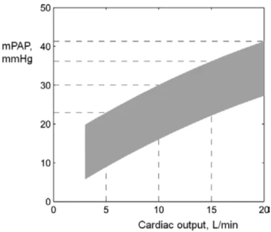
Figure 1. Limits of normal of mean pulmonary artery pressure
(mPAP) as a function of cardiac output (CO) at exercise in healthy young adults, constructed from multi-point noninvasive (n=113) and invasive (n=24) measurements reported in ref 7. All baseline resting measurements were within the limits of normal. No slope of linearized mPAP-CO relationships exceeded 3 mmHg/L/min. Slopes of mPAP-CO and derived distensibility calculations were identical in noninvasive and invasive studies. Stippled lines indi-cate upper limits of mPAP at specified cardiac outputs.
Figure 2. Modeled mean pulmonary arterial pressure-cardiac
output (mPAP-Q) relationships during dynamic exercise with progressively increased distensibility coefficients (α). The normal range of α values is 1-2%. Authorization for this adaptation has been o btained both from the owner of the copyright in the origi-nal work and from the owner of copyright in the translation or adaptation. Adapted from reference 7, reprinted with permission of the American Thoracic Society. Copyright © 2013 American Thoracic Society.
1472 Circulation September 24, 2013
exercise in older subjects5 and at very low levels of exercise in
heart failure patients.5,16
Cardiac and pulmonary diseases may affect mPAP by increased PVR, increased LAP, and sometimes an increased CO. The limited data available point to a prominent effect of increased PVR as a cause of steeper mPAP/CO plots. A dis-proportionate increase in mPAP, ie, more than predicted from the resting PVR equation, may also be due to pulmonary vaso-constriction related to low mixed venous PO2, acidosis and sympathetic nervous system activation.5,17 Exercise-induced
pulmonary vasoconstriction typically causes extrapolated pressure intercepts of mPAP/CO plots to be negative.16
When a sufficient number of mPAP-CO coordinates are generated, it becomes possible to identify patterns.11,12,16 Both
linear and “take-off” patterns of mPAP-CO relationships have been observed in patients with left heart failure.11,16 A take-off
pattern is explained by increased LAP or pulmonary vasocon-striction. A “plateau pattern” may emerge, particularly when CO is replaced by O2 uptake (Vo2), based on the rationale that both variables are linearly related. An inability to increase CO at still-increasing Vo2 at the highest level of exercise may explain “plateauing” of the mPAP-Vo2 relationship in PAH.12
Recent studies have begun to clarify the clinical correlates of exercise-induced PH.11,12 Invasive and noninvasive
stud-ies have accumulated, showing that the slope of linearized mPAP-CO relationships should not exceed 3 mm Hg·L−1·min−1
(Figure 1 and the Table6,9–12,16,18–32). This corresponds to the
former definition of exercise-induced PH (mPAP >30 mm Hg) provided that CO remains <10 L/min or total pulmonary resis-tance (mPAP/CO) at maximum exercise is <3 Wood units.
Although further work is needed to improve exercise test-ing protocols for the diagnosis and follow-up of PH, several guiding principles and limitations deserve mention. Exercise should be dynamic, avoiding the increase in systemic vascular resistance and abrupt changes in intrathoracic pressure that occur with resistive exercise and might present with unpre-dictable effects on the pulmonary circulation. Even with dynamic exercise, however, increased ventilation causes pul-monary vascular pressures to vary greatly between inspiration and expiration, as reflected by respiratory swings of esopha-geal pressures. Therefore, measuring pulmonary vascular pressures at end expiration, when esophageal pressures are positive (particularly in obstructive lung disease), may lead to a false diagnosis of exercise-induced PH. It is therefore pref-erable to average the reading of pulmonary vascular pressures
over the entire respiratory cycle. Postexercise measurements are thought to be unreliable because of the rapid return of pulmonary vascular pressures and flows to the baseline rest-ing state. Even though workload or Vo2 and CO are linearly related, they should not be used as surrogates considering that a determinant of pulmonary vascular pressure is flow and the individual variability of workload or Vo2 versus CO relationships is high. Finally, recently developed noninvasive echocardiographic measurements of mPAP-CO relationships have been shown to closely approximate invasively measured mPAP-CO values,7,9,25 and may be feasible in highly dedicated
centers with very experienced echocardiographers, but they require more validation to demonstrate clinical relevance.
Accepting a cutoff value of a slope of mPAP/CO plots >3 mm Hg·L−1·min−1 or a total PVR >3 Wood units at maximum
exercise does not clarify whether the abnormal pressure is attrib-utable to pulmonary vasoconstriction, remodeling, or upstream transmission of a high LAP or PCWP. Thus, a better definition of the limits of normal of LAP at exercise is also needed. In the meantime, it can be safely assumed that the functional state of the pulmonary circulation is best described by >4- to 5-point (mPAP−LAP)/CO plots to calculate a slope (Figure 4).33
Heart Failure With Reduced
LV Ejection Fraction
Heart failure with reduced LVEF (HFrEF) is commonly associated with the development of pulmonary hypertension (HFrEF-PH). HFrEF-PH purports a poor prognosis, particu-larly when it is accompanied by RV dysfunction.34 Single
rest-ing right heart catheter or echocardiography measurements tend to vary with diurnal patterns, autonomic tone, and timing of medication exposures (ie, diuretics), which can confound conclusions about the degree of right ventricular-pulmonary vascular (RV-PV) dysfunction present in HFrEF.35 In contrast,
exercise permits multiple measurements across varying load-ing conditions to assess RV contractile reserve and the ability of the pulmonary vasculature to accommodate increased flow.36
A recent study established that exercise in HFrEF elicits a steep increase in mPAP that is attributable to exaggerated increases in both TPG and PCWP.11 A steep linear increment
in mPAP in response to exercise was independently associated with reduced exercise capacity (peak Vo2) and reduced survival in patients with HFrEF. Failure to augment mPAP throughout exercise, resulting in a plateau pattern of mPAP versus work during late exercise, was indicative of dynamic RV dysfunction
Figure 3. Relationship between pulmonary
capil-lary wedge pressure (PCWP) and transpulmonary gradient (TPG) across the normal range of stroke volumes (SV) achieved during exercise. DPG indicates diastolic pulmonary pressure gradient, which is the gradient between pulmonary arte-rial diastolic pressure and PCWP. An increase in PCWP is normally associated with a proportion-ately greater increase in mean pulmonary artery pressure (mPAP) than that of diastolic pulmonary artery pressure. As PCWP increases, the DPG may decrease (A) or remain unchanged (B). The different impact of increased PCWP on the TPG and the DPG is explained by the effects of SV and of PCWP on pulmonary arterial compliance.
at UNIV MEDECINE LEIG 228272 on November 18, 2014
http://circ.ahajournals.org/
and portended a particularly poor prognosis.11 These findings
complement previous studies showing that mPAP and PVR responses to exercise are more closely related to outcomes than resting hemodynamic measurements in HFrEF.11
Exercise echocardiography has also been used in the evalu-ation of HFrEF-PH, although Doppler measures of right- and particularly left-side pressures can be challenging to ascertain during exercise and require further validation. Tumminello et al found that peak exercise pulmonary artery systolic pressure (PASP) in patients with HFrEF was related principally to mitral regurgitation and contractile reserve, whereas Maréchaux et al identified intraventricular dyssynchrony and impaired contrac-tile reserve to be independent predictors of exercise-induced changes in PASP.38 Notably, very low tricuspid annular plane
systolic excursion (<14 mm) predicted a more modest maxi-mum increase in PASP during exercise, a finding that mirrors
invasive data suggesting that marked impairment in RV perfor-mance during exercise leads to a limited capacity to augment PASP and mPAP.11 The aforementioned
echocardiography-based studies focused on peak exercise PASP or peak exercise changes in PASP, which are attenuated if CO augmentation is modest during exercise. Therefore, it is preferable to interpret exercise mPAP augmentation patterns relative to increases in blood flow through the pulmonary circulation (mPAP/CO as described previously; see the Table).
Several mechanisms may account for the heightened mPAP/CO observed in HFrEF. Increased left-side pressures during exercise caused by HFrEF are to be expected, but exag-gerated increases in TPG are also observed in HFrEF. This may be attributable to maximally recruited pulmonary vasculature at rest on account of elevated resting LV pressures, thus limiting passive recruitment and distension during exercise observed in
Table. Review of Exercise Stress Tests of the Pulmonary Circulation in Normal Subjects and in Patients With Chronic Obstructive Pulmonary Disease, Scleroderma, Left Heart Failure, and Pulmonary Arterial Hypertension.
Condition Author n Age, y
Exercise Protocol Work Rate, W CO Rest, L/min CO Ex, L/min mPAP Rest, mm Hg mPAP Ex, mm Hg ΔmPAP/ΔCO Normal
Hickam et al18 8 23±7 Leg press 20±7 7.1±1.8 10.4±3.5 11±3 13±1 0.9
Lonsdorfer et al19 7 31±8 Cycle 276±50 5.4±1.5 20.0±3.3 14±2 27±6 0.9
Degre et al20 11 41±5 Cycle NA 5.0±0.9 13.3±1.7 11±4 23±5 1.4
Slonim et al21 5 22±2 Leg press 20±2 4.9±0.6 7.2±0.8 15±2 19±2 1.6
Wagner et al22 8 30±6 Cycle 180 6.9±2.0 20.5±2.5 13±3 30±6 1.2 Damato et al23 24 31±6 Treadmill 160±23 5.1±0.8 17.5±2.0 15±5 29±8 1.1 Reeves et al24 9 NA Cycle 240 6.7±1.2 24.7±3 15±2 29±8 0.8 Tolle et al12 16 46±15 Cycle 156±43 5.8±1.0 15.5±3.2 14±3 27±4 1.4 Argiento et al25 25 36±14 Cycle 170±51 4.7±1.0 18.0±4.2 14±3 30±7 1.4 Argiento et al9 113 37±13 Cycle 175±50 5.3±1.2 15±3 18±3.6 33±7 1.5 Kovacs et al6 16 >50 Cycle NA 6.2±1.5 13.1±1.7 15±4 32±7 2.5 COPD
Hickam et al18 5 51±10 Leg press 10 6.2±1.0 7.8±2.3 18±5 25±2.5 4.0
Saito et al26 14 67±7 Cycle 27 4.3±0.7 8.8±1.1 19±4 41±10 4.6
COPD+PH Blanco et al27 20 64±7 Cycle NA 4.9±1.0 9.0±2.2 27±10 56±14 7.0*
COPD-PH COPD+PH Hilde et al28 Hilde et al28 26 72 64±6 62±8 Cycle 74±25 50±20 5.2±1.0 5.6±1.2 10.4±2.5 9.4±2.8 18±3 29±4 37±8 48±7 4.6 7.2* Scleroderma SSc-PH Saggar et al29 57 51±13 Cycle NA 5.2±1.4 9.4±3.1 18±4 35±8 4.5
Left heart failure
HFrEF Lewis et al30 13 47±9 Cycle 83±10 3.2±0.2 4.8±0.3 28±4 36±4 5.0
HFrEF Lewis et al11 60 60±12 Cycle 75±28 3.7±0.7 7.6±2.1 28±3 50±3 5.6
HFrEF Mancini et al31
Survivors 49 53±10 Cycle NA 4.0±1.1 7.7±2.1 28±11 46±12 4.9 Nonsurvivors 16 52±8 4.1±0.8 7.1±2.3 31±10 55±8 8.0 HFrEF Janicki et al16 42 NA Treadmill NA 3.7±1.3 5.9
HFpEF Borlaug et al10 32 65±13 Cycle 47±19 5.2±1.1 9.1±1.9 19±4 43±7 6.1
PAH
Janicki et al16 9 NA Treadmill NA 3.4±0.7 8.9±2.7 43±16 81±16 6.1
Blumberg et al32 16 55±8 Cycle 25–50 3.7±1 5.8±2.4 45±8 70±13 11.9
CO indicates cardiac output; COPD, chronic obstructive lung disease; Ex, exercise; HFpEF, heart failure with preserved ejection fraction; HFrEF, heart failure with reduced left ventricular ejection fraction; mPAP, mean pulmonary arterial pressure; ΔmPAP/ΔCO, the slope of the pulmonary arterial pressure versus cardiac output during exercise; PAH, pulmonary arterial hypertension; PH, pulmonary hypertension; and SSc, systemic scleroderma.
1474 Circulation September 24, 2013
normal subjects. Elevations in PCWP also lower pulmonary artery compliance at a given PVR, leading to increased pul-monary artery wave reflections and augmentation in PASP and thus mPAP.13,39 Subnormal Pao
2 during exercise and an
imbal-ance between endothelium-derived vasoactive and vasocon-strictive mediators may also contribute to increased pulmonary vascular tone in HFrEF.40 In support of the latter mechanism,
inhibition of cyclic GMP hydrolysis with the phosphodiester-ase 5 inhibitor sildenafil has been shown to improve exercise PVR and exercise capacity in HFrEF.30,36
A greater knowledge of compensatory responses of the RV–pulmonary vascular unit during exercise in patients with HFrEF may improve our understanding of its functional role in determining exercise capacity while aiding in earlier diag-nosis of PH complicating HFrEF and informing targeted ther-apeutic interventions.36
Heart Failure With Preserved
LV Ejection Fraction
Heart failure with preserved LV ejection fraction (HFpEF) constitutes ≈40% of the heart failure population and is associ-ated with a similar prognosis to HFrEF.41 HFpEF, like HFrEF,
is associated with a steep increase in mPAP during exercise (Table). Echocardiography-based mechanistic studies indicate that exercise mPAP in HFpEF, unlike HFrEF, is most closely associated with degree of diastolic dysfunction.42 In both
HFrEF and HFpEF, it is important not to overlook comorbid conditions that can contribute to PH such as chronic obstruc-tive pulmonary disease (COPD), obstrucobstruc-tive sleep apnea, val-vular heart disease, or chronic thromboembolic PH.
Current guideline-based diagnostic criteria for HFpEF include objective evidence of elevated cardiac filling pressures based on cardiac catheterization, echocardiography, or natriuretic peptide assays.42,43 These criteria lead to potential
underdiag-nosis of HFpEF in symptomatic patients without hypervolemia. Borlaug et al reported invasive hemodynamic responses to exer-cise in 55 euvolemic subjects with exertional dyspnea and nor-mal resting hemodynamic measurements.10 A PCWP threshold
of ≥25 mm Hg during supine cycle ergometry or weightlifting with the arms was used to diagnose HFpEF. HFpEF subjects experienced significantly greater exercise-induced increases in mPAP (ie, from 19±4 to 43±7 mm Hg; Table) than subjects with noncardiac dyspnea (ie, from 15±4 to 23±5 mm Hg) despite achieving lower peak COs. Exercise PASP is easily amenable to noninvasive measurement. In the same study, exercise PASP identified HFpEF with 96% sensitivity and 95% specificity using a cut point of 45 mm Hg. Exercise PASP outperformed resting PASP, natriuretic peptide levels, and echocardiographic indicators for diagnosing HFpEF.10
Similarly, Kitzman et al reported in 1991 that compensated outpatients with HFpEF had normal resting PCWP but marked increases in exercise PCWP, suggesting that HFpEF may ini-tially manifest with only intermittent elevations in cardiac filling pressures.43 Both studies indicate that exercise
hemody-namic measurements are of incremental value for the diagnosis of HFpEF and may provide a window into earlier diagnosis of the condition. However, at present, there is no consensus on the appropriate partition value to define an abnormal exer-cise PCWP or LV end-diastolic pressure, with suggested cut-off values ranging from 15 to 25 mm Hg. Furthermore, before labeling elevations in exercise hemodynamic measurements as early forms of HFpEF, more information is needed on the natu-ral history of patients with exercise elevations in mPAP and PCWP to determine the rate at which they go on to develop adverse outcomes known to be associated with overt HFpEF.
In addition to assisting with the diagnosis of HFpEF in the setting of normal resting hemodynamic measurements, exercise may be of particular value is assessing the entity of PH out of proportion to LV dysfunction, alternatively called PH–LV dysfunction or mixed PH.45 In mixed PH, the
partitioning of relative contributions of PCWP and TPG to exercise mPAP may help to further phenotype patients with dyspnea and to inform targeted interventions directed at either the LV or the pulmonary vasculature. Indeed, a recent trial of phosphodiesterase 5 inhibition in HFpEF subjects with a high burden of resting precapillary PH and RV dysfunction showed marked improvement in mPAP, PVR, and quality of life with sildenafil treatment.45 In contrast, in a broader
HFpEF population in whom neither PH nor RV dysfunction was mandated by inclusion criteria, sildenafil did not improve exercise capacity.46 Whether exercise hemodynamic responses
can identify HFpEF subjects with earlier forms of abnormal RV–pulmonary vascular reserve capacity who will benefit from RV–pulmonary vascular–directed interventions is being actively investigated.
Pulmonary Arterial Hypertension
Pulmonary arterial hypertension (PAH) is a rare disease associated with remodeling of the small pulmonary arteries, Figure 4. Linear mean pulmonary arterial pressure (mPAP)-flow
relationships based on averages of serial measurements of mean PAP and cardiac output during incremental exercise. Normal young adults (□), patients with scleroderma with PAP in the lower normal range (◊) and borderline-PH range (♦), and patients with resting PAH (•) demonstrate approximately linear pressure-flow responses during exercise. Despite similar resting mPAP and CO in normals and the two scleroderma subgroups, exercise elicits a steeper increment in PAP vs. CO in scleroderma borderline-PH group (♦). The mPAP-CO slope of > 3.0 (dashed reference line) represents an abnormal pulmonary vascular response to exercise and may be indicative of early pulmonary vasculopathy. Adapted from reference 33. Authorization for this adaptation has been obtained both from the owner of the copyright in the original work and from the owner of the copyright in the translation or adaptation. Reprinted with per-mission of Advances in Pulmonary Hypertension. Copyright © 2013
Advances in Pulmonary Hypertension.
at UNIV MEDECINE LEIG 228272 on November 18, 2014
http://circ.ahajournals.org/
leading to an increase in PVR, adaptive right heart hyper-trophy, and right heart failure.1,2 Numerous pathogenic
fac-tors and diseases affecting the pulmonary vascular bed have been identified that result in a similar clinical phenotype with impaired exercise capacity, quality of life, and prognosis. The diagnosis of PAH requires confirmation by direct assessment of invasive hemodynamic parameters at rest.1,2
As reviewed by Bossone et al, there have been a limited number of studies that have evaluated invasive hemodynam-ics during exercise in PAH.5 Whereas healthy subjects can
achieve a more than 10-fold increase in VO2 during exercise due to a complex cardiovascular, ventilatory and peripheral response, in PAH patients peak VO2 is significantly reduced. Patients with PAH are mainly limited by a reduced CO at rest and during exercise and have steep mPAP-CO relationships.16
RV dysfunction is the most important factor contributing to functional impairment and mortality.
Despite the lack of adequate standards, there is increas-ing enthusiasm for performincreas-ing noninvasive assessment of exercise hemodynamics to screen for PAH and to assess prognosis. As reviewed by Bossone and Naeije,4 the
clini-cal phenotype of normal pulmonary arterial pressure at rest and high response during exercise has been evaluated with recumbent exercise echo Doppler studies in subjects at risk for PAH in 10 studies involving 824 subjects (427 with con-nective tissue disease, 54 with congenital heart disease, and 343 relatives of idiopathic PAH cases).5 Although the
mea-surement of PASP during symptom-limited exercise or at given workloads is of interest as a screening tool for PAH, it is not adequate for determining whether the exaggerated exercise-induced increase in PASP in persons with dyspnea or fatigue is attributable to a precapillary (PAH) or postcapil-lary (pulmonary venous hypertension) source. It may be help-ful for selecting high-risk patients who would benefit from direct measurement of invasive hemodynamics at rest and with exercise such as those with a family history of PAH or connective tissue diseases (discussed below).
Grünig et al demonstrated the use of recumbent exercise echo Doppler for identifying asymptomatic family members of idiopathic PAH patients carrying a predisposing mutation in the bone morphogenetic protein receptor II gene in 2 fami-lies47 and subsequently led a multicenter study in 291 relatives
of 109 PAH patients and 191 age-matched control subjects from 6 centers in European countries.48 During exercise (mean
maximal workload of 148 W in control subjects and 124 W in relatives), 10% of control subjects but 31.6% of the relatives (P<0.0001) exceeded the 90th percentile of mean maximal tri-cuspid regurgitation velocity of >3.08 m/s (corresponding to PASP of >43 mm Hg) seen in control subjects.
Tolle et al conducted a landmark study demonstrating that the pattern and severity of the invasive hemodynamic response to cycle ergometry exercise (symptom limited with peak heart rate ≥80% of predicted or respiratory exchange ratio of ≥1) in exercise-induced PAH are intermediate between those of the normal subject and those of the patient with resting PAH and provide support for the hypothesis that exercise-induced PAH is a mild yet symptomatic phase of the disease.8 Cardiopulmonary
exercise testing was used to diagnose the cause of dyspnea or fatigue in 255 patients; of these patients, the concluding
diagnosis was PAH in 93 and 16 were considered normal. Of the PAH group, 78 had exercise-induced PAH and 15 had evi-dence of PAH at rest (mPAP ≥25 mm Hg). The mean (2 SD) mPAP with exercise was 27.4 mm Hg (7.4 mm Hg) in the nor-mal group, 36.6 mm Hg (11.4 mm Hg) in the exercise-induced group, and 48.4 mm Hg (22.2 mm Hg) in those with resting PAH, whereas the PCWP and mRAP did not differ between groups. Consistent with findings in HFrEF, a plateau pattern in PAP versus Vo2 (or CO) in exercise-induced PAH patients was associated with worse functional capacity.12 Interestingly,
rest-ing mPAP, includrest-ing those in an “indeterminate” range of 21 to 25 mm Hg, did not reliably predict exercise-induced PAH.
Exercise hemodynamics in idiopathic PAH on PAH-specific treatment correlates much better with improved exercise tol-erance than resting hemodynamics. Extending earlier obser-vations by their group, in 2008, Provencher et al evaluated 42 patients with idiopathic PAH at baseline and after PAH-specific treatments for an average of 5 months.49 With a 43-m
improvement in 6-minute walk distance, on multivariate anal-ysis, improvement was related to changes in cycle exercise (40 W) hemodynamics; specifically, the peak cardiac index was achieved at a lower mPAP, consistent with exercise vasodila-tion. Whether a change in pressure-flow relationship response (∆mPAP/∆CO; Figures 1 and 2) at a given workload, target heart rate, or percent of aerobic capacity will improve risk stratification and treatment decisions is an important area of future research in PAH.
Exercise Hemodynamics in Scleroderma
Patients with scleroderma are considered an at-risk group for developing PH, which when present is the major cause of death.1,2 They may have latent or asymptomatic PAH,
associ-ated with hypoxemia and lung disease, as well as LV dia-stolic dysfunction or pulmonary venous hypertension from hypertension or scleroderma cardiomyopathy.50,51 Although
the definition of PH requires an mPAP ≥ 25 mm Hg, there is evidence that scleroderma patients with a baseline “bor-derline” resting mPAP of 21 to 24 mm Hg and with exagger-ated PAP response to exercise are at higher risk of developing progressive pulmonary vascular disease.52,53 This suggests
that the hemodynamic response to exercise in scleroderma, as assessed by echo Doppler and heart catheterization, may detect those who are likely to develop rapidly progressive PAH in a preclinical phase that may respond to therapy. The importance of such efforts is supported by several studies. RV diastolic dysfunction has been found in patients with sclero-derma and normal resting but abnormal exercise PH (latent PH) scleroderma.50 Kovacs and colleagues52 reassessed
exer-cise invasive hemodynamics (50 W and maximum exerexer-cise) in 10 borderline PAH patients with scleroderma after a 1-year observation period; a progression was noted in pertinent hemodynamic variables (mPA and PVR). Furthermore, in the UK PH registry, one fifth of scleroderma patients with exer-cise PH developed resting PAH after ≈2.3 years.53
The frequency, spectrum, and significance of a hyperten-sive PAP response to exercise in scleroderma are currently uncertain. The PAP response may reflect a normal variant (eg, related to age) without clinical significance or a manifestation of abnormal pulmonary arterial vasodilator reserve and may
1476 Circulation September 24, 2013
be composed of precapillary or postcapillary pathological rises in pressure. Saggar et al54 assessed the invasive hemodynamic
response to recumbent submaximal cycle exercise in 57 patients with dyspnea and clinical scleroderma who had normal resting hemodynamics but decreased Dlco or a FVC:Dlco percent pre-dicted ratio >1.4. Those with an LV ejection fraction <50% or more than mild LV diastolic dysfunction were excluded. They found 4 distinct groups: normal in 26%, exercise pulmonary venous hypertension in 21% (mPA >30 mm Hg, PCWP >18 mm Hg, and TPG <15 mm Hg), exercise out-of-proportion PH in 16% (mPA >30 mm Hg, PCWP >18 mm Hg, and TPG ≥15 mm Hg), and exercise PAH in 37% (mPA >30 mm Hg, PCWP <18 mm Hg, and TPG >15 mm Hg). A baseline mPAP <14 mm Hg was not associated with exercise PH or exercise out-of-proportion PH, whereas a resting mPAP >20 mm Hg has a 90% positive predictive value for exercise PH, exercise out-of-proportion PH, or pulmonary venous hypertension, and a concurrent PVR ≥ 2 Wood units had a positive predictive value of 100%. For those with an mPAP of 14 to 20 mm Hg, a low normal PVR had a negative predictive value of 100%, and a PVR ≥ 2 Wood units had a positive predictive value of 89% for predicting exercise PH and exercise out-of-proportion PH. In a similar study of 54 patients with scleroderma at risk for PH, of the 17 patients with an RV systolic pressure <35 mm Hg at rest and at least a 20-mm Hg increase in RV systolic pressure dur-ing stress echo Doppler, durdur-ing the heart catheterization with exercise conducted by arm exercise with weights, 1 patient had no PH, 3 patients had evidence of LV diastolic dysfunction, and 13 patients had exercise-induced PAH.51
Finally, a recent uncontrolled, open-label study suggested that chronic oral administration of ambrisentan may be asso-ciated with significant improvement in exercise cardiopulmo-nary hemodynamics and exercise capacity (6MW) in patients with SSc-spectrum and normal resting PAP but exercise PH.29 These improvements included a significant decrease in
exercise PVR, total pulmonary resistance, and mPAP, and an increase in CO and SV.29 The clinical significance of these
observations needs to be validated by a larger placebo con-trolled randomized study.
Further work defining normal versus abnormal ΔPAP/ΔCO specifically for patients with scleroderma, with and without interstitial lung disease, needs to be conducted. Nonetheless, an exercise evaluation of scleroderma-PAH patients may allow earlier diagnosis, earlier initiation of therapy, and per-haps a more favorable outcome.
Valvular Heart Disease
PH at rest and exaggerated during recumbent exercise testing is associated with an adverse outcome in patients with aortic and mitral valvular disease.55 The presence of PH at rest or
during exercise should therefore be included in perioperative risk assessment of mitral and aortic valve replacement.56,57 In
older patients with severe mitral valve stenosis and PH, the perioperative mortality increases to 20%; therefore, the indica-tion of valve surgery has to be proven carefully.56 Preoperative
PH was also an independent risk factor for long-term survival in patients with aortic valve replacement.57,58
Patients with valvular disease and normal or mildly ele-vated PASP at rest may develop severe PH during exercise,
which may be unrelated to the resting pressure.37,59 The level
of increase in PASP depends on the ability to successfully recruit the pulmonary vasculature to accommodate increased blood flow with exercise, the contribution (or proportion) of the reduction in cross-sectional area of the pulmonary circula-tion, the reduction in pulmonary vascular compliance, and the increase in LAP, each of which may be abnormal at rest.37
Mitral Valve Disease
Although the number of studies of exercise in mitral valve disease is limited, they consistently demonstrate that exercise-induced PH is associated with a poor outcome. Functional mitral regurgitation secondary to chronic LV dysfunction is associated with dynamic changes in PASP (as estimated by the transtricuspid pressure gradient) during exercise, and there is a correlation between the rise in PASP during exercise and the increase in mitral regurgitation.37,59,60 The increase in PASP is
more pronounced in patients with exercise-limiting dyspnea and in those hospitalized for acute pulmonary edema,37 and
the acute increase in PASP during exercise independently pre-dicts cardiac death and heart failure admission.60 The optimal
cut point for predicting an increase in cardiac events was a 21-mm Hg increase in PASP. Considering the adverse prognos-tic implications of exercise-induced changes in mitral regurgita-tion, for patients with moderate functional mitral regurgitation in whom surgical coronary revascularization is planned, an exaggerated increase in mitral regurgitation, PASP, and dys-pnea during exercise echocardiography may aid in the decision of whether to include mitral valve intervention.61 The PASP
increases markedly during exercise in a large proportion (46%) of patients with asymptomatic degenerative mitral regurgitation (prolapse or flail), and the change in PASP correlates with the change in effective regurgitant orifice and regurgitant volume.62
In this cohort, development of PH during exercise is associ-ated with a 3-fold increased risk of developing symptoms at 3 years, whereas resting PH was not an independent predictor of outcome.62 The best cutoff value of PASP during exercise
for predicting events was 56 mm Hg. Although there are no prospective prognostic data, a similar threshold of PASP with exercise (>60 mm Hg) has been recommended by the American College of Cardiology/American Heart Association guidelines for mitral valve intervention in patients with asymptomatic moderate to severe mitral stenosis (Class IIa indication; Level
of Evidence, C), a recommendation not supported in the more recent European Society of Cardiology guidelines.61
Aortic Valve Disease
A resting PASP >50 mm Hg is present in ≈6% of asymptom-atic patients with severe aortic stenosis.63 This suggests that the
impact of severe aortic stenosis on LV diastolic function and left atrial geometry and function may generally be counterbal-anced effectively by increasing left atrial compliance and pul-monary vasodilator reserve. Using the definition of exercise PH as a PASP >60 mm Hg, Lancellotti et al63 recently reported that
exercise PH is frequent (55% of the cohort) in the elderly with asymptomatic severe aortic stenosis (aortic valve area <0.6 cm2/m2) and preserved LV ejection fraction. The rise in PASP
was determined predominantly by the degrees of LV relaxation and left atrial compliance. For a given increase in LV filling at UNIV MEDECINE LEIG 228272 on November 18, 2014
http://circ.ahajournals.org/
pressure, patients with smaller changes in left atrial dimen-sions (ie, exhausted left atrial compliance reserve) during exer-cise displayed a higher increase in PASP during exerexer-cise; this relationship was even stronger as recruitable left atrial function diminished, consistent with reduced atrial emptying associated with reduced left atrial function. Most important, exercise PH in asymptomatic severe AS was associated with an alarming rate of cardiac death (12%) and reduced overall 3-year car-diac event-free survival (22±7% versus 55±9%; P=0.014), and exercise PH was an independent predictor of cardiac events.63
Chronic Obstructive Lung Disease
The prevalence of PH (defined as echo Doppler PASP >35 mm Hg or mPAP >20 mm Hg on right heart catheterization) ranges between 30% and 70% in COPD (group 3 PH).64 PH
in COPD is highly correlated with a decrease in functional capacity and mortality. A close relationship exists between the severity of hypoxemia and mPAP and PVR in COPD, suggest-ing that alveolar hypoxia causes constriction of the resistance vessels and over time induces pulmonary vascular remodel-ing. However, severe PH (mPA >35-40 mm Hg) occurs in <5% of persons with COPD and typically is associated with less severe obstruction, more hypoxemia and hypocapnia, and lower Dlco.64 The latter is characterized as disproportionate
or out-of proportion PH. In a series of COPD patients with an mPA ≥40 mm Hg, 60% were related to other associated conditions, including appetite suppressants, collagen vascular disease, portal hypertension, LV disease, chronic thromboem-bolic PH, and obstructive sleep apnea.65
In the middle-aged and elderly, a greater extent of emphy-sema on computed tomography scanning and more severe air-flow obstruction are linearly related to impaired LV filling, reduced stroke volume, and lower CO without changes in the ejection fraction.66 In addition, a low forced expiratory
vol-ume in the first second of expiration (FEV1) and obstructive airway disease are associated with a 2- to 3-fold long-term (15-year) risk of incident heart failure in middle-aged men and women.67 Thus, in COPD, it is highly likely LV diastolic
dys-function is a contributor to reduced exercise capacity and PH at rest and/or during exercise.
Patients with COPD whose exercising mPA is >30 mm Hg are more prone to develop resting PH (mean, 6 years) compared with patients whose exercising mPA is within normal limits.68
An exercise study was conducted in 98 COPD patients with rest-ing PH (27% with ≥25 mm Hg) and without restrest-ing PH (73% with <25 mm Hg) and no LV dysfunction or comorbidities. The prevalence of PH was 5%, 27%, and 53% in Global Initiative for Chronic Obstructive Lung Disease (GOLD) stages II, III, and IV, respectively.28 Exercise resulted in an abnormal hemodynamic
response (defined as ΔmPAP/ΔCO >3 mm Hg·L−1·min−1) in the
resting PH group, ΔmPAP/ΔCO 7.17±4.88 mm Hg·L−1·min−1,
as well as in the no PH group, ΔmPAP/ΔCO 4.55±3.33 mm Hg·L−1·min−1. Similar results were obtained in both groups
for ΔmPAP/Δwatts. These results support that the exercise-induced rise in mPAP should be interpreted relative to the increase in CO or possibly watts, rather than relying on single absolute value of mPAP. However, caution is needed in the eval-uation of mPAP-CO slope in COPD because intrathoracic pres-sure is more positive than negative during exercise in COPD.
Conclusions and Future Directions
There is growing evidence that measurement of PAP during dynamic exercise provides valuable incremental information to resting hemodynamic measurements alone. Recent studies have established that an exaggerated increment in PAP dur-ing exercise is closely associated with impaired functional capacity in HF,11 PAH,12,49 exercise-induced PH,12 and
valvu-lar heart disease.37,62 Additional recent studies have shown that
exercise PAP responses potently predict adverse outcomes in HF11 and valvular heart disease55,60,62,63 and also predict future
PH progression in patients with COPD.68 However, studies to
date have been conducted in relatively small cohorts, using heterogenous exercise protocols, measurement methods, and definitions of abnormal hemodynamic responses to exercise.
From the studies summarized here, we conclude that ideally multipoint assessments of mPAP relative to CO should be per-formed during dynamic exercise, with an mPAP-CO slope >3 mm Hg·L−1·min−1 signaling an abnormal pulmonary vascular
response to exercise. Determination of LAP or PCWP during exercise is also important to ascertain precapillary and post-capillary contributions to mPAP during exercise, but optimal cut points for abnormal exercise LAP or PCWP have not been defined.10 Future investigations are needed to further validate
noninvasive methods for the evaluation of the RV and pul-monary vasculature during exercise and to further define the functional and prognostic significance of PAP-Q responses to exercise in various cardiopulmonary diseases. Finally, understanding the natural history of abnormal exercise P-Q responses and their modifiability with interventions (ie, oxy-gen therapy, exercise training, pulmonary vasodilators) will help determine whether exercise hemodynamic measurements can be used to diagnose early forms of PH and to inform the selection of targeted interventions.
Disclosures
None.
References
1. Galie N, Hoeper MM, Humbert M, Torbicki A, Vachiery JL, Barbera JA, Beghetti M, Corris P, Gaine S, Gibbs JS, Gomez-Sanchez MA, Jondeau G, Klepetko W, Opitz C, Peacock A, Rubin L, Zellweger M, Simonneau G. Guidelines for the diagnosis and treatment of pulmonary hypertension: The task force for the diagnosis and treatment of pulmonary hypertension of the european society of cardiology (esc) and the european respiratory society (ers), endorsed by the international society of heart and lung trans-plantation (ishlt). European Heart Journal. 2009;30:2493–2537. 2. McLaughlin VV, Archer SL, Badesch DB, Barst RJ, Farber HW, Lindner
JR, Mathier MA, McGoon MD, Park MH, Rosenson RS, Rubin LJ, Tapson VF, Varga J, Harrington RA, Anderson JL, Bates ER, Bridges CR, Eisenberg MJ, Ferrari VA, Grines CL, Hlatky MA, Jacobs AK, Kaul S, Lichtenberg RC, Lindner JR, Moliterno DJ, Mukherjee D, Pohost GM, Rosenson RS, Schofield RS, Shubrooks SJ, Stein JH, Tracy CM, Weitz HH, Wesley DJ, ACCF/AHA. ACCF/AHA 2009 expert consensus docu-ment on pulmonary hypertension: A report of the american college of cardiology foundation task force on expert consensus documents and the american heart association: Developed in collaboration with the american college of chest physicians, american thoracic society, inc., and the pulmo-nary hypertension association. Circulation. 2009;119:2250–2294. 3. Sun XG, Hansen JE, Oudiz RJ, Wasserman K. Exercise
pathophysi-ology in patients with primary pulmonary hypertension. Circulation. 2001;104:429–435.
4. Wood P. Pulmonary hypertension with special reference to the vasocon-strictive factor. British Heart Journal. 1958;20:557–570.
1478 Circulation September 24, 2013
5. Bossone E, Naeije R. Exercise-induced pulmonary hypertension. Heart
failure clinics. 2012;8:485–495
6. Kovacs G, Berghold A, Scheidl S, Olschewski H. Pulmonary arterial pres-sure during rest and exercise in healthy subjects: A systematic review. Eur
Respir J. 2009;34:888–894.
7. Naeije R, Vanderpool R, Dhakal BP, Saggar R, Saggar R, Vachiery JL, Lewis GD. Exercise-induced pulmonary hypertension: Physiological basis and methodological concerns. American Journal of Respiratory and
Critical Care Medicine. 2013;187:576–583.
8. Reeves JT, Linehan JH, Stenmark KR. Distensibility of the normal human lung circulation during exercise. Am J Physiol Lung Cell Mol Physiol. 2005;288:L419-425.
9. Argiento P, Vanderpool RR, Mule M, Russo MG, D’Alto M, Bossone E, Chesler NC, Naeije R. Exercise stress echocardiography of the pul-monary circulation: Limits of normal and sex differences. Chest. 2012;142:1158–1165.
10. Borlaug BA, Nishimura RA, Sorajja P, Lam CS, Redfield MM. Exercise hemodynamics enhance diagnosis of early heart failure with preserved ejection fraction. Circ Heart Fail. 2010;3:588–595.
11. Lewis GD, Murphy RM, Shah RV, Pappagianopoulos PP, Malhotra R, Bloch KD, Systrom DM, Semigran MJ. Pulmonary vascular response pat-terns during exercise in left ventricular systolic dysfunction predict exer-cise capacity and outcomes. Circ Heart Fail. 2011;4:276–285.
12. Tolle JJ, Waxman AB, Van Horn TL, Pappagianopoulos PP, Systrom DM. Exercise-induced pulmonary arterial hypertension. Circulation. 2008;118:2183–2189.
13. Naeije R, Vachiery JL, Yerly P, Vanderpool R. The transpulmonary pres-sure gradient for the diagnosis of pulmonary vascular disease. Eur Respir
J. 2013;41:217–223.
14. Stickland MK, Welsh RC, Petersen SR, Tyberg JV, Anderson WD, Jones RL, Taylor DA, Bouffard M, Haykowsky MJ. Does fitness level modulate the cardiovascular hemodynamic response to exercise? J Appl Physiol. 2006;100:1895–1901.
15. West JB. Vulnerability of pulmonary capillaries during exercise. Exerc
Sport Sci Rev. 2004;32:24–30.
16. Janicki JS, Weber KT, Likoff MJ, Fishman AP. The pressure-flow response of the pulmonary circulation in patients with heart failure and pulmonary vascular disease. Circulation. 1985;72:1270–1278.
17. Kafi SA, Melot C, Vachiery JL, Brimioulle S, Naeije R. Partitioning of pulmonary vascular resistance in primary pulmonary hypertension. J Am
Coll Cardiol. 1998;31:1372–1376.
18. Hickam JB, Cargill WH. Effect of exercise on cardiac output and pulmo-nary arterial pressure in normal persons and in patients with cardiovascu-lar disease and pulmonary emphysema. J Clin Invest. 1948;27:10–23. 19. Lonsdorfer-Wolf E, Richard R, Doutreleau S, Billat VL,
Oswald-Mammosser M, Lonsdorfer J. Pulmonary hemodynamics during a strenu-ous intermittent exercise in healthy subjects. Med Sci Sports Exerc. 2003;35:1866–1874.
20. Degre S, de Coster A, Messin R, Denolin H. Normal pulmonary pres-sure-flow relationship during exercise in the sitting position. Int Z Angew
Physiol. 1972;31:53–59.
21. Slonim NB, Ravin A, Balchum OJ, Dressler SH. The effect of mild exer-cise in the supine position on the pulmonary arterial pressure of five nor-mal human subjects. J Clin Invest. 1954;33:1022–1030.
22. Wagner PD, Gale GE, Moon RE, Torre-Bueno JR, Stolp BW, Saltzman HA. Pulmonary gas exchange in humans exercising at sea level and simu-lated altitude. J Appl Physiol. 1986;61:260–270.
23. Damato AN, Galante JG, Smith WM. Hemodynamic response to treadmill exercise in normal subjects. J Appl Physiol. 1966;21:959–966.
24. Reeves JT, Groves BM, Sutton JR, Wagner PD, Cymerman A, Malconian MK, Rock PB, Young PM, Houston CS. Operation everest ii: Preservation of cardiac function at extreme altitude. J Appl Physiol. 1987;63:531–539. 25. Argiento P, Chesler N, Mule M, D’Alto M, Bossone E, Unger P, Naeije R.
Exercise stress echocardiography for the study of the pulmonary circula-tion. Eur Respir J. 2010;35:1273–1278
26. Saito S, Miyamoto K, Nishimura M, Aida A, Saito H, Tsujino I, Kawakami Y. Effects of inhaled bronchodilators on pulmonary hemodynamics at rest and during exercise in patients with copd. Chest. 1999;115:376–382 27. Blanco I, Gimeno E, Munoz PA, Pizarro S, Gistau C, Rodriguez-Roisin R,
Roca J, Barbera JA. Hemodynamic and gas exchange effects of sildenafil in patients with chronic obstructive pulmonary disease and pulmonary hypertension. Am J Respir Crit Care Med. 2010;181:270–278.
28. Hilde JM SI, Hansteen V, Nissen M, Melsom MN, Hisdal J, Humerfelt S, Steine K. Hemodynamic responses to exercise in patients with copd. Eur
Respir J. 2012.
29. Saggar R, Khanna D, Shapiro S, Furst DE, Maranian P, Clements P, Abtin F, Dua S, Belperio J. Brief report: Effect of ambrisentan treat-ment on exercise-induced pulmonary hypertension in systemic sclero-sis: A prospective single-center, open-label pilot study. Arthritis Rheum. 2012;64:4072–4077.
30. Lewis GD, Lachmann J, Camuso J, Lepore JJ, Shin J, Martinovic ME, Systrom DM, Bloch KD, Semigran MJ. Sildenafil improves exercise hemodynamics and oxygen uptake in patients with systolic heart failure.
Circulation. 2007;115:59–66.
31. Mancini D, Katz S, Donchez L, Aaronson K. Coupling of hemody-namic measurements with oxygen consumption during exercise does not improve risk stratification in patients with heart failure. Circulation. 1996;94:2492–2496.
32. Blumberg FC, Riegger GA, Pfeifer M. Hemodynamic effects of aerosol-ized iloprost in pulmonary hypertension at rest and during exercise. Chest. 2002;121:1566–1571.
33. Lewis GD. Pulmonary vascular response patterns to exercise. Advances in
Pulmonary Hypertension. 2010;9:92–101.
34. Ghio S, Gavazzi A, Campana C, Inserra C, Klersy C, Sebastiani R, Arbustini E, Recusani F, Tavazzi L. Independent and additive prognostic value of right ventricular systolic function and pulmonary artery pressure in patients with chronic heart failure. J Am Coll Cardiol. 2001;37:183–188. 35. Gibbs JS, Cunningham D, Shapiro LM, Park A, Poole-Wilson PA, Fox
KM. Diurnal variation of pulmonary artery pressure in chronic heart fail-ure. Br Heart J. 1989;62:30–35.
36. Lewis GD, Shah R, Shahzad K, Camuso JM, Pappagianopoulos PP, Hung J, Tawakol A, Gerszten RE, Systrom DM, Bloch KD, Semigran MJ. Sildenafil improves exercise capacity and quality of life in patients with systolic heart failure and secondary pulmonary hypertension. Circulation. 2007;116:1555–1562.
37. Tumminello G, Lancellotti P, Lempereur M, D’Orio V, Pierard LA. Determinants of pulmonary artery hypertension at rest and during exercise in patients with heart failure. European heart journal. 2007;28:569–574. 38. Marechaux S, Pincon C, Le Tourneau T, de Groote P, Huerre C, Asseman
P, Van Belle E, Neviere R, Bauters C, Deklunder G, Lejemtel TH, Ennezat PV. Cardiac correlates of exercise induced pulmonary hypertension in patients with chronic heart failure due to left ventricular systolic dysfunc-tion. Echocardiography (Mount Kisco, N.Y. 2008;25:386–393.
39. Tedford RJ, Hassoun PM, Mathai SC, Girgis RE, Russell SD, Thiemann DR, Cingolani OH, Mudd JO, Borlaug BA, Redfield MM, Lederer DJ, Kass DA. Pulmonary capillary wedge pressure augments right ventricular pulsatile loading. Circulation. 2012;125:289–297.
40. Sylvester JT, Shimoda LA, Aaronson PI, Ward JP. Hypoxic pulmonary vasoconstriction. Physiological reviews. 2012;92:367–520.
41. Owan TE, Redfield MM. Epidemiology of diastolic heart failure. Progress
in cardiovascular diseases. 2005;47:320–332.
42. Paulus WJ, Tschope C, Sanderson JE, Rusconi C, Flachskampf FA, Rademakers FE, Marino P, Smiseth OA, De Keulenaer G, Leite-Moreira AF, Borbely A, Edes I, Handoko ML, Heymans S, Pezzali N, Pieske B, Dickstein K, Fraser AG, Brutsaert DL. How to diagnose diastolic heart failure: A consensus statement on the diagnosis of heart failure with nor-mal left ventricular ejection fraction by the heart failure and echocardiog-raphy associations of the european society of cardiology. Eur Heart J. 2007;28:2539–2550.
43. Kitzman DW, Higginbotham MB, Cobb FR, Sheikh KH, Sullivan MJ. Exercise intolerance in patients with heart failure and preserved left ven-tricular systolic function: Failure of the frank-starling mechanism. J Am
Coll Cardiol. 1991;17:1065–1072.
44. Lam CS, Roger VL, Rodeheffer RJ, Borlaug BA, Enders FT, Redfield MM. Pulmonary hypertension in heart failure with preserved ejection fraction: A community-based study. Journal of the American College of
Cardiology. 2009;53:1119–1126.
45. Guazzi M, Vicenzi M, Arena R, Guazzi MD. Pulmonary hypertension in heart failure with preserved ejection fraction: A target of phosphodiester-ase-5 inhibition in a 1-year study. Circulation. 2011;124:164–174. 46. Redfield MM, Chen HH, Borlaug BA, Semigran MJ, Lee KL, Lewis G,
LeWinter MM, Rouleau JL, Bull DA, Mann DL, Deswal A, Stevenson LW, Givertz MM, Ofili EO, O’Connor CM, Felker GM, Goldsmith SR, Bart BA, McNulty SE, Ibarra JC, Lin G, Oh JK, Patel MR, Kim RJ, Tracy RP, Velazquez EJ, Anstrom KJ, Hernandez AF, Mascette AM, Braunwald E, Trial R. Effect of phosphodiesterase-5 inhibition on exercise capac-ity and clinical status in heart failure with preserved ejection fraction: A randomized clinical trial. JAMA: the journal of the American Medical
Association. 2013;309:1268–1277.
at UNIV MEDECINE LEIG 228272 on November 18, 2014
http://circ.ahajournals.org/
47. Grunig E, Janssen B, Mereles D, Barth U, Borst MM, Vogt IR, Fischer C, Olschewski H, Kuecherer HF, Kubler W. Abnormal pulmonary artery pressure response in asymptomatic carriers of primary pulmonary hyper-tension gene. Circulation. 2000;102:1145–1150.
48. Grunig E, Weissmann S, Ehlken N, Fijalkowska A, Fischer C, Fourme T, Galie N, Ghofrani A, Harrison RE, Huez S, Humbert M, Janssen B, Kober J, Koehler R, Machado RD, Mereles D, Naeije R, Olschewski H, Provencher S, Reichenberger F, Retailleau K, Rocchi G, Simonneau G, Torbicki A, Trembath R, Seeger W. Stress doppler echocardiography in relatives of patients with idiopathic and familial pulmonary arterial hypertension: Results of a multicenter european analysis of pulmonary artery pressure response to exercise and hypoxia. Circulation. 2009;119:1747–1757. 49. Provencher S, Herve P, Sitbon O, Humbert M, Simonneau G, Chemla D.
Changes in exercise haemodynamics during treatment in pulmonary arte-rial hypertension. Eur Respir J. 2008;32:393–398.
50. Huez S, Roufosse F, Vachiery JL, Pavelescu A, Derumeaux G, Wautrecht JC, Cogan E, Naeije R. Isolated right ventricular dysfunction in systemic sclerosis: Latent pulmonary hypertension? Eur Respir J. 2007;30:928–936. 51. Steen V, Chou M, Shanmugam V, Mathias M, Kuru T, Morrissey R.
Exercise-induced pulmonary arterial hypertension in patients with sys-temic sclerosis. Chest. 2008;134:146–151.
52. Kovacs G, Maier R, Aberer E, Brodmann M, Scheidl S, Troster N, Hesse C, Salmhofer W, Graninger W, Gruenig E, Rubin LJ, Olschewski H. Borderline pulmonary arterial pressure is associated with decreased exercise capacity in scleroderma. Am J Respir Crit Care Med. 2009;180: 881–886.
53. Condliffe R, Kiely DG, Peacock AJ, Corris PA, Gibbs JS, Vrapi F, Das C, Elliot CA, Johnson M, DeSoyza J, Torpy C, Goldsmith K, Hodgkins D, Hughes RJ, Pepke-Zaba J, Coghlan JG. Connective tissue disease-associ-ated pulmonary arterial hypertension in the modern treatment era. Am J
Respir Crit Care Med. 2009;179:151–157.
54. Saggar R, Khanna D, Furst DE, Shapiro S, Maranian P, Belperio JA, Chauhan N, Clements P, Gorn A, Weigt SS, Ross D, Lynch JP, 3rd. Exercise-induced pulmonary hypertension associated with systemic scle-rosis: Four distinct entities. Arthritis Rheum. 2010;62:3741–3750. 55. Kiefer TL, Bashore TM. Pulmonary hypertension related to left-sided
car-diac pathology. Pulm Med. 2011;2011.
56. Bonow RO, Carabello BA, Chatterjee K, de Leon AC, Jr., Faxon DP, Freed MD, Gaasch WH, Lytle BW, Nishimura RA, O’Gara PT, O’Rourke RA, Otto CM, Shah PM, Shanewise JS. 2008 focused update incorporated into the acc/aha 2006 guidelines for the management of patients with valvular heart disease: A report of the american college of cardiology/american heart association task force on practice guidelines (writing committee to revise the 1998 guidelines for the management of patients with valvular heart disease). Endorsed by the society of cardiovascular anesthesiolo-gists, society for cardiovascular angiography and interventions, and soci-ety of thoracic surgeons. Journal of the American College of Cardiology. 2008;52:e1-142.
57. Melby SJ, Moon MR, Lindman BR, Bailey MS, Hill LL, Damiano RJ, Jr. Impact of pulmonary hypertension on outcomes after aortic
valve replacement for aortic valve stenosis. J Thorac Cardiovasc Surg. 2011;141:1424–1430.
58. Roselli EE, Abdel Azim A, Houghtaling PL, Jaber WA, Blackstone EH. Pulmonary hypertension is associated with worse early and late outcomes after aortic valve replacement: Implications for transcatheter aortic valve replacement. J Thorac Cardiovasc Surg. 2012;144:1067–1074 e1062. 59. Lancellotti P, Tribouilloy C, Hagendorff A, Moura L, Popescu BA,
Agricola E, Monin JL, Pierard LA, Badano L, Zamorano JL. European association of echocardiography recommendations for the assessment of valvular regurgitation. Part 1: Aortic and pulmonary regurgitation (native valve disease). Eur J Echocardiogr. 2010;11:223–244.
60. Lancellotti P, Troisfontaines P, Toussaint AC, Pierard LA. Prognostic importance of exercise-induced changes in mitral regurgitation in patients with chronic ischemic left ventricular dysfunction. Circulation. 2003;108:1713–1717.
61. Vahanian A, Alfieri O, Andreotti F, Antunes MJ, Baron-Esquivias G, Baumgartner H, Borger MA, Carrel TP, De Bonis M, Evangelista A, Falk V, Iung B, Lancellotti P, Pierard L, Price S, Schafers HJ, Schuler G, Stepinska J, Swedberg K, Takkenberg J, Von Oppell UO, Windecker S, Zamorano JL, Zembala M. Guidelines on the management of valvular heart disease (version 2012). Eur Heart J. 2012;33:2451–2496. 62. Magne J, Lancellotti P, Pierard LA. Exercise pulmonary
hyperten-sion in asymptomatic degenerative mitral regurgitation. Circulation. 2010;122:33–41.
63. Lancellotti P, Magne J, Donal E, O’Connor K, Dulgheru R, Rosca M, Pierard LA. Determinants and prognostic significance of exercise pulmo-nary hypertension in asymptomatic severe aortic stenosis. Circulation. 2012;126:851–859.
64. Minai OA, Chaouat A, Adnot S. Pulmonary hypertension in copd: Epidemiology, significance, and management: Pulmonary vascular dis-ease: The global perspective. Chest. 2010;137:39S–51S.
65. Chaouat A, Bugnet AS, Kadaoui N, Schott R, Enache I, Ducolone A, Ehrhart M, Kessler R, Weitzenblum E. Severe pulmonary hypertension and chronic obstructive pulmonary disease. Am J Respir Crit Care Med. 2005;172:189–194.
66. Barr RG, Bluemke DA, Ahmed FS, Carr JJ, Enright PL, Hoffman EA, Jiang R, Kawut SM, Kronmal RA, Lima JA, Shahar E, Smith LJ, Watson KE. Percent emphysema, airflow obstruction, and impaired left ventricular filling. N Engl J Med. 2010;362:217–227.
67. Agarwal SK, Heiss G, Barr RG, Chang PP, Loehr LR, Chambless LE, Shahar E, Kitzman DW, Rosamond WD. Airflow obstruction, lung func-tion, and risk of incident heart failure: The atherosclerosis risk in commu-nities (aric) study. Eur J Heart Fail. 2012;14:414–422.
68. Kessler R, Faller M, Weitzenblum E, Chaouat A, Aykut A, Ducolone A, Ehrhart M, Oswald-Mammosser M. “Natural history” of pulmonary hypertension in a series of 131 patients with chronic obstructive lung dis-ease. Am J Respir Crit Care Med. 2001;164:219–224.
KEY WORDS: exercise test ◼ hemodynamics ◼ hypertension, pulmonary