Deoxycitidine Kinase Is Associated With Prolonged Survival After
Adjuvant Gemcitabine for Resected Pancreatic Adenocarcinoma
Raphaël Maréchal, MD1; John R. Mackey, MD2; Raymond Lai, MD3; Pieter Demetter, MD4; Marc Peeters, MD5; Marc Polus, MD6; Carol E. Cass, MD2; Isabelle Salmon, MD4; Jacques Devière, MD1; and Jean-Luc Van Laethem, MD1
1Department of Gastroenterology and Hepato-Pancreatology, Gastrointestinal Cancer Unit, Erasme University Hospital, Free University of
Brussels, Brussels, Belgium; 2Department of Oncology, University of Alberta, Cross Cancer Institute, Edmonton, Alberta, Canada; 3Department of Pathology and Laboratory Medicine, Cross Cancer Institute, University of Alberta, Edmonton, Alberta, Canada; 4Department of Pathology, Erasme University Hospital, Free University of Brussels, Brussels, Belgium; 5Department of
Hepato-Gastroenterology, Digestive Oncology Unit, University Hospital Ghent, Gent, Belgium; 6Department of Medical Oncology, Sart-Tilman
University Hospital Center, Liège, Belgium
ABSTRACT
BACKGROUND: Gemcitabine (2',2'-difluorodeoxycytidi∩e) administration after resection of pancreatic cancer improves both disease-free survival (DFS) and overall survival (OS). Deoxycytidine kinase (dCK) mediates the rate-limiting catabolic step in the activation of gemcitabine. The authors of this report studied patient outcomes according to the expression of dCK after a postoperative gemcitabine-based chemoradiation regimen.
METHODS: Forty-five patients with resected pancreatic adenocarcinoma received adjuvant gemcitabine based-therapy in the context of multicenter phase 2 studies. Their tumors were evaluated retrospectively for dCK protein expression by immunohistochemistry. A composite score based on the percentage of dCK-positive cancer cells and the intensity of staining was generated, and the results were dichotomized at the median values. RESULTS: The median follow-up was 19.95 months (95% confident interval [CI], 3.3-107.4 months). The lymph node (LN) ratio and dCK protein expression were significant predictors of DFS and OS in univariate analysis. On multivariate analysis, dCK protein expression was the only independent prognostic variable (DFS: hazard ratio [HR], 3.48; 95% CI, 1.66-7.31; P = .001; OS: HR, 3.2; 95% CI, 144-7.13; P = .004).
CONCLUSIONS: dCK protein expression was identified as an independent and strong prognostic factor in patients with resected pancreatic adenocarcinoma who received adjuvant gemcitabine therapy. The authors concluded that it deserves prospective evaluation as a predictive biomarker for patient selection.
KEYWORDS : pancreas ; cancer ; gemcitabine ; surgery ; deoxycytidine kinase.
Patients with pancreatic adenocarcinoma have a poor prognosis despite curative-intent surgical resection, with a 5-year overall survival (OS) rate of approximately 20%. Randomized trials suggest that the outcome of these patients is improved by the administration of gemcitabine.1,2 Gemcitabine is a 2'2'-difluoro-2'-deoxycytidine nucleoside analogue that inhibits DNA replication and repair and is a prodrug that is phosphorylated by deoxycitidine kinase (dCK) to its mononucleotide in the rate-limiting step of its cellular anabolism. Subsequent nucleotide kinases convert gemcitabine monophosphate to its active metabolites, gemcitabine diphosphate and gemcitabine triphosphate.3,4 The de novo DNA synthesis pathway is blocked through inhibition of ribonucleotide reductase (RRM) by gemcitabine diphosphate.5
In addition to its cytotoxic effect, gemcitabine is a potent radiosensitiser. Recent in vivo studies have confirmed these observations and have demonstrated significant tumor growth delay with the combination of gemcitabine and ionizing radiation in animal models.6-9 These results have prompted a variety of adjuvant clinical trials using gemcitabine in combination with radiation therapy.7-13 In the Radiation Therapy Oncology Group study RTOG 9704,14 gemcitabine was compared with bolus 5-fluorouracil (5-FU) as adjuvant chemotherapy after pancreatic cancer resection. In that phase 3 study, all patients received radiation with a concurrent, continuous infusion of 5-FU sandwiched between chemotherapy during 1 month (gemcitabine or 5-5-FU) before and 3 months of
chemotherapy afterward. Specific to tumors located in the pancreatic head, patients in the gemcitabine group had a trend toward better median survival (20.5 months vs 16.9 months in the 5-FU group; P = .09). It is noteworthy that the authors of that study subsequently focused their attention on gemcitabine sensitivity and on human equilibrative nucleoside transporter 1 (hENT1), an equilibrative nucleoside transporter (NT) that is the primary
gatekeeper for intracellular uptake of gemcitabine. hENT1 protein expression has been associated significantly with improvements in OS and disease-free survival (DFS) in patients with pancreatic cancer who received gemcitabine, but not in patients who received 5-FU.15 That study demonstrated that hENT1 is a useful predictive biomarker rather than simply a prognostic biomarker.
Recently, our team demonstrated that both hENT1 expression and human concentrative NT 3 (hCNT3) expression were predictive of patient outcomes in a cohort of patients with resected pancreatic adenocarcinoma who received an adjuvant combination of gemcitabine and radiation.16 We hypothesized that the level of dCK within pancreatic adenocarcinoma also may be a determinant of gemcitabine efficacy and may refine the identification of those patients who will derive a particular benefit from this therapy. For this reason, we investigated the expression of dCK and sought associations with patient outcomes in a series of patients with resected adenocarcinoma who were treated on adjuvant gemcitabine-based clinical trials.
MATERIALS AND METHODS Patient Specimens
Paraffin-embedded tissue specimens from 45 primary ductal adenocarcinomas of the pancreas were obtained from the Surgical Pathology archives of the Belgian centers that included patients in 2 phase 2 Belgian multicentric studies.11,12 For each patient, 1 representative block of the infiltrating primary carcinoma was selected. Clinicopathologic and treatment data were obtained for each patient from the medical records. The project was approved by the relevant institutional review boards.
Adjuvant Treatment Plan
Treatment was planned to start within 8 weeks after surgery. Each patient was assigned to receive 2 cycles of gemcitabine 1000 mg/m2 weekly as a 30-minute infusion for 3 of 4 weeks on Days 1, 8, 15, 29, 36, and 43. After a 1-week rest, chemoradiation was started. Gemcitabine 300 mg/m2 as a 30-minute infusion was given weekly for 5 consecutive weeks and was administered 4 hours before radiation. Patients received 40 gays (Gy) (n = 15) to 50.4 Gy (n = 30) according to trial design.11,12
Immunohistochemistry
Rabbit polyclonal antibodies were raised against a synthetic peptide corresponding to residues 246 through 260 of the human dCK protein.17 Tissues on slides were deparaffinized in xylene and rehydrated in decreasing concentrations of ethanol to water. Endogenous peroxidase was quenched in 3% H2O2 for 10 minutes. Antigen
retrieval was performed using the RHS-2 (Milestone Inc., Atlanta, Ga) Microwave Rapid Histoprocessor in high pH Target Retrieval Solution (Dako, Glostrup, Denmark) followed by rinsing in water for 10 minutes. Tissues were incubated with the dCK polyclonal antibody at a dilution of 1:1200 at 4°C overnight in a humidified container. Slides were washed 2 times in phosphate-buffered saline (PBS) for 5 minutes. For the secondary antibody, the Antirabbit EnVision+ System-HRP (Dako) was used to incubate the tissues at room temperature for 30 minutes. Slides were washed twice in PBS, and the tissues were incubated with 3,3',diaminobenzidine (Dako) for 3 minutes; then, the slides were rinsed in water for 10 minutes followed by a soak in 1% copper sulfate for 5 minutes. Hematoxylin was used to counterstain the tissues. The slides were dipped 3 times in saturated lithium carbonate and rinsed in water, and the tissues were dehydrated in increasing concentrations of ethanol and xylene, then coverslipped. Tonsil was used as a positive control. The primary antibody was replaced with PBS for the negative control.
Evaluation of dCK Staining
Quantitative scoring using light microscopy was performed by a single pathologist (R.L.) who was blinded to clinical characteristics and outcomes. Cytoplasmic and nuclear staining was scored separately. Cytoplasmic staining was used for the evaluation of dCK protein expression. Immunohistochemical results were scored only in invasive adenocarcinoma cells. Staining of dCK protein was assigned a score from 0 to 2 based on staining intensity (no staining = 0, weakly positive staining = 1, and strongly positive staining = 2). The percentage of adenocarcinoma cells stained at each intensity level was recorded for each specimen. A final score was determined by multiplying the intensity score and the percentage of the positive cells in the specimen, as described previously.16 Therefore, the weighted scores ranged between 0 and 200.
Statistical Methods
DFS was calculated from the date of curative-intent radical resection to the date of first recurrence or last follow-up, and OS was calculated from the date of surgery to the date of death or last follow-up. The Kaplan-Meier method was used to plot DFS and OS, and the log-rank test was used to compare curves. A Cox proportional hazards multivariate model was used to corroborate the association between clinical and pathologic factors and tumor expression of dCK related to the efficacy of adjuvant radiochemotherapy in terms of DFS and OS. Multivariate analyses used a step-down procedure based on the likelihood ratio test. A P value ≤0.1 in univariate analysis was required to consider the variable for multivariate analysis. Data were analyzed using the SPSS software package (version 10.0; SPSS, Inc., Chicago, Ill). Statistical significance was prespecified at P<.05. RESULTS
Patient Treatment and Outcome
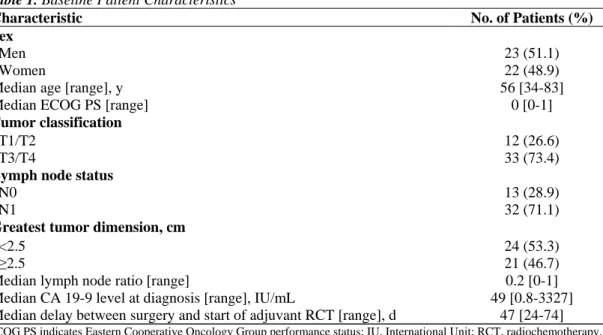
In total, 45 patients who underwent curative (R0) resection for adenocarcinoma of the pancreatic head were studied. These included 23 men and 22 women. The median performance was 0 (range, 0-1), and the a median patient age was 58 years (range, 34-83 years). Characteristics of the study patients are listed in Table 1. Adjuvant chemoradiation regimens were tolerated well and were completed by 43 of 45 patients (95%). World Health Organization grade 3/4 hematologic toxicities were reported in 10 of 45 patients (21%), and grade 3/4 nonhematologic toxicities were reported in 3 of 45 patients (7%). The median follow-up after surgery was 21.9 months (range, 3.3-107.4 months). Overall, the median DFS and OS were 13 months (range, 1-107.4 months) and 21.9 months (range, 3-107.4 months), respectively. At the last follow-up, 30 patients had died of disease recurrence, and 15 patients remained alive.
Table 1. Baseline Patient Characteristics
Characteristic No. of Patients (%)
Sex
Men 23 (51.1)
Women 22 (48.9)
Median age [range], y 56 [34-83]
Median ECOG PS [range] 0 [0-1]
Tumor classification
T1/T2 12 (26.6)
T3/T4 33 (73.4)
Lymph node status
N0 13 (28.9)
N1 32 (71.1)
Greatest tumor dimension, cm
<2.5 24 (53.3)
≥2.5 21 (46.7)
Median lymph node ratio [range] 0.2 [0-1]
Median CA 19-9 level at diagnosis [range], IU/mL 49 [0.8-3327]
Median delay between surgery and start of adjuvant RCT [range], d 47 [24-74]
ECOG PS indicates Eastern Cooperative Oncology Group performance status; IU, International Unit; RCT, radiochemotherapy.
dCK Immunostaining:
Among the 45 resected pancreatic cancer samples, all tumor samples had detectable cytoplasmic labeling for dCK with heterogeneous staining intensity (intensity score, 1+ and/or 2+); staining intensity varied within some tumors and between individual tumors. Nuclear staining was observed in 25 of 45 samples (55.5%) and was restricted exclusively to carcinoma cells that exhibited high cytoplasmic intensity staining (score, 2+). Positive labeling also was observed within normal lymphocytes, acinar cells, and islets, although the most consistent labeling for dCK was noted within lymphocytes. Thus, lymphocytes served as a useful positive internal control for evaluating staining patterns in the neoplastic cells.
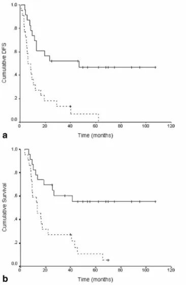
Only cytoplasmic immunostaining was considered for statistical analysis. We dichotomized dCK cytoplasmic staining based on the median staining score. By using this criterion, the cutoff score was 140, and patients were divided in 2 groups: 1) low dCK expression, with a staining score <140, and 2) high dCK expression, with a staining score ≥140 (Fig. 1, top).
Figure 1. These charts illustrate (Top) disease-free survival (DFS) and (Bottom) overall survival according to
deoxycitidine kinase expression.
Correlation Between Patient Outcomes and dCK Expression
Univariate analysis
The results of univariate analysis are summarized in the Table 2. Both OS and DFS were associated significantly with dCK expression. The median OS was 13.2 months (95% confidence interval [CI], 5.7-20.7 months) for patients with low dCK expression and was not reached during follow-up for patients with high dCK expression (hazard ratio [HR], 3.44; 95% CI, 1.60-7.44; P = .0008) (Table 2; Fig. 1, bottom). Similarly, the 3-year survival rate was 27.1% ± 9.5% versus 52.2% ± 10.4% (P = .03) for the low and high dCK expression groups,
respectively. DFS also was longer in patients who had high dCK expression compared with patients who had low dCK expression (HR, 3.61; 95%CI, 1.74-7.75; P = .001; median DFS: low dCK expression, 6.3 months [95%CI, 2.9-9.6 months]; high dCK expression, 46.8 months [95%CI, 38.6-77.5 months]; P = .0003) (Table 2; Fig. 1, bottom).
Table 2. Univariate Analysis of Overall and Disease-Free Survival
OS DFS
Variable OR (95% CI) P OR (95% CI) P
Age, y <56, n=22 1.00 ≥56, n=23 1.23 (0.61-1.47) .31 1.19 (0.69-1.27) .18 Sex Women, n=22 1.00 1.00 Men, n=23 1.21 (0.76-1.43) .45 1.12 (0.84-1.52) .51 ECOG PS 0, n=40 1.00 1.00 1, n=5 1.09 (0.76-1.62) .63 1.15 (0.69-1.74) .72 CA 19-9 at diagnosis, lU/mL <49, n=23 1.00 1.00 ≥49, n=22 1.82 (0.86-3.85) .14 1.57 (0.78-3.18) .23
Greatest tumor dimension, cm
<2.5, n=24 1.00 1.00
≥2.5, n=21 1.88 (0.94-3.73) .07 1.81 (0.87-3.70) .10
Lymph node metastasis
No, n=13 1.00 1.00
Yes, n=32 1.80 (0.73-4.46) .19 1.59 (0.71-3.57) .26
Lymph node ratio
<0.2, n= 22 1.00 1.00
≥0.2, n=23 2.01 (0.94-4.24) .06 1.81 (0.89-3.64) .11
dCK expression
High, n=23 1.00
Low, n=22 3.44 (1.60-7.44) .002 3.61 (1.74-7.51) .001
OS indicates overall survival; DFS, disease-free survival; ECOG PS, Eastern Cooperative Oncology Group performance status; dCK, deoxycytidine kinase.
Table 3. Overall Survival and Disease-Free Survival Multivariate Analysis
Variable OR 95% CI P
DFS
Lymph node ratio
<0.2, n= 22 1.00
≥0.2, n=23 1.46 0.69-3.06 .284
Greatest tumor dimension, cm
<2.5, n=24 1.00 ≥2.5, n=21 1.64 0.82-3.28 .16 dCK expression High, n=23 1.00 Low, n=22 3.61 1.74-7.51 .001 OS
Lymph node ratio
<0.2, n= 22 1.00
≥0.2, n=23 1.53 0.68-3.41 .299
Greatest tumor dimension, cm
<2.5, n=24 1.00
≥2.5, n=21 1.60 0.83-3.73 .206
dCK expression
High, n=23 1.00
Low, n=22 3.45 1.60-7.44 .002
Multivariate analysis
A multivariate model was used to identify independent prognostic factors. The model included all
histopathologic variables that had significant prognostic value in univariate analysis (greatest tumor dimension, lymph node ratio) (see Table 2) and the dCK abundance value. This analysis revealed that dCK abundance was the only independent risk factor for death (OS: HR, 3.61; 95%CI, 1.74-7.51; P = .001) and was the sole independent risk factor for recurrence (DFS: HR, 3.22; 95%CI, 1.56-6.65]; P = .002) (Table3).
DISCUSSION
Although evidence is emerging for the benefit of postoperative gemcitabine in the setting of resected pancreatic adenocarcinoma, such treatment carries some toxicity and is not without financial costs. Means with which to predict which patients are most likely to benefit from such treatment are needed, and determining the mediators of gemcitabine resistance may facilitate further improvements in patient outcomes. Recently, it was
demonstrated that immunohistochemical assessment of the NT protein hENT1 has predictive value of hENT1 immunohisto-chemistry for assessing the benefit from gemcitabine adjuvant chemotherapy in patients with early stage pancreatic cancer.15 However, after intracellular entry mediated by NTs, several enzymes involved in gemcitabine metabolism, such as dCK, cytidine deaminase, RRM1, and RRM2, also may have a key role in altering intracellular disposition of the drug and determining response to gemcitabine. These proteins have had limited evaluation as biomarkers in pancreatic carcinoma. Single, small, retrospective, clinical studies have suggested the poor prognostic value of high levels of RRM1 or RRM2 gene expression and low deoxycytidine kinase protein expression in patients with pancreatic cancer who were treated with gemcitabine.18-20
Our data demonstrate a strong and statistically significant correlation between low levels of dCK protein expression in pancreatic cancer and poor clinical outcomes after gemcitabine-based adjuvant therapy. This finding remained significant on multivariate analyses after adjusting for standard clinicopathologic prognostic factors. It is noteworthy that dCK protein expression was the only significant independent markers of DFS and OS and conferred better prognostic information than lymph node involvement, the lymph node ratio, and the greatest tumor diameter. These results suggest that, for resected pancreatic cancer patients who are treated with gemcitabine, dCK evaluation could provide additional and potentially more powerful information that is not provided readily by standard prognostic factors. Only 1 previous study evaluated the prognostic value of dCK expression in pancreatic adenocarcinoma.20 In a retrospective cohort of 32 patients who received gemcitabine (either as a single agent or in combination) for either metastatic or resected disease, the investigators reported low dCK expression was associated significantly with decreased OS. The results of our study, which was conducted in a homogeneous population of patients who received treatment in the context of prospective trials evaluating the adjuvant gemcitabine regimen, substantiate and enhance the results of that earlier report.20
In our study, dCK immunostaining was predominantly cytoplasmic, although nuclear staining was observed in specimens that exhibited high levels of cytoplasmic dCK protein. This immunostaining pattern is consistent with previous reports.21,23 Furthermore, the reported literature supports a cytoplasmic cellular localization of dCK on the basis of immunochemistry, immunoblotting of cellular fractions, enzymatic activity studies, and kinetic isotope incorporation experiments.21-27
Limitations of our current study include its retrospective nature and the lack of controls who did not receive gemcitabine. Consequently, the predictive value of dCK in gemcitabine-treated pancreatic carcinoma (ie, the ability to identify those patients most likely to benefit from gemcitabine) could not be assessed formally. In conclusion, dCK expression was identified as an independent prognostic factor in patients with resected pancreatic cancer who received gemcitabine-based therapy. In our efforts to guide treatment decision for patients with resected pancreatic cancer, we might define gemcitabine sensitivity and resistance better in an integrated analysis of the expression of dCK, hENT1, and hCNT3 in a larger cohort of similar patients, ideally within the context of a randomized controlled trial. In this manner, it may be possible to more precisely define those subgroups of patients who derive particular benefit from adjuvant gemcitabine and those patients who warrant the investigation of experimental therapies.
CONFLICT OF INTEREST DISCLOSURES
We thank Cheryl Santos for excellent technical assistance.
REFERENCES
1. Oettle H, Post S, Neuhaus P, et al. Adjuvant chemotherapy with gemcitabine vs observation in patients undergoing curative-intent resection of pancreatic cancer: a randomized controlled trial. JAMA. 2007;297:267-277.
2. Neuhaus P, Riess H, Post S, et al. CONKO-001: Final results of the randomized, prospective, multicenter phase III trial of adjuvant chemotherapy with gemcitabine versus observation in patients with resected pancreatic cancer (PC) [abstract]. J Clin Oncol. 2008;26(15S). Abstract 4504.
3. Huang P, Plunkett W. Induction of apoptosis by gemcitabine. Semin Oncol. 1995;15:2403-2413.
4. Ruiz van Haperen VW, Veerman G, Vermorken JB, et al. 2',2'-Difluoro-deoxycitidine (gemcitabine) incorporation into RNA and DNA of tumour cell lines. Biochem Pharmacol. 1993;46:762-766.
5. Plunkett W, Huang P, Searcy CE, et al. Gemcitabine: preclinical pharmacology and mechanisms of action. Semin Oncol. 1996;23:3-15.
6. Lawrence TS, Change EY, Hohn TM, et al. Radiosensitization of pancreatic cancer cells by 2',2'-difluoro-2'-deoxycytidine. Int J Radiat
Oncol Biol Phys. 1996;34:867-872.
7. Milas L, Fujii T, Hunter NR, et al. Enhancement of tumor radioresponse in vivo by gemcitabine, Cancer Res. 1999;59: 107-114.
8. Mason KA, Milas L, Hunter NR, et al. Maximizing therapeutic gain with gemcitabine and fractionated radiation. Int J Radiat Oncol Biol
Phys. 1999;44:1125-1135.
9. Blackstock AW, Bernard SA, Rchards F, et al. Phase I trial of twice-weekly gemcitabine and concurrent radiation in patients with advanced pancreatic cancer. J Clin Oncol. 1999;17:2208-2212.
10. Murphy JD, Adusumilli S, Kent A, et al. Full-dose gemcitabine and concurrent radiotherapy for unresectable pancreatic cancer. Int J
Radiat Oncol Biol Phys. 2007;68:801-808.
11. Van Laethem JL, Demols A, Gay F, et al. Postoperative adjuvant gemcitabine and concurrent radiation after curative resection of pancreatic head carcinoma: a phase II study. Int J Radiat Oncol Biol Phys. 2003;56:974-980.
12. Demols A, Peeters M, Polus M, et al. Adjuvant gemcitabine and concurrent continuous radiation (45 Gy) for resected pancreatic head carcinoma: a multicenter Belgian phase II study. Int J Radiat Oncol Biol Phys. 2005;62:1351-1356.
13. Van Laethem JL, Van Cutsem E, Hammel P, et al. Adjuvant chemotherapy alone versus chemoradiation after curative resection for pancreatic cancer: feasibility results of a randomised EORTC/FFCD/GERCOR phase II/III study (40013/22012/ 0304) [abstract]. J Clin
Oncol. 2008;26(15S). Abstract 4514.
14. Regine WF, Winter KA, Abrams RA, et al. Fluorouracil vs gemcitabine chemotherapy before and after fluorouracilbased chemoradiation following resection of pancreatic adenocarcinoma: a randomized controlled trial. JAMA. 2008; 299:1019-1026.
15. Farrell JJ, Elsaleh H, Garcia M, et al. Human equilibrative nucleoside transporter 1 levels predict response to gemcitabine in patients with pancreatic cancer. Gastroenterology. 2009;136:187-195.
16. Marechal R, Mackey JR, Lai R, et al. Human equilibrative nucleoside transporter 1 and human concentrative nucleoside transporter 3 predict ' after adjuvant gemcitabine therapy in resected pancreatic adenocarcinoma. Clin Cancer Res. 2009;15:2913-2919.
17. Hatzis P, Al-Madhoon AS, Jullig M, Petrakis TG, Eriksson S, Talianidis I. The intracellular localization of deoxycytidine kinase. J Biol
Chem. 1998;273:30239-30243.
18. Nakahira S, Nakamori S, Tsujie M, et al. Involvement of ribonucleotide reductase M1 subunit overexpression in gemcitabine resistance of human pancreatic cancer. Int J Cancer. 2007;120:1355-1363.
19. Itoi T, Sofuni A, Fukushima N, et al. Rbonucleotide reductase subunit M2 mRNA expression in pretreatment biopsies obtained from unresectable pancreatic carcinomas. J Gastroenterol. 2007;42:389-394.
20. Sebastiani V, Rcci F, Rubio-Viquiera B, et al. Immunohistochemical and genetic evaluation of deoxycytidine kinase in pancreatic cancer: relationship to molecular mechanisms of gemcitabine resistance and survival. Clin Cancer Res. 2006; 12:2492-2497.
21. Hubeek I, Peters GJ, Broekhuizen AJ, et al. Immunocytochemical detection of deoxycytidine kinase in haematological malignancies and solid tumours. J Clin Pathol. 2005;58:695-699.
22. Arner ES, Eriksson S. Mammalian deoxyribonucleoside kinases. Pharmacol Ther. 1995;67:155-186.
23. Keszler G, Spasokoukotskaja T, Csapo Z, et al. Activation of deoxycytidine kinase in lymphocytes is calcium dependent and involves a conformational change detectable by native immunostaining. Biochem Pharmacol. 2004;67: 947-955.
24. Spyrou G, Reichard P. Compartmentation of dCTP pools. Evidence from deoxyliponucleotide synthesis. J Biol Chem. 1987;262:16425-16432.
25. Spyrou G, Reichard P. Intracellular compartmentation of deoxycytidine nucleotide pools in S phase mouse 3T3 fibro- blasts. J Biol
Chem. 1989;264:960-964.
26. Spasokukotskaja T, Spyrou G, Staub M. Deoxycytidine is salvaged not only into DNA but also into phospholipid precursors. Biochem
Biophys Res Commun. 1988;155:923-929.
27. Spasokukotskaja T, Taljanidisz J, Sasvari-Szekely M, Staub M. Deoxycytidine is salvaged not only into DNA but also into phospholipid precursors. III. dCOP-diacylglycerol formation in tonsillar lymphocytes. Biochem Biophys Res Commun. 1991;174:680-687.
![Rôles et mécanismes d'action du récepteur AT[indice inférieur 2] de l'angiotensine II d'un modèle cellulaire neuronal au tissu prostatique humain l'importance du contexte](data:image/gif;base64,R0lGODlhAQABAIAAAP///wAAACH5BAEAAAAALAAAAAABAAEAAAICRAEAOw==)