Université de Montréal
Characterization of the membrane transporter OATP1A2
activity towards different classes of drugs
par Jennifer Lu
Faculté de Pharmacie
Thèse présentée à la Faculté des Études Supérieures en vue de l’obtention du grade de doctorat
en Sciences Pharmaceutiques Option pharmacologie
Décembre, 2016
i
Résumé
Les transporteurs membranaires sont des éléments importants dans le devenir, l’efficacité, et la toxicité du médicament. Ils influencent la pharmacocinétique et la pharmacodynamie de ces derniers. Plusieurs interactions médicamenteuses observées cliniquement sont attribuables à la fois aux enzymes responsables du métabolisme des médicaments et aux transporteurs membranaires. Il est connu qu’une variabilité existe entre différents individus dans la réponse à un médicament et les polymorphismes génétiques retrouvés dans les gènes codant pour les transporteurs membranaires peuvent partiellement expliquer cette variabilité.
OATP1A2 est un transporteur membranaire exprimé sur des organes importants, comme le cerveau et le rein. Plusieurs médicaments utilisés en clinique sont des substrats d’OATP1A2 et l’expression localisée de ce transporteur suggère un rôle important dans le devenir du médicament. Donc, mon projet de doctorat consistait à caractériser l’activité d’OATP1A2 en relation avec ses substrats et inhibiteurs, et de plus, à évaluer l’impact de différents variants génétiques d’OATP1A2 sur leur transport.
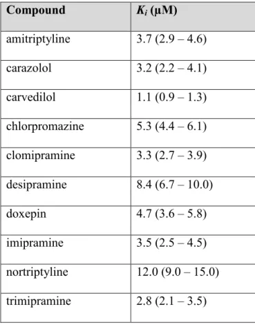
Dans le premier article, la rosuvastatine a été utilisée comme substrat-type pour étudier le transport d’OATP1A2. Les expériences ont été menées en introduisant la rosuvastatine en compétition avec différent β-bloqueurs, une classe de médicaments rapportée dans la littérature comme substrats d’OATP1A2. Parmi les β-bloqueurs évalués, le carvédilol était l’inhibiteur le plus puissant. Dans la deuxième partie de l’étude, des médicaments ayant une structure similaire au carvédilol, tels que les antidépresseurs tricycliques, ont été évalués quant à leur potentiel d’inhibition sur OATP1A2. Une relation structure-activité a été définie à l’aide de ces données. Nous avons démontré que des composés tricycliques avec une courte chaîne aliphatique pouvaient inhiber OATP1A2.
ii
Dans le deuxième article, OATP1A2 a été étudié en considérant son expression et son rôle au sein de la barrière hémato-encéphalique (BHE). Des études précédentes ont démontré qu’OATP1A2 est exprimé sur la membrane luminale des cellules endothéliales formant la BHE. Nos données démontrent que les triptans, une classe de médicaments couramment utilisées pour traiter la crise migraineuse, sont des substrats d’OATP1A2 et que les composés tricycliques identifiés comme inhibiteurs d’OATP1A2 dans nos études précédentes peuvent inhiber le transport des triptans par OATP1A2. Ces résultats sont importants puisque: 1) il a été suggéré que les triptans peuvent agir au niveau du système nerveux central en se liant aux récepteurs trouvés sur les neurones centraux; 2) comme les triptans sont des molécules hydrophiles, un mécanisme de transport facilité est nécessaire pour qu’ils pénètrent la BHE et OATP1A2 pourrait être l’élément clé; 3) l’inhibition d’OATP1A2 par les composés tricycliques pourrait limiter l’accès des triptans à leur site d’action.
Le troisième article caractérise l’activité associée à deux variants génétiques d’OATP1A2 (OATP1A2*2 et *3). Leur capacité à transporter les triptans et leur potentiel d’inhibition par les médicaments tricycliques ont été évalués. Des résultats supplémentaires caractérisant OATP1A2, mais sans liens directs avec les trois articles, seront présentés en annexe.
Dans l’ensemble, les résultats présentés dans cette thèse servent à caractériser le transporteur membranaire OATP1A2 en relation avec ses substrats et inhibiteurs, et en fonction de ses variants génétiques.
Mots-clés : Transporteurs de médicaments, OATP1A2, interactions médicamenteuses,
triptans, barrière hémato-encéphalique, rosuvastatine, antidépresseurs tricycliques, carvédilol, polymorphisme d’un seul nucléotide.
iii
Abstract
Drug transporters are important determinants in drug disposition, efficacy, and toxicity. They influence the pharmacokinetics and pharmacodynamics of drugs. Several clinically-observed drug-drug interactions are mediated through drug metabolizing enzymes and drug transporters. It is well known that there is an interindividual variability in the response to medications and polymorphisms found in genes encoding for drug transporters partially account for it.
OATP1A2 is a membrane drug transporter expressed on important organs, such as the brain and the kidney. A wide spectrum of drugs used in the clinic are substrates of OATP1A2. Its localisation suggests an essential role in drug disposition. Thus, my PhD project consisted of characterizing the activity of OATP1A2 in regards to its substrates, inhibitors, and different protein variants due to genetic polymorphisms.
In the first article, rosuvastatin was used as the probe substrate to study OATP1A2 transport activity. Experiments were conducted by putting rosuvastatin in competition with different β-blockers, a class of drugs known in the literature to be transported by OATP1A2. One of the drugs evaluated, carvedilol, inhibited OATP1A2 with much more potency than the others. In the second part of the study, drugs with a structure similar to carvedilol, such as tricyclic antidepressants, were tested for their potential to inhibit OATP1A2. A structure-activity relationship was defined using the data. It was demonstrated that drugs composed of a tricyclic ring with a short aliphatic amine chain were potent OATP1A2 inhibitors.
In the second article presented, OATP1A2 was studied in the context of its localization at the blood-brain barrier (BBB). OATP1A2 expression at the luminal membrane of the endothelial cells making up the BBB was demonstrated in the literature. Our article showed that triptans, a class of commonly used anti-migraine drugs, were OATP1A2 substrates. The tricyclic drugs previously evaluated were shown to potently inhibit triptan transport through OATP1A2.
iv
These findings are important for three reasons: 1) it has been postulated that triptans may act at the central nervous system by binding to receptors found on central neurons; 2) as triptans are hydrophilic molecules, a facilitated transport mechanism is required for them to penetrate the BBB and OATP1A2 may be the key player; and 3) the inhibition of OATP1A2 by the tricyclic drugs may limit the entrance of triptans to their site of action.
The third article characterized the transport activity of two OATP1A2 protein variants (OATP1A2*2 and *3). Their capacities to transport triptans and their potential of being inhibited by tricyclic drugs were evaluated. Additional data characterizing OATP1A2 but considered out of the scope of the three articles will be presented in appendices.
In overall, the central theme of this thesis looks into the characterization of the OATP1A2 membrane drug transporter in regards to its substrates, inhibitors, and proteins variants.
Keywords: Drug transporters, OATP1A2, drug-drug interactions, triptans, blood-brain barrier,
v
Table of Contents
Résumé ... i
Abstract ... iii
Table of Contents ... v
List of tables ... viii
List of figures ... ix List of abbreviations ... x Acknowledgments... i Preface... iii SECTION 1: INTRODUCTION ... 1 1.1 TRANSPORTERS ... 2 1.1.1 GENERAL INTRODUCTION ... 2 1.1.2 DRUG TRANSPORTERS... 6
1.1.3 ENDOGENOUS ROLES OF DRUG TRANSPORTERS ... 8
1.2 OATP1A2 ... 10
1.2.1 OATP1A2 CHARACTERISTICS ... 10
1.2.2 OATP1A2 AT THE BLOOD-BRAIN BARRIER ... 14
1.3 INTERINDIVIDUAL VARIABILITY IN RESPONSE TO DRUGS ... 19
1.3.1 DRUG-DRUG INTERACTIONS ... 20
1.3.2 SINGLE-NUCLEOTIDE POLYMORPHISMS ... 23
1.4 Rationale, Hypothesis, and Objectives ... 28
SECTION 2: MANUSCRIPTS... 32
SECTION 2.1 ... 33
ARTICLE #1 ... 33
Effects of β-blockers and tricyclic antidepressants on the activity of human organic anion transporting polypeptide 1A2 (OATP1A2) ... 33
2.1.1 OBJECTIVES ... 34
2.1.2 INTRODUCTION ... 34
vi
2.1.4 DISCUSSION ... 79
SECTION 2.2 ... 80
ARTICLE #2 ... 80
Effects of tricyclic compounds on the transport of anti-migraine triptans through human organic anion transporting polypeptide 1A2 (OATP1A2) ... 80
2.2.1 OBJECTIVES ... 81 2.2.2 INTRODUCTION ... 81 2.2.3 ARTICLE ... 82 2.2.4 DISCUSSION ... 126 SECTION 2.3 ... 128 ARTICLE #3 ... 128
Impact of single nucleotide polymorphisms found in human organic anion transporting polypeptide 1A2 (OATP1A2) on triptans transport... 128
2.3.1 OBJECTIVES ... 129 2.3.2 INTRODUCTION ... 129 2.3.3 ARTICLE ... 129 2.3.4 DISCUSSION ... 162 SECTION 3: CONCLUSION ... 163 3. CONCLUSION ... 164 REFERENCES ... 169 APPENDICES ... 185 APPENDIX 1 ... 186
LIST OF PUBLICATIONS AND ABSTRACTS ... 186
PUBLICATIONS ... 187 ABSTRACTS ... 187 APPENDIX 2 ... 191 ADDITIONAL RESULTS ... 191 Objective ... 192 Introduction ... 192
Material and Methods ... 192
viii
List of tables
Table 1: List of the 52 SLC families. ... 3
Table 2: List of ABC families. ... 4
Table 3: List of transporters involved in the disposition of drugs. ... 6
Table 4: List of OATP1A2 substrates and inhibitors. ... 11
Table 5: Expression of drug transporters at the human blood-brain barrier. ... 16
Table 6: Transporter mediated drug-drug interactions observed in clinical studies ... 21
Table 7: List of nonsynonymous SLCO1A2 genetic polymorphisms. ... 27
Table 8: IC50 values from the inhibition of almotriptan, naratriptan, and zolmitriptan uptake through OATP1A2 by various compounds ... 192
ix
List of figures
Figure 1: Different routes of transport across the BBB. ... 15 Figure 2: Inhibition of OATP1A2-mediated transport of almotriptan by different
compounds. ... 194 Figure 3: Inhibition of OATP1A2-mediated transport of naratriptan by different
compounds. ... 196 Figure 4: Inhibition of OATP1A2-mediated transport of zolmitriptan by different
x
List of abbreviations
ABC: ATP-binding cassette
ADME: absorption, distribution, metabolism, and excretion ATP: Adenosine triphosphate
BBB: blood-brain barrier
BCRP: breast cancer resistance protein CLint: intrinsic clearance
CNS: central nervous system CYP450: cytochromes P450 DDI: drug-drug interaction DNA: deoxyribonucleic acid
DPDPE: Deltorphin II, [D-Pen2,5]enkephalin FDA: Food and Drug Administration
HEK293: human embryonic kidney cells 293 HGNC: HUGO Gene Nomenclature Committee
HMG-CoA reductase: 3-hydroxy-3-methyl-glutaryl-coenzyme A reductase HPLC: high pressure liquid chromatography
IC50: half maximal inhibitor constant
ITC: International Transporter Consortium Ki: inhibitory constant
Km: Michaelis constant
LC-MS/MS: liquid chromatography-tandem mass spectrometry logD: log10(coefficient of distribution)
logP: log10(coefficient of partition) MCT: monocarboxylate transporter MDR1: multidrug resistance protein 1 MRI: magnetic resonance imaging mRNA: messenger ribonucleic acid
xi NBD: nucleotide binding domains
OAT: organic anion transporter
OATP1A2: organic anion transporting polypeptide 1A2 OCT: organic cation transporter
PET: positron-emission tomography P-gp: P-glycoprotein
PXR: pregnane X receptor S.D.: standard deviation SLC: solute carrier
SNP: single nucleotide polymorphism
SPECT: single-photon emission computed tomography T3: triiodothyronine
T4: thyroxine
TMD: transmembrane domain UV: ultraviolet
i
To my parents and my fiancé Vincent, Nothing would be possible without them
i
Acknowledgments
I would like to thank my Ph.D. research director, Dr. Jacques Turgeon, for giving me the opportunity to pursue one of my most important goals in life. The idea of pursuing a doctoral degree in the pharmaceutical field stem in my mind since I was 16. Fourteen years later, it’s with the feeling of great accomplishment that I assembled this document together. I would like to thank my co-director, Dr Véronique Michaud, for her remarkable help, especially towards the finalizing steps of my Ph.D. Thank you both for giving me the opportunity to learn and to grow. Thank you both for supporting me. Above all, thank you both for sharing your knowledge with me.
Thank you to the members of the jury for taking their time to evaluate my work. Thank you to the funding agency, Fonds de recherché du Québec Santé (FRQS), and Montreal University for their financial support throughout the numerous years.
A Ph.D. project is never accomplished alone. Many people provided me their help either by being directly involved in the project or through moral support. Fleur Gaudette and François Bélanger were the two key players for help.
A special thank to my brother Henry Leung for his tremendous help in the lab and his good humor. I keep in mind all the pranks we played on people. They were quite immature but hilarious nonetheless. Thank you Ju-Jing Tan for your moral support and for listening to all my stories and my ranting.
Throughout my six years at the lab, many people came and go from the lab. I will keep a memory of each person who was a member of the lab: Jade Huguet, Liliam Gabriela Guilarte Moya, Sophie Gravel, Alexia Grangeon, Sarah Maximos, Valérie Clermont, Roxanne Pelletier, and our numerous summer students.
ii
A huge thanks to the 8th floor crew for bringing such an entertaining student life and with
whom I shared a lot of good times and good laughs. These memories will remain very dear to me.
Thank you to my family and my future family-in-law for their immeasurable support. Thank you to my parents for finally accepting my choice of career.
Keeping the best for last, I can never thank enough my fiancé, Vincent Morin, for his eternal support and for always believing in what I do. Thank you for being part of my life. Now that this chapter is finished, I am looking forward to starting a new chapter with you. Je t’aime mon amour.
Although it was not always a smooth process getting there and getting through it, I am proud of what was accomplished. The knowledge, experience, and hard labor can never be taken away.
iii
Preface
As we are coming to understand that a drug’s pharmacokinetic and pharmacodynamic profile depends not only on drug metabolizing enzymes but also on its interaction with specific drug transporters in the body, we are moving forward into a new era in drug development where smarter and more specific strategies will be developed. Indeed, research in the drug transporter field will help us understand how to specifically target a drug to its active site, ameliorating its pharmacokinetic profile, increasing efficacy, reducing toxicity and adverse events.
Characterizing drug transporters will help in understanding the interindividual variability in the response to drugs. Single nucleotide polymorphisms and drug-drug interactions mediated via transporters may modify the pharmacokinetic profile of drugs; thus, modifying the drug response. More and more examples of variability in drug disposition due to the activity of drug transporters are being demonstrated in the literature. To move forward in understanding drug response variability mediated by drug transporters, it is crucial to investigate the fundamental characteristics of known drug transporters. The work presented in this thesis aimed at characterizing the OATP1A2 drug transporter.
My thesis will first introduce the broad family of transporters, and then it will focus on the OATP1A2 drug transporter. Its potential role at the blood-brain barrier will be presented. The concept of how drug-drug interactions and single-nucleotide polymorphisms found in drug transporters influence the intervindividual variability in the response to drugs will be presented. The second section contains three scientific articles representing the work done during my doctorate. For each article, the study objectives will be listed, followed by a short introduction, then the article will be presented and discussed. Additional data generated following the theme of this thesis but considered out of the scope of the three articles will be presented in appendices.
iv
The first two articles characterize the OATP1A2 transporter in regards to its various substrates and inhibitors. The second article places OATP1A2 in the context of the blood-brain barrier and CNS-active drugs were evaluated. The articles are: 1) Effects of β-blockers and tricyclic antidepressants on the activity of human organic anion transporting polypeptide 1A2 (OATP1A2) and 2) Effects of tricyclic compounds on the transport of anti-migraine triptans through human organic anion transporting polypeptide 1A2 (OATP1A2). The third article investigates two common protein variants of OATP1A2 for their transport activity: Impact of single nucleotide polymorphisms found in human organic anion transporting polypeptide 1A2 (OATP1A2) on the transport of triptans.
Finally, my thesis ends with an overall discussion on the future directions to take in understanding the role of OATP1A2 at the blood-brain barrier and wraps up with a short conclusion.
1
2
1.1 TRANSPORTERS
1.1.1 GENERAL INTRODUCTION
Transporters are transmembrane proteins that span biological membranes and mediate the translocation of substrates across membranes. At the plasma membrane, they control the uptake or efflux of endogenous compounds (sugars, amino acids, nucleotides, and ions) and xenobiotics in and out of the cells. On membranes forming subcellular organelles, they regulate entrance of substrates into organelles. They are classified into two superfamilies: 1) solute carrier (SLC) and 2) ATP-binding cassette (ABC). In humans, the SLC superfamily is composed of 395 members divided into 52 families (SLC1 – SLC52) based on the number of α-helices and sequence homology (Table 1) [1]. SLCs that function by moving solutes down their concentration gradient across membranes using the electrochemical potential difference as the driving force are classified as facilitated transporters. SLCs transporting substrates against their concentration gradient using the ion gradient, such as sodium or proton, generated by ATP-dependent pumps are classified as secondary-active transporters. SLCs using the gradient generated by secondary-active transporters to function are classified as tertiary-active transporters. The ABC superfamily is composed of 49 members divided into seven families (ABCA – ABCG) (Table 2). ABC transporters use the energy of adenosine triphosphate (ATP) hydrolysis to move molecules against their concentration gradient and they are also classified as active transporters.
3
Table 1: List of the 52 SLC families.
The total numbers of members in each family classified in 2004 and in 2013 are shown on the right. The families known to transport drugs are shown in boxes. Table adapted from Hediger et al. (Mol Aspects Med 2013).
The HGNC Solute Carrier Family Series Total 2004 Total 2013
SLC1: The high affinity glutamate and neutral amino acid transporter family 7 7 SLC2: The facilitative GLUT transporter family 14 14 SLC3: The heavy subunits of the heteromeric amino acid transporters 2 2 SLC4: The bicarbonate transporter family 10 10 SLC5: The sodium glucose cotransporter family 8 12 SLC6: The sodium- and chloride-dependent neurotransmitter transporter family 16 21 SLC7: The cationic amino acid transporter/glycoprotein-associated amino-acid transporter family 14 14 SLC8: The Na+/Ca2+ exchanger family 3 3 SLC9: The Na+/ H+ exchanger family 8 13 SLC10: The sodium bile salt cotransport family 6 7 SLC11: The proton coupled metal ion transporter family 2 2 SLC12: The electroneutral cation-Cl cotransporter family 9 9 SLC13: The human Na+-sulfate/carboxylate cotransporter family 5 5 SLC14: The urea transporter family 2 2 SLC15: The proton oligopeptide cotransporter family 4 5 SLC16: The monocarboxylate transporter family 14 14 SLC17: The vesicular glutamate transporter family 8 9 SLC18: The vesicular amine transporter family 3 4 SLC19: The folate/thiamine transporter family 3 3 SLC20: The type III Na+-phosphate cotransporter family 2 2 SLC21/SLCO: The organic anion transporting family 11 12 SLC22: The organic cation/anion/zwitterion transporter family 18 23 SLC23: The Na+-dependent ascorbic acid transporter family 4 4 SLC24: The Na+/(Ca2+–K+) exchanger family 5 6 SLC25: The mitochondrial carrier family 27 53 SLC26: The multifunctional anion exchanger family 10 11 SLC27: The fatty acid transport protein family 6 6 SLC28: The Na+-coupled nucleoside transport family 3 3 SLC29: The facilitative nucleoside transporter family 4 4 SLC30: The zinc efflux family 9 10 SLC31: The copper transporter family 2 2 SLC32: The vesicular inhibitory amino acid transporter family 1 1 SLC33: The Acetyl-CoA transporter family 1 1 SLC34: The type II Na+-phosphate cotransporter family 3 3 SLC35: The nucleoside-sugar transporter family 17 30
4
SLC36: The proton-coupled amino acid transporter family 4 4 SLC37: The sugar-phosphate/phosphate exchanger family 4 4 SLC38: The System A & N, sodium-coupled neutral amino acid transporter family 6 11 SLC39: The metal ion transporter family 14 14 SLC40: The basolateral iron transporter family 1 1 SLC41: The MgtE-like magnesium transporter family 3 3 SLC42: The Rh ammonium transporter family (pending) 3 3 SLC43: Na+-independent, system-L like amino acid transporter family 2 3 SLC44: Choline-like transporter family 5 SLC45: Putative sugar transporter family 4 SLC46: Folate transporter family 3 SLC47: Multidrug and Toxin Extrusion (MATE) family 2 SLC48: Heme transporter family 1 SLC49: FLVCR-related transporter family 4 SLC50: Sugar efflux transporters 1 SLC51: Transporters of steroid-derived molecules 2 SLC52: Riboflavin transporter family 3
Total 298 395
Table 2: List of ABC families.
The total numbers of members in each family are shown on the right. The families known to transport drugs are shown in boxes. Table adapted from Vasiliou et al. (Hum. Genomics 2009).
Subfamily name Aliases Number of genes
ABCA ABC1 12 ABCB MDR 11 ABCC MRP 13 ABCD ALD 4 ABCE OABP 1 ABCF GGN20 3 ABCG White 5 Total 49
5
As the transporter field is continuously growing, the gene nomenclature system was developed and approved by the HUGO Gene Nomenclature Committee to facilitate gene classification [2]. The SLCs genes are named using the root symbol SLC, followed by a numeral to indicate the family (e.g., SLC1, solute carrier family 1), followed by a letter to indicate the subfamily (e.g. SLC1A), and ending with a number to indicate the individual transporter gene (e.g. SLC1A1). SLC transporters are classified in the same family based on their biochemical function and when their amino acid sequence has at least 20% identity [3]. The SLC21 family is an exception to this classification method because this research field has evolved rapidly. Hagenbuch and Meier have developed an updated evolutionary-based nomenclature system in order to accommodate a species-independent classification system with less ambiguity [4]. SLC21 is replaced by the root symbol SLCO (Slco for other animal species) followed by a number, a letter (capital for human genes and small letter for other animal species), and another number to designate the family, subfamily, and individual transporter gene, respectively (e.g. SLCO1A2 for humans and Slco1a2 for animals). Members with more than 40% amino acid sequence identity are grouped into the same family and those with more than 60% amino acid sequence identity are grouped into the same subfamily.
Whereas for ABC genes, the nomenclature is based on divergent evolution from a common ancestor and sequence similarity [5]. Family members share 30-50% sequence homologies [6]. They are named by the root symbol ABC, followed by a letter (A to G) to designate the family, and a number to designate individual members [2]. In general, the ABC transporter consists of two nucleotide binding domains (NBDs), also known as ATP binding cassettes, and two transmembrane domains (TMDs) [5]. In order to power translocation of substrates against their gradient, the two NBDs have to work together to bind and hydrolyse ATP molecules. Several highly conserved motifs, crucial for the ATPase activity, are found in the NBD: Walker A and Walker B sequences, the ABC signature motif, the H loop and the Q loop. The TMD, made up of several hydrophobic α-helices, is responsible for substrate recognition and translocation. Some ABC proteins (e.g. BCRP) are considered as “half-transporters” as their gene encode a single NBD and a single TMD. To gain functionality, the protein subunits need to homodimerize, heterodimerize or oligomerize.
6
1.1.2 DRUG TRANSPORTERS
Drug transporters are important determinants in drug absorption, distribution, organ-specific targeting, metabolism, excretion, efficacy, and toxicity. It is evident that drug transporters influence the pharmacokinetics and pharmacodynamics of a drug. Among the growing numbers of transporters, only a few members, organized in 13 families (Table 3), have been identified as essential in drug disposition [7].
Table 3: List of transporters involved in the disposition of drugs.
Table adapted from You & Morris (2014).
Gene family Gene name Protein name
SLC22 SLC22A1 OCT1 SLC22A2 OCT2 SLC22A3 OCT3 SLC22A4 OCTN1 SLC22A5 OCTN2 SLC22A21 OCTN3 SLC22A16 CT2 SLC22A6 OAT1 SLC22A7 OAT2 SLC22A8 OAT3 SLC22A11 OAT4 SLC22A10 OAT5 SLC22A20 OAT6 SLC22A12 URAT1
SLC21/SLCO SLCO1A2 OATP1A2
SLCO1B1 OATP1B1 SLCO1B3 OATP1B3 SLCO1C1 OATP1C1 SLCO2A1 OATP2A1 SLCO2B1 OATP2B1 SLCO3A1 OATP3A1 SLCO4A1 OATP4A1 SLCO4C1 OATP4C1 SLCO5A1 OATP5A1 SLCO6A1 OATP6A1 SLC15 SLC15A1 PEPT1 SLC15A2 PEPT2 SLC15A4 PHT1
7 SLC15A3 PHT2 SLC16 SLC16A1 MCT1 SLC16A7 MCT2 SLC16A8 MCT3 SLC16A3 MCT4 SLC5 SLC5A8 SMCT1 SLC5A12 SMCT2 SLC28 SLC28A1 CNT1 SLC28A2 CNT2 SLC28A3 CNT3 SLC29 SLC29A1 ENT1 SLC29A2 ENT2 SLC29A3 ENT3 SLC29A4 ENT4 SLC47 SLC47A1 MATE1 SLC47A2 MATE2 SLC51 SLC51A OST-α SLC51B OST-β SLC10 SLC10A1 NTCP SLC10A2 ASBT ABCB ABCB1 MDR1/P-gp ABCB11 BSEP ABCC ABCC1 MRP1 ABCC2 MRP2 ABCC3 MRP3 ABCC4 MRP4 ABCC5 MRP5 ABCC6 MRP6 ABCC10 MRP7 ABCC11 MRP8 ABCC12 MRP9
ABCG ABCG2 BCRP/ABCG2
While drug transporters are found on every tissue of the human body, they are particularly highly expressed on the epithelia of tissues functioning as barriers for drug entry such as the intestine, liver, kidney, blood-brain barrier (BBB), and placenta. This expression pattern corresponds well to their function as cells gatekeepers. Epithelial cells are typically polarized into apical and basolateral membranes. For example, the brain capillary endothelial cells that make up the BBB are polarized into apical/luminal and basolateral membranes which correspond to the side facing the blood and the central-nervous system (CNS), respectively. The expression pattern of drug transporters usually differs on the two membranes. The polarization is essential for directing substrates towards the same direction. In addition, their
8
presence on plasma membranes of epithelial cells can cause variability in drug concentrations between the plasma and the target organ.
Drug transporters represent one of the rate-limiting steps in drug disposition. In drug development, studies have predominantly focused on transporters expressed on the intestine, liver, kidney, and BBB as they are the most common sites of drug-drug interactions [8].
Irregularity in transporter expression and activity may lead to inter-individual variability in the response to drugs. Drug-drug interactions (DDIs) and genetic polymorphisms on drug transporters can contribute to this variability. Due to the emerging role that transporters play in DDIs, experts from academia, industry, and the US Food and Drug Administration (FDA) were united to form the International Transporter Consortium (ITC) in 2007. Their mission is to determine which transporter is clinically important in DDIs, establish standardized protocols for the in vitro and in vivo study of DDIs, and establish a consensus on current knowledge of clinically relevant drug transporters [9]. They generate decision trees that guide industry into when to perform clinical studies for DDI with new molecular entities. Up to now, the ITC suggested that substrates of multidrug resistance protein 1 (MDR1; also known as P-glycoprotein (P-gp) or ABCB1), organic anion-transporting polypeptide 1B1 (OATP1B1), organic anion-transporting polypeptide 1B3 (OATP1B3), organic anion transporter 1 (OAT1), organic anion transporter 3 (OAT3), organic cation transporter 2 (OCT2), breast cancer resistance protein (BCRP; also known as ABCG2) should be evaluated further. These recommendations are based on the observation that clinically significant interactions were observed with drugs that are substrates of these transporters. This list will most likely expand as more studies are being done in the field of transporter related DDI.
1.1.3 ENDOGENOUS ROLES OF DRUG TRANSPORTERS
Transporters perform many physiological roles as they have endogenous substrates, such as metabolites, nutrients, antioxidants, gastrointestinal microbiome products, bile salts, neuro-active molecules, hormones and signalling molecules [10]. Some transporters are key players
9
in certain metabolic processes, such that sequence variants in the genes encoding these transporters can lead to diseases. For example, ABCC2 (MRP2) located on the hepatocyte canalicular membrane is the main efflux transporter for the elimination of bilirubin conjugates into the bile [7]. Mutations that abolish the cell surface expression or activity of ABCC2 lead to Dubin-Johnson syndrome which is characterized by a dark liver and an increase in conjugated bilirubin in the blood [11, 12]. OATP1B1 and OATP1B3 transporters, located on the sinusoidal membrane of hepatocytes, are responsible for the uptake of conjugated bilirubin from the blood. Mutations causing complete loss of both transporters result in Rotor syndrome, a rare benign autosomal recessive disease, characterized by conjugated hyperbilirubinaemia and jaundice [13]. In contrast to patients with Dubin-Johnson syndrome, patients with Rotor syndrome do not have a pigmented liver because the conjugated bilirubin cannot re-enter the hepatocyte through OATP1B1 nor OATP1B3 [14].
Drug transporters are also implicated in other diseases and their expression is altered in various diseased states. For example, several members of the OATPs are up- or downregulated in different cancers and it has been suggested that their expression state might affect cancer development [15]. SLCO1A2 mRNA expression is upregulated 8 times in breast cancerous tissue compared to adjacent normal tissue [16]. OATP1A2 protein expression has been confirmed in breast carcinoma cells from patients but not in nonneoplastic epithelial cells, stroma, or adipocytes adjacent to the tumour [17]. It has been suggested that OATP1A2 on breast cancer cells contribute to the hormone-induced progression of breast cancer since steroid hormones are substrates of this transporter. Some ABC transporters are also overexpressed in tumors, and they may contribute to resistance to chemotherapy. In fact, repeated administration of anti-cancer drugs is associated with chemotherapy resistance and treatment failure [18]. Once drug resistance has developed, the tumor is typically cross-resistant to multiple drugs even if their structures are unrelated. Overexpression of ABC transporters, such as P-gp/MDR1, BCRP/ABCG2, and members of the MRP, contributing to pumping drugs out of the cancerous cells is the main reason for multi-drug resistance [19].
10
1.2 OATP1A2
1.2.1 OATP1A2 CHARACTERISTICS
OATP1A2 (previously known as OATP-A, OATP1, and OATP) is a member of the SLC21/SCLO family. It is made up of 12 transmembrane domains with a large fifth extracellular loop and both the C- and termini oriented towards the cytoplasmic space. N-glycosylation sites, important for targeting the protein to the plasma membrane, are found in extracellular loops 2 and 5 [20]. OATP1A2 exists in unglycosylated and several glycosylated forms and molecular weights between 60 and 150 kDa have been found [21-23]. The OATP proteins are distinguished by their signature amino acid sequence (D-X-RW-(I,V)-GAWW-X-G-(F,L)-L) which is found between extracellular loop 3 and the transmembrane domain 6 for OATP1A2 [24]. Transport through OATP1A2 is considered to be bidirectional as it moves substrates down its gradient across the membrane regardless of the orientation. It has been suggested that the OATPs function like a rocker-switch type of mechanism to translocate substrates through a central pore [25]. The mechanism of transport is recognized to be sodium-independent but the driving force is still unknown.
OATP1A2 mRNA expression is nearly ubiquitous. It has been detected at various expression levels in the human lung, brain, intracranial artery, optic nerve, retina, spinal cord, prostate, testis, lymph node, pituitary gland, duodenum, esophagus, kidney, liver, spleen, tonsil, [26-28]. However, mRNA is not necessarily representative of protein expression. OATP1A2 protein expression, confirmed by either immunofluorescence or Western blot, is found on the luminal membrane of the brain capillary endothelial cells which make up the blood-brain barrier, apical membranes of cholangiocytes in the liver, apical membrane of the distal nephrons in the kidney, the apical membrane of enterocytes in the duodenum, apical cell layers of the urothelium, placenta, red blood cells, brain neurons, and retina [2, 21-23, 27, 29-31].
11
A lot of attention has been drawn to OATP1A2 due to its postulated location on the intestine and its possible role in mediating drug absorption. Many food-drug interactions were attributed to the inhibition of intestinal OATP1A2 by flavonoids found in fruits and vegetables. It has been demonstrated that fexofenadine bioavailability is decreased when co-administered with fruit juices (grapefruit, apple, orange) in healthy volunteers and in vitro data supports the role of OATP1A2 in mediating this interaction [29, 32, 33]. Misaka et al. demonstrated that green tea reduced plasma concentrations of the β-blocker nadolol and their findings are also supported by in vitro results [34, 35]. However, all this excitement has declined lately because recent studies could not detect OATP1A2 in the intestine by liquid chromatography-tandem mass spectrometry (LC-MS/MS) nor the mRNA transcript [36, 37]. Thus, OATP1A2 expression in the small intestine remains controversial. The previously observed food-drug interaction may be attributed to the inhibition of another intestinal transporter, such as OATP2B1. Indeed, grapefruit juice, orange juice, and their constituents can also inhibit OATP2B1 [38].
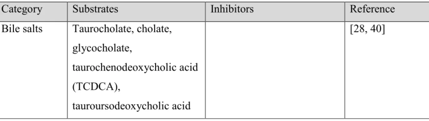
OATP1A2 transports a wide spectrum of substrates including endogenous molecules, xenobiotics, and clinically relevant drugs. A list of currently known OATP1A2 substrates and inhibitors classified into categories is presented in Table 4.
Table 4: List of OATP1A2 substrates and inhibitors.
Adapted from Franke et al. (Pharmacogenomics 2009) [39].
Category Substrates Inhibitors Reference
Bile salts Taurocholate, cholate, glycocholate,
taurochenodeoxycholic acid (TCDCA),
tauroursodeoxycholic acid
12 (TUDCA)
Hormones Estrone-3-sulfate (E3S), estradiol 17β-glucuronide (E217βG), dehydroepiandrosterone (DHEAS), triiodothyronine (T3), thyroxine (T4), Steroid hormones [23, 40, 41]
Peptides Deltorphin II, [D
-Pen2,5]enkephalin (DPDPE),
BQ-123, substance P, vasoactive intestinal peptide
[21, 30, 40]
Organic anions
Bromosulfophthalein (BSP), sodium fluorescein, all-trans-retinol [2, 28, 40, 42] Organic cations methyl-quinine, N-methyl-quinidine, APD-ajmalinium, [43]
Drugs Imatinib, quinine,
fexofenadine, methotrexate, atorvastatin, pitavastatin, pravastatin, rosuvastatin, rocuronium, celiprolol, acebutolol, atenolol, nadolol, sotalol, labetalol, EDDP, docetaxel, mirabegron, glibenclamide, triptans (almotriptan, eletriptan, frovatriptan, rizatriptan, sumatriptan, zolmitriptan), aliskiren, Chloroquine, hydroxychloroquine, multikinase inhibitors (lapatinib, bosutinib,
cediranib, afatinib, erlotinib, foretanib, gefitinib,
nilotinib, pelitinib, sunitinib, vandetanib), rifampicin, clarithromycin, everolimus, sirolimus, tacrolimus, cyclosporine [26, 27, 31, 41, 43-63]
13 tebipenem pivoxil,
levofloxacin, trospium chloride, doxorubicin
Toxins Ouabain, microcystin [40, 64]
Eicosanoids Prostaglandin E2 (PGE2) [40]
Flavonoids Naringin, hesperidin,
apigenin, kaempferol, quercetin, epicatechin gallate, epigallocatechin gallate
[29, 34, 35, 65]
OATP1A2 activity is modulated by different proteins. The chaperon proteins PDZK1 and NHERF1 enhance OATP1A2 stability at the plasma membrane and decrease the transporter internalization [66]. Post-translational modifications of OATP1A2 regulate its activity. Phosphorylation by protein kinase C or casein kinase 2 increases OATP1A2 internalization through clathrin mediated endocytosis [67, 68]. Five putative N-glycosylation sites were identified in OATP1A2 and such modification targets the transporter to the plasma membrane [23]. The transporter is also regulated at the transcriptional level. It has been shown that the vitamin D receptor and the xenobiotic sensor pregnane X receptor (PXR) upregulate the transcription of SLCO1A2 gene [16, 69].
Several animal transporters share a certain degree of homology with the human OATP1A2. Bovine Oatp1a2 share 83% homology with its human counterpart but the bovine protein differs with its 11 predicted transmembrane domains and multiple binding sites [70-72]. Five rat proteins (Oatp1a1, Oatp1a3, Oatp1a4, Oatp1a5, and Oatp1a6) share between 66-72% homology with OATP1A2. The pattern of expression differs among the five members. Four mouse proteins (Oatp1a1, Oatp1a4, Oatp1a5 and Oatp1a6) share between 66-73% homology with OATP1A2 and their pattern of expression is also variable. Multiple members are found in the rodent due to gene duplication. Pig Oatp1a2 (84% homology) contains 12 transmembrane
14
domains and its mRNA has been found in the liver, brain, and intestine [73, 74]. Dog Oatp1a2 share 87% homology with OATP1A2 and its mRNA has been found in the liver and kidney [73]. Rodents are not considered good animal models for the study of the OATP1A2 transporter due to the multiplicity in protein members and their low sequence homology. Further studies are needed to determine if other animals are more representative.
1.2.2 OATP1A2 AT THE BLOOD-BRAIN BARRIER
The brain is separated from the circulating blood by the BBB and from the cerebrospinal fluid by the choroid plexus. This delimitation is important to protect the brain from potentially harmful agents, regulate ions level, confine central neurotransmitters, prevent leakage of plasma proteins, and tightly control nutrients and metabolites essential for the brain [75]. The BBB is formed by the endothelial cells lining the brain microvessels. The surface area formed by the BBB represents a vast interface for exchange and is in average between 12 and 18 m2 in
adults [76]. The structure of the endothelial cells is maintained by astrocytes feet and pericytes. The particularity with these endothelial cells is the organization of the proteins in the tight junctional complexes found between the cells that maintains the BBB “tightness” and integrity. The tight junctions prevent paracellular diffusion of ions and macromolecules between endothelial cells. The effectiveness of the tight junctions results in a high transendothelial electrical resistance of the BBB (1500 to 2000 Ω · cm2) [77]. Under
physiological conditions, the BBB is almost impermeable to endogenous and exogenous substances.
The BBB tightly controls the access of substances to the brain in order to maintain a stable environment for the CNS. Specific receptors, ions channels, and transporters are expressed to regulate the entrance of nutrients. Several mechanisms of transport exist for molecules to cross the BBB: cell migration, passive diffusion, carrier-mediated efflux, carrier-mediated influx, receptor-mediated transcytosis, and adsorptive-mediated transcytosis (Figure 1). Lipid-soluble molecules and certain gases (oxygen and carbon dioxide) can diffuse passively through the
15
BBB. Substrates attempting to cross the BBB can be pumped out of the endothelial cells by the ABC transporters located on the luminal membrane. In contrast, SLC transporters may move substrates from the circulating blood into the CNS. Many vital molecules, such as glucose, amino acids and nucleosides, are directed to the brain through the SLC transporters. Several macromolecules, such as transferrin and insulin, bind to cell surface receptors and are taken up in the cells by endocytosis [78, 79]. Positively charge proteins, such as albumin and the SynB5 peptide, can also be transported by vesicles but in a non-specific and non-receptor mediated manner. The cationic macromolecules adsorb to the endothelial cell surface and induce endocytosis. The endocytosed macromolecules then move through the cell before being release at the abluminal membrane [80, 81]. Mononuclear leukocytes, monocytes and macrophages are able to cross the BBB by a process of diapedesis directly through the endothelial cells [82].
Figure 1: Different routes of transport across the BBB.
16
Several CNS diseases, such as multiple sclerosis and Alzheimer’s disease, may involve the disruption of the BBB [83, 84]. The tight junctions between endothelial cells are affected and the expression of transporters and enzymes may be modified. Consequently, the entry of immune cells, endogenous molecules, and xenobiotics is facilitated.
Targeting the CNS in drug therapy is challenging due to the limited permeation of the BBB and the blood-cerebrospinal fluid barrier. The level of difficulty is increased when the drug target is within cellular compartments of the brain parenchyma (i.e., astrocytes, microglia, oligodendrocytes, and neurons). Accessibility of pharmaceuticals to the brain is therefore highly dependent on influx and efflux transporters.
Drug transporters found on the luminal membrane of the BBB are potential entry sites for drugs or toxins. Those found on both the luminal and abluminal membranes may allow a direct flow of their substances from the blood to the CNS. Drug transporters detected at the human BBB are listed in Table 5.
Table 5: Expression of drug transporters at the human blood-brain barrier.
Adapted from Stieger et al. (Clin Pharmacokinet 2015) [85].
Transporter Gene Protein expression confirmed
OATP1A2 SLCO1A2 [21, 23, 30, 86] OATP1C1 SLCO1C1 [87] OATP2B1 SLCO2B1 [30] OCT1 SLC22A1 [88] OCT2 SLC22A2 [88] OCT3 SLC22A3 [89] OCTN2 SLC22A5 [90] ENT1 SLC29A1 [91]
17 MATE1 SLC47A1 [89] MCT1 SLC16A1 [92] BCRP/ABCG2 ABCG2 [91] MDR1/P-gp MDR1 [91] MRP1 ABCC1 [93] MRP4 ABCC4 [91, 93] MRP5 ABCC5 [93]
Toxins may also gain access to the brain through drug transporters. For example, 126 patients undergoing haemodialysis in Brazil developed symptoms of acute neurotoxicity and subacute hepatotoxicity following the use of water from a lake with cyanobacteria overgrowth [94]. Among these patients, 60 subsequently died. Microcystins, a class of toxins produced by cyanobacteria, were detected in the patients’ serum, dialysis filters, and water-treatment column. It was later demonstrated that microcystins are transported in oocytes by OATP1B1, OATP1B3, and OATP1A2 [64]. OATP1B1 and OATP1B3, found on the sinusoidal membrane of hepatocytes, may be responsible for the hepatotoxicity; whereas, OATP1A2 may play a role in neurotoxicity. Microcystins caused damage to oocytes but only in the presence of OATP1A2. In addition, neurons in the brain express OATP1A2 [30]. As a result, microcystins are able to exert their toxicity in neurons after crossing the BBB.
In terms of normal physiology, drug transporters are also implicated in the passage of endogenous compounds and removal of neurotoxins to and from the brain, respectively. Thyroid hormones play a pivotal role in the development and differentiation of the brain as well as the maintenance and metabolic regulation of the adult CNS [95]. Triiodothyronine (T3)
and thyroxine (T4) are substrates of OATP1C1 and OATP1A2 and they might access the brain
through one of these routes [40]. Monocarboxylate transporter 1 (MCT1) may be involved in bringing lactate and ketone bodies (β-hydroxybutyrate and acetoacetate) to the brain to use as energy substrates when the level of glucose is reduced, such as conditions of prolonged starvation, diabetes, or under hypoglycaemia [96].
18
The numerous ABC transporters found at the luminal side of the BBB play a neuro-protective role. This is exemplified in a phase I clinical trial where PSC 833, a second-generation MDR1 inhibitor, was given concomitantly with etoposide, an anticancer drug. Severe ataxia was observed in patients who received the highest doses of PSC 833 [97]. This adverse event was attributed to a higher permeation of etoposide to the brain caused by the inhibition of MDR1 efflux by PSC 833. While ABC transporters protect the brain from potential neurotoxins, they also represent a considerable challenge for the delivery of CNS-active drugs to their target site. Consequently, the development of drugs treating brain diseases such as brain tumors and bacterial or viral infections has been severely hindered.
On the other hand, SLC transporters at the BBB may have an opposite role. OATP1A2 expression on the luminal membrane of the endothelial cells suggests an important function of this transporter on brain penetration of drugs. Deltorphin II and [D-penicillamine2,5]enkephalin
(DPDPE) are δ-opioid receptor agonists that have previously been considered as potential central analgesics in humans [98]. To cause analgesia, they need to reach the δ-opioid receptor located within the CNS. Since they are peptides, they don’t easily penetrate the BBB. Nonetheless, they have been found to enter the animal brain in a saturable manner, suggesting the involvement of a transporter [99-101]. They have been found to be OATP1A2 substrates and it has been suggested that deltorphin II and DPDPE can cross the human BBB using OATP1A2 [21].
Triptans (e.g. almotriptan, eletriptan, frovatriptan, naratriptan, rizatriptan, sumatriptan, and zolmitriptan) are commonly used anti-migraine medications that are believe to act at the CNS. All triptans, with the exception of eletriptan, are hydrophilic; thus, limiting their penetration to the brain by passive diffusion. Studies have found that zolmitriptan is not only capable of crossing the human BBB but it can also bind to its target receptor at the CNS [102, 103]. OATP1A2 has been suggested as a key player in triptans distribution to the brain as they are substrates [55].
19
In conclusion, the expression of different drug transporters at the brain suggests a highly coordinated and regulated environment for the permeation of xenobiotics to the brain. It is becoming more and more recognized that drug transporters at the BBB play a crucial role in the pharmacokinetic and pharmacodynamic profiles of a drug. Therefore, it is important to understand the molecular properties of drug transporters expressed at the BBB in the development of CNS-active drugs in order to optimize their concentrations and effects at the target site.
1.3 INTERINDIVIDUAL VARIABILITY IN RESPONSE
TO DRUGS
A major setback in treating patients suitably in the clinic is the impact of interindividual variability in the response to medications. This heterogeneity in drug response influences the efficacy, safety and toxicity of the medication. The origin of the variability is multifactorial and comprises of genetic, environmental (dietary constituents), physiological (age and gender), epigenetic, and pathological factors.
Adverse reactions to drugs is an important public health concern. Conclusions from a meta-analysis revealed that the incidence of suffering from an adverse reaction (including non-serious, non-serious, and fatal reactions) related to a medication is around 15.1% in hospitalized patients and that adverse drug reactions is the 4th cause of mortality in the United States [104,
105]. In addition, an estimate of 6.5% of hospital admission is related to adverse drug reactions [106]. At the other end of the spectrum, there are patients who do not benefit from a medication due to a lack of efficacy. In 1969, the FDA initiated a post-marketing surveillance system on drug safety. They revealed that the most common undesirable side effect encountered until 2002 is the lack of drug efficacy [107].
20
For those reasons, it is crucial to understand the underlying influences on the response to drugs in order to improve their safety and efficacy profile. The concept of personalized medicine is born from these efforts. Personalized medicine is an emerging approach for treating diseases that takes into accounts the patient’s genetic background, environmental influences, and lifestyle. More broadly, it can be defined as the tailoring of medical treatment to the individual characteristics, needs and preference during all stages of care, including prevention, diagnosis, treatment and follow up [108].
The next two sections introduce how drug transporters are involved in the interindividual variability in the response to drugs.
1.3.1 DRUG-DRUG INTERACTIONS
Drug-drug interactions (DDIs) can result when one drug alters the pharmacokinetic of another drug or its metabolites. The additive pharmacodynamic effect of multiple drugs can also lead to a DDI. The underlying mechanisms of a DDI affecting the pharmacokinetic of a drug are caused by either inhibition or induction of drug metabolizing enzymes [e.g. cytochromes P450 (CYP450)] or drug transporters. Dietary supplements and some foods may alter drug metabolism and/or transport. The result of those interactions is a sudden alteration in metabolism or transport in individuals who otherwise would have tolerated a particular dose of that drug. The known safety and efficacy of that drug is changed.
The desirable and undesirable effects of a drug are related to its concentration at various sites of action, which is usually related to the blood or tissue concentration of the drug. The blood and tissue concentrations are determined by the drug’s absorption, distribution, metabolism, and excretion (ADME). DDIs related to metabolism are well-recognized but effects related to transporters are being documented with increasing frequency and are, therefore, important to consider in drug development. Transporters can affect the safety and efficacy profile of a drug by affecting the concentration of a drug or its metabolites in various tissues.
21
Several DDIs of clinical significance with mechanisms attributed to the inhibition or induction of transporters have been reported in the literature over the years. A few examples are listed in
Table 6 [7, 109].
Table 6: Transporter mediated drug-drug interactions observed in clinical studies
Transporter Perpetrator drug / compound
Victim drug Pharmacokinetic effect of victim drug Reference OATP1A2/ OATP2B1 (intestinal) Grapefruit juice (6’,7’-Dihydroxyberg amottin, bergamottin)
Fexofenadine p.o. ↓AUC, Cmax [29, 32, 33, 38] Orange (hesperidin, tangeritin, nobiletin) and apple juice (quercetin, kaempferol)
Aliskiren p.o. ↓AUC, Cmax [110]
OATP1B1 (hepatic)
Gemfibrozil Pravastatin p.o. ↑AUC, Cmax; ↓CLr [111] Cyclosporine Pravastatin p.o. ↑AUC, Cmax [112, 113] Cyclosporine Rosuvastatin p.o. ↑AUC, Cmax [114] OAT1/
OAT3 (renal)
Probenecid Furosemide p.o. ↑AUC, Cmax, T½, Tmax; ↓CLt, CLr, CLnr
iv. ↑AUC, T½; ↓CLt, CLr
[115-117]
Probenecid Methotrexate iv. ↑Cserum, T½ [118] OCT2 Cimetidine Metformin p.o. ↑AUC, Cmax; ↓CLr [119]
22
(renal) Cimetidine Ranitidine p.o. ↑AUC, T½; ↓CLr [120] Cimetidine Dofetilide p.o. ↑AUC, Cmax, T½;
↓CLt, CLr, CLnr
[121]
Cimetidine Pindolol p.o. ↑AUC, Cmax; ↓CLr [122] Trimethoprim Procainamide p.o. ↑AUC; ↓CLt, CLr [123]
ABCB1 (P-gp) (intestinal) St-John’s wort (hypericin, pseudohyperici n) [inducer]
Digoxin p.o. ↓ AUC, Cmax, Ctrough [124]
Quinidine Digoxin iv. ↑ T½, Cserum; ↓CLt, CLr, CLnr
[125, 126] Clarithromycin Digoxin p.o. ↑AUC, Cmax, T½; ↓CLrng [127] Ritonavir Digoxin iv. ↑AUC, Vd, T½;
↓CLt, CLr, CLnr [128] ABCG2 (BCRP) (intestinal) Omeprazole/ pantoprazole
Methotrexate p.o. ↑AUC, Cmax; ↓CLr [129, 130] Elacridar Topotecan p.o. ↑AUC, Cmax, F [131] ↑: Increased; ↓: Decreased; AUC: area under the plasma/serum concentration time curve; CLnr: Nonrenal clearance; CLr: Renal clearance; CLrng: Renal nonglomerular clearance; CLt: Total clearance; Cmax: Maximal plasma/serum concentration; Cserum: Serum concentration; Ctrough: Minimum plasma/serum concentration during steady state; F: Bioavailability; iv.: Intravenous administration of drug; p.o.: Oral administration of drug; T½: Plasma/serum concentration half-life; Vd: Volume of distribution.
Although an interaction is observable when fexofenadine is administered concomitantly with fruit juices, it is not likely to be mediated by OATP1A2 as previously considered. As mentioned earlier, the interaction may be mediated by OATP2B1 instead. OATP2B1 is found in the intestine and is also capable of transporting fexofenadine [36, 132].
23
DDIs are not always as straightforward as the examples listed above. Sometimes, multiple mechanisms are involved, such that multiple enzymes and/or transporters are implicated. Other times, when a cocktail of medication is given, more than one drug may inhibit the same enzyme or transporter. As the transporter field is a relatively new but rapidly evolving field, other less characterized transporters may also be involved in mediating DDIs.
Transporter-mediated DDIs have the potential to seriously influence drug efficacy and toxicity. Understanding DDIs will help us better prescribe medications and make a better use of our resources available. For instance, it can help in adjusting the dosage, determine if additional therapeutic monitoring if required, and establishing contraindication to concomitant use. Extensive research has led to contraindicating the consumption of grapefruit juice and orange juice with certain medications as described on the drug’s label (e.g. fexofenadine). Dosage adjustments were done following the description of a clinically significant DDI between the two HIV protease inhibitors, ritonavir and saquinavir. Coadministration of ritonavir with saquinavir increased saquinavir bioavailability dramatically without affecting the pharmacokinetics of ritonavir [133]. As a result, ritonavir is now used as a booster to increase the bioavailability of other protease inhibitors instead of being used alone.
1.3.2 SINGLE-NUCLEOTIDE POLYMORPHISMS
Another factor leading to interindividual differences in the response to drugs is found in the genes encoding drug transporters. These genes are polymorphic; as a result, their protein expression level and transport efficiency is variable.
A single-nucleotide polymorphism (SNP) is a genetic variation where a single nucleotide is altered. SNPs are the most common type of genetic variations [134]. For a variation to be defined as a polymorphism, it has to occur at a frequency higher than 1% in the population. Most SNPs do not affect an individual health and normal development. Yet, some SNPs have been found to play a role in an individual’s response to certain drugs, response to environmental factors, and susceptibility of developing particular diseases [134].
24
Polymorphisms found in the genes implicated in the pharmacodynamics and especially the pharmacokinetics of a drug are responsible for 50% of the variability observed in drug responses [135].
Pharmacogenomics, also interchangeably referred to pharmacogenetics, is the field of study that concentrate on understanding how genes affect an individual’s response to medications. By understanding how genetic variations influence the drug’s pharmacokinetics, the drug’s dosage can be tailored in advance to each patient in order to improve efficacy and safety. However, not all drugs will benefit from such optimisation process. The cost-efficiency of genotyping is not recognized for drugs with a large therapeutic window, good safety profile, and those where multiples genes are implicated in their pharmacokinetics. Drugs with a narrow therapeutic index are most likely to profit from this approach. In recent years, pharmacogenomic tests have become available but their usage remained limited. The main reason is the lack of scientific proof in the improvement of patients’ care with genotyping [136]. In spite of this setback, this field is still in its early stages and additional unknown cofactors may have influenced the output. Further studies are necessary to determine which drug can profit from genotyping.
Two approaches are used to investigate the importance of genetic variations in drug response. The phenotype-to-genotype approach associates an observable drug response to a gene and polymorphisms in that gene. The genotype-to-phenotype approach examines all naturally occurring polymorphisms of that gene before conducting functional in vitro experiments and/or clinical assessments. The second method is most frequently used for drug transporters. For example, the ABCB1 variants, 267G>T/A and 3435C>T, were first characterized in vitro before being associated with drug pharmacokinetics, response, and toxicity [7]. Unfortunately, genotype-to-phenotype studies have been less successful at identifying polymorphisms that result in substantial clinical effects. This could be explained by the overlap in drug transporters substrates and the complexity of a drug response which is likely to involve multiples genes.
25
Many SNPs found in drug transporter genes have been reported in the public database Pharmacogenetics Knowledge Base (PharmGKB), which documents genotypic and phenotypic pharmacogenetic data (www.pharmgkb.org). Nonsynonymous SNPs found in the coding region can influence transporter function. SNPs found in untranslated regions may influence mRNA stability and translation, while SNPs found in the promoter may influence transcription and gene/protein expression.
In 2003, a study screened 24 drug transporters genes for polymorphisms in exonic and flanking intronic regions in 247 DNA samples from an ethnically diverse population (100 European Americans, 100 African Americans, 30 Asians, 10 Mexicans, and 7 Pacific Islanders) and made several interesting observations [137]. The number of synonymous (silent) and nonsynonymous (resulting in an amino acid change) SNPs identified was similar (175 and 155, respectively). However, genetic variation was three- to fourfold more frequent at synonymous positions than at nonsynonymous position. It is suggestive of a selective pressure to suppress major changes in transporter function. Also, more SNPs were found in the loop domains than in the transmembrane regions. Similar to other studies, genetic variations differ across ethnic groups. Among the 680 SNPs identified, 421 were population-specific and were mostly found at low frequency.
Functional analysis of nonsynonymous variants has been investigated mostly using in vitro heterologous expression systems. One very peculiar observation is that the polymorphism affecting the transporter activity is substrate dependent. In other words, the same variant may have enhanced activity with certain substrates but reduced transport for others. Furthermore, in vitro findings are not always correlated with clinical data. Consequently, providing in vitro as well as clinical evidences should be stressed when establishing an association between a genotype and its effects on drug disposition.
26
There are cases where in vitro findings have been validated with clinical data. ABCB1 and ABCG2 are efflux transporters expressed in the intestine and they are implicated in limiting drug absorption. When their activity is modified by a SNP, it is reasonable to suppose that the bioavailability of drugs transported by ABCB1 or ABCG2 is also affected. ABCB1 A893S and A893T variants showed increased transport of fexofenadine in membrane vesicles preparation from cells expressing those variants [138]. These two variants are thus hyperfunctional compared to the wild-type. Subjects with the ABCB1 2677G>T and 2677G>A genotypes, leading to the A893S and A893T variants, have a decreased plasma exposure after a single dose administration of fexofenadine [139, 140]. In addition, in vitro experiments demonstrated that cells expressing the ABCG2 Q141K variant have increased intracellular levels of topotecan compared to ABCG2 wild-type [141]. The Q141K variant is thus hypofunctional. In the same study, patients who are heterozyogous for ABCG2 421C>A, the SNP that gives rise to the Q141K variant, have an increased bioavailability compared to patients with the ABCG2 421CC genotype when topotecan is administered orally. Furthermore, the OATP1B1 V174A variant membrane expression is reduced compared to the wild-type [142]. OATP1B1 is expressed exclusively on the sinusoidal membrane of hepatocytes and it is implicated in the uptake of drugs from the blood into the liver. In the clinic, subjects heterozygous for SLCO1B1 521T>C have an increased exposure to pravastatin than subjects with the SLCO1B1 521TT genotype and homozygous subjects have an even higher exposure to pravastatin than heterozygous subjects [143].
Several nonsynonymous SNPs have been identified in the SLCO1A2 gene from a collection of ethnically diverse genomic DNA samples and a few variant proteins have been characterized in vitro using overexpression systems (Table 7).
27
Table 7: List of nonsynonymous SLCO1A2 genetic polymorphisms.
Nucleotide position BP change AA position AA change
Effects of the transporter activity
38 T>C 13 I>T ↔ for E3S, Deltorphin II, DPDPE [23] ↑ for E3S and methotrexate [44] 382 A>T 128 N>Y ↔ for E3S, Deltorphin II, DPDPE [23]
↔ for E3S and methotrexate [44] 404 A>T 135 N>I ↓ for E3S, Deltorphin II, DPDPE [23]
↔ for E3S and methotrexate [44] 502 C>T 168 R>C ↓ for E3S and methotrexate [44] 516 A>C 172 E>D ↓ for E3S, Deltorphin II, DPDPE [23]
↓ for E3S and methotrexate [44]
550 G>A 184 E>K ↓ for E3S, imatinib, methotrexate [144] 553 G>A 185 D>N ↓ for E3S, imatinib, methotrexate [144] 559 G>A 187 A>Y ↓ for Deltorphin II; ↔ for E3S and
DPDPE [23]
↔ for E3S and methotrexate [44]
763 G>A 255 V>I ↔ for E3S, imatinib, methotrexate [144] 775 A>C 259 T>P ↓ for E3S, imatinib, methotrexate [144] 830 C>A 277 T>N ↔ for E3S and methotrexate [44] 833 A>− 278 N>del ↓ for E3S and methotrexate [44] 841 A>G 281 I>V ↔ for E3S and methotrexate [44] 862 G>A 288 D>N ↓ for E3S, imatinib, methotrexate [144] 968 T>C 323 L>P ↔ for E3S and methotrexate [44] 1063 A>G 355 I>V ↔ for E3S and methotrexate [44] 2003 C>G 668 T>S ↔ for E3S, Deltorphin II, DPDPE [23]
↔ for E3S and methotrexate [44]
BP: base pair; AA: amino acid; DPDPE: Deltorphin II, [D-Pen2,5]enkephalin; E3S:
28
Since OATP1A2 is found on organs important in drug disposition, such as the BBB and the kidney, it is of interest to investigate this transporter further. In addition, OATP1A2 is expressed on different cell types and transports a broad spectrum of substrates, rendering it a good candidate for the distribution of drugs in organs. It must be remembered that the efficacy of drugs with intracellular target sites is related to their ability to penetrate their target organ.
1.4 Rationale, Hypothesis, and Objectives
PK and PD concepts were initially developed assuming that the free concentration of a drug freely distributes across cell membranes and that equilibrium was reached across all tissues. The discovery of influx and efflux drug transporters with substrate specificity and selectivity expressed on all tissues is modifying these PK/PD concepts. Drug transporters are becoming increasingly recognized as important determinants in drug disposition, efficacy, and toxicity. Previous sections illustrate the role they play in interindividual variability in the response to drugs by demonstrating variable activity when carrying a SNP and by mediating DDIs.
OATP1A2 is a membrane drug transporter expressed on important organs, such as the brain and the kidney. Its localisation suggests an essential role in drug disposition such that a DDI or genetic variability may affect the local concentrations of the drug and ultimately the PD effects may be modified. Previous studies, assuming the localisation of OATP1A2 in the small intestine, demonstrated that OATP1A2’s activity in transporting drugs used in the clinic is modifiable by flavonoids found in fruit juices and green tea. These studies put in evidence OATP1A2’s potential for being inhibited. Although OATP1A2’s intestinal expression is controversial, its expression on the luminal membrane of the endothelial cells forming the BBB is well established. A wide spectrum of drugs used in the clinic are substrates of OATP1A2 and some of these drugs may depend on OATP1A2 to reach their site of action (e.g. hydrophilic anti-migraine triptan drugs crossing the BBB to reach their receptors in the brain).
29
This leads us to propose the following central hypothesis: OATP1A2 is a key determinant in drug concentrations for organs in which they are expressed and thus, may be essential for drug PD effects.
The primary objectives of my PhD project consist of:
1. Characterizing the transport activity of OATP1A2 in regards to its substrates and inhibitors.
a. Characterizing the HEK293-OATP1A2 and the HEK293-VC cells using rosuvastatin as the probe substrate.
b. Identifying subsequent OATP1A2 substrates and inhibitors using rosuvastatin as the probe substrate in competition experiments.
2. Characterizing the activity of different OATP1A2 protein variants due to genetic polymorphisms.
Data obtained from the first objective of the project defined a structure-activity relationship where drugs composed of a tricyclic ring with a short aliphatic amine chain are potent OATP1A2 inhibitors. This leads to a second more specific hypothesis: tricyclic compounds, such as tricyclic antidepressant drugs, may block OATP1A2 located at the BBB; thus preventing the passage of CNS-active hydrophilic drugs depending on OATP1A2 to reach their site of action.
Triptans are commonly used in the treatment of acute migraine attacks. It has been postulated that triptans may act at the central nervous system by binding to receptors found on central neurons. As these molecules are hydrophilic, facilitated transport is required for their passage across the BBB and OATP1A2 may be the key player. Although triptans are successful in treating migraine attacks, a proportion of patients fail to respond to their action. We believe that certain cases of non-responder may be explained by the failure of the drug in penetrating
30
the brain. The two factors potentially involved in restricting triptans access to the brain are DDIs through OATP1A2 or genetic variability affecting the activity of the transporter.
The objectives of the second hypothesis consist of:
1. Characterizing the transport of hydrophilic anti-migraine triptan drugs through OATP1A2.
2. Characterizing the potential for tricyclic compounds to inhibit triptans transport via OATP1A2.
3. Characterizing the potential for tricyclic compounds to inhibit triptans transport via OATP1A2 at clinically significant concentrations.
4. Characterizing the transport of triptans through OATP1A2 genetic variants.
5. Characterizing the potential for tricyclic compounds to inhibit triptans transport through OATP1A2 genetic variants.
Compounds with a tricyclic chain and aliphatic amine chain, such as tricyclic antidepressants and the β-blocker carvedilol, are expected to inhibit the transport of triptans through OATP1A2 at high potency. The inhibition is still expected with the most potent inhibitors at concentrations observed in the clinic. The relevance of such an interaction is that the concentration of the victim drug might fall below its therapeutic window in the brain. Consequently, the antimigraine activity may be abolished.
There are several lines of evidence supporting a mechanism of action of triptans in the CNS: 1) 5-HT1B and 5-HT1D receptors proteins, the receptors to which triptans bind to, are found on
trigeminal sensory neurons; 2) activation of the trigeminal nucleus neurons by electrical stimulation is inhibited after administration of a triptan in animal models; 3) some patients experience CNS adverse events related to triptans; 4) studies using positron emission tomography (PET) show that zolmitriptan can penetrate the brain at therapeutic doses and can