HAL Id: dumas-01241515
https://dumas.ccsd.cnrs.fr/dumas-01241515
Submitted on 10 Dec 2015HAL is a multi-disciplinary open access archive for the deposit and dissemination of sci-entific research documents, whether they are pub-lished or not. The documents may come from teaching and research institutions in France or abroad, or from public or private research centers.
L’archive ouverte pluridisciplinaire HAL, est destinée au dépôt et à la diffusion de documents scientifiques de niveau recherche, publiés ou non, émanant des établissements d’enseignement et de recherche français ou étrangers, des laboratoires publics ou privés.
Neurovascular outcomes and predictive factors following
carotid body tumor surgical resection
Éléa Lamblin
To cite this version:
Éléa Lamblin. Neurovascular outcomes and predictive factors following carotid body tumor surgical resection. Human health and pathology. 2015. �dumas-01241515�
AVERTISSEMENT
Ce document est le fruit d'un long travail approuvé par le
jury de soutenance et mis à disposition de l'ensemble de la
communauté universitaire élargie.
Il n’a pas été réévalué depuis la date de soutenance.
Il est soumis à la propriété intellectuelle de l'auteur. Ceci
implique une obligation de citation et de référencement
lors de l’utilisation de ce document.
D’autre part, toute contrefaçon, plagiat, reproduction illicite
encourt une poursuite pénale.
Contact au SICD1 de Grenoble :
thesebum@ujf-grenoble.fr
LIENS
LIENS
Code de la Propriété Intellectuelle. articles L 122. 4
Code de la Propriété Intellectuelle. articles L 335.2- L 335.10
http://www.cfcopies.com/V2/leg/leg_droi.php1
UNIVERSITE JOSEPH FOURIER FACULTE DE MEDECINE DE GRENOBLE
Année : 2015 N°
Neurovascular outcomes and predictive factors
following carotid body tumor surgical resection
THESE
PRESENTEE POUR L’OBTENTION DU DOCTORAT EN MEDECINE DIPLÔME D’ETAT
LAMBLIN ELEA
Née le 23 juin 1985, à Lille
THESE SOUTENUE PUBLIQUEMENT A LA FACULTE DE MEDECINE DE GRENOBLE* Le 11 mars 2015
Devant le jury composé de :
Président du jury : M. le Professeur Emile REYT
Directeur de thèse : M. le Professeur Christian Adrien RIGHINI Membres : M. le Professeur Sébastien SCHMERBER M. le Professeur Jean-Luc MAGNE Mme. Le Docteur Nelly WION M. le Docteur Ihab ATALLAH
*La Faculté de Médecine de Grenoble n’entend donner aucune approbation ni improbation aux opinions émises dans les thèses ; ces opinions sont considérées comme propres à leurs auteurs.
6
Remerciements :
A nos maîtres et membres du jury
Monsieur le Professeur Emile REYT, président du jury :
Vous nous faîtes l’honneur de présider la soutenance de cette thèse. Vous avez su nous accompagner avec bienveillance et professionnalisme tout au long de ces années, depuis l’externat jusqu’à intégrer la grande famille des oto-rhino-laryngologistes. Nous avons été honoré de participer à un congrès international sous votre direction. Votre disponibilité, vos qualités professionnelles, pédagogiques et humaines sont un exemple pour tous. Soyez assuré de notre respect le plus sincère.
Monsieur le Professeur Christian Adrien RIGHINI, directeur de thèse :
Vous nous avez fait l’honneur de nous confier ce travail dont les thèmes vous sont chers. Nous vous remercions pour votre enseignement clinique rigoureux et sommes heureux d’avoir pu bénéficier de votre enseignement chirurgical. Que cet humble travail soit le témoignage de notre reconnaissance.
Monsieur le Professeur Sébastien SCHMERBER :
Nous sommes heureux d’avoir pu bénéficier à votre contact de vos compétences et de votre excellence chirurgicale dans le domaine de l’otologie. Vous avez su nous transmettre avec enthousiasme cette passion. Nous vous remercions pour votre accompagnement, vos conseils et pour la confiance que vous nous avez toujours donnée.
Monsieur le Professeur Jean-Luc MAGNE :
Vous nous faîtes l’honneur de juger ce travail. Nous vous remercions d’y apporter vos compétences et votre expertise. C’est en travaillant dans votre service comme externe que notre vocation pour la chirurgie s’est faite évidence. Nous vous prions de croire à l’expression de notre plus grande admiration.
Madame le Docteur Nelly WION :
Nous vous remercions d’avoir accepté d’être membre du jury. Veuillez trouver en ce travail l’expression de mes remerciements les plus sincères.
Monsieur le Docteur Ihab Atallah :
Merci pour ton aide précieuse dans l’élaboration de ce travail. Ton optimisme, ta rigueur pointilleuse et ton humilité sont exemplaires. J’ai eu beaucoup de plaisir à travailler et apprendre à tes côtés, entre sonate et requiem. Je te souhaite une belle carrière.
7
A mes maîtres et aux médecins rencontrés lors de mon parcours :
En Neurochirurgie : merci au Professeur Emmanuel GAY et à toute son équipe de m’avoir accompagnée au tout début de l’internat dans cette spécialité passionnante.
En Chirurgie thoracique et endocrinienne : un semestre passé dans un service exemplaire, tenu avec tant de justesse par le Professeur BRICHON, à l’école de l’anatomie avec le Professeur CHAFFANJON. Une équipe où il faisait si bon vivre entourée d’Emmanuel COCHET, Hélène BLAISE, Caroline DUCOS et Augustin PIRVU.
En ORL au CHU de Grenoble : Nombreux m’ont connue externe et font partie de ceux qui m’ont donnée envie de faire de l’ORL. A Alexandre KARKAS qui nous manque tant depuis qu’il a quitté le service, tu as su me transmettre ta passion, je te souhaite une magnifique carrière à la hauteur de tes qualités et de tes ambitions! Merci à Alice pour ton implication dans tout ce que tu entreprends, ta présence dans les bons moments et les plus difficiles aussi. Docteur DUMAS, je serai toujours ébahie par votre intérêt passionné pour la recherche en vestibulo, cela force mon admiration! A Anne RIVRON et Joëlle TROUSSIER pour votre professionnalisme. Merci à Romain TOURNIAIRE, je suis heureuse que tu es trouvé ta place, entouré de ta (grande) famille. Merci à Nils MOREL d’accorder sa confiance à une de ses anciennes externes ! A Éric (ça y est tu es dans cette case !) : j’ai commencé et fini mon internat avec toi avec plaisir et bonne humeur.
En ORL au CH d’Annecy : Docteur Bernard FONLUPT, je vous serai toujours reconnaissante pour votre compréhension et votre écoute lorsqu’il y en a eu besoin.
En ORL au CH de Chambéry : Au binôme avec lequel il fût si agréable de travailler 6 mois, merci de m’avoir accordé votre confiance : Docteur Patrick MANIPOUD et Docteur Christophe GUICHARD. Merci également au Docteur François GUIRAUD de m’avoir aidé à démystifier la vestibulologie (pas encore l’allerguo !), à Patrick NICOLLET et Pierre SAUMUR pour votre gentillesse. Maura, c’était un plaisir de partager quelques mois avec toi, enchantés par ton bel accent et ta joie de vivre !
A l’INM à Montpellier : Vous m’avez ouvert la porte d’un monde qui m’était inconnu et qui m’a passionné autant que vous l’êtes : merci à Jean-Luc PUEL, Jérôme BOURIEN et Jing WANG.
8
A toutes les équipes qui font de chaque service une famille où chacun est indispensable:
Merci aux anesthésistes-réanimateurs : et particulièrement à Marie Pierre, Yves et Edith. Merci aux infirmières et aides-soignantes : A l’équipe de neurochir avec qui il a fait bon débuter. A l’image de leurs médecins, l’ambiance familiale avec l’équipe de Chir tho restera gravée dans ma mémoire. A Corinne à Annecy et toute son équipe. A l’équipe de choc de Chambéry (Manu, Océane, Tammy, Sandrine, Francoise…). A l’équipe du 6 ème (Corinne, Sandra, Marine, Marion, Lydie, Laurence..), celle des consult’ (Ghislaine qu’on aime entendre arriver, Martine « la moins jeune »...). Et à celle du bloc, devenue une équipe soudée et efficace grâce à Katie, même si Karine n’aura jamais quatre braââaas : il est très agréable de travailler ensemble, restez toutes!!
Merci aux secrétaires : A Zora et Faustine.
Discrètement et avec beaucoup de patience, merci à l’équipe des secrétaires d’ORL de faire toujours de votre mieux : Christelle Brichet (merci de nous élever aux bonbons !! et de répondre toujours présente à mes réclamations !), Christelle Chabert, Caroline, Nadège, Ludivine…
A mes co-internes :
Des co-internes en or mais qui sont désormais loin : Ulysse, Alexandru et Nuhman.
Des co-internes devenus grands : Renaud MONTESSUY qui a su également être mon chef sur les astreintes de neurochirurgie à Annecy comme Vivien MENDES, soyez fiers de votre service !
Le meilleur de tous (car tu étais le seul, t’emballes pas !) : Christol ! Un montpelliérain et presque grenoblois d’adoption, ton dynamisme et tes macro-siestes font de toi quelqu’un d’unique !
A tous mes collègues d’ORL : sans vous le quotidien serait beaucoup plus difficile, merci de votre bonne humeur (et de me supporter quand je râle !). A Rachel (oups, promis ce sera la dernière fois !), Akil pour être un parfait médiateur et un sacré gai luron, Anne pour ton rire, Cindy (après de longues heures de dissection, elles trouvèrent le bout du tunnel !), Jenny (une
9
maman-interne si courageuse !), Ashley Bagouz le-plus-gentil-mais-qui-ski-de-travers et Padawan (j’adoooôre qd tu m’appelles au milieu de la nuiiiiit, à quand une sortie ski ensemble ?? de nuit ?)! Aux binômes des grands débuts de l’internat et bientôt presque à nouveau tous réunis: N’Éric et Junêê, Alexandra et Etienne. Ludo je ne te connais que peu encore, mais on aura le temps très bientôt !
A la plus ORL des non-ORL : Virginie, c’est un plaisir de bosser avec toi !
A ceux qui n’ont pas embrassé notre belle spécialité mais qui de près ou de loin, s’y sont frotté : Cécile, Julie (et pour les parties « sportives » de Badminton !), Marco et Olivier.
A mes amis :
Celles de toujours : Nina, mon enfance n’aurait pas été la même sans toi, à tous nos souvenirs, Sam : que ton grain de folie perdure, Ségo une aussi bonne amie qu’une maman ! Sophie qui fait de la résistance contre la surpopulation planétaire ! A vos hommes et vos enfants, aux futures rando, les mioches dans le dos !
Gentiane et Tibo : vous avoir pour amis est un trésor, voir grandir vos enfants un plaisir, je vous souhaite contre vents et marrées que vos projets continuent à si bien réussir !
Les meilleurs potes de médecine : éclatés aux quatre coins de France (ou du monde), vous retrouver chaque we est une bouffée d’oxygène : Sarah (ton énergie te fera faire 3 fois le tour du monde, revient maintenant !), Camille & David (trop près on ne se voit pas assez ! surtout les réunions mariage…), Coline (trop loin... heureusement que vous savez organiser avec Pierre les meilleurs we ou vacances que l’on puisse imaginer), Dom (retrouve vite ta grande forme que l’on retourne gravir des sommets ensemble !) et Sophie (profiter d’une bière en terrasse à Paris avec toi est toujours très agréable, merci de loger si souvent la provinciale !). En souvenir de toutes ces soirées, des colocations, des vacances, des sorties sportives et de tout le bon temps à venir : bientôt une semaine en bateau avec les Pierrots en capitaines ?
Les potes de sous-conf : Adrien ou l’homme de la nature, tu m’as fait découvrir la vie depuis -10° à + 90°, quelles belles expériences en Suède avec toi et Anna ! Professeur Thevenon, ton exil en pays bourguignon commence à être long, revenez nombreux !
Et tous les autres copains : Les soirées n’auraient jamais été telles sans vous, on aime toujours à se retrouver, et cela encore pour longtemps ! Aline et Chouchou (toujours là pour
10
boire un coup !), Jérôme et Lise (Annecy c’est pas si loin ?), Anaïs et Guitou (bon voyage !), Jé et Julie (toujours un bon plan à Lyon !), Elo et Ibé, Amé et Tof, Ptit Ben et Aurélie, Marie B (à ta folie !), Matou, Major, Marie P, Sophie Niderprim …
Ainsi que les Montpelliérains : Vous le savez cette année avec vous tous ne fût que découvertes enrichissantes et belles rencontres : Antoine, Marie, Charlène, Florian, Gilles…
A ma famille :
Pierrot : D’un coin à l’autre du monde, d’une idée à l’autre, d’un plat épicé à un dessert oriental, du cochon au mouton, en bateau ou en ski, j’aime tout ce que l’on partage et ce que tu m’as fait découvrir, allant toujours de surprises en surprises, je veux continuer à partager cette vie avec toi pour tout le plaisir et le réconfort que tu m’apportes !
A mes parents : Merci de m’avoir toujours accompagnée, d’avoir été toujours présents dans les bons moments et les plus difficiles, de croire en moi dans mes projets les plus fous, et pour votre amour inconditionnel. Se savoir si bien entourée d’une maman aimante et curieuse et d’un papa toujours dispo pour bricoler ou faire du sport sont les meilleurs atouts que votre fille puisse espérer !
A ma sœurette et mon frérot : Héloïse tu as réalisé tes rêves et je suis très fière de toi ! Pierrot parce que tu m’appelles encore Lala en me disant que tu m’aimes…
A ma mamie : super Mamie, ta présence est si apaisante et réconfortante. Sur les routes de nos aïeux, ces voyages ensemble nous ont tant liées ! Merci pour tout.
A ma famille du « ch’Nord » : Mon parrain Alain, et ses enfants : pour les souvenirs d’enfance à Valenciennes. Eugénie & Loic, depuis l’Afrique du Sud, c’est une amitié redécouverte et cela fait plaisir de vous avoir à Grenoble maintenant !
A Hélène qui nous manque à tous.
A ma belle-famille : dans laquelle j’ai été si bien accueillie. Isa, merci pour tous ces repas du dimanche soir et d’avoir un fils « exceptionnel », Pascal : on aime passer du temps avec toi de part les montagnes, Emilie, Majid & le petit Hani : polyglottes et si accueillants depuis l’Allemagne, jusqu’en Israël, déjà tant de souvenirs ! A Jeanine Nave, présente ce jour ! A Julien et à tous les cousins – cousines exceptionnels : Adou, Sam & les loulous, Audrey, Romain & Elise, Raphaëlle, Sarah, Samuel et Gabriel…
11
Table des matières
I- Préambule ... 12 1. Embryologie ... 12 2. Epidémiologie ... 12 3. Génétique... 12 4. Histoire naturelle ... 13 5. Bilan pré thérapeutique... 14 6. Classification ... 14 7. Traitement ... 15 8. Complications post-opératoires ... 15 9. Problématique ... 16 10. Objectifs ... 16 II- Article ... 17 1. Abstract ... 18 2. Introduction ... 19
3. Patients and Methods ... 21
4. Results ... 23
5. Discussion ... 31
6. Conclusion ... 36
7. References ... 37
12
I- Préambule
1. Embryologie
Décrit pour la première fois en 1743 par Haller les paraganglions du glomus carotidien sont situés à la face postéro-médiane de la carotide commune au niveau de la bifurcation carotidienne. Les paraganglions cervico-faciaux dérivent embryologiquement des cellules de la crête neurale, et sont présents sur tout le trajet de migration depuis la base du crâne jusqu’à la crosse de l’aorte (paraganglions tympaniques, jugulaires, vagaux, glomus carotidiens,
laryngés, aortico-pulmonaires). Ils appartiennent au système neuroendocrinien diffus (SNED).
2. Epidémiologie
Les paragangliomes cervico-faciaux sont des tumeurs rares (1/30 000 à 1/ 100 000) se développant aux dépends des paraganglions parasympathiques. La localisation la plus fréquente se trouve au niveau du glomus carotidien (60 %). Ces tumeurs sont nommées paragangliomes du glomus carotidien ou chémodectomes du fait de leur fonction chémoréceptrice.
3. Génétique
Environ 30% des paragangliomes sont génétiquement déterminés : la survenue à un âge plus précoce et les formes multiples sont alors plus fréquentes. Le syndrome paragangliome/ phéochromocytome (PGL), dû à l’inactivation du complexe II mitochondrial ou succinate déshydrogénase (SDH) par la mutation d’une de ses sous unités est l’expression clinique la
13
PGL 1 (mutation gène SDHD): forme fréquente, multiple, familiale, association fréquente avec un phéochromocytome
PGL 2 (mutation gène SDHA2) : forme rare familiale
PGL 3 (mutation gène SDHC) : forme très rare avec paragangliome unique, bénin
PGL 4 (mutation gène SDHB) : fréquence des formes malignes, familiale, association à un phéochromocytome et cancer du rein.
mutation Expression clinique
1 SDHD Paragangliomes cervicaux multiples (glomus carotidien + vagal) 2 SDHD Parangangliomes du glomus carotidien bilatéraux
3 SDHD Parangangliomes du glomus carotidien bilatéraux + phéochromocytome + médiastinal 4 SDHD Parangangliomes du glomus carotidien bilatéraux
5 SDHD Parangangliomes du glomus carotidien bilatéraux
6 SDHD Paragangliomes cervicaux multiples (glomus carotidien + vagal) 7 SDHD Paragangliomes multiples (glomus carotidien +aorto-pulmonaire) 8 SDHB Paragangliome du glomus carotidien unique
9 SDHB Parangangliomes du glomus carotidien bilatéraux 10 SDHB Paragangliome du glomus carotidien unique
Mutations génétiques et expressions cliniques retrouvées parmi les 49 patients sur 18 bilans génétiques effectués dans cette série.
Les paragangliomes peuvent également s’intégrer dans d’autres syndromes génétiques plus rares (Néoplasie endocrinienne multiple type I ou II, maladie de Von Hippel Lindau, neurofibromatose de type 1).
4. Histoire naturelle
Les paragangliomes sont des tumeurs à croissance lente, principalement bénignes. Cependant les paragangliomes du glomus carotidien peuvent présenter une forme maligne (6%) et du fait de leur localisation anatomique peuvent évoluer avec un risque de compression des structures neurovasculaires adjacentes (vaisseaux carotidiens et nerfs crâniens).
14
5. Bilan pré thérapeutique
Le bilan doit être exhaustif avant toute décision thérapeutique.
Bilan pré thérapeutique des paragangliomes du glomus carotidien (d’après Gimenez-Roqueplo A-P, Le
Syndrome Paragangliome/Phéochromocytome Héréditaire, 2006 et Burnichon N, Abermil N, Buffet A, Favier J, Gimenez-Roqueplo A-P. The genetics of paragangliomas. Eur Ann of Otorhinolaryngol Head and Neck Dis. 2012;129(6):315-8)
6. Classification
La classification de Shamblin (1971) est la plus utilisée. Elle permet de distinguer trois stades anatomo-cliniques en fonction de l’engainement vasculaire (décrit initialement sur les constations peropératoires puis radiologiques):
- Stade I : lésion de petite taille sans engainement des vaisseaux carotidiens
- Stade II : lésion engainant partiellement les axes artériels, et présentant une adhérence modérée avec l’adventice, permettant leur dissection en sous-adventitiel
- Stade III : lésion plus volumineuse avec engainement circonférentiel d’au moins un des vaisseaux carotidiens, nécessitant une résection carotidienne avec pontage.
15
7. Traitement
L’exérèse chirurgicale est le traitement de référence et le seul traitement curatif. Les principes
fondamentaux de la chirurgie reposent sur une exérèse complète de la tumeur par une voie de cervicotomie large permettant un bon contrôle des éléments vasculo-nerveux. L’intervention doit être programmée afin qu’une équipe de chirurgiens vasculaire soit disponible, la possibilité de prélever une veine saphène doit être prévue, après un repérage par échographie-doppler. L’exérèse du paragangliome est réalisée en sous adventitielle en terminant par la
région du bulbe carotidien. La résection du paragangliome doit être complétée par l’exérèse des ganglions péri tumoraux.
Les autres possibilités de stratégie thérapeutique sont principalement la radiothérapie ou l’abstention thérapeutique et doivent être considérées en cas de contre-indication chirurgicale
ou anesthésique, ou de traitement complémentaire à la chirurgie (chirurgie incomplète, récidive).
8. Complications post-opératoires
La morbidité vasculaire post-opératoire a considérablement diminuée ces dernières décennies (<1% AVC) grâce aux améliorations des techniques opératoires (dissection sous adventitielle, réduction du temps de clampage, héparinisation). Or la morbidité neurologique périphérique reste élevée : entre 14 et 49% des patients présentent en post-opératoire immédiat un déficit neurologique périphérique, et 6 à 23% seront définitifs. Ainsi peuvent persister des troubles de déglutition, de phonation, ou respiratoire qui peuvent impacter durablement la qualité de vie des patients.
16
9. Problématique
Il est reconnu que la classification de Shamblin, basée sur l’engainement vasculaire est directement liée au risque vasculaire per-opératoire. Cependant des séries préalables montrent des résultats controversés concernant le lien entre la classification de Shamblin et la survenue de déficits neurologiques périphériques post-opératoires.
10. Objectifs
Nous souhaitons par une analyse rétrospective de tous les patients opérés entre 1980 et 2011 d’un paragangliome du glomus carotidien, reporter notre expérience dans la prise en charge
de cette pathologie rare. Ainsi nous ferons une analyse descriptive des complications neuro-vasculaires per et post-opératoires précoces (<1 mois) et tardives ainsi que des séquelles à 18 mois.
Enfin, nous chercherons à montrer si la classification de Shamblin et/ou la taille de la tumeur peuvent être des facteurs prédictifs des complications vasculaires d’une part et des complications neurologiques périphériques d’autre part.
17
II-
Article
Neurovascular outcomes and predictive factors following carotid body
tumor surgical resection
E. LAMBLIN1,2(MD-MSc), I. ATALLAH1,2,3(MD-PhD),E. REYT1,2(MD),
S. SCHMERBER1,2(MD-PhD), J- L. MAGNE2,4(MD), B. BOUSSAT5(MD-MSc), C. A. RIGHINI1,2,3(MD-PhD)
1
Department of Otolaryngology-Head and Neck Surgery, Grenoble University Hospital, 1 avenue du maquis du Grésivaudan, 38043 Grenoble Cedex 9, France
2
Joseph Fourier University, Grenoble I, BP 53, 38041 Grenoble Cedex 9, France 3
INSERM U823, Albert Bonniot Institute, BP 170, 38042 Grenoble Cedex 9, France 4
Thoracic Vascular and Endocrine Surgery Department, Grenoble University Hospital, 1 avenue du maquis du Grésivaudan, 38043 Grenoble Cedex 9, France
5
Department of Research and Clinical Investigation, Grenoble University Hospital, 1 avenue du maquis du Grésivaudan , 38043 Grenoble Cedex 9, France
The authors declare no conflict of interest.
Corresponding author: E. LAMBLIN
Department of Otolaryngology-Head and Neck Surgery, Grenoble University Hospital, 1 avenue du marquis du Grésivaudan, 38043 Grenoble Cedex 9, France
Email address: ELamblin@chu-grenoble.fr
18
1. Abstract
Objectives: Our retrospective study aims to report our experience in the management of carotid body tumors and to evaluate the early and late postoperative neurovascular complications according to the Shamblin classification and tumor size.
Study Design: We performed a retrospective study on 54 carotid body tumor resections in 49 patients, between 1980 and 2011. The data was analyzed including early and late postoperative complications and sequelae at 18 months.
Results: The median follow-up was 17 months (IQR, 10-67 months). Carotid body tumors were classified according to Shamblin classification: group I (n=17), group II (n=25), and group III (n=12). Early postoperative complications occurred in 31 cases, including 30 cases of cranial nerve deficit. Cranial nerve deficit occurred in 83% of Shamblin III tumors and the mean tumor size was significantly larger in patients with deficit (4cm ±1.4 versus 2.9 ±1.3; p<0.01). Shamblin III tumors and tumor >3.2 cm were considered as predictive factors for early postoperative peripheral neurological complications. There was no cranial nerve deficit recovery in 8 patients, even after 18 months of follow-up; no predictive factor was identified. Conclusion: Surgical resection remains the only curative treatment with low vascular morbidity. However early postoperative nerve deficits are still frequent, most of them are temporary and they show recovery within 18 months. Tumor size and Shamblin classification correlate with neurovascular complications.
Keywords: carotid body tumor, paraganglioma, surgery, neurovascular complications, Shamblin classification.
19
2. Introduction
Paragangliomas are rare tumors that are usually benign. They develop from diffuse neuro endocrine system cells, which are distributed from the skull base to the pelvic floor. Cervical paragangliomas account for 0.6% of head and neck tumors1. Carotid body tumors (CBT) are the most frequent parasympathetic ganglia tumors occurring in the global population, at a rate of 1/30,000 to 1/100,0002.
Complete surgical resection is the only curative treatment for CBT, even in slow-growing tumors, because of the risk of local compression and the slight risk of associated malignancies3–6. Breakthroughs in genetic screening, imaging technology as three-dimensional time-of-flight Magnetic Resonance Angiography7 (3D TOF MRA), and surgical techniques have resulted in a significant decrease in mortality (< 1%) and vascular morbidity for CBT resection (perioperative stroke < 1%)8. However, morbidity related to cranial nerve deficits is still high and ranges from 14 to 49% for early postoperative deficit3,5, and from 6 to 23% for permanent nerve deficit, but the latter was only reported in a few recent studies 9–11.
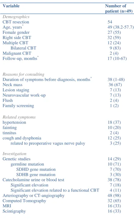
It is widely accepted that the Shamblin classification (Fig. 1), which is based on the involvement of carotid vessels is directly related to the risk of vascular damage12,13. Previous studies reported controversial results regarding the relationship between the Shamblin classification and postoperative cranial nerve deficits9–11,13,14.
20
Fig 1. The Shamblin classification of carotid body tumors allows predicting the difficulty of surgical resection.
Group I tumors are local and easily resected. Group II tumors adhere to or partially surround the carotid arteries. Group III tumors completely encase at least one of the arteries. ECA, External carotid artery; ICA, internal carotid artery; X, vagal nerve; XII, hypoglossal nerve.
Other treatment strategies include preoperative embolization, adjuvant, salvage or exclusive radiation therapy (conventional, stereotactic or intensity modulated radiotherapy), or observation15–19.
We analyzed the criteria that could predict neurovascular complications following CBT surgical resection. The aim of our study was to report our experience in the management of CBT and to evaluate the early and late neurovascular postoperative complications according to the Shamblin classification and tumor size.
21
3. Patients and Methods
Patient inclusion
We performed a retrospective study that included all patients that underwent CBT surgery at Grenoble University Hospital (France) between January 1980 and December 2011. Data consisted of patient characteristics, surgical management, early and late postoperative complications (before and after one month postoperatively), permanent peripheral neurological deficit at 18 months, and long-term follow-up. Patients were classified according to Shamblin by imaging (on CT or MRI). The tumor size was defined by measuring the longest axis on pathological examination.
Surgery
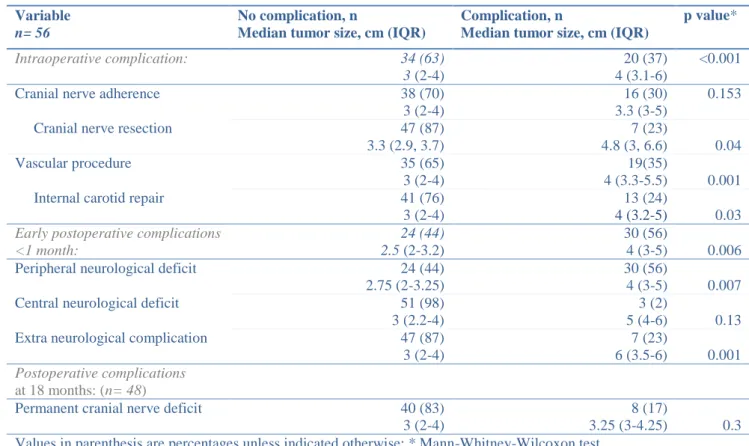
CBT resections were usually performed by a surgical team that included an otolaryngologist and a vascular surgeon. A curved skin incision was performed to obtain a large surgical field for neurovascular structure dissection. The vagus, spinal, and hypoglossal nerves were identified early in the course of the dissection. The proximal and distal portions of the common, internal, and external carotid arteries (ICA and ECA) were localized before tumor resection (Fig. 2). Then a careful subadventitial plane dissection was performed craniocaudally up to the carotid bulb where the glomus artery arises from the posterior part of the carotid bifurcation20. A primary repair with a venous patch or a venous bypass graft was performed by a vascular surgeon when vascular reconstruction was planned, especially in large tumors or those that are adherent to the ICA. The removal of adjacent lymph nodes was usually performed for all patients, since 2000.
22
Fig 2. View of the neurovascular structures during right CBT resection. XII, vagus nerve; ECA, external carotid
artery; CC, common carotid artery; ICA, internal carotid artery; T, tumor.
Statistical analyses
We reported baseline patient characteristics as medians and interquartile ranges (IQR; i.e., 25th and 75th percentiles) for continuous variables, and percentages for categorical variables. Univariate analysis was used to identify the Shamblin classification or tumor size as possible predictive factors for neurovascular complications. The data was analyzed with a Chi2 test or Fisher's exact test for categorical variables and Kruskal-Wallis test or Mann-Whitney-Wilcoxon test for continuous variables. A receiver operating characteristics (ROC) curve analysis was performed to determine the accuracy of tumor size as a predictor for peripheral neurological deficit.Two sided p values <0.05 were considered as statistically significant. All statistical analyses were performed with R version 3.0.1.
23
4. Results
Patient characteristics
Characteristics of patients, symptoms, and preoperative data are listed in table 1. Forty-nine patients underwent 54 CBT resections. 36 cases showed a lateral neck swelling that was present for a median period of 38 months (IQR, 1-48 months). The median length of follow-up was 17 months (IQR, 10-67 months). Forty-three patients presenting 48 CBT were followed-up for 18 months or more.
The most common diagnostic imaging was arteriography or CT-angiography. However, ultrasonography or fine needle aspiration was performed primarily for 12 patients. This group of patients underwent cervical exploration without any complication (10 before 1992, referred to our department by other hospitals). These patients underwent CBTs resection in a second time following an additional radiologic work-up that allowed to establish the diagnosis.
Most patients with multiple paraganglioma (12 patients) presented with bilateral CBT. Other associated localizations were also found: 3 tympano-jugular, 1 vagal, 1 mediastinal, 1 pancreatic, and 1 adrenal (pheochromocytoma). Six patients with bilateral CBTs underwent bilateral resections: the smallest was usually the first to be excised. In some cases treatment consisted of contralateral radiotherapy (because of post-operative ipsilateral vagal palsy) or routine follow-up.
Genetic studies were systematically offered to all patients and families, including search for germline mutations of the succinate dehydrogenase subunits D, B (SDHD, SDHB). Ten patients had mutations, mostly in the SDHD gene. Eight of them presented multiple paragangliomas with SDHD mutation in 7 cases. Four patients showed a significant high level of catecholamine that was related to functional CBTs.
24 Variable Number of patient (n=49) Demographics CBT resection 54 Age, years* 49 (38.2-57.7) Female gender 27 (55) Right side CBT Multiple CBT Bilateral CBT Malignant CBT Follow-up, months*
Reasons for consulting
32 (59) 12 (24) 9 (83) 2 (4) 17 (10-67)
Duration of symptoms before diagnosis, months* 38 (1-48)
Neck mass 36 (67) Lesion staging 7 (13) Neurovascular work-up 7 (13) Flush 2 (4) Family screening 1 (2) Related symptoms hypertension fainting tinnitus
cough and dysphonia
related to preoperative vagus nerve palsy
Investigation
Genetic studies germline mutation SDHD gene mutation SDHB gene mutation Catecholamine urine or blood test Significant elevation
Significant elevation related to a functional CBT Arteriography or CT-angiography Computed Tomography MRI Scintigraphy 18 (37) 10 (20) 2 (4) 12 (25) 3 (25) 14 (29) 10 (71) 7 (70) 3 (30) 38 (78) 7 (18) 4 (11) 48 (98) 32 (65) 16 (33) 16 (33)
Values in parenthesis are percentages unless indicated otherwise;
*
values are medians (interquartile range). CBT, carotid body tumor; SDHD-B, succinate dehydrogenase sub unit D-B; CT, computed tomography; MRI, magnetic resonance imaging
Table 1. Clinical features and preoperative data of the study group.
All CBTs were histologically confirmed as paragangliomas. Two patients presented regional metastases that were incidental findings in lymph nodes adherent to the tumor. The tumor could not be completely resected in 4 patients with Shamblin III because of the extension to
25
the skull base around the vessels (2 patients with a tumor size of 6 cm), or due to a positive surgical margin (2 patients).
The tumor size was significantly larger in higher Shamblin grades (p < 0.01, Fig. 3).
Fig. 3.Boxplots representing tumor size according to the Shamblin classification (Kruskall-Wallis test: *, p<0.01).
Surgical management
All patients underwent surgical resection. Preoperative tumor embolization was performed in 2 cases treated prior to 1992. The median surgical time was 135 minutes (IQR, 120-180 minutes). A positive correlation between both Shamblin classification and tumor size with surgical time was observed (Fig. 4).
26
Fig. 4. Relationship between both Shamblin classification (a) and tumor size (b) with length of surgery. a:
boxplot representing length of surgery operating time depending on Shamblin classification. b: bi plot points graph representing length of surgery depending on tumor size. Mean length of surgery correlated with Shamblin group (Kruskal-Wallis test: *, p<0.05) and tumor size (Pearson correlation test: r=0.50; p<0.001).
Intraoperative complications occurred in 20 cases (37%). Seven patients (13%) required nerve resection: 3 resections of the vagus and superior laryngeal nerve and 1 of the hypoglossal nerve. Vascular procedures were performed in 19 cases (35%): 18 external carotid artery ligations were performed and internal carotid artery repairs were required in 13 cases (9 venous bypass grafts, 2 venous patches, 1 suture, and 1 ligation). The Shamblin classification and the tumor size were positively correlated to intraoperative complications, including nerve resection and vascular repair (p<0.05, Table 2 and 3).
27 Variable Shamblin I n= 17 Shamblin II n= 25 Shamblin III n= 12 p value* Intraoperative complications: 1 (6) 8 (32) 11 (92) <0.001†
Cranial nerve adherence 3 (18) 7 (28) 6 (50) 0.201†
Cranial nerve resection 1 (6) 1 (4) 5 (42) 0.008†
Vascular procedure 0 (0) 9 (36) 10 (83) <0.001†
Internal carotid repair 0 (0) 4 (13) 9 (75) <0.001†
Early postoperative complications: 5 (29) 16 (64) 10 (83) 0.003
Peripheral neurological deficit 5 (29) 15 (60) 10 (83) 0.013
Central neurological deficit 0 (0) 0 (0) 3 (25) 0.008†
Extra neurological complication 0 (0) 2 (8) 5 (42) 0.003†
Postoperative complications at 18 months: n=17 n=22 n=9
Permanent cranial nerve deficit 1 (6) 4 (18) 3 (33) 0.179†
Values in parenthesis are percentages unless indicated otherwise; *χ2 test, except †Fisher test.
Table 2. Neurovascular complications according to the Shamblin classification.
Variable
n= 56
No complication, n
Median tumor size, cm (IQR)
Complication, n
Median tumor size, cm (IQR)
p value* Intraoperative complication: 34 (63) 3 (2-4) 20 (37) 4 (3.1-6) <0.001
Cranial nerve adherence 38 (70)
3 (2-4)
16 (30) 3.3 (3-5)
0.153
Cranial nerve resection 47 (87)
3.3 (2.9, 3.7) 7 (23) 4.8 (3, 6.6) 0.04 Vascular procedure 35 (65) 3 (2-4) 19(35) 4 (3.3-5.5) 0.001
Internal carotid repair 41 (76)
3 (2-4)
13 (24)
4 (3.2-5) 0.03
Early postoperative complications <1 month:
24 (44) 2.5 (2-3.2)
30 (56)
4 (3-5) 0.006
Peripheral neurological deficit 24 (44)
2.75 (2-3.25)
30 (56)
4 (3-5) 0.007
Central neurological deficit 51 (98)
3 (2.2-4)
3 (2)
5 (4-6) 0.13
Extra neurological complication 47 (87)
3 (2-4)
7 (23)
6 (3.5-6) 0.001
Postoperative complications
at 18 months: (n= 48)
Permanent cranial nerve deficit 40 (83)
3 (2-4)
8 (17)
3.25 (3-4.25) 0.3 Values in parenthesis are percentages unless indicated otherwise; * Mann-Whitney-Wilcoxon test.
28
Early postoperative complications < 1 month
The postoperative complications consisted of neurovascular accidents in 3 patients (1 case experienced a stroke after internal carotid artery ligation and 2 patients presented a transient ischemic attack), and peripheral neurological deficit in 30 cases. The deficit involved several nerves in 18 cases, and a complete deficit in 11 cases. The most commonly involved nerves were the vagus nerve (37%), the hypoglossal nerve (32%), the sympathetic chain (13%), the glossopharyngeal nerve (10%), and superior laryngeal nerve (6%). Other early postoperative non-neurological events included local complications (1 case of abscess and 2 cases of hematoma requiring drainage), vascular complications (1 graft thrombosis requiring revascularization), or general complications (1 case of pulmonary embolism, 1 case of gastrointestinal bleeding, 1 case of pneumonia, and 2 cases of hypertension). No mortality was observed in the perioperative period. The incidence of early postoperative complications, including peripheral neurological deficits and postoperative non-neurological events, was significantly higher in patients with a higher Shamblin classification and a larger tumor size (Table 2 and 3).
We compared the incidence of deficits predicted according to the tumor size cut-off value, to determine the accuracy of the tumor size in predicting early peripheral neurological deficits. The best sensitivity and specificity was found for a tumor size ≥ 3.2cm (table 4). The positive
predictive value for peripheral neurologic deficit occurrence for a tumor size > 3.2 cm was 76%.
29
a b
Table 4. Accuracy of a tumor size ≥ 3.2 cm in predicting early postoperative peripheral neurological deficits. 4a:
Performance measures at the optimal cut-off value (3.2cm) 4b: Receiver operating characteristic (ROC) curve figure at the optimal cut-off value.
Late postoperative complications
The outcomes were assessed after 18 months for 43 patients (48 CBT resections), 5 patients who presented with initial nerve deficits were lost to follow-up. The nerve deficits were transient in 17 cases (68% with initial deficits recovered completely). The median time for recovery was 6 months (IQR, 2-12 months). Speech therapy was systematically prescribed for 30 patients with vagus nerve involvement. Nevertheless, 8 cases (17% of all CBT resection population) of cranial nerve deficits were permanent. Persistent symptoms showed swallowing difficulties in 4 cases, dysphonia in 3 cases, and 1 case of tongue paralysis. The univariate analysis for permanent cranial nerve deficits did not reveal any significant predictive factors (Table 2 and 3).
One patient died 10 months after surgery from a stroke. In 3 patients, other late post-operative non-neurological complications were observed (persistent uncontrolled hypertension, pneumonia, and graft thrombosis). Three patients complained of first bite syndrome21.
Measure Value by test
for tumor size = 3.2 cm
Sensitivity 63 (57- 70) Specificity 75 (69- 81)
PPV 76 (70- 82)
NPV 62 (55- 69)
Values are percentages (95 per cent confidence intervals); PPV positive predictive value; NPV negative predictive value; AUC area under the receiver operating characteristic curve
0 .0 0 0 .2 5 0 .5 0 0 .7 5 1 .0 0 Se n si ti vi ty 0.00 0.25 0.50 0.75 1.00 1 - Specificity
30
Follow-up
One patient presented post-operative local residual tumor progression 1 year following resection. This case showed incomplete excision without additional treatment. The patient underwent surgery 6 years ago, without any signs of recurrence till the present time.
Two patients presented local recurrences associated with distant localizations. The first patient showed a local recurrence associated with mediastinal localization, one year after a bilateral Shamblin III CBTs resection with regional lymph node metastasis. The cervical recurrence was treated by radiation (because of contralateral permanent vagus nerve deficit), and the mediastinal metastasis was removed, without any recurrence during follow-up for 10 years. The second case showed local recurrence associated with multiple cervical localizations 27 years following resection: he presented positive surgical margins and incidental regional lymph node metastasis on initial pathological examination. No additional treatment was initiated for this patient given his age and multiple cervical localizations.
Bone and liver metastases occurred in one case 4 years after a bilateral resection of functional CBTs. Clinical course was rapidly unfavorable for this patient despite radiotherapy and the patient died few weeks later.
31
5. Discussion
The clinical evaluation of a lateral neck mass should be performed cautiously to avoid missing a rare differential diagnosis such as that of a paraganglioma. More than two thirds of patients with carotid body tumors present with a pulsatile lateral neck mass at the time of the diagnosis. This mass has usually a restricted mobility in the vertical axis 22 and can be present for several years.
The Doppler ultrasound sensitivity is too low for the diagnostic work-up of paragangliomas. MRI (especially 3D TOF MR angiography7) provides more data than CT scan in case of uncertain diagnosis. However the gold standard is still Digital Subtraction Angiography7 for the detection of small paragangliomas, because it reveals the specific vascular supply of the paraganglioma by the ascending pharyngeal artery23. Fine needle aspiration or open biopsy should be avoided.
The clinical value of embolization is still controversial9,19,24–26. We do not usually perform preoperative embolization (only 2 patients before 1992) even though some authors have recently suggested that it is not associated with an increased incidence of stroke26. Many authors have suggested that embolization is little beneficial for the outcome and management of the CBT and recent studies do not show any decrease in the morbidity rate, especially for
cranial nerve deficit24. On the other hand, it could facilitate the resection of large adherent tumors, especially the ones located high in the neck with a low need for carotid clamping and subsequent reduced blood loss9,25.
According to recent studies, 30% of CBT are hereditary and present mainly SDHD gene mutation9,27. Systematic genetic studies should be considered for all patients, due to the high prevalence of mutations in patients presenting with an apparently sporadic paraganglioma
32
(from 6 to 25%)15,28,29. Several authors30,31 have suggested that multifocal CBT were frequently observed in SDHD mutation carriers (71% in this study). The results of a recent meta-analysis demonstrated that the risk of malignant paraganglioma was higher in case of SDHB gene mutation31.
Two patients presented with histologically documented regional lymph node metastases and one with distant metastases32. Malignancy may only be diagnosed when there is metastasis to non neuroendocrine tissue 31,33, since there are no accurate histopathological characteristics of malignant forms. It should be noted that the diagnoses of regional metastasis were incidental in both cases: cervical lymph node metastases are not uncommon feature in patients with CBT. We performed regional lymph node removal in the surgical site to identify possible occult metastasis, for patient with apparently benign CBT15,33. Boedecker et al15 recommended selective neck dissection for levels II and III, but the extent of lymphadenectomy remains to be defined. Our results were consistent with those of previous studies indicating that malignancy ranged around 6% for CBT, and that distant metastasis were less frequent than in other head and neck paraganglioma sites33,34. The authors of two large studies focusing on malignant head and neck paraganglioma reported that patients with regional metastasis had significantly better five-year overall survival rate (76.8-82.4%) than those with distant metastasis (11.8-41.4%). For patients with regional and distant metastasis, there is no evidence that postoperative radiotherapy improves the five-year overall survival rate. On the other hand, in cases of incomplete removal, adjuvant radiation therapy may slow tumor growth and prolong patient survival33,34.
Three patients showed residual tumor progression or recurrences 1 and 27 years after surgery. Elshaik et al35 reported a long median time before recurrence of head and neck paraganglioma (36 months, range 15-350): long follow-up is needed to detect late recurrences. Their results
33
also confirmed that surgery allowed to control local recurrence but there are few studies have assessed the role of radiotherapy in the local control of CBT recurrence 35.
Complete surgical excision is the aim of any operative procedure and is the only curative option. Nevertheless radiation therapy in patients who cannot undergo surgery because of advanced age or comorbidity, aggressive or unresectable tumors can be discussed15,18. The goal of radiotherapy is disease control or growth inhibition rather than tumor elimination. Indeed a recent review18 on CBT management shows no significant differences in tumor control and deaths between surgery and radiotherapy, but a higher rate of major complication and cranial nerve palsies in surgical series. Other factors to be considered are radiation side effects: radiation induced malignancy (1% at 10 years), making surgery more challenging with higher risk of neurovascular complication. Some adverse effects could be minimized by the use of intensity- modulated radiotherapy. Tumor doubling time is very slow; hence conservative treatment, such as a wait-and-see strategy, can also be discussed in elderly patients.
The most important aspect of the Shamblin classification12 is the carotid artery vessel wall involvement, assuming that tumor size is related to the arterial involvement 22. But a non-linear relationship has been reported in a few studies14,36. The Shamblin classification was significantly associated with tumor size in our study. The Shamblin classification was initially based on intraoperative and pathological findings12, but in our study it was assessed according to preoperative radiological data. Nevertheless, the Shamblin groups were distributed as usually reported: one third in group 1, almost one half in group 2, and one fourth in group 39,10. Other accurate imaging criteria should also reflect the artery involvement such as those based on the angle of the circumferential encasement of the ICA, or the identification of a lucent zone between the tumor and the ICA14,36,37. We could not collect these features due to the inconsistency of preoperative radiological data. The tumor size was assessed according to
34
the longest axis calculated in pathological reports. The assessment of the tumor volume by imaging could also provide a more objective preoperative criterion9,27.
Twenty-four percent of cases required ICA reconstruction in our study. This was consistent with published rates ranging from 6 to 52%8,19,38.We included all types of ICA reconstructions, even minor ones. This could account for the difference with the results of recent studies since there was limited information on the specifics of vascular repair14. Vascular damage decreased over the past few decades, especially for ICA ligation3. Consequently, postoperative stroke is no longer a major risk: only one patient in our study experienced a stroke, consequently to an ICA ligation in 1991. Seventeen percent of patients presented a stroke3 in the oldest large series published in 1988. Furthermore, vascular repair has become safer with improved surgical techniques. Thus, early proximal and distal control of the carotid vessels, subadventitial tumor dissection, intravenous heparinization when vascular repair is needed, and the short carotid clamping time reduce postoperative vascular complications. The higher Shamblin classification and larger tumor size in our study were accurately correlated with a higher rate of vascular damage. Hence, both items could be used to estimate the risk of vascular repair11.
Peripheral neurological morbidity has remained high and unchanged for 50 years3 despite improved surgery techniques. Peripheral neurological deficit occurred in 56% of our cases, in the early postoperative period. We demonstrated that early neurological deficit was also related with both the Shamblin classification and tumor size. Hence, the Shamblin classification, as well as the tumor size, could also be used to predict the risk of early cranial nerve deficit9. Some authors suggested using an arbitrary 4 cm tumor size limit to predict a higher risk of neurological morbidity 13,20. We showed that a tumor size ≥3.2 cm was the best cut-off value defined by the ROC analysis to predict early neurological deficit. The larger the
35
tumor the higher the risk for nerve deficits, therefore an early diagnosis and surgery for tumor < 3 cm are crucial 19.
Nerve deficit was transient in 68% of cases with initial deficit, andpermanent nerve deficit was observed clinically after 18 months in 17% of operated cases. Appropriate rehabilitation is important, including early speech therapy. We did not observe any nerve deficit recovery at follow-up after 18 post-operative months; however authors do not always report the deficit recovery time 39 or only with a follow-up of 12 months9. We did not find any predictive factors for permanent nerve deficits at 18 months. Power et al 9 reported that permanent cranial nerve deficit at 1 year was correlated to the Shamblin group. Further investigation is required with studies taking 18 months as the cut-off value for permanent nerve deficit.
36
6. Conclusion
Surgical resection remains the only curative treatment with low vascular morbidity. However immediate postoperative nerve deficits are still frequent but most of them are temporary as they recover within 18 months. Tumor size and Shamblin classification correlated with neurovascular complications. Early detection of CBTs is primordial for the operative outcome. Other objective imaging criteria should be assessed in order to optimize planning for multidisciplinary management.
37
7. References
1. Maves MD. Vascular tumors of the head and neck. In Head and Neck-Otolaryngology. In: Bailey BJ, Jonhnson JT, Kohut RI, Pillsbury HC.,eds.Tardy Medical Management. Philadelphia JB Lippincott; 1993:1397-1409
2. Baysal BE, Farr JE, Rubinstein WS, et al. Fine mapping of an imprinted gene for familial non chromaffin paragangliomas, on chromosome 11q23. Am J Hum Genet. 1997;60(1):121-132. 3. Hallett JW, Nora JD, Hollier LH, Cherry KJ, Pairolero PC. Trends in neurovascular complications
of surgical management for carotid body and cervical paragangliomas: a fifty-year experience with 153 tumors. J Vasc Surg. 1988;7(2):284-291.
4. Wang S, Wang M, Barauskas T, Calcaterra T. Surgical management of carotid body tumors.
Otolaryngol Head Neck Surg. 2000;123(3):202-206.
5. Sajid MS, Hamilton G, Baker DM. A multicenter review of carotid body tumour management.
Eur J Vasc Endovasc Surg. 2007;34(2):127-130.
6. Nora JD, Hallett JW Jr, O’Brien PC, Naessens JM, Cherry KJ Jr, Pairolero PC. Surgical resection of carotid body tumors: long-term survival, recurrence, and metastasis. Mayo Clin Proc Mayo Clin. 1988;63(4):348-352.
7. Berg R. Imaging and management of head and neck paragangliomas. Eur Radiol. 2005;15(7):1310-1318.
8. Smith JJ, Passman MA, Dattilo JB, Guzman RJ, Naslund TC, Netterville JL. Carotid body tumor resection: does the need for vascular reconstruction worsen outcome? Ann Vasc Surg. 2006;20(4):435-439.
9. Power AH, Bower TC, Kasperbauer J, et al. Impact of preoperative embolization on outcomes of carotid body tumor resections. J Vasc Surg. 2012;56(4):979-989.
10. Makeieff M, Raingeard I, Alric P, Bonafe A, Guerrier B, Marty-Ane C. Surgical Management of Carotid Body Tumors. Ann Surg Oncol. 2008;15(8):2180-2186.
11. Lim JY, Kim J, Kim SH, et al. Surgical treatment of carotid body paragangliomas: outcomes and complications according to the shamblin classification. Clin Exp Otorhinolaryngol. 2010;3(2):91-95.
12. Shamblin WR, ReMine WH, Sheps SG, Harrison EG Jr. Carotid body tumor (chemodectoma). Clinicopathologic analysis of ninety cases. Am J Surg. 1971;122(6):732-739.
13. Luna-Ortiz K, Rascon-Ortiz M, Villavicencio-Valencia V, Herrera-Gomez A. Does Shamblin’s classification predict postoperative morbidity in carotid body tumors? A proposal to modify Shamblin’s classification. Eur Arch Otorhinolaryngol. 2005;263(2):171-175.
14. Arya S, Rao V, Juvekar S, Dcruz AK. Carotid body tumors: objective criteria to predict the Shamblin group on MR Imaging. Am J Neuroradiol. 2008;29(7):1349-1354.
15. Boedeker CC. Paragangliomas and paraganglioma syndromes. GMS Curr Top Otorhinolaryngol
38
16. Chino JP, Sampson JH, Tucci DL, Brizel DM, Kirkpatrick JP. Paraganglioma of the Head and Neck: Long-term Local Control with Radiotherapy. J Clin Oncol June 2009. 2009;32(3):304-307. 17. Hinerman RW, Amdur RJ, Morris CG, Kirwan J, Mendenhall WM. Definitive radiotherapy in the
management of paragangliomas arising in the head and neck: A 35-year experience. Head Neck. 2008;30(11):1431-1438.
18. Suárez C, Rodrigo JP, Mendenhall WM, et al. Carotid body paragangliomas: a systematic study on management with surgery and radiotherapy. Eur Arch Otorhinolaryngol. 2014;271(1):23-34. 19. Fruhmann J, Geigl JB, Konstantiniuk P, Cohnert TU. Paraganglioma of the carotid body: treatment strategy and SDH-gene mutations. Eur J Vasc Endovasc Surg Off J Eur Soc Vasc Surg. 2013;45(5):431-436.
20. Makeieff M, Thariat J, Reyt E, Righini C-A. Treatment of cervical paragangliomas. Eur Ann
Otorhinolaryngol Head Neck Dis. 2012;129(6):308-314.
21. Laccourreye O, Werner A, Garcia D, Malinvaud D, Tran Ba Huy P, Bonfils P. First bite syndrome.
Eur Ann Otorhinolaryngol Head Neck Dis. 2013;130(5):269-273.
22. Davidge-Pitts K, Pantanowitz D. Carotid body tumor. Surg Annu. 1984;16:203-227.
23. Van Den Berg R, Rodesch G, Lasjaunias P. Management of Paragangliomas. Interv Neuroradiol. 2002;8(2):127-134.
24. Litle VR, Reilly LM, Ramos TK. Preoperative embolization of carotid body tumors: when is it appropriate? Ann Vasc Surg. 1996;10(5):464-468.
25. Zhang T, Jiang W, Li Y, Li B, Yamakawa T. Perioperative Approach in the Surgical Management of Carotid Body Tumors. Ann Vasc Surg. 2012;26(6):775-782.
26. Vogel TR, Mousa AY, Dombrovskiy VY, Haser PB, Graham AM. Carotid body tumor surgery: management and outcomes in the nation. Vasc Endovascular Surg. 2009;43(5):457-461. 27. Kruger AJ, Walker PJ, Foster WJ, Jenkins JS, Boyne NS, Jenkins J. Important observations made
managing carotid body tumors during a 25-year experience. J Vasc Surg. 2010;52(6):1518-1523. 28. Fakhry N, Niccoli-Sire P, Barlier-Seti A, Giorgi R, Giovanni A, Zanaret M. Cervical paragangliomas: is SDH genetic analysis systematically required? Eur Arch Otorhinolaryngol. 2008;265(5):557-563.
29. Lima J, Feijão T, Silva AF da, et al. High frequency of germline succinate dehydrogenase mutations in sporadic cervical paragangliomas in northern spain: mitochondrial succinate dehydrogenase structure-function relationships and clinical-pathological correlations. J Clin
Endocrinol Metab. 2007;92(12):4853-4864.
30. Neumann HP, Pawlu C, Peczkowska M, et al. Distinct clinical features of paraganglioma syndromes associated with SDHB and SDHD gene mutations. JAMA J Am Med Assoc. 2004;292(8):943-951.
31. Van Hulsteijn LT, Dekkers OM, Hes FJ, Smit JWA, Corssmit EPM. Risk of malignant paraganglioma in SDHB-mutation and SDHD-mutation carriers: a systematic review and meta-analysis. J Med Genet. 2012;49(12):768-776.
39
32. Righini C, Pecher M, Halimi S, Magne J, Reyt E. Malignant carotid paraganglioma. A case report.
Ann Otolaryngol Chir Cervicofac. 2003;120(2):130-138.
33. Lee JH, Barich F, Karnell LH, et al. National cancer data base report on malignant paragangliomas of the head and neck. Cancer. 2002;94(3):730-737.
34. Sethi RV, Sethi RKV, Herr MW, Deschler DG. Malignant head and neck paragangliomas: Treatment efficacy and prognostic indicators. Am J Otolaryngol. 2013; 34(5):431-438.
35. Elshaikh MA, Mahmoud-Ahmed AS, Kinney SE, et al. Recurrent head-and-neck chemodectomas: a comparison of surgical and radiotherapeutic results. Int J Radiat Oncol. 2002;52(4):953-956. 36. Kaddah RO, Haggag M, Lotfi U. Impact of geometric concepts in Multislice CT angiography and
MRI on surgical outcome of carotid body tumors. Egypt J Radiol Nucl Med. 2011;42(3–4):373-380.
37. Knight TT, Gonzalez JA, Rary JM, Rush DS. Current concepts for the surgical management of carotid body tumor. Am J Surg. 2006;191(1):104-110.
38. Luna-Ortiz K, Rascon-Ortiz M, Villavicencio-Valencia V, Granados-Garcia M, Herrera-Gomez A. Carotid body tumors: review of a 20-year experience. Oral Oncol. 2005;41(1):56-61.
39. Kotelis D, Rizos T, Geisbüsch P, et al. Late outcome after surgical management of carotid body tumors from a 20-year single-center experience. Langenbecks Arch Surg. 2008;394(2):339-344.
40
III- Conclusion
THESE SOUTENUE PAR : Eléa LAMBLIN
TITRE : Neurovascular outcomes and predictive factors following carotid body tumor surgical resection
La prise en charge thérapeutique des paragangliomes du glomus carotidien doit se faire après un bilan complet dans le cadre d’une prise en charge multidisciplinaire (ORL, chirurgiens vasculaires, endocrinologues, radiologues et généticiens).
Le traitement de référence est l’exérèse chirurgicale avec exérèse des ganglions adjacents péri tumoraux du fait de la fréquence de découverte de métastases ganglionnaires occultes (4%).
La chirurgie expose le patient à des risques per- opératoires : dans 24 % des cas un geste est réalisé sur la carotide interne, et dans 13 % des cas un sacrifice nerveux est nécessaire. Les complications neuro-vasculaires post-opératoires immédiates (<1 mois) décrites sont la survenue d’accidents neuro-vasculaires dans 3 cas (1 AVC et 2 AIT), et la présence de déficits neurologiques périphériques dans 30 cas. Ces évènements indésirables per et post-opératoires immédiats sont directement liés au stade de la classification de Shamblin ainsi qu’à la taille de la tumeur. Ainsi, il est montré qu’une lésion de taille supérieure à 3.2 cm exposait le patient à la survenue d’un déficit neurologique périphérique dans 74% des cas. Cependant ces déficits étaient transitoires dans 68 % des cas, et seulement 8 patients présentaient un déficit permanent à 18 mois de suivi.
Ces résultats soulignent l’importance d’un diagnostic précoce de ces tumeurs à un stade de développement limité, passant avant tout par un dépistage génétique systématique chez tous les patients.
VU ET PERMIS D'IMPRIMER Grenoble, le 11/02/2015
41
SERMENT
D’HIPPOCRATE
En présence des Maîtres de cette Faculté, de mes chers condisciples et devant l’effigie d’HIPPOCRATE,
Je promets et je jure d’être fidèle aux lois de l’honneur et de la probité dans l’exercice de la Médecine.
Je donnerai mes soins gratuitement à l’indigent et n’exigerai jamais un salaire au-dessus de mon travail. Je ne participerai à aucun partage clandestin d’honoraires.
Admis dans l’intimité des maisons, mes yeux n’y verront pas ce qui s’y passe ; ma langue taira les secrets qui me seront confiés et mon état ne servira pas à corrompre les mœurs, ni à favoriser le crime.
Je ne permettrai pas que des considérations de religion, de nation, de race, de parti ou de classe sociale viennent s’interposer entre mon devoir et mon patient.
Je garderai le respect absolu de la vie humaine.
Même sous la menace, je n’admettrai pas de faire usage de mes connaissances médicales contre les lois de l’humanité.
Respectueux et reconnaissant envers mes Maîtres, je rendrai à leurs enfants l’instruction que j’ai reçue de leurs pères.
Que les hommes m’accordent leur estime si je suis fidèle à mes promesses.
Que je sois couvert d’opprobre et méprisé de mes confrères si j’y manque.
42
Résumé
Titre : Complications neuro-vasculaires après traitement chirurgical des paragangliomes du glomus carotidien. A propos de 54 cas.
Objectifs : Evaluer les complications neuro-vasculaires post-opératoires précoces et tardives après traitement chirurgical de paragangliome du glomus carotidien (PG). Evaluer la classification de Shamblin et la taille de la lésion comme facteurs prédictifs de survenue des complications neurologiques et vasculaires.
Matériel et méthode : Etude rétrospective incluant tous les patients opérés pour un PG de 1980 à 2011. Analyse des données médicales préopératoires, complications post-opératoires immédiates (<1 mois), et tardives (séquelles à 18 mois).
Résultats : Cinquante-quatre PG ont été réséqués chez 49 patients avec une médiane de suivi post-opératoire de 17 mois (EI, 10-67 mois). Selon la classification de Shamblin : 17 PG appartenaient au groupe I, 25 au groupe II et 12 au groupe III. Il existait des complications post-opératoires immédiates dans 31 cas, dont 30 paralysies d’au moins un nerf crânien. Il s’agissait de PG de groupe III selon la classification de Shamblin dans 83 % des cas, et la taille de la lésion était significativement plus importante chez ces patients présentant des complications neurologiques périphériques immédiates (4cm ±1.4 versus 2.9 ±1.3; p<0.01). Huit patients présentaient un déficit d’au moins un nerf crânien persistant après 18 mois : aucun facteur prédictif n’a été mis en évidence.
Conclusion : La chirurgie est le traitement de référence des PG avec une morbidité vasculaire faible. La morbidité reste principalement liée aux complications neurologiques périphériques, majoritairement transitoires et dont la fréquence est corrélée à la classification de Shamblin et la taille de la lésion.
Mots clés : paragangliome du glomus carotidien, chémodectome, chirurgie, complications neuro-vasculaires, classification de Shamblin.