HAL Id: tel-00353429
https://pastel.archives-ouvertes.fr/tel-00353429
Submitted on 15 Jan 2009HAL is a multi-disciplinary open access
archive for the deposit and dissemination of sci-entific research documents, whether they are pub-lished or not. The documents may come from teaching and research institutions in France or abroad, or from public or private research centers.
L’archive ouverte pluridisciplinaire HAL, est destinée au dépôt et à la diffusion de documents scientifiques de niveau recherche, publiés ou non, émanant des établissements d’enseignement et de recherche français ou étrangers, des laboratoires publics ou privés.
Nicolas Le Moigne
To cite this version:
Nicolas Le Moigne. Swelling and dissolution mechanisms of cellulose fibres. Mechanics [physics.med-ph]. École Nationale Supérieure des Mines de Paris, 2008. English. �NNT : 2008ENMP1581�. �tel-00353429�
Ecole Doctorale 364 : Sciences Fondamentales et Appliquées
N° attribué par la bibliothèque
|__|__|__|__|__|__|__|__|__|__|
T H E S E
pour obtenir le grade de
Docteur de l’Ecole Nationale Supérieure des Mines de Paris
Spécialité «Science et Génie des Matériaux»
présentée et soutenue publiquement par
M. Nicolas Le Moigne
le 9 Décembre 2008
MECANISMES DE GONFLEMENT ET DE DISSOLUTION
DES FIBRES DE CELLULOSE
SWELLING AND DISSOLUTION MECHANISMS OF CELLULOSE FIBRES
Directeur de thèse : Patrick Navard
Jury :
M. Pedro Fardim
Rapporteur
M. Jürgen Puls
Rapporteur
M. Herman Höfte
Examinateur
M. Patrick Navard
Examinateur
M. Jürgen Engelhardt
Membre invité
Mme Kristina Jardeby
Membre invité
M. Michel Pierre
Membre invité
Contents
Introduction francophone 3
Chapter I: State of the Art – Review on cellulose biosynthesis, structure and dissolution mechanisms
Introduction 9
1.1 Biosynthesis of cellulose fibres 10
1.2 Structure of cellulose fibres 15
1.3 Swelling and dissolution mechanisms of cellulose fibres 32
Chapter II: Gradient in dissolution capacity of successively deposited cell wall layers in
cotton fibres. 43
Chapter III: Restricted dissolution capacity of native and regenerated cellulose fibres under
uniaxial elongational stress. 55
Chapter IV: Dissolution mechanisms of wood cellulose fibres in NaOH-water. 67
Chapter V: Structural changes and alkaline solubility of wood cellulose fibres after
enzymatic peeling treatment. 91
Chapter VI: Contraction and rotation of natural and regenerated cellulose fibres during
swelling and dissolution. 109
Chapter VII: Kinetics of dissolution of cellulose and cellulose derivatives under shear. 121
Conclusions 131
Articles and Communications 135
Résumé du travail de thèse
Contexte de l’étude
À la différence des réserves minérales et fossiles qui furent formées il y a plusieurs millions d’années, les polysaccharides, comme la cellulose et l’amidon, représentent une ressource quasi inépuisable de matériaux naturels qui se renouvelle année après année. Parmi la grande variété de polysaccharides synthétisés dans la nature, la cellulose constitue le polymère naturel le plus abondant. On la trouve en composé majoritaire au sein des fibres dans les plantes, les algues et les champignons mais elle est aussi synthétisée par certains organismes tels que les cyanobactéries.
La plupart des polymères de grande diffusion sont synthétisés à partir des ressources pétrolières. Du fait de la limitation des stocks et des nouvelles normes environnementales, la recherche s’est concentrée ces dernières années sur la valorisation de matériaux polymères produits par la nature de façon pérenne, comme la cellulose. Cependant, leur usage et leur transformation présentent plusieurs contraintes principalement liées à leurs structures morphologiques et chimiques très spécifiques. Bien qu’utilisée depuis des siècles dans l’industrie textile et la papeterie, la cellulose fait toujours l’objet de nombreuses études scientifiques et techniques visant à améliorer, diversifier et simplifier son utilisation. Dans la majorité des cas, la cellulose ne peut être utilisée sous sa forme fibrillaire naturelle, excepté pour les applications en fibres de renforcement ou textile (cas du coton, du lin, du chanvre…). Elle ne peut pas non plus être fondue comme la plupart des polymères semi cristallins et sa transformation doit donc être précédée d’une étape de dissolution ou de dérivation. Cependant, sa structure complexe n’offre pas une grande accessibilité aux réactifs et de nombreuses études ont été menées afin de mieux comprendre comment démanteler ou modifier cette structure pour améliorer son accessibilité tout en préservant au maximum les propriétés originelles du matériau. Bien qu’améliorant la dissolution, les prétraitements chimiques, physiques ou enzymatiques utilisés aujourd’hui entraînent le plus souvent une forte dégradation de la structure qui a pour principale conséquence une forte diminution des propriétés des produits transformés (fibres textiles, éponges, films…) et ne sont pas toujours aussi performant que l’industrie le souhaite.
En dépit d’une bonne connaissance et d’une bonne maîtrise de la dissolution et de la dérivation d’un point de vue chimique, les prétraitements dits d’ « activation » sont optimisés de façon relativement empirique. Le but de nos travaux est d’identifier les principaux paramètres impliqués dans la dissolution de la cellulose afin d’« activer » la cellulose de façon plus efficace afin d’enlever ou de modifier seulement ce qui est nécessaire. Notre approche scientifique est basée sur les nombreuses connaissances accumulées sur la biosynthèse et la structure des fibres de cellulose et d’étudier leurs mécanismes de gonflement et de dissolution dans différentes conditions (qualité du solvant, type de fibres, tension…) afin d’analyser leur capacité de dissolution aux différentes échelles structurales.
Principaux résultats obtenus
L’état de l’art nous a permis de décrire les mécanismes de biosynthèse et la composition et la structure des trois principales échelles structurales des fibres de cellulose, c’est à dire moléculaire, agrégée et macrostructurale, et de leurs principaux constituants, la cellulose, les hémicelluloses, les protéines, les pectines, la lignine (Chapitre I). Lors de cette étude bibliographique, nous avons pu constater que malgré l’étendue des connaissances, la dynamique complète de synthèse et de déposition de la cellulose sur les parois des fibres ainsi que le rôle que jouent les différents composés non cellulosiques dans la structuration des parois ne sont pas totalement élucidés. Les études existantes sur les mécanismes de gonflement et de dissolution des fibres de cellulose montrent une forte influence de la qualité du solvant et de la morphologie originelle des fibres. Le comportement des différentes échelles structurales en terme de gonflement et de dissolution n’est quant à lui pas clairement établi.
Nos travaux se sont concentrés sur l’étude du gonflement et de la dissolution des fibres de cellulose en faisant varier la qualité du solvant (N-methylmorpholine-N-oxide avec différentes quantités d’eau et solutions aqueuses de NaOH à 8%), les conditions de dissolution, comme la tension, et l’origine des fibres (coton, bois, fibres régénérées ou dérivées). Les mécanismes de gonflement et de dissolution ont été étudiés par des observations microscopiques à haute résolution. Une séparation sélective des fractions solubles et insolubles a été réalisée par centrifugation. La distribution de masse molaire, la cristallinité, la composition en sucre, l’allomorphie et la quantité de chaque fraction ont été analysées. A partir de ces résultats, nous avons pu mieux décrire les mécanismes de gonflement et de dissolution des fibres de cellulose et ainsi identifier les principaux paramètres gouvernant la dissolution.
Notre apport principal est de montrer que la capacité de dissolution de la cellulose doit être considérée aux différentes échelles structurales des fibres :
De l’échelle macrostructurale à l’échelle agrégée :
Nous avons montré qu’il existe un gradient de dissolution au sein des fibres de coton entre les trois principales parois (primaire, secondaire S1, secondaire S2) déposées successivement au cours de la biosynthèse. Les parois les plus anciennement déposées (parois primaire et S1) sont les plus difficiles à dissoudre (Chapitre II), laissant supposer que la grande quantité de matériaux non cellulosiques présents dans ces parois constitue une gêne à la dissolution. Nous avons ensuite démontré de façon claire qu’outre les classiques raisons thermodynamiques contrôlant la dissolution d’un polymère, la suppression des parois primaire et S1 par pelage enzymatique (Chapitre V) ainsi que la déstructuration des parois par explosion à la vapeur, hydrolyse en milieu acide ou par traitement enzymatique (chapitre IV et V) permettent d’améliorer la dissolution des fibres de cellulose. Un facteur très important et totalement nouveau est le fait que les mouvements de rotation et de contraction des fibres et la convection du solvant autour des fibres doivent aussi être favorisés au cours de la dissolution (chapitre VI et VII). Cependant, certaines fractions restent insolubles et leur capacité de dissolution doit être analysée à une échelle plus fine de la structure. L’un des résultats importants est la présence de quantités plus élevées d’hémicelluloses dans les fractions insolubles par rapport aux fractions solubles suggérant que la cellulose provenant des fractions insolubles est peut être plus intimement incorporée dans la matrice d’hémicelluloses (Chapitre IV).
De l’échelle agrégée à l’échelle moléculaire :
Nous avons montré qu’une fraction importante de chaînes de cellulose, similaires en longueur, peuvent appartenir soit à la fraction soluble soit à la fraction insoluble, prouvant que la masse molaire n’est pas un facteur discriminant pour la dissolution de la cellulose (Chapitre IV). Ce résultat est à mettre en relation avec l’environnement chimique des chaînes de cellulose qui est plus riche en hémicelluloses dans les fractions insolubles. De plus, un phénomène intéressant a été mis en évidence en faisant varier les conditions de tension durant la dissolution. Nous avons en effet montré que la capacité de dissolution des fibres de cellulose est limitée si elles sont soumises à une contrainte d’élongation axiale (Chapitre III).
Ce résultat suggère que l’accessibilité du solvant aux chaînes de cellulose n’est pas le seul paramètre influent et que la dissolution est fortement dépendante de la possibilité de mobilité conformationelle des chaînes. De tels mouvements nécessitent, dans le cas des fibres de cellulose, une mobilité à grande échelle des chaînes impliquant que leurs extrémités soient libres. Lorsque le réseau de liaisons hydrogènes présent au sein des fibres de cellulose est mise en tension de façon macroscopique, la mobilité conformationelle moléculaire est réduite ce qui limite la capacité de dissolution des fibres.
Principaux paramètres gouvernant la dissolution :
Nos résultats suggèrent donc que plusieurs paramètres moléculaires et structuraux ainsi que des paramètres procédés doivent être bien contrôlés afin d’améliorer la dissolution des fibres de cellulose :
(i) Les paramètres de procédés : nous avons montré que la convection du solvant
et la possibilité de mouvements des fibres dans le solvant permettent d’éviter la formation d’une couche très visqueuse diminuant fortement l’efficacité du solvant,
(ii) Les paramètres structuraux : la suppression des parois externes, la
déstructuration des parois internes et la suppression sélectives des hémicelluloses sont des paramètres important améliorant la dissolution,
(iii) Les paramètres moléculaires : l’amélioration de la mobilité conformationelle des chaînes est un paramètre important.
Les traitements d’activation préalables à la dissolution doivent donc être orientés vers une déstructuration plus sélective des fibres et une suppression des hémicelluloses qui semblent jouer un rôle important sur la solubilité. Dans ce sens, les traitements enzymatiques sont de bons candidats car leur action est locale et peut être très spécifique en comparaison aux méthodes classiques d’explosion à la vapeur, de broyage ou de dérivation qui entraînent une dégradation globale des chaînes de cellulose. Cependant, l’un des points bloquants est de pouvoir permettre aux enzymes de pénétrer au sein de la structure complexe des fibres afin de supprimer des composants de façon sélective. L’activation doit aussi permettre de favoriser la mobilité moléculaire en cassant le réseau de liaisons hydrogène, en limitant sa reformation et
en démantelant de façon efficace le réseau hémicellulosique. Ces actions pourraient être contrôlés avant ou au cours la dissolution.
En dépit des connaissances avancées dans les domaines de la modification chimique et de la dissolution des fibres de cellulose, le gonflement et la dissolution de la cellulose restent encore aujourd’hui un domaine scientifique complexe qui n’a pas livré tous ses secrets. Une avancée significative pourrait être réalisée par une meilleure compréhension des mécanismes de biosynthèse. En effet, ceci permettrait de mieux contrôler ou modifier les ressources naturelles de cellulose pour les intégrer plus largement dans le monde industriel.
State of the art – Review on cellulose biosynthesis, structure and dissolution mechanisms.
Introduction
As the most important skeletal component in plants, cellulose is an almost inexhaustible polymeric raw material. Mineral and fossil reserves were deposited and formed over billions of years and are, therefore, limited in stock. On the other hand, polysaccharides, especially cellulose and starch, are reproduced in abundance year after year by means of solar energy.
Most of current polymeric materials are synthesized from limited petrochemical resources. Due to the lack of availability of the source materials and the new environmental requirements, recent research has focused on the development of new materials which can be produced from naturally occurring polymers, such as cellulose. However, a limitation to their use is the difficulty involved in their processing and fabrication because of their specific structure. Although hugely used in the textile and paper industry, the challenge that faced generations of scientists and industries is to find a cheap, simple and non polluting dissolving process. In fact, in most cases (except for cotton and bacterial cellulose), cellulose cannot be used in its natural form; it has to be dissolved or derivatized. One of the crucial aspects in cellulose processing is the capacity of the fibres to be “accessible” to reagents in order to undergo subsequent transformation stages. The lack of accessibility of cellulose is related to its complex structure when it comes from cell walls, and many studies have been conducted to understand in what way the structure could be dismantled or modified to enhance the dissolution with a preservation of the original solid state of cellulose. However, aside improving the dissolution, these treatments, called “activation”, are always accompanied of strong degradations of the solid state leading to a lowering of the product properties.
Despite a good industrial “know how” to dissolve and derivatize cellulose, the swelling and dissolution mechanisms that leads to the solution state are not fully elucidated. The knowledge of the biosynthetic processes and the structure of cellulose fibre are necessary to better describe the dissolution. On the other hand, an extensive understanding of cellulose swelling and dissolution should give new information on the cellulose structure and the biosynthesis and open the way to design better activation treatments, solvents or reagents.
The present state of the art deals with firstly a short review on cellulose biosynthesis, secondly a description of the particular three structure levels of cellulose fibres and finally a review of several studies focused on swelling and dissolution mechanisms of cellulose fibres.
1.1. Biosynthesis of cellulose fibres
Plant cell walls are complex polysaccharides composites mostly made of cellulose microfibrils but also composed of matrix materials as hemicelluloses, pectins, proteins and lignin. Cellulose has mainly an architectural function and provides strength to plants while the others components help the building of the cell wall and control the cell environment. The synthesis of these different constituents occurs in the cell and is mainly governed by two apparatus the Cellulose Synthase Complex which produces cellulose and the Golgi which produces the matrix polysaccharides [1].
1.1.1. The plant cell
The plant cell is composed of four main entities the cell wall, the cytoplasm delimitated by the plasma membrane, the nucleus which contains the DNA and the organelles as the mitochondria, the Golgi apparatus or the chloroplasts (Figure 1). The chlorophyll molecules present in the chloroplasts are able to convert solar energy (photons), CO2 and water (present in the vacuole) into glucose molecules, O2 and water through the photosynthesis mechanism. One part of these glucose molecules is then stored in the cell wall in the form of macromolecules as cellulose, hemicelluloses and pectins while the other part is used in the mitochondria to produce useable energy, called ATP. Chloroplasts are also able to produce their own ATP. The ATP molecules are consumed by many enzymes and cellular processes including biosynthetic reactions, mobility and cell division. A constant amount of about 8000 available ATP molecules is present in each cell [2].
Figure 1. Schematic representation of the plant cell with the main entities involved in the biosynthesis [from http://www.biologycorner.com/bio1/cell.html, 2008].
The glucose molecules made for storage are used by the cell in an activated form called UDP-glucose (Uridine diphosphate glucose). The UDP-glucose is the substrate for the cellulose synthesis by the Cellulose Synthase Complex present in the plasma membrane. Others sugars as NDP-sugars are used in the Golgi apparatus to produce the others matrix components as hemicelluloses and pectins [1].
1.1.2. The Cellulose Synthase Complex and the Golgi
Despite an earlier assumption of its existence by Preston in 1964 [3], first evidence of the
Cellulose Synthase Complex (CSC) were provided by Brown and Montezinos in 1976 [4] and
afterwards by Mueller and Brown in 1980 [5]. By means of freeze fracture and microscopy, they were able to observe and identify the CSC, firstly called “rosette TCs”, in corn root cells as a complex of six particles associated with cellulose microfibrils. However, it was Kimura et al. [6] who finally gave the confirmation of the presence of cellulose synthases in the CSC structure. In higher plants and certain green alga, the CSC is a rosette of about 25 to 30 nm consisting of six subunits each composed of six CESA proteins [1] (see Figure 2).
Figure 2. Electron micrographs of plant cell walls from freeze fracture preparations showing a CSC, (left) exoplasmatic face of the plasma membrane, (middle) CSC in the depression form by the plasma membrane (scale bars are both 100 nm), (right) CSC at a higher magnification (scale bar 10 nm) with the six subunits of the rosette. (Illustration taken from[7])
The CSC is thus a complex of 36 CESA proteins each able to produce one cellulose chain. As shown in Figure 3a and b, the UDP-glucose substrate is used by the CESA proteins to produce β-1,4-glucan chains that coalesce as cellulose microfibrils [1] by means of Van der Waals forces and hydrogen bonds [8, 9] (see also chapter 1.2.2). Numerous CESA proteins exist depending of the plant sources and a different set of CESA occur in the primary and secondary walls [1, 10, 11] (see chapter 1.2.3 for details on wall structure).
Figure 3. Synthesis of cellulose and matrix polysaccharides in the CSC and the Golgi (illustration taken from [1]
). (a, b) rosette in higher plants are composed of 36 CESA proteins able to produce glucan chains by using UDP-glucose as a substrate, (c) the matrix polysaccharides synthesized in the Golgi are delivered by the secretory vesicle to the inner cell wall surface.
As previously mentioned the rosette structure of the CSC occurs mostly in higher plants. However, several CSC structure can be found as diagonal rows of particles in Vaucheria hamata and linear CSC in Acetobacter [12] (Figure 4). These CSC structural variations have a strong influence on the microfibrils size. Typical microfibrils consist of 36 to 90 glucan chains in land plants and some green algae [13] while few microfibrils can contain up to 1400 glucan chains in certain green algae as Valonia macrophysa.
Figure 4. CSCs in Vaucheria hamata (left, labelled A and B) and Linear CSCs in Acetobacter (right, labelled D and E). (Illustrations taken from[12])
The formation of the cell wall is engineered by the numerous CSCs present in the plasma membrane which produce and deposit cellulose microfibrils on the cell surface. To achieve
this, the CSCs are thought to be intimately linked and guided by microtubules and to move along the plasma membrane while they deposit the cellulose microfibrils (Figure 5). A meaningful evidence of this movement was given recently by Paredez et al. [14]. In fact, they were able to obtain an in vivo visualization of the cellulose deposition by fluorescently labelling the CESA.
plasma membrane Primary wall Inside of the cell Cell wall Cellulose Synthase Complex (CSC) Microtubule plasma membrane Cellulose microfibril plasma membrane Primary wall Inside of the cell Cell wall Cellulose Synthase Complex (CSC) Microtubule plasma membrane Cellulose microfibril
Figure 5. Building of the cell wall. The CSCs localized in the plasma membrane synthesize and depose the cellulose microfibrils along the cell wall. Several distinct cell wall layers are produced by this mechanism.
The movement of the CSC is supposed to be induced by the polymerization and the crystallisation of the glucan chains. A recent modelling approach of this mechanism has been developed by Diotallevi and Mulder [7]. They modelled how the CSCs move by means of the cellulose polymerization as a motor (Figure 6). In this model, they identify the polymerization and the crystallisation as the combined driving forces, and the chains stiffness and the membrane elasticity as force transducers. Their first analytical results were in agreement with the recently reported speed value of the CSC [14] (5.8 × 10⁻⁹ m/s). The modelling tools are indeed promising in the description and understanding of cell wall deposition.
t t+1
Figure 6. Two snapshots (t, t+1) of the movement of one CSC along the plasma membrane involved by the polymerization and the crystallization of the cellulose chains [7]. Each filament represents six cellulose chains.
As was stressed by Emons et al. [15], the intimate relationship between microtubules and CSC are not fully understood yet. In fact, they pointed out that highly structured arrays can be observed without a parallelism between microtubules and cellulose microfibrils showing that CSC can also move in ordered patterns in the absence of underlying microtubules. On this basis, a geometrical approach to microfibrils deposition was proposed by Emons and Mulder [16]
(see also chapter 1.2.2).
The parallel production of matrix polysaccharides by the Golgi is also subjected to several questions. It is known that the Golgi apparatus synthesizes matrix polysaccharides as hemicelluloses and pectins by means of CSL proteins which use NDP-sugar as a substrate [1]. The polysaccharides are then delivered to the inner cell wall surface by secretory vesicle (Figure 3c). However, much less is known about the topology of the CSL proteins and the way they use the NDP-sugar to produce the polysaccharides.
Finally, after completion of the cell wall deposition, the cell dies and leaves behind a well organized and complex polysaccharide structure, i.e. the cellulose fibre.
1.2. Structure of cellulose fibres
In the preceding chapter, we have seen briefly how the plant cell is able to produce different components by means of the CSCs and the Golgi. These biosynthetic processes lead to the building of a cellulose fibre and end with the death of the cell. In this chapter, we will describe how cellulose is organized together with all the non-cellulosic components considering the different levels of the fibre structure. The structure of cellulose fibres is usually described at three levels [17,18] :
(i) The molecular level (Å), described by the chemical constitution, the steric conformation, the molecular mass distribution, the hydroxyl groups, the intramolecular interactions, etc… of the cellulose macromolecule.
(ii) The aggregated level (nm), described by the aggregation of the cellulose macromolecules into elementary crystals, fibrils and microfibrils (intermolecular interactions, crystal lattices, degree of order, etc).
(iii) The macrostructural level (nm to µm), described by the organization of the microfibrils and macrofibrils into layers and walls (existence of distinct cell wall layers in native cellulose fibres or of skin-core structures in regenerated cellulose fibres, presence of different non-cellulosic components, voids and pores, etc).
1.2.1. Molecular level
There is a rather good knowledge of the chemical constitution, the steric conformation, the molecular mass, the position and function of the three functional hydroxyl groups and their intra- and intermolecular interactions.
a) Chemical structure
The cellulose chemical formula is C6H10O5. The widely accepted macromolecular structure of cellulose is given in Figure 7. The glucose base units are linked together by β-1,4 glycosidic bonds formed between the carbon atoms C-1 and C-4 of adjacent glucose units.
Figure 7. Molecular structure of cellulose. (Illustration taken from [18])
Terminal hydroxyl groups are present at both ends of the cellulose chain molecule. The C-1 hydroxyl group has a reducing activity while the C-4 hydroxyl group at the other end of the chain is non-reducing. Three reactive hydroxyl groups are also present in each of the anhydroglucose (AGU) base unit at C-2, C-3 and C-6 positions.
There are two possible conformations of the AGU depending on the relative position of the hydroxyls with respect to the plane of the pyranose ring [19] (consisting of five carbons and one oxygen). In the axial conformation (1C4-chair) the hydroxyls are placed alternately above and below the ring plane and in the equatorial conformation (4C1-chair) they are positioned in the ring plane with the hydrogen atoms in a vertical position. AGU in the cellulose chain generally adopt the thermodynamically more stable 4C1-chair conformation [17]. The possible AGU conformations and the steric conformation formula of the cellulose molecule are shown in Figure 8.
a
b
Figure 8. Possible conformations of the AGU (a) and conformation of the cellulose molecules (b). (Illustration taken from [17, 19])
b) Chain length and molecular mass distribution
An AGU (C6H10O5) has a molecular mass of 162 g/mol. The average molecular mass of a cellulose substrate has an average degree of polymerization (DP) equals to n × 162.
Native cellulose has DP varying upon the cellulose source. It can be higher than 10,000 for cotton. Depending on the severity of cooking and pre-treatment, cellulose used in dissolving wood pulps has an average DP of 600 to 1200 and 100 to 200 for cellulose powders [18] (e.g. microcrystalline cellulose prepared by acidic hydrolysis). Cellulose chains are polydisperse and the molecular mass distribution strongly depends on sources as shown in Figure 9.
Figure 9. Chain length distribution of various celluloses sources (P is the degree of polymerisation): a) Cotton; b) Other cotton specie; c) China grass ; d) Flax ; e) Ramie; f) Balsam ; g) White fir ; h) Birch. (Illustration taken from [20])
c) Intramolecular hydrogen bonding
The three hydroxyls group of the AGU are able to interact and form hydrogen bonds by means of intra and inter molecular interactions. The strength of these hydrogen bonds is around 25 kJ/mol (Van der Waals forces 0.15 kJ/mol; O-H covalent bond 460 kJ/mol) [17].
From infrared spectroscopy, X-ray diffraction and nuclear magnetic resonance (NMR) investigations, it was shown that intramolecular hydrogen bonds are formed between O-3-H and O-5’ of the adjacent glucose unit [21]. Thereafter, Blackwell et al. [22] assumed the existence of a second intramolecular hydrogen bond between O-2-H and O-6’. The two possible intramolecular hydrogen bonds are shown in Figure 10.
Figure 10. Intramolecular hydrogen-bonding according to (A) Liang and Marchessault [21] and (B) Blackwell et al.[22]. (Illustration taken from [17])
Intramolecular hydrogen bonding is responsible for the stiffness of the cellulose molecule. The chair conformation of the pyranose ring and the β-glucosidic linkage also favour the linear nature and the stiffness of the chain [17].
1.2.2. Aggregated Level
When synthesised in the plasma membrane, cellulose chains have a high tendency to align and aggregate in larger entities, i.e. elementary fibrils then microfibrils and macrofibrils, due to the regular build-up of the cellulose molecule, the stiffness of the chain and especially their high hydrogen bonding capacity.
a) Fibrillar structure
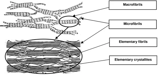
As was discussed in chapter 1.1.2, in higher plants, cellulose is synthesized in the plasma membrane by a ring of six complexes of six hexagonally arranged cellulose synthase enzymes (CESAs): the cellulose synthase complex (CSC) also called “rosette”. In such a reactor, six adjacent β-1,4-glucan chains produced by each of the six CESAs complexes are extruded and probably start associating by Van der Walls forces then form hydrogen bonds [8, 9], thus building the first basic fibrillar unit called “elementary fibril”. Larger and larger associations are then built up to the formation of the complete wall structure.
As shown in Figure 11, the elementary fibrils constituted of six glucan chains, aggregate in microfibrils having typical cross sections of 3,5 to 10 nm and consisting of 36 to 90 glucan chains in land plants and some green algae [13], i.e. six or more elementary fibrils. However, microfibrils can contain until 1400 glucan chains in certain green algae as Valonia macrophysa. In fact, the dimension of the microfibrils is highly dependant of the CSC structure [13] and the latter can undergo strong variation depending on the plant and algae
species (see chapter 1.1.2). The aggregation of the elementary fibrils in microfibrils forms crystalline units called “elementary crystallites”. The size of these crystals is typically 5-6 nm in diameter for bacterial cellulose and 4-5 nm for cotton [23]. Their length is 10-20 nm for regenerated cellulose fibres and longer for cotton and other natural cellulosic fibres [17]. Thereafter, the microfibrils organized in macrofibrils to form the cell wall.
It has to be noticed that there is some confusion in the literature concerning the description of the different aggregated entities, i.e. the elementary fibrils, the microfibrils and the macrofibrils. Often, the microfibrils are considered as the smallest morphological unit [24] instead of the elementary fibril (aggregate of six glucan chains). Considering the fact that microfibrils undergo large dimension variations due to the varied existing structure of the CSC, we will consider the elementary fibril as the smaller basic cellulose unit synthesized. Larger entities as micro and macrofibrils are thus only aggregates of this elementary entity.
Figure 11. Elementary cellulose fibrils synthesis and the different levels of organisation in the cell wall of plant. (Scheme reconstructed with illustrations taken from [1, 25])
Many hypotheses have been suggested for the internal build-up of the fibrillar structure over the years. The model accepted today was proposed by Hearle in 1958 and supported by recent WAXS and 13C CP-MAS NMR results [18]. This model assumes low ordered (“amorphous”) and highly ordered (“crystalline”) regions arranged in a fringed fibrillar fashion. As can be seen on Figure 12, elementary fibrils aggregate in elementary crystallites and form microfibrils and macrofibrils. Several imperfections occur in this structure as the
M Miiccrrooffiibbrriill E Elleemmeennttaarryyffiibbrriill ββββ ββββ--11,,44--gglluuccaanncchhaaiinn C Ceellllmmeemmbbrraannee C CSSCCoorr " "rroosseettttee"" C CEESSAAss M Maaccrrooffiibbrriill
less ordered regions in the microfibrils, the interstice between the microfibrils and the larger voids between the microfibrils and the macrofibrils [17]. The cohesion of these different entities is ensured by an hydrogen bond network.
Figure 12. Representation of the fringed fibrillar model of the fibre structure. (Scheme reconstructed with illustrations taken from [17])
However, considering several studies on “amorphous” cellulose, O’Sullivan [23] suggested that “amorphous” cellulose probably still possess a degree of order. This hypothesis is in accordance with the fact that CSC produces chains that all keep a parallel straight conformation up to coagulating on the cell wall because of their stiffness, their high hydrogen bonding capacity [17, 18] and also topological constraints due to the proximity of the many chains exiting from the CSC orifices [5, 7, 26, 27].
b) Cellulose polymorphs
Cellulose can adopt different crystalline forms – it is a polymorphic material. Native crystalline cellulose is called “cellulose I” and is composed of two main forms Iα and Iβ [28]. Higher plant, e.g wood and cotton, are principally made with Iβ form. Iα is more present in primitive organism like bacteria. Iα phase has a triclinic P-1 structure with one cellulose chain per unit cell and Iβ presents a monoclinic unit cell with two chains per unit (space group P-21) [29]
. Iα is metastable and can be transformed by annealing into the thermodynamically more stable Iβ phase [18].
Elementary fibrils
Elementary crystallites Microfibrils Macrofibrils
Iα cellulose have a unit cell with width parameters (a= 6.74 Å, b= 5.93 Å, c= 10.36 Å, γ-angle= 81°) shorter than those of the Iβ form (a= 7.85 Å, b= 8.27 Å, c=10.38 Å, γ-angle= 96.3°) [30]. As shown on Figure 13, both forms adopt a packing arrangement, in which the molecules are organized in a parallel manner.
Figure 13. Representation of the model of cellulose Iβ
[31]
, intrasheet (100) through the center of the unit cell. (Illustration taken from [30])
Cellulose can also adopt other polymorphic crystal structures. When native celluloses are treated at room temperature with strong alkaline solutions, or when native cellulose is precipitated from a dissolved state, such as the Lyocell process, cellulose adopts a modified crystal structure giving a specific X-ray diffraction pattern (see Figure 15). This modification is known as cellulose II (sometimes called “hydratecellulose”, “mercerized cellulose”, “regenerated cellulose” [17]) and is the most important form from an industrial point of view. The crystal lattice structure of this polymorph has a monoclinic unit cell P21 – like cellulose-I – with axis dimensions of 8.1 Å for the a-axis, 9.05 Å for the b-axis, 10.31 Å for the c-axis, and 117.1° for the γ-angle [30]. Cellulose II unit has an antiparallel arrangement of the chains with two cellulose molecules rotated by 180° around their axis that form a 2/1 helix (Figure 14). The reasons that lead to the irreversible transition from cellulose I and cellulose II without an intermediate stage of dispersion of the cellulose molecules is not yet well understood [32]. It has to be noticed that cellulose II can be produced by Nature by a mutant strain of Gluconacetobacter xylinum[24].
Figure 14. Representation of the model of cellulose II [33] intrasheet (010) through the center of the unit cell. (Illustration taken from [30])
Besides cellulose I and II modifications, two other polymorphs lattice structures of much less practical importance are known, called cellulose III and cellulose IV. Cellulose III is formed by the treatment of native cellulose fibres with liquid ammonia and has a parallel chain arrangement [30]. Cellulose IV can be obtained from cellulose III sources by a treatment in hot glycerol baths under stretch [24]. The chain arrangement of the form IV2 of this polymorph is antiparallel as in cellulose II [30]. The existence of these two modifications is supported by some distinct differences in the X-ray diffraction pattern and in the RMN13C spectra (see Figure 15).
Figure 15. X-ray diffraction pattern (left) and RMN Spectra (right) for the different polymorphs of cellulose. (Illustration taken from [34])
c) Intermolecular hydrogen bonding
As was shown in chapter 1.2.1, cellulose molecules could have one or two intramolecular hydrogen bond. This leaves the possibility for an interlinkage by another intermolecular hydrogen bond between the hydroxyl O-3-H and the hydroxyl O-6 of the neighboring chain (Figure 16). The packing of the cellulose chains in cellulose I is thus very dense and the hydroxyls inside the crystals are not easily accessible.
Figure 16. Intermolecular (and intramolecular) hydrogen bond patterns of cellulose I and cellulose II. (Illustration taken from [18]).
In cellulose II, the position of the hydroxyls favours the formation of intermolecular hydrogen bonds (Figure 16). The intramolecular bonds are similar to cellulose I. Consequently, cellulose II is more densely packed and the cellulose molecules are strongly linked with an average bond length shorter than in cellulose I.
1.2.3. Macrostructural Level
a) Cell wall structure of native celluloses fibres
Cellulose microfibrils and macrofibrils are the basic fibrillar elements of the cell wall building. During the period of cell growth, the fibres possess only a very thin cell wall, named
“primary wall”, which envelops the protoplasm. After completion of the growth in length, the thickness of the cell wall increases by the deposition of cellulose microfibrils in a second relatively thick layer, the “secondary wall”, on the inner side of the primary wall. At the end of the biosynthesis, the plant cell die and the central channel of the cell is more or less narrowed depending of the maturity of the fibre.
The cell wall morphology is composed of cellulose layers differing in their fibrillar texture, as shown schematically in Figure 17 for a cotton hair (or fibre as it is often called) and a wood fibre. These two materials consist of cellulose layers with different thickness and microfibrils orientations. Wood Primary wall Random, 0.1 – 0.5 µm S1 wall 50 - 90°, 0.3 µm S2 wall 10 - 30°, 1 – 5 µm tertiary wall 60 - 90°, < 0.1 µm Middle lamella Wart layer Cotton Cuticle Primary wall Random, 0.1 – 0.5 µm S1 wall 20 - 30°, 0.2 µm S2 wall 35 - 45°, 1 – 8 µm Lumen Reversal Wood Primary wall Random, 0.1 – 0.5 µm S1 wall 50 - 90°, 0.3 µm S2 wall 10 - 30°, 1 – 5 µm tertiary wall 60 - 90°, < 0.1 µm Middle lamella Wart layer Wood Primary wall Random, 0.1 – 0.5 µm S1 wall 50 - 90°, 0.3 µm S2 wall 10 - 30°, 1 – 5 µm tertiary wall 60 - 90°, < 0.1 µm Middle lamella Wart layer Cotton Cuticle Primary wall Random, 0.1 – 0.5 µm S1 wall 20 - 30°, 0.2 µm S2 wall 35 - 45°, 1 – 8 µm Lumen Reversal
Figure 17. Schematic drawing of the macrostructural morphology of a cotton fibre (A) and a wood fibre (B) with : cuticle (rich in pectins and waxes), lumen, middle lamella (mainly lignin), primary wall, reversal of the fibril spiral, S1-secondary wall, S2-secondary wall (main body), tertiary wall, wart layer. For each wall, the thickness and the microfibrils orientation are drawn according to [17, 18, 35, 36].
In the outer cell layer, i.e. the primary wall (P), the microfibrils are almost randomly positioned over a thickness of 500 nm. The secondary wall (S) consists of two layers S1 and S2. In the case of cotton, the thickness of the S1 layer is about 200 nm, and for a wood fibre, it is 300 nm. The microfibrils are parallel and densely packed in a flat S-helix with variable angles function of the nature of fibres (50 to 70° for wood fibres and 20 to 30° for cotton fibres). The S2 layer is several µm-thick depending of the maturity of the fibre and contains most of the cellulose mass [17, 18, 37] (see table 1). The microfibrils from cotton are well aligned with periodical reversal in a so-called S-Z fashion and orientated around 35-45° to the fibre axis. For other natural sources like wood, flax, hemp… the microfibrils are positioned in an extended helix at a high degree of orientation towards the fibre axis (10 to 30° for wood fibres, see also Table 1).
Table 1. Variation of the cellulose content and the spiral angle for some plant cell walls in the S2 layer. (Illustration taken from [37])
The last layer deposited closest to the fibre lumen, called tertiary layer (T) in the case of wood fibres and S3 layer in the case of cotton, is not always present and is very thin (< 100 nm). Its microfibrils are aligned in a flat helix forming a small angle with the cross-sectional axis (60 to 90° for wood fibres).
b) Cell wall components of native cellulose fibres and fine structure of the walls
The primary wall is the most complex part of the cell wall and contains many components. Cellulose (15-30%) and hemicelluloses (25% ; Xyloglucan, Xylan and glucomannan) are present as in the secondary wall but the primary wall may also contains pectins (30% ; Homogalacturonan, RG type I, RG type II [1]), proteins (20% ; strongly glycosyled HRPG, slightly glycosyled PRP, not glycosyled GRP, AGP [38]), lignin and phenolic acids (feluric, coumaric) [39]. The cellulose microfibrils are randomly positioned and linked to hemicelluloses by hydrogen bonds that help the positioning of the microfibrils. A gelled matrix is formed by pectins associated with Ca2+ ions. It determines the porosity and the pH of the wall. Proteins, as Hydroxyprolin Rich Glyco Proteins (HRGPs) so-called expansins or extensins, and enzymes, as endoglucanases, help the elongation of the primary wall, repositioning the cellulose microfibrils by disrupting the hydrogen interactions with hemicelluloses [40] (see Figure 18). At the end of the elongation stage, structural proteins form a secondary network by covalent and hydrogen interactions and block the plasticity of the primary wall [39, 41]. In addition, proteins are supposed to be crosslinked to pectins and also phenolic acids by forming protein-phenolic-protein interactions [42]. In general, the different components present in the primary wall undergo strong interactions as revealed by dynamic FT-IR spectroscopy [43]. The complex structure of the primary wall is shown in Figures 18 and 19.
Fix cut Stick Stretch Fix cut Stick Stretch
Figure 18. Role of the proteins and enzymes during the elongation stage. (Illustration taken from [44])
Figure 19. Complexity of the primary wall at the end of the elongation stage. (Illustration taken from [41]
)
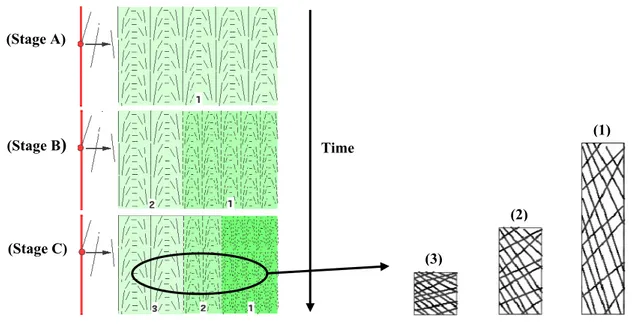
In addition to this complex structure, the arrangement of the cellulose microfibrils is not homogeneous across the thickness of the primary wall (Figure 20). During their synthesis by the protoplasm (in red), microfibrils are ordered in a regular manner (Stage A, zone 1). During the elongation, microfibrils are orientated and are pushed back towards outside (Stages B and C) [41]. The oldest zones are thinned in consequence of the elongation (Stage C, zone 1).
Figure 20. Orientation of the cellulose microfibrils during the elongation stage. (Illustrations taken from [41]) (Stage A) (Stage B) (Stage C) Time (3) (2) (1) HRPGs and enzymes Cellulose microfibrils Hemicelluloses Pectins Structural proteins
The secondary wall is mostly composed of cellulose (50-85%), hemicelluloses (0-25%) and lignin (0-25%) depending to plant species. In the case of cotton fibres, the secondary wall contains almost only cellulose (around 99%). Components are linked together by hydrogen bonds (cellulose-hemicelluloses) and crosslink in the case of cellulose or lignin-hemicelluloses. Several authors assume that the hemicelluloses and lignin are dispersed in layers between the cellulose microfibrils [23, 36, 45]. The cellulose microfibrils are arranged helically as discussed in the preceding chapter on cell wall structure.
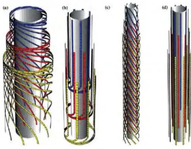
c) Geometrical model for cell wall formation
As was shown in chapter 1.1.2, new tools as computer modelling are now used to describe the synthesis and the morphology of cellulose fibre. Mulder and Emons [16]build a geometrical model to explain how the CSCs, the distance between cellulose microfibrils being deposited and the cell geometry, determine wall texture. In this model they have proposed that the default mechanism, which determines the orientation of cellulose microfibrils as they are deposited in the absence of other influences, has geometrical origin. The first results showed that several known cell wall textures can be reproduced: axial, helical, helicoidal and crossed wall texture as shown in Figure 21.
Figure 21. Different cell wall textures as predicted by the geometrical model of Mulder et al. [16]. (a) helicoidal texture, (b) crossed polylamellate texture, (c) helical texture, (d) purely axial texture.
d) Pore structure
In addition to the fibrillar morphology of the fibre cell wall, there is a system of pores, capillaries, voids, and interstices [18]. There is a wide distribution of the pores in terms of size and shape. A total pore volume and an average pore size are thus not enough to give a complete and precise description of the pore system. Some data on pores of various cellulose samples measured by two techniques are given in table 2 and 3.
Table 2. Data on pores of cellulose samples calculated by SAXS measurements. (Illustration taken from [18])
Table 3. Void characteristics in native cellulose (cotton, spruce…) and regenerated (viscose rayon, viscose staple) measured by mercury porosimetry. (Illustration taken from [18])
The total pore volume and pore size distribution are very sensitive to pre-treatments. Mercerization leads to a decrease in pore diameter and an enhancement of micropore surface while enzyme treatments enlarge the existing pores [46]. Acid hydrolysis enhances the pore system by removing amorphous cellulose from the surface and revealing the macrofibrillar structure of cellulose fibres [47] (see Figure 22).
Figure 22. (A) Untreated cotton linters, (B) Cotton linters after 1.0% cellulose was hydrolyzed, (C) Cotton linters after 11.8% cellulose was hydrolyzed, the macrofibrillar structure is revealed. (Illustration taken from [47])
A B C
a The term porosity denotes the total percentage of pores including the
Drying results in an irreversible reduction of the pore volume due to the closing of the smallest pore, a mechanism called hornification (see Figure 23).
Figure 23. “Conceptual model of pore closure”. When water is evaporated (a > b > c), the pores collapse due to the capillary forces with the high surface tension of water (illustration taken from [48]). Formation of hydrogen bonds block the pore and prevent its re-opening.
Due to the pore and void system, the surface area of cellulose fibres exceeds by far the geometrical outer surface of the fibre. The inner surface area is considered as a key parameter with regard to accessibility and reactivity in dissolution and derivatization processes [18].
e) Particular twisting and kidney shape of cotton fibres
Several twists can be observed along the cotton fibre (Figure 24 a and 24 b) and the cross-sections reveal a “kidney” or “bean” shape (Figure 24 c). At the end of the biosynthesis, before boll opening, the lumen is filled with a liquid containing the cell nucleus and the protoplasm. The twists and the “kidney” shape of the dried fibre are due to the removal of this liquid, also called dehydration or desiccation [49, 50].
Althought the spiral reversal S-Z changes only 1 to 3 times per mm, the number of twists in cotton fibres is higher (between 3.9 and 6.5 per mm). However, the lateral alignment of the fibrillar reversals in the concentric cellulose layers and the ultimate twists in the dried fibres are related. Basra et al. [50] explain that the probability for reversals in the cellulose layers to coincide laterally increases with fibre maturity and increasing secondary wall thickness. When a sufficient number of reversals coincides, stress leads to twist formation. The twist is thus the region where reversals overlap among concentric layers. When cotton fibres are placed under wet conditions, an untwisting occurs in function of the quality of the swelling agent [35].
(a) (b) (c) Figure 24. Twists (a, b) and kidney shape (c) in cotton fibres. (Illustrations taken from [49, 51, 52])
The kidney shape is more or less pronounced in function of the fibre maturity. For a high maturity fibre, the shape is almost ellipsoidal due the thickness of the secondary wall. The influence of maturity can be seen in Figure 25. The blue circle shows a cotton fibre cross-section with a thick and well-developed wall. This is a mature cotton fibre. The red circle shows a fibre cross-section with a thin and poorly developed wall. This is an immature cotton fibre.
Figure 25. Cross-sections of mature (blue circle) and immature (red circle) cotton fibres. (Illustration taken from [53]).
The dehydration involves mechanical strains and resulting deformations along the fibre. Several authors postulate the existence of a bilateral structure due to an asymmetry in the distributions of stresses which occur during the dehydration of cotton. In fact, fibres undergo an important shape modification from a swollen cell tube to a fibre [49, 54]. Observation of ultrathin sections shows that the density of chain packing is not homogeneous in the secondary wall (Figure 26).
Figure 26. Ultrathin sections of cotton fibres perpendicular to fibre axis showing the fine structure of different zones of the secondary wall (illustration taken from [49]). Zone A, B, C correspond to the different zones as defined in Figure 27.
In the cross sections of mature fibres, different zones were thus distinguished as regard to the organization of their fine structure as shown in Figure 27. They were also thought to differ in their accessibility to reagents and their behaviour during swelling [49].
Figure 27. (left) Relations between mechanical strains (full arrows) and deformations (dotted arrows) produced by collapsing of the cotton fibre during initial drying, (right) model showing the changes in packing density and orientation of the fibrillar membranes in a cotton fibre.(Illustration taken from [49])
Conclusions
Many studies have been performed to understand the cellulose structure at all its morphological levels, and the whole structure and organization are now well understood. New methods such as computer modelling are helping to better describe cellulose structure and synthesis. However, several key issues like the full dynamics of cell wall deposition and the role of the various non-cellulosic components in the structuration of the cell wall are mostly unknown. Several aspects are still under question, like the understanding of the formation of the hydrogen bonding network and the transition between cellulose I and cellulose II. The origins of the accessibility and reactivity of cellulose are closely linked to these different aspects.
Primary and secondary walls reveal large differences in their structural organization in terms of components and arrangement of cellulose microfibrils which should have a strong impact on the swelling and dissolution mechanisms of cellulose fibres.
Wood and cotton fibres are rather different in their microfibrils organization, in the presence of lignin in wood and in the absence of hemicelluloses in the secondary wall of cotton. Taking this into consideration, the walls of wood and cotton should show different swelling and dissolution behaviours in a solvent.
1.3. Swelling and dissolution mechanisms of cellulose fibres
Due to their complex and multi-scale structure, the mechanisms of swelling and dissolution of cellulose fibres are not homogeneous. As will be shown, several proposals have been made for explaining the non homogeneous swelling and dissolution of cellulose fibres and cellulose derivatives. This phenomenon has to be related with the fibre source but also with the solvent quality.
1.3.1. Heterogeneous mechanisms of swelling and dissolution of cellulose
fibres and cellulose derivatives
The most spectacular effect of the heterogeneous swelling and dissolution of cellulose fibre is the ballooning phenomenon. The swelling can take place in some selected zones along the fibres. This localised swelling gives the impression of having “balloons” growing as shown on Figure 28. This heterogeneous ballooning swelling has been observed long ago, first by Nägeli in 1864 [55], then by Pennetier in 1883 [56], Fleming and Thaysen in 1919 [57], Marsh et al. in 1941[58], Hock in 1950 [59] or Rollins and Tripp in 1952 [60, 61] (Numerous other studies are also reported in reference [35]).
Figure 28. (a) Cellulose fibre swollen by alkali and carbon bisulfite, (b) Ballon formation in raw cotton. (Illustrations taken from [58, 59]respectively)
In 1954, Ott et al. [62] proposed an explanation of this phenomenon. It must be noted that similar explanations were suggested in all the preceding references. For a wide range of cellulose fibres, the microfibrils of the secondary wall are aligned in a helical manner with respect to the long axis of the fibre. Ott et al. deduces that the swelling is greater transversely than lengthwise (as it is generally for fibres where the orientation of the cellulose chains is mainly in the fibre direction). Consequently, they proposed that “when raw cotton fibres are placed in certain swelling agent, the radial expansion of the cellulose in the secondary wall
causes the primary wall to burst. As the expanding swollen cellulose pushes its way through these tears in the primary wall, the latter rolls up in such a way as to form collars, rings or spirals which restrict the uniform expansion of the fibre and forms balloons”. Ott et al. have also reported that an irregular swelling can be visible with fibre without the primary wall (removed by alkaline digestion) due to the S1 wall which restricts lateral expansion.
All the authors assume that the ballooning phenomenon has structural origins, i.e. linked to morphological variations between the different walls. However, the explanation cited above assumes that cellulose is in a swollen state in each of the balloons, which remains to be demonstrated. In addition, no data exist on the respective roles of the primary and secondary wall layers in this process. Finally, the different stages that lead to the ballooning phenomenon have not been well described. In paper I of our study, we will describe by means of high resolution observations the state of the cellulose inside the balloons and the swelling and dissolution mechanisms of the different walls.
The main regions implied in the ballooning have been described in details by Cuissinat and Navard [63, 64]. As shown on Figure 29, three zones have been identified: the membrane, the inside of the balloons and the unswollen sections. However, the exact origin of these three zones was not reported.
Unswollen sections between the balloons Membrane
Inside of the balloons
Figure 29. Identification of the three zones of a wood fibre swollen by ballooning in water- 8% NaOH. (Adapted from [63, 64])
Stawitz and Kage [65] have also reported heterogeneous swelling and dissolution in carboxymethyl cellulose (CMC) fibres. The mechanisms of swelling are described on Figure 30. Fibres are swelling by ballooning with a helical structure around the balloons (1 to 3). The breakage of the helical structure and the unswollen sections between the balloons leads to a high homogeneous swelling (4 to 6). The highly swollen sections are then tear into thin sections and finally into fragments (7 to 10). In paper II of our study, we will see that the
model of Stawitz and Kage for CMC is also valid for all wood based fibres in NaOH-water systems. By selecting well defined samples and solvent, we were able to describe in more details in what way the observed features are swelling and dissolving and the exact origins of these phenomena.
Figure 30. The swelling of Carboxymethyl cellulose (CMC). (Illustration taken from [65])
1.3.2. Influence of the solvent quality on swelling and dissolution
mechanisms of cellulose fibres
Chanzy et al. [66] have investigated the swelling and dissolution of various cellulose fibres, both native and regenerated, in N-methylmorpholine N-oxide (NMMO) with different amounts of water. Three domains of “water concentration” were found to be important. When the amount of water was low (NMMO- 16% water), cellulose fibres, such as ramie, cotton and wood were dissolving rapidly by fragmentation without significant swelling. At higher water concentration (e.g., NMMO- 18-20% water), the ramie fibres exhibited a heterogeneous swelling: a ballooning phenomenon was observed in localized places. In the case of wood and cotton fibres, the ballooning was well defined and the difference of swelling with ramie fibre was attributed to a difference of organization of the cellulose microfibrils within the various species. After the removal of the swelling agent, they observed that the initial ramie fibres were converted into an unoriented cellulose II crystalline structure. Chanzy et al. called this region “irreversible swelling”. With more water (e.g., NMMO- 20% and more), ramie fibres were sometimes both having cellulose I and II after the removal of the swelling agent but most of the time the cellulose I structure of the cellulose crystals was conserved.
The regions of dissolution, irreversible and reversible swelling of cellulose in the NMMO- water solvents as defined by Chanzy et al. are schematically represented in Figure 31. Region
A corresponds to the dissolution of the ramie fibres by fragmentation without swelling. Region B and C corresponds the irreversible swelling and reversible swelling ,respectively. When water amounts were below 28% (D), no visible changes were observed which thus corresponds to a region of non activity.
Figure 31. Partial phase diagram [66] of the system NMMO-water on native cellulose fibres showing the various regions of dissolution : (A) by fragmentation without swelling, (B) irreversible swelling, (C) reversible swelling, (D) non activity.
The designation “reversible or irreversible swelling” corresponds to the cellulose organisation within the cellulose crystals but not to structural and morphological modifications that cannot be recovered after the swelling experiments. In the irreversible zone (B), only cellulose II was present after the removal of the swelling agent by washing with water and subsequent drying and cellulose I structure cannot be seen. In the reversible zone (C), the swelling experiments do not lead to a cellulose II lattice and the cellulose I crystal organisation is conserved after washing and drying.
The most important result in this study is that by changing the water amount from 16% to 20 % w/w, an important change from the dissolution by fragmentation of cellulose to reversible swelling was observed. In addition, pronounced differences were found in the crystalline structures of the fibres in function of the amount of water.
Based on the preceding study, Cuissinat and Navard [63, 64] performed observations by optical microscopy of free floating fibres between two glass plates for a wide range of solvent quality. They have identified four main dissolution modes for wood and cotton fibres in function of the quality of the solvent, in NMMO:
Mode 1: Fast dissolution by fragmentation (under 17% water).
Mode 2: Swelling by ballooning and dissolution (from 18 to 24 % water).
Mode 3: Swelling by ballooning and no complete dissolution (from 25 to 30 % water). Mode 4: Low homogeneous swelling and no dissolution (above 35 % water).
These different dissolution mechanisms are summarized in Figure 32. These mechanisms have been also observed with ionic liquids solvent [67] and for a wide range of plant fibres [68] and some cellulose derivatives if the derivatization occurred without dissolution [69]. From all these studies, it is shown that the key parameter in the dissolution mechanisms is the morphological structure. If the original wall structure of the native fibre is preserved, the dissolution mechanisms are similar for wood, cotton, other plant fibres and some cellulose derivatives, the solvent quality driving the type of mechanism that will occur for a given fibre type.
Observation by optical microscopy between two glass plates
No complete Dissolution
Bad quality solvent (e.g. water- 8% NaOH or NNMO- 25 to 35% water)
Swelling by ballooning No complete
Dissolution
Bad quality solvent (e.g. water- 8% NaOH or NNMO- 25 to 35% water)
Swelling by ballooning
Moderate quality solvent (e.g. NNMO- 18 to 24% water)
Swelling by ballooning
Moderate quality solvent (e.g. NNMO- 18 to 24% water)
Swelling by ballooning
Non solvent (e.g. NMMO- 35% water and over)
Homogeneous swelling No Dissolution
Non solvent (e.g. NMMO- 35% water and over)
Homogeneous swelling No Dissolution
Solvent quality Good quality solvent
(e.g. NMMO- under 17% water)
Fragmentation
Dissolution
Good quality solvent (e.g. NMMO- under 17% water)
Fragmentation
Dissolution
Figure 32. Swelling and dissolution mechanisms of wood and cotton fibres in function of the solvent quality. (Adapted from [63, 64])
Conclusions
The swelling and dissolution mechanisms of cellulose fibre are strongly influenced by the solvent quality but the exact origins of such phenomena are not well understood. The roles of the different levels of the cellulose structure are not well established. By controlling the quality of the solvent, the dissolution conditions (as the tension) and the fibre sources, it will be shown that it is possible to reveal the characteristic mechanisms of the swelling and dissolution at the different length scales of the cellulose structure from the macromolecule to the walls of the fibre. This will be demonstrated in the next parts of this dissertation.
References
[1] Lerouxel, O.; Cavalier, D. M.; Liepman, A. H.; Keegstra, K. Biosynthesis of plant cell wall polysaccharides – a complex process. Curr. Opin. Plant Biol. 2006, 9, 1–10.
[2] Zaffran, J. Initiation à la biologie végétale. ed. Ellipses, Paris, 1998.
[3] Preston, R. D. Formation of Wood in Forest Trees. ed. Zimmerman M.H., Academic Press, New York, 1964, 169–188.
[4] Brown Jr, R. M.; Montezinos, D. Cellulose microfibrils: visualization of biosynthetic and orienting complexes in association with the plasma membrane. Proceedings of the National Academy of Sciences of the USA, 1976, 73, 143–147.
[5] Mueller, S. C.; Brown Jr, R. M. Evidence for an intramembrane component associated with a cellulose microfibril synthesizing complex in higher plants. J. Cell Biol., 1980, 84, 315–326.
[6] Kimura, S.; Laosinchai, W.; Itoh, T.; Cui, X.; Linder, C. R.; Brown Jr, R. M. Immunogold labeling of rosette terminal cellulose-synthesizing complexes in the vascular plant Vigna angularis. Plant Cell, 1999, 11, 2075–2085.
[7] Diotallevi, F.; Mulder, B. The cellulose synthase complex: a polymerization driven supramolecular motor. Biophys. J., 2007, 92, 2666–2673.
[8] Cousins, S. K.; Brown Jr, R. M. Cellulose I microfibril assembly: computational molecular mechanics energy analysis favours bonding by Van de Waals forces as the initial step in crytallization. Polymer, 1995, 36, 3885–3888.
[9] Cousins, S. K.; Brown Jr, R. M. X-ray diffraction and ultrastructural analyses of dye-altered celluloses support Van der Walls forces as the initial step in cellulose crystallization. Polymer, 1997, 38, 897–902.
[10] Robert, S.; Mouille, G.; Höfte, H. The mechanism and regulation of cellulose synthesis in primary walls: lessons from cellulose deficient Arabidopsis mutants. Cellulose, 2004, 11, 351–364.
[11] Taylor, N.G.; Gardiner J.C.; Whiteman, R.; Turner, S.R. Cellulose synthesis in the Arabidopsis secondary cell wall. Cellulose, 2004, 11, 329–338.