Parallel magnetic resonance imaging reconstruction problems using wavelet representations
209
0
0
Texte intégral
Figure
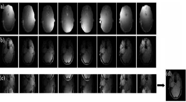
![Figure 2.10: Example of motion artifacts; right: blur artifacts; left: phantom artifacts [Gerardin, 2006].](https://thumb-eu.123doks.com/thumbv2/123doknet/2914337.75902/43.892.251.671.783.998/figure-example-motion-artifacts-artifacts-phantom-artifacts-gerardin.webp)
+7

Documents relatifs