HAL Id: tel-01637706
https://pastel.archives-ouvertes.fr/tel-01637706
Submitted on 17 Nov 2017HAL is a multi-disciplinary open access archive for the deposit and dissemination of sci-entific research documents, whether they are pub-lished or not. The documents may come from teaching and research institutions in France or abroad, or from public or private research centers.
L’archive ouverte pluridisciplinaire HAL, est destinée au dépôt et à la diffusion de documents scientifiques de niveau recherche, publiés ou non, émanant des établissements d’enseignement et de recherche français ou étrangers, des laboratoires publics ou privés.
Mechanisms of action of satiating gut peptides in the
regulation of food intake through vagal afferent pathways
Charlotte Ronveaux
To cite this version:
Charlotte Ronveaux. Mechanisms of action of satiating gut peptides in the regulation of food in-take through vagal afferent pathways. Life Sciences [q-bio]. AgroParisTech, 2015. English. �NNT : 2015AGPT0002�. �tel-01637706�
i
Doctorat ParisTech
T H È S E
pour obtenir le grade de docteur délivré par
L’Institut des Sciences et Industries
du Vivant et de l’Environnement
(AgroParisTech)
Spécialité :
Science de vie et santé
Directeur de thèse : Helen RAYBOULD Co-encadrement de thèse : Daniel TOME
Jury
Mme Claire GAUDICHON, Professeur, AgroParisTech Présidente Mme Inge DEPORTERE, Professeur, University of Leuven Rapporteur M. Thomas LUTZ, Professeur, University of Zurich Rapporteur M. Mihai COVASA, Directeur de recherche, INRA, Institut Micalis Examinateur
M. Daniel TOME, Professeur, AgroParisTech Co-encadrement de thèse
Mme Helen RAYBOULD, Professeur, University of California- Davis Directrice de thèse
présentée et soutenue publiquement par
Charlotte Ronveaux
le 6 Janvier 2014Mechanisms of action of satiating gut peptides in the regulation of food
intake through vagal afferent pathways
Acknowledgements
The completion of my dissertation would not have been possible without the guidance of my mentors, help from lab members, and support from my family and friends.
I would like to express my deepest gratitude to my advisor, Helen Raybould, for her excellent guidance, patience, and encouragement through the past three years. Helen you are a wonderful mentor and I am grateful to be continuing on in your lab.
Thank you to Daniel Tomé for your help and guidance through my dissertation, and for the encouragement to do my PhD in Helen’s lab.
I would like to thank Will de Lartigue for being an amazing and caring mentor. Thank you for your time, patience and guidance through my dissertation. I am very grateful to have worked so closely with you, I have learned so much.
Thank you Kristi Hamilton for being such a great friend and amazing coworker. I greatly appreciate the times you let me talk through ideas, vent when things weren’t working and always putting a positive spin on things.
Thank you to all the past and present members of the lab who have been here to help in more ways than one; Jim, Gaelle, Shirley, Liz, Sofia, Myrta, Analise, Ricky and Mathilde.
I would like to express my gratitude towards my family and friends for all their love and support. To my parents, thank you for your unconditional love and encourgement and most importantly, thank you for teaching me to never give up.
iv
I would like to especially thank Tim for being there during my good days and bad. Your kindness, humor and unconditional support have made everything easier. I wouldn’t know what to do without you.
Thesis Summary
As the initial interface for nutrient sensing, digestion and absorption, the gastrointestinal (GI) tract plays a critical role in the regulation of energy homeostasis. Information that arises from the GI tract is key to normal physiological responses controlling gut function and regulating food intake. Vagal afferent neurons (VAN) are a major pathway by which information about ingested nutrients reaches the central nervous system to influence GI function and food intake behavior. VAN express receptors for many of the regulatory peptides released from the gut that are involved in regulation of food intake and body weight.
This dissertation addresses the role of two gut peptides, leptin and glucagon-like peptide-1, acting at the level of VAN, to inhibit food intake. First, the mechanism of action of glucagon-like peptide-1 (GLP-1) on VAN is addressed. GLP-1-induced satiation requires a postprandial state; the data support that feeding changes the localization of GLP-1Rs from the cytoplasm to the neuronal cell membrane. Further, ghrelin and its receptor GHSR1 expressed by VAN is involved in regulating GLP-1 receptor translocation. Second, the importance of leptin receptor expression by VAN in the development of hyperphagia and obesity was demonstrated by selective knockout of the leptin receptor (LepR) in VAN; mice express an obesogenic phenotype.
Obesity and its resultant health consequences are a major worldwide health problem. Effective or preventative treatments for obesity are limited. Our findings have filled the gap in our knowledge of the mechanism of GLP-1 and leptin signaling on VAN. Understanding the physiology regulating feeding behavior is imperative in developing non-invasive anti-obesity treatments.
vi
Resumé
Le tractus gastro-intestinal, interface initiale pour la détection, la digestion et l'absorption des nutriments, joue un rôle critique dans la régulation de l'homéostasie énergétique. Les signaux qui proviennent du tractus gastro-intestinal sont nécessaires au contrôle de la fonction intestinale et de la régulation de la prise alimentaire. Les neurones afférents vagaux (NAV) sont une voie importante via laquelle les informations sur les nutriments ingérés atteignent le système nerveux central pour influencer ces deux fonctions. Les NAVs expriment les récepteurs pour la plupart des peptides régulateurs libérés par l'intestin impliqués dans la régulation de la prise alimentaire et du poids corporel.
Cette thèse porte sur le rôle de deux peptides de l'intestin, la leptine et le glucagon-like peptide-1 (GLP-1), qui agissent au niveau des NAVs pour inhiber la prise alimentaire. Tout d'abord, nous expliquons le mécanisme d'action du GLP-1 sur les NAVs. La satiété induite par le GLP-1 nécessite un état post-prandial ; les données confirment que le statut nutritionnel régule la localisation du GLP-1R du cytoplasme vers la membrane des cellules neuronales. De plus, la ghréline et son récepteur GHSR1, exprimés par les NAVs, sont impliqués dans la régulation de la translocation du GLP-1R. Deuxièmement, ’utilisation de souris knockout pour le recepteur a la leptine sur les NAVs nous a permis de montrer l’importance de ce recepteur dans la physiopathologie de l’obésité et de l’hyperhagie. En effet, ces souris KO présentent un phénotype obésogène.
L'obésité et ses conséquences sur la santé sont des problèmes majeurs de santé dans le monde entier. Les traitements efficaces de prévention ou de l'obésité sont
limités. Nos résultats ont apporté des connaissances sur le mécanisme du GLP-1 et sur la signalisation de la leptine au niveau es NAVs. Comprendre la physiologie de la régulation de la prise alimentation est impératif dans le développement des traitements non-invasifs contre l’obésité.
Resumé substantiel de thèse
L’obésité est devenue une pandémie mondiale dont la prévalence a augmenté au cours des dernières décennies. L’obésité est associée à des comorbidités graves telles que le diabète de type 2, l’hypertension, et les maladies cardiovasculaires. C’est un problème de santé publique pour lequel on observe un manque de traitements simples et efficaces. Le contrôle de la prise alimentaire et du poids corporel est un mécanisme complexe qui implique l’intégration de nombreux signaux périphériques au niveau du système nerveux central. Ainsi les signaux humoraux d’un certain nombre de tissus comme le pancréas, le foie ou le tube digestif influencent l'homéostasie énergétique. En raison des données récentes montrant une grande efficacité de la chirurgie bariatrique contre l’obésité, une grande attention est actuellement portée sur le rôle de l’intestin dans cette pathologie.
Suite à l’absorption de nutriments lors d’un repas, des signaux nerveux et hormonaux, témoin de la quantité et de la qualité des aliments ingérés, sont émis le long du tractus digestif. Les hormones sécrétées dans la muqueuse intestinale peuvent agir soit directement par voie humorale sur l’hypothalamus, soit via les récepteurs présents sur les terminaisons vagales. L’activation de cette voie afférente vagale va relayer des signaux efférents jusqu’au tissus cibles pour contrôler la fonction intestinale et le comportement alimentaire. Les neurones afférents vagaux (NAV) sont la principale voie de communication entre le tube digestif et le cerveau. Le nerf vague est le lien entre les signaux périphériques de l’intestin et le système nerveux central dans le contrôle de la fonction digestive et du comportement alimentaire. Une vagotomie chirurgicale ou la destruction de neurones par la capsaïcine chez les rongeurs entraîne une augmentation
x
de la prise alimentaire et inhibe les signaux de satiété. La signalisation vagale contribue donc à la régulation de la prise alimentaire.
Cette thèse vise à comprendre le mécanisme d'action de deux hormones anorexigènes, le Glucagon like peptide-1 (GLP-1) et la leptine, dans le contrôle de la prise alimentaire par les NAVs. Nous avons divisé cette présentation en deux parties. Tout d'abord, nous identifierons le mécanisme d'action du GLP-1 sur le nerf vague. Nous avons démontré que, contrairement à tout autre récepteur couplé à la protéine G qui augmente le niveau d'expression de sa protéine, le récepteur du GLP-1 modifie sa localisation cellulaire en fonction du statut nutritionnel. De plus, nous étudierons les médiateurs impliqués dans cette translocation en réponse à la prise alimentaire. Dans une deuxième partie, nous montrerons l'importance de la résistance à la leptine au niveau des NAVs en invalidant spécifiquement les récepteurs de la leptine sur le nerf vague. Nous avons également comparé la différence entre les souris mâles et femelles KO au niveau de la composition corporelle et de la prise alimentaire. Ce travail vise à approfondir nos connaissances sur les mécanismes physiologiques de la régulation de l'homéostasie énergétique.
Première Partie: GLP-1
GLP-1 et son récepteur se trouvent au niveau périphérique et central. Au niveau périphérique, le GLP-1 est dérivé du gène pré-proglucagon présent dans les cellules L intestinales et les cellules A du pancréas. Le GLP-1 est sécrété par les cellules L en réponse à la prise alimentaire. En condition de jeun, les concentrations du GLP-1 sont très faibles et augmentent avec l'ingestion de nutriments. De nombreuses études ont
montré que le GLP-1 est rapidement dégradé dans sa forme inactive par la dipeptidyl peptidase IV (DDP-IV).
Le GLP-1 peut traverser la barrière hémato-encéphalique mais étant dégradé très rapidement il est peu probable qu'une quantité importante de GLP-1 périphérique actif puisse atteindre le cerveau. Dans des conditions physiologiques et en raison de la courte demi-vie plasmatique du GLP-1, il agit plus probablement localement dans la paroi intestinale pour influencer la prise alimentaire. Les NAVs sont un site principal d'action pour les signaux anorexigène dérivés de l’intestin. L'administration systémique de GLP-1 augmente l'activité électrophysiologique des neurones du ganglion noueux. De plus, une désafférentation sous-diaphragmatique atténue les effets rassasiant du GLP-1 chez l'homme. En revanche, les souris GLP-1R knockout (KO) présentent un poids corporel et une prise alimentaire normale, ce qui remet en question le rôle physiologique du GLP-1 endogène dans le contrôle de la prise alimentaire.
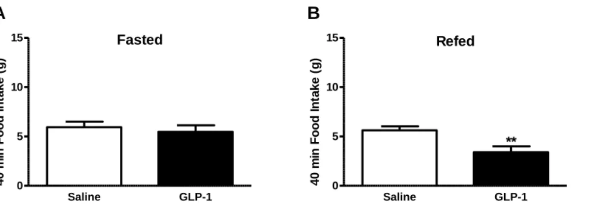
En effet, il existe des incohérences dans la littérature en ce qui concerne le rôle du GLP-1 natif périphérique dans ce contrôle. La plupart des méthodes utilisées dans ces études n’utilisent pas le GLP-1 dérivé de l’intestin mais utilise soit des analogues du GLP-1 qui échappent à la dégradation et peuvent ainsi facilement atteindre les récepteurs centraux soit administrent de façon prolongée le GLP-1 natif en perfusion intraveineuse. Dans notre étude, nous avons cherché à imiter les actions du GLP-1 natif par l'administration d'une injection aiguë de GLP-1 périphérique chez les rats soit à jeun soit à jeun puis re-nourris avant l'injection. Nous avons ainsi démontré que le GLP-1 périphérique nécessite un état postprandial pour induire la satiété.
xii
Des travaux antérieurs ont montré que les NAVs changent leur phénotype neurochimique en fonction de l'état nutritionnel. Les NAV présentent deux phénotypes : orexigène ou anorexigène. Dans un état à jeun, l'expression des récepteurs anorexigènes est diminuée tandis que l'expression des récepteurs orexigènes est augmentée. A l'opposé, ces changements sont inversés par la prise alimentaire par un mécanisme dépendant de la CCK. Étant donné que le GLP-1 ne diminue pas la prise alimentaire dans un état réalimenté, nous avons supposé que l'expression de la protéine du GLP-1R changeait selon le statut nourris ou à jeun.
Nous avons montré que ni l’expression d'ARNm ni la protéine du GLP-1R ne sont altérées sur les NAVs par l'alimentation, mais que la localisation du GLP-1R sur les corps cellulaires des NAVs est modifiée. La localisation du GLP-1R sur la membrane plasmique des neurones des ganglions noueux augmente de manière significative en fonction de la consommation énergétique par rapport à l'état à jeun. Ces résultats signifient que le GLP-1R exprimé sur les neurones afférents vagaux se déplace vers la membrane plasmatique dans la période postprandiale. Le déplacement du récepteur vers la membrane fournit une explication possible au fait que l'administration exogène de GLP-1 inhibe la prise alimentaire seulement après un repas.
Mécanisme de la translocation du GLP-1R
Afin de comprendre le mécanisme de GLP-1 sur les NAVs, nous avons voulu comprendre les facteurs qui sont impliqués dans la translocation du GLP-1R. Il a été montré que les hormones dérivées de l’intestin agissent en synergie pour réguler la prise alimentaire, en particulier au niveau du nerf vague. Mais le mécanisme de
translocation du GLP1-R vers la membrane plasmatique n’est pas encore connu. Deux hypothèses sont possibles : soit un signal est libéré suite à la consommation alimentaire qui amène le récepteur à la surface, soit la diminution d’un signal en réponse à la mise à jeun maintient le récepteur dans le cytoplasme. Nous avons décidé d'étudier les interactions entre le GLP-1 et la CCK et entre le GLP-1 et la ghréline.
Nous avons démontré in vivo que la ghréline, plutôt que la CCK, module les effets rassasiant du GLP-1 natif systémique. Lorsque la ghréline endogène est bloquée, le GLP-1 diminue de façon significative la consommation énergétique dans un état de jeûne chez le rat. Dans un état de jeûne, le GLP-1 augmente l'activité des NAVs en présence d'un antagoniste du récepteur de la ghréline. Par ailleurs, nous avons confirmé, en culture, que la translocation du GLP-1R varie en fonction de la composition nutritionnelle du milieu. Le blocage de la ghréline en condition nourrie restaure le GLP-1R au niveau de la membrane plasmique. In vitro, la ghréline inhibe la translocation du GLP-1R vers la membrane plasmatique, mécanisme atténué en présence d'un antagoniste du récepteur de la ghréline. Les données indiquent que la ghréline conserve les récepteurs dans le cytoplasme par l'intermédiaire de l'AMPc et de la voie de la MAPK p38 dans un état à jeun.
Nous avons ainsi approfondi notre connaissance sur le mécanisme de régulation de la translocation du GLP-1 sur le NAV. La ghréline est un médiateur majeur dans la signalisation du pouvoir rassasiant du GLP-1 sur les NAVs.
xiv
Deuxième Partie: Leptine
Récemment, beaucoup d'attention a été portée sur la leptine, une hormone dérivée du tissu adipeux et de l'intestin, qui contrôle l'apport énergétique, la graisse corporelle, la reproduction et l'homéostasie glucidique. La leptine est un régulateur clé de l'homéostasie énergétique; elle diminue la prise alimentaire et le poids corporel chez les rongeurs et chez l'homme. De plus, les taux circulants de leptine sont corrélés avec l'adiposité. Les récepteurs de la leptine sont exprimés dans des sites centraux et
périphériques, et l’expression la plus élevée se trouve dans le noyau arqué (ARC) de
l'hypothalamus. Un déficit de la leptine et de son récepteur induit une augmentation de la prise alimentaire et de l'adiposité et entraine une obésité morbide chez les rongeurs et l’homme. L’hyperphagie et l'obésité sont associées à la résistance à la leptine caractérisée par l'incapacité de répondre à la leptine exogène et endogène. Il a été démontré que la résistance à la leptine dans l’ARC de l'hypothalamus est un facteur clé de l'obésité.
Résistance à la leptine sur les NAVs
La signalisation de la leptine sur les NAVs joue un rôle important dans la régulation de l'homéostasie énergétique et le développement d'une résistance à la leptine sur les NAVs conduit à un phénotype obèse. Dans des modèles d’obésité induite par un régime riche en lipide, la résistance à la leptine sur les NAVs entraine une augmentation du poids corporel et de la prise alimentaire. En parallèle, en réponse à la leptine, la phosphorylation de STAT-3, marqueur de la signalisation de la leptine, est diminuée sur les NAVs. La résistance à la leptine ne se produisant qu’après l'augmentation de la prise alimentaire, du poids corporel et de l'adiposité, nous avons
supposé que la résistance à la leptine spécifiquement sur les NAVs jouait un rôle important dans la physiopathologie de l'obésité.
Notre hypothèse était que la résistance à la leptine sur les NAVs induit l’hyperphagie et conduit à un phénotype obèse. En utilisant un système Nav1.8Cre-LoxP, nous avons développé une souris KO conditionnel qui invalide le récepteur de la leptine seulement dans les neurones afférents primaires. Les souris mâles KO présentent une augmentation du poids corporel, de la prise alimentaire et de l'adiposité par rapport à un témoin sauvage. Les souris KO ont une sensibilité réduite à la CCK et ont été incapables de répondre à la leptine. La CCK est un médiateur prédominant du “switch” de phénotype des NAVs, et l'absence du récepteur de la leptine exprimée par NAV compromet l'activation de NAV par la CCK. Nous avons étudié si le phénotype de
sensibilité à l’obèsité chez les souris KO était dû à la perte de plasticité des NAVs. En
effet, le phénotype des NAVs des souris KO est bloqué dans un état orexigénique : les récepteurs orexigènes sont constamment exprimés indépendamment de l'état de l'alimentation.
Pour déterminer si la résistance à la leptine sur les NAVs est nécessaire pour le développement de l'obésité, nous avons nourris les souris KO et les souris sauvages (WT) avec un régime riche en lipide pendant trois mois. Comme attendu, la consommation d’un régime hyperlipidique a augmenté le poids corporel et la grasse masse des souris WT comparées aux souris WT nourries avec un régime contrôle. En ce qui concerne les souris KO, après 21 semaines, elles présentent un poids corporel et une masse grasse plus importante que les souris WT nourris avec régime témoin. En
xvi
revanche, avec un régime riche en lipide, le poids corporel et la masse grasse des souris KO étaient moins importants des souris WT.
Nous avons démontré que la résistance à la leptine sur les NAVs est suffisante pour induire une augmentation du poids même nourri avec un régime contrôle. En revanche, la consommation d'un régime hyperlipidique n’augmente pas le poids corporel chez les souris KO. Nous en avons donc conclu que d'autres facteurs sont impliqués dans l’augmentation du poids corporel induit par un régime riche en lipide et qu'il y a d’autres facteurs qui augmentent la prise alimentaire avant la résistance à la leptine sur les NAV.
Nous présumons que les souris KO acquièrent des mécanismes compensatoires pour corriger la perte des récepteurs de la leptine sur les NAVs au cours du développement les empêchant ainsi de prendre plus de poids lors d’une consommation d’un régime hyperlipidique.
Différences des sexes dans la régulation de la prise alimentaire
Les fluctuations des concentrations d'hormones gonadiques influencent le comportement alimentaire et la composition corporelle. L'oestrogène influence l'homéostasie métabolique en régulant l'appétit et la prise alimentaire. Les rattes ovariectomisées (OVX) sont hyperphagiques et présentent une augmentation du poids corporel et de l'adiposité. En outre, les rattes OVX n’ont pas le même comportement alimentaire que les témoins. Le traitement à l'oestrogène normalise ce comportement et rétablit la consommation alimentaire et la composition corporelle.La perte du récepteur de la leptine spécifiquement sur les NAVs conduit à un phénotype obésogène quel que soit le sexe: les souris KO femelles et mâles voient leur poids corporel et leur adiposité augmentés par rapport aux témoins. De plus, les souris KO augmentent leur apport alimentaire pendant la phase de nuit par rapport aux WT. De plus, l’absence du récepteur à la leptine sur les NAV atténue l’effet rassasiant de la CCK et de la leptine.
Nous avons démontré que la perturbation de la signalisation de la leptine induit des différences de composition corporelle et de comportement alimentaire entre les souris femelles et mâles. Le poids corporel des souris est significativement augmenté chez les males par rapport aux femelles. En revanche, la masse grasse est significativement plus importante chez les femelles que chez les mâles. Les souris KO femelles et mâles mangent plus que les souris WT, mais leur comportement alimentaire est très différent. Les souris KO femelles diminuent la quantité du repas mais augmentent leur fréquence par rapport aux souris WT. A l’opposé, les souris KO mâles mangent des repas plus copieux à une plus faible fréquence.
Nous avons mis en évidence que la résistance à la leptine sur les NAV mène à l'obésité par différents comportements alimentaires chez les souris mâles et femelles. Des travaux supplémentaires sont nécessaires pour comprendre les mécanismes conduisant à ces différences entre mâles et femelles. Comprendre le rôle des peptides de l'intestin et la façon dont leur signalisation module la prise alimentaire dans les deux sexes est nécessaire afin de développer un traitement non-invasif contre l’obésité.
xviii
Conclusion
Nous avons démontré que le GLP-1 périphérique joue un rôle important dans le contrôle de la prise alimentaire et que la signalisation du GLP-1 sur les NAVs est fonctionnelle et est régulée par la ghréline. Il apparaît aussi clairement que la résistance à la leptine sur les NAVs est nécessaire et suffisante pour induire l'obésité. L’absence de signalisation de la leptine sur les NAVs induit l'obésité d’une manière différente chez les femelles et les mâles. Il est nécessaire de comprendre la physiopathologie de maladies métaboliques pour lutter contre les symptômes et développer des traitements efficaces et des mesures préventives. Comprendre les dysfonctionnements des voies de détection des nutriments par les afférences vagales est essentiel pour le développement de thérapies contre l'obésité. Les études présentées dans cette thèse soulignent l'importance de la voie afférente vagale dans la réponse aux signaux hormonaux dérivés de l’intestin.
List of Publications and Scientific Communications
Publications
Ronveaux CC, De Lartigue G., Raybould HE (2014) Ability of GLP-1 to decrease food intake is dependant on Nutrient Status. Physiology and Behavior. 135(222-9).
De Lartigue G. Ronveaux CC, Raybould HE (2014) Leptin receptor knockout in vagal afferent neurons drives hyperphagia and weight gain. Molecular Metabolism. 3(6):595-607 (Featured on the cover).
Ronveaux CC, Tome D, Raybould HE (2015) Glucagon-like-peptide-1 interacts with ghrelin and leptin to regulate glucose metabolism and food intake through vagal afferent neuron signaling. Journal of Nutrition (Ahead of Print).
In Preparation
Ronveaux CC, De Lartigue G., Raybould HE. Ghrelin inhibits translocation of GLP-1R to the plasma membrane of vagal afferent neurons. In preparation (2014).
M. Arnold, Ronveaux CC, de Lartigue G., Langhans W., Raybould HE. (2014) Vagal afferent neurons in rats and mice express the monocarboxylate transporter-2 (MCT2). In
preparation (2014).
Prebiotic bovine milk oligosaccharides improves gut barrier function in high-fat diet induced obesity (2014). Hamilton MK, Ronveaux CC, Barile D, Mills D, and Raybould HE. In preparation (2014).
xx
Presentations
(*denotes presenter)
Oral Presentations
Ghrelin inhibits translocation of GLP-1Rs to the plasma membrane on vagal afferent neurons. Ronveaux CC.*, de Lartigue G., Raybould HE. Society for the Study of Ingestive Behavior. Seattle Washington. August 2014. (Received ABIES Internataional Travel Award and New Investigator Travel Award).
Deletion of leptin signaling in vagal afferent neurons results in an obese phenotype. Ronveaux CC.*, de Lartigue G., Raybould HE. Interdisciplinary Graduate and Professional Student Symposium. Davis, California. April 2014.
Ability of GLP-1 to decrease food intake is nutrient dependent. Ronveaux CC.*, de Lartigue G., Raybould HE. Society for the Study of Ingestive Behavior. New Orleans, Louisana. August 2013.
GLP-1R expression on vagal afferent neurons change according to nutritional status. Ronveaux CC.*, de Lartigue G., Raybould HE. Interdisciplinary Graduate and Professional Student Symposium. Davis California. April 2013. (Received Dean’s Prize for Best Oral Presentation in Veterinary Medicine Award).
Posters
Progressive Increase in Large Intestine Transcellular but Not Paracellular Permeability Correlates with Plasma Endotoxemia in Diet-Induced Obese Rats Digestive. Boudry G*, Hamilton MK, de Lartigue G, Ronveaux CC, Raybould HE. Disease Week (DDW), Orlando, May 2013.
Prebiotic bovine milk oligosaccharides improves gut barrier function in high-fat diet induced obesity. Hamilton MK*, Ronveaux CC, Barile D, Mills D, and Raybould HE. October 2014.
Table of Contents
Acknowledgements……….………..iii
Thesis summary………...v
Resumé………..vi
Resumé substaniel de thèse………...ix
Première Partie: GLP-1
………...x
Mécanisme de la translocation du GLP-1R
……….xii
Deuxième Partie: Leptine
………..………xiv
Résistance a la leptine sur les NAVs
………...………xiv
Différences des sexes dans la régulation de la prise alimentaire
………..xvi
Conclusion
………...xviii
List of Publications and Scientific Communications………..xix
List of Figures………...………….4
Abbreviations………8
Chapter 1: Literature Review on gut-derived peptides and their mechanism of action on vagal afferent neurons ... 11
1.1 Introduction ... 13 1.2 Nutrient sensing in the GI tract ... 15
1.2.1 Recent advances in understanding nutrient sensing ... 15
1.3 Importance of gut-derived hormones on regulation of food intake ... 19
1.3.1 Glucagon like peptide-1 ... 19 1.3.2 Ghrelin ... 20 1.3.3 Leptin ... 21 1.3.4 Cholecystokinin ... 23
1.4 The Vagus Nerve ... 26
1.4.1 Phenotypic Changes of VAN According to Feeding Status ... 29 1.4.2 VAN phenotype in diet induced obesity ... 32 1.4.3 Summary ... 34
1.5 GLP-1 secretion in the GI tract ... 34 1.6 The insulinotropic activity of GLP-1 ... 39 1.7 GLP-1 and the control of food intake ... 43
1.7.1 Evidence that Ghrelin Modulates GLP-1-induced Actions ... 47 1.7.2 Evidence that Leptin Interacts with GLP-1 actions ... 49 1.7.3 Summary ... 51
1.8 The role of leptin in obesity ... 52
1.8.1 Summary ... 55
1.9 Sex differences influence energy homeostasis ... 56 1.10 Conclusion ... 60 1.11 Objectives of Dissertation... 60 1.12 References ... 62
2
Chapter 2: Experimental Chapter on investigating the mechanism of action of gut-derived peptides on vagal afferent neurons ... 77 Chapter 2.1: Ability of Glucagon like Peptide-1 to Decrease Food Intake is Dependent on Nutritional Status ... 79 2.1.1 Abstract ... 80 2.1.2 Highlights ... 81 2.1.3 Introduction ... 82 2.1.4 Methods ... 84 2.1.5 Results ... 92 2.1.6 Discussion ... 106 2.1.7 References ... 112
Chapter 2.2: Ghrelin inhibits translocation of Glucagon like Peptide-1 Receptors to the plasma membrane of vagal afferent neurons ... 117
2.2.1 Abstract ... 118 2.2.2 Highlights ... 120 2.2.3 Introduction ... 121 2.2.4 Methods ... 123 2.2.5 Results ... 133 2.2.6 Discussion ... 151 2.2.7 References ... 154
Chapter 2.3: Deletion of leptin signaling in vagal afferent neurons results in hyperphagia and obesity ... 157 2.3.1 Abstract ... 158 2.3.2 Highlights ... 159 2.3.3 Introduction ... 160 2.3.4 Methods ... 162 2.3.5 Results ... 166 2.3.6 Discussion ... 192 2.3.7 References ... 198
Chapter 2.4: Knockdown of Leptin Receptor on vagal afferent neurons drives obesity
differently in female than male mice ... 205
2.4.1 Abstract ... 206 2.4.2 Highlights ... 207 2.4.3 Introduction ... 208 2.4.4 Methods ... 210 2.4.5 Results ... 213 2.4.6 Discussion ... 230
2.4.7 References ... 235
Chapter 3: Discussion and Conclusion ... 239
3.1 Introduction ... 240 3.2 Objectives and aims ... 240 3.3 Summary of findings ... 242 3.4 Main findings ... 244 3.5 Limitations ... 246 3.6 Possible future research ... 248 3.7 Conclusion ... 249 3.8 References ... 250
4
List of Figures
CHAPTER 1
Figure 1.1 Schematic of the topography of enteroendocrine cells... 6 Figure 1.2 Leptin signaling leads to various biological activites ... 22 Figure 1.3 Biological activities of cholecystokinin ... 25 Figure 1.4 The vago-vagal loop ... 28 Figure 1.5 Neurochemical phenotype of VAN changes according to feeding status ... 31 Figure 1.6 Chronic high fat diet leads to a dysregulation in VAN phenotype ... 33 Figure 1.7 Differential posttranslational processing of pre-proglucagon ... 35 Figure 1.8 GLP-1 signaling pathway ... 38 Figure 1.9 GLP-1 and GIP effects on glucose homeostasis... 42 Figure 1.10 Gut-derived GLP-1 communication to the CNS ... 46 Figure 1.11 Anorexigenic effects of leptin in the hypothalamus ... 54
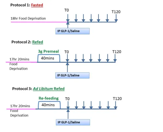
CHAPTER 2.1
Figure 2.1.1 Protocol for investigating whether GLP-1-induced satiation is dependant on feeding status………...………..………...86 Figure 2.1.2 Administration of GLP-1 in fasted and refed animals……….93 Figure 2.1.3 Effects of GLP-1 on 40min food intake in fasted and refed animals………94 Figure 2.1.4 Administration of GLP-1 in ad libitum refed animals………...95 Figure 2.1.5 Specificity of GLP-1R antibody on tissue sections………..97 Figure 2.1.6 Specificity of GLP-1R antibody on MIN6 cells……….98 Figure 2.1.7 Specificity of GLP-1R antibody with immunoblots………..99 Figure 2.1.8 Measurement of protein levels of GLP-1R in VAN in fasted and refed
animals by immunofluorescence……….101 Figure 2.1.9 Measurement of protein levels of GLP-1R in VAN in fasted and refed
Figure 2.1.10 GLP-1R gene expression in VAN in fasted and refed animals…….…..103 Figure 2.1.11 Localization of GLP-1R in fasted and refed VAN………..105
CHAPTER 2.2
Figure 2.2.1 Protocol A - Role of ghrelin in GLP-1-induced satiation………...126 Figure 2.2.2 Protocol B - Role of CCK on GLP-1-induced satiation using CCK1R
antagonist………...125 Figure 2.2.3 Protocol C - Role of CCK on GLP-1-induced satiation using CCK1R KO mice……….128 Figure 2.2.4 Blockade of ghrelin on GLP-1-induced satiation………...135 Figure 2.2.5 Blockade of ghrelin on GLP-1 signaling on VAN………..136 Figure 2.2.6 GLP-1R localization after blockade of ghrelin in VAN………..137 Figure 2.2.7 Validation of membrane-only surface stain protocol………...139 Figure 2.2.8 GLP-1Rs translocate according to feeding status in vitro………141 Figure 2.2.9 Ghrelin blockade on GLP-1Rs translocation in vitro……….143 Figure 2.2.10 Pathways of ghrelin-induced GLP-1R translocation………144 Figure 2.2.11 Blockade of CCK1R on food intake………...146 Figure 2.2.12 Blockade of CCK1R on GLP-1-induced satiation………147 Figure 2.2.13 GLP-1 administration in 12h fasted CCK1R KO mice………....149 Figure 2.2.14 GLP-1 administration in 6h fasted CCK1R KO mice ... 150
CHAPTER 2.3
Figure 2.3.1 Generation of conditional sensory-neuron-LepR KO by Cre-loxP system ... .168 Figure 2.3.2 Verification of conditional sensory-neuron-LepR KO by Cre-loxP system.
... 168 Figure 2.3.3 Analysis of CRE in nodose ganglia and brain ... 169 Figure 2.3.4 Verification of LepR KO in peripheral tissues ... 170 Figure 2.3.5 Leptin receptor in VAN of Nav1.8/LepRfl/fmice and WT mice ... 172 Figure 2.3.6 Leptin-induced phosphorylation of pSTAT3 in Nav1.8/LepRfl/ and WT mice
6
Figure 2.3.7 Analysis of energy homeostatic parameters ... 177 Figure 2.3.8 Analysis of fat pads in Nav1.8/LepRfl/fl vs. WT mice ... 178 Figure 2.3.9 Analysis of body composition by DEXA………..179 Figure 2.3.10 Energy expenditure analysis by CLAMS ... 180 Figure 2.3.11 Food intake Analysis over 24 h measured by CLAMS ... 181 Figure 2.3.12 Meal pattern analysis by CLAMS ... 182 Figure 2.3.13 Table on meal parameters measured by metabolic cages ... 183 Figure 2.3.14 Satiating effects of CCK and leptin in Nav1.8/LepRfl/fl and WT mice ... 187 Figure 2.3.15 Analysis of VAN plasticity of CART and MCH ... 188 Figure 2.3.16 Analysis of VAN plasticity of Y2R and CB1 ... 189 Figure 2.3.17 Analysis of weight gain and adiposity in response to a HF diet ... 191
CHAPTER 2.4
Figure 2.4.1 Analysis of energy homeostatis parameters of female Nav1.8/LepRfl/fl mice compared to WT mice ... 215 Figure 2.4.2 Analysis of body composition of female mice by DEXA ... 216 Figure 2.4.3 Analysis of energy expenditure of female mice by CLAMS ... 217 Figure 2.4.4 Food intake analysis over 24 h in female Nav1.8/LepRfl/fl and WT mice . 218 Figure 2.4.5 Meal pattern analysis in female Nav1.8/LepRfl/fl vs. WT mice ... 219 Figure 2.4.6 Meal pattern analysis in female Nav1.8/LepRfl/fl vs. WT mice ... 220 Figure 2.4.7 Satiating effects of CCK and Leptin in female Nav1.8/LepRfl/fl mice ... 222 Figure 2.4.8 Absolute change of body composition between male and female
Nav1.8/LepRfl/fl mice ... 225 Figure 2.4.9 Comparison of meal patterns between male and female WT mice ... 226 Figure 2.4.10 Comparison of meal pattern between male and female WT mice ... 227 Figure 2.4.11 Absolute change of meal pattern between male and female
Figure 2.4.12 Absolute change of meal pattern between male and female
8
Abbreviations
-MSH: alpha-melanocyte-stimulating
hormone
AgRP: agouti gene-related protein ARC: arcuate nucleus
cAMP: cyclic adenosine monophosphate
CART: cocaine amphetamine regulated transcript
CB1: cannabinoid receptor type 1 CB1R: cannabinoid receptor type 1 receptor
CCK: cholecystokinin CCK-1R: CCK-1 receptor CCK-2R: CCK-2 receptor
CLAMS: comprehensive lab animal monitoring system
CNS: central nervous system CREB: cAMP responsive element binding protein
DEXA: dual-energy X-ray absorptiometry
DIO: diet-induced obesity
DMEM: Dulbecco's modified Eagle's medium
DS: Donkey serum
DPP-IV: dipeptidyl peptidase-4 EEC: enteroendocrine cell
EGR-1: early response gene-1 ER: estrogen receptor
Ex-4: exendin 4
GAPDH: Glyceraldehyde 3-phosphate dehydrogenase
GPCR: G protein coupled receptor GFP: green fluorescent protein
GHS-R: growth hormone secretagogue receptor
GI: gastrointestinal
GIP: glucose-dependent insulinotropic peptide
GIPR: glucose-dependent insulinotropic peptide receptor
GLP-1: glucagon like peptide 1 GLP-1R: GLP-1 receptor
GOAT: ghrelin O-acyltransferase HBSS: Hank's Balanced Salt Solution HF: high fat
ICV: intracerebroventricular IP: intraperitoneal
KO: knockout
LepR: leptin receptor
MAPK: mitogen-activated protein kinase MCH: melanin concentrating hormone
MCH1R: MCH receptor MIN6: mouse insulinoma 6 NPY: neuropeptide Y
NTS: nucleus of solitary tract OVX: ovariectomy
PBS: phosphate buffered saline PM: plasma membrane
POMC: pro-opiomelanocortin PYY: peptide tyrosine tyrosine SDA: Subdiaphragmatic vagal deafferentation
STAT-3: Signal transducer and activator of transcription 3
TBST: Tris-Buffered Saline and Tween 20
VAN: vagal afferent neurons WT: wildtype
Chapter 1: Literature Review on gut-derived peptides
and their mechanism of action on vagal afferent neurons
1.1 Introduction
Obesity is one of the most significant global public health issues. 2/3 of Americans are either overweight or obese. In France, in 2012, the percentage of the population being obese was at 14.5%. Its prevalence is increasing worldwide; in the last 30 years the obese population has more than doubled in the United States, France, and United Kingdom (1). Obesity has been linked to numerous health complications such as diabetes and heart disease. The causes of obesity are likely to be complex and multifactorial, but can be characterized as the overconsumption of high-calorie foods and reductions in energy expenditure. In the last 30 years, the food environment has changed in that the availability of energy dense and palatable foods have increased significantly. In parallel, energy expenditure has decreased due to advancements in transportation modes. In addition, there are genetic mutations that result in an obese phenotype.
Although obesity is influenced by a complex interaction between diet, physical exercise and the environment, the scope of this dissertation will primarily focus on diet-induced physiological changes. Chronic high fat (HF) diet leads to hyperphagia, obesity and metabolic diseases such as insulin resistance (2). The mechanism by which HF diet alters energy homeostasis is not well understood. Diet-induced obesity (DIO) results in modulations in neuronal and tissue responsiveness to nutrients that are essential for regulating food intake and energy homeostasis. Obese individuals have elevated circulating leptin suggesting that individuals do not respond to endogenous leptin. Indeed, chronic high fat feeding leads to leptin resistance in mice (3). Diet-induced obese models have blunted responses to satiating signals such as cholecystokinin (CCK) (4). Altogether, evidence suggests that diet is one of the factors responsible for inducing obesity through physiological changes. Although balancing food intake and
14
energy expenditure would lead to weight loss, DIO leads to physiological changes that drive obese subjects to overeat.
Control of food intake involves many organs interacting in both the peripheral and central system that respond to nutrients with pleiotropic biological actions including gastric motility, food intake inhibition, and body weight regulation. Among them are the gastrointestinal tract and the vagus nerve, which will be the main focus of this dissertation. The vagus nerve is the main conduit by which gut-derived signals is communicated to the central nervous system (CNS) via the Nucleus of the solitary tract (NTS). Afferents in the NTS can communicate to higher order neurons such as the hypothalamus to influence food intake. Vagotomies or destruction of vagal afferent neurons via capsaicin treatment in rodents leads to hyperphagia and impaired responses to gut peptides involved in feeding compared to sham operated animals (5).
The vagus nerve is unique in that it innervates all tissues that are involved in digestion and absorption of nutrients, where activation of vagal afferents is necessary for the preparation and the onset of a meal. Furthermore, vagal afferent neurons (VAN) express receptors for many of the regulatory peptides and molecules released by enteroendocrine (EEC) cells that line the mucosal epithelia of the gut. EEC cells release humoral peptides that activate vagal afferent fibers, which communicate signals from the periphery to the brainstem activating the vago-vagal pathway. Gut-derived hormones, such as CCK and leptin, have been implicated in the regulation of long-term energy homeostasis.
Diet-induced obese models have altered gut-brain signaling present on the vagus nerve, which coincides with hyperphagia. Although much progress has been done using diet-induced obese rodent models, the origins of overnutrition are still unclear and need to be elucidated. Therefore we have focused on investigating the mechanism of action of
anorexigenic hormones on vagal afferent neurons. Understanding the underlying drivers of hyperphagia is important in developing effective non-invasive treatments against obesity.
1.2 Nutrient sensing in the GI tract
The gastrointestinal (GI) tract plays an important role in the regulation of energy homeostasis. It is the initial interface of the body at which nutrients are digested, absorbed and assimilated. EECs are specialized chemosensing epithelial cells dispersed along the mucosa of the GI tract. EECs are the first level of integration of information from the gut lumen that respond to the presence or absence of nutrients and secrete a variety of gut-derived hormones such as CCK. They are strategically located in the mucosa of the GI tract to taste and sense luminal contents and release hormones basolaterally to enter into the circulation (Figure 1.1). Gut-derived hormones either act directly on central receptors or they bind to their receptors found on vagus nerve fiber terminals to activate vago vagal pathway. In turn, efferent signals will target peripheral targets to control gut functions and ingestive responses.
1.2.1 Recent advances in understanding nutrient sensing
ECCs are chemosensing cells that finely coordinate nutrient sensing with metabolic and behavioral functions such as regulating energy homeostasis and glucose metabolism. ECCs release hormones such as CCK, glucagon-like-peptide 1 (GLP-1) and peptide YY (PYY) in response to nutrient ingestion (6). These signals are important in regulating appetite and body weight; in a prepandial state, circulating levels of orexigenic peptides are high to stimulate food intake and conversely, in postprandial states, anorexigenic peptides are secreted to inhibit food intake.
16
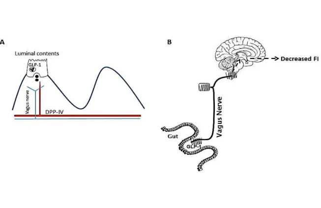
Figure 1.1 Schematic of the topography of enteroendocrine cells EECs are located in the mucosa of the gastrointestinal epithelium to taste and sense nutrients passing through the lumen. Peptides known to regulate food intake are released on the basolateral side and act either on nearby neural pathways or can enter directly into the circulation. Schematic taken from Cummings D.E and Overduin J, 2007 (44).
EECs are scattered throughout the epithelium of the GI tract and represent less than 1% of the epithelial cell population making the study of native EECs difficult. However, the development of transgenic mice expressing green fluorescent protein (GFP) under the control of specific EECs gene promotors has greatly enhanced the ability to study gut EECs in cell culture. GFP is used as a marker of gene expression and protein localization in living organisms without the use of an exogenous substrate. GFP with enteroendocrine promoters label hormone precursors, chemosensor receptors and granular proteins.
Given that EECs are difficult to study, much of what we know about EECs and nutrient sensing stems from cell lines such as STC-1 and GLUTag cells. These cells have been helpful in understanding intracellular signaling, however, they are transformed cells whereby their exact protein content or responses to stimuli may not replicate in vivo EEC function. Physiological and anatomical characterization of ECCs has been greatly enhanced through GFP-tagged mice. Traditionally, ECCs have been classified according to hormonal content, morphology and localization in the GI tract leading to the concept of “one cell type, one hormone”. However, results from transgenic mice with tagged EECs have questioned this concept. GFP-tagged mice with enteroendocrine promoters have demonstrated that multiple hormones are synthesized and released from a single ECC revealing the pluritrophic actions of EECs (7, 8). For example, studies using CCK-GFP mice have demonstrated that CCK is coexpressed with GLP-1, glucose-dependent insulinotrophic peptide (GIP), PYY, secretin and neurotensin (9, 10). Surprisingly, Egeord et al. found that promoters for CCK and GLP-1 were scattered throughout the gut rather than segregated in either the proximal or distal gut (8). The hormone content of individual cells differs according to their localization. For example, EECs from the proximal gut contain high levels of CCK and low levels of
GLP-18
1. Conversely, EECs cell from the distal gut have high levels of GLP-1 and low levels of CCK (9). An extensive study on CCK-containing cells demonstrated that the coexpression of hormones, such as CCK and ghrelin, was consistent between immature and fully differentiated cells (10).
The anatomical arrangement within each cell has been extensively studied in the last few years; PYY and GLP-1 secretory granules have been demonstrated to be contained at the base of L-cells. Furthermore L cells contain a prominent basal cytoplasmic process (7, 11). GLP-1 and PYY granules are contained in separate storage organelles within the same cell and these organelles can be selectively mobilized according to different stimuli (12).
Gastrointestinal functions are modulated by changes in luminal contents to protect against harmful substances. EECs act as primary chemoreceptors by releasing signaling peptides in response to changes in the luminal environment. Transgenic GFP-mice has been possible to look at the mechanism of nutrient sensing directly on native EECs. For example, using duodenal extract from CCK-GFP mice, it has been demonstrated that amino acid-induced CCK release acts through calcium sensing receptor (12). There are several molecular responses stimulated by nutrient sensing such as protein transport, glucose sensors and G–protein coupled receptors (GPCR). Convincing evidence suggests that EECs in the GI tract express several G-protein coupled receptors that are involved in chemosensing (13). These receptors are stimulated by glucose, calcium and short and long chain fatty acids. Many gut-derived proteins have been shown to act specifically through GPCR. It has been established that GPCR are involved in nutrient sensing of fatty acid (12, 13). In mouse intestine, GPR120 is colocalized with GLP-1(13). Furthermore, fatty acid induced secretion of GLP-1 is attenuated in GPR120 knockout mice (13). GPR40 gene and protein expression was
identified in CCK-expressing ECCs. GPR40 was shown to directly mediate fatty acid-induced CCK secretion, which was further attenuated by the deletion of GPR40 (14).
1.3 Importance of gut-derived hormones on regulation of food
intake
Gut-derived hormones are secreted in the absence or presence of nutrients to control food intake. They are localized in the epithelium of the GI tract in an ideal location to respond to luminal contents. Their patterns of release alter according to nutrient availability in ways that could affect short and long-term feeding behavior. Studies using exogenous administrations of gut peptides have revealed the functional importance of signals that arise from the GI tract in regulating energy homeostasis.
1.3.1 Glucagon like peptide-1
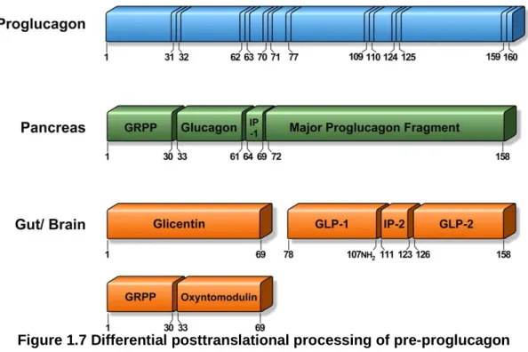
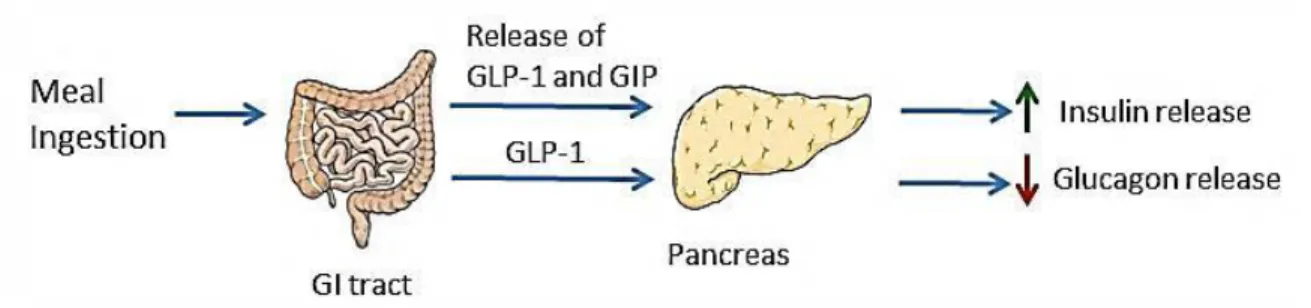
GLP-1 is derived from the expression of the transcriptional product of the
pre-proglucagon gene in intestinal L-cells and pancreatic α-cells. GLP-1 is released by
L-cells predominately found in the distal gut in response to nutrient ingestion. Glucose and fat are the most potent stimulators of GLP-1 stimulation (15). GLP-1 receptors (GLP-1R) are found throughout the peripheral and CNS, where its highest expression is found in the lung, distal gut and brain (16, 17). GLP-1 was considered until recently for its role as an incretin hormone and ability to restore glucose homeostasis in type 2 diabetic patients. In fact, GLP-1 analogs and DPP-IV inhibitors are on the market for the treatment of type 2 diabetes. Emerging evidence suggest a possible physiological role for GLP-1 in regulating food intake. Exogenous administration of GLP-1 or its long-acting analogs dose-dependently inhibit food consumption and the satiating effects of GLP-1 are attenuated by prior administration of GLP-1R antagonist (18, 19). Given that GLP-
20
food intake is unclear and this is discussed in further detail in the GLP-1 and control of
food intake section.
GLP-1 has specifically been implicated in the pathogenesis of obesity. DIO leads to blunted GLP-1 release; decreased postprandial plasma concentrations and blunted anorexigenic response to GLP-1R activation (20, 21). Duca et al. have demonstrated that DIO results in impaired GLP-1 satiation signaling; DIO rats have an attenuated response to GLP-1R antagonist exendin 4 (Ex-4) compared to controls. Furthermore, DIO rats have decreased GLP-1 protein expression in the intestinal epithelium indicating a reduction in intestinal nutrient response from L-cells (22). Evidence suggests that GLP-1 would be an effective treatment for obesity. Plasma concentrations of GLP-1 dramatically increase in patients following bariatric surgery (23). Infusions of GLP-1 will induce satiation in lean adults by suppressing food intake and meal size. Interestingly, this effect is preserved in obese subjects implying GLP-1 as a prime candidate for anti-obesity treatments (24).
1.3.2 Ghrelin
The discovery of ghrelin opened a new frontier to research in energy homeostasis in that ghrelin is currently the only circulating hormone that can stimulate food intake and adiposity in humans and rodents in comparison to the large family of identified anorexigenic gut hormones. Ghrelin is a 28-amino acid polypeptide produced mainly by endocrine X/A-like cells in rodents and P/D1 in humans located in the gastric epithelium (25). Although the stomach is the main site of secretion, ghrelin is also secreted by the pituitary, hypothalamus, lung, heart and pancreas. Acetylated ghrelin, which is converted by ghrelin O-acyltransferase (GOAT) enzyme, acts on the growth hormone secretatgogue receptor (GHS-R) which is constitutively expressed (26, 27).
GHS-R is predominately found in the pituitary and the hypothalamus (28). The biological functions of ghrelin are widespread; it plays a role in lipid metabolism, glucose homeostasis and growth hormone release (28, 29). Additionally, ghrelin stimulates appetite, body weight and adiposity.
Interestingly, circulating ghrelin concentrations are decreased in obese individuals compared to their lean controls (30). Given that ghrelin plasma levels are negatively correlated with insulin and leptin plasma levels, the downregulation in ghrelin may be a consequence of elevated insulin or leptin. However, antagonizing ghrelin receptor can decrease food intake in obese Zucker rats (31) possibly blocking the constitutive receptor activity (27). Given that ghrelin is involved in meal initiation makes it an attractive target in understanding feeding and energy homeostasis.
1.3.3 Leptin
Leptin is a 167-amino acid peptide that is mainly expressed in white adipose tissue but also found in a variety of tissues such as placenta, stomach, skeletal muscle and pituitary glands. Leptin is produced and secreted predominately by adipose tissue and the stomach (32). Circulating leptin levels positively correlate with body adiposity which reflects long term energy stores (33). Leptin is a gut and adipose tissue-derived hormone that regulates a range of biological functions and processes; including energy intake and expenditure, body fat, neuroendocrine systems, autonomic function, and insulin and glucose balance (34) (Figure 1.2). It exerts its effect by binding to its receptor, LepR, which is located throughout the CNS (35). The highest expression of the long isoform of LepR is found in the
22
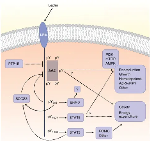
Figure 1.2 Leptin signaling leads to various biological activites
Leptin binds to its receptor to regulate energy intake and energy expenditure, glucose homeostasis as well as reproduction and growth. Schematic taken from Myers et al. 2008 (33).
hypothalamus, an area known to regulate energy homeostasis. Six isoforms of LepR have been identified in rats; the long form has been implicated in the suppression of food intake and stimulation of energy expenditure (34).
The presence of leptin was predicted before it was cloned in 1994. Coleman et
al. performed parabiosis studies in which they joined ob/ob mice, those lacking leptin,
and db/db mice, those lacking leptin receptor. They concluded that ob/ob mice were missing a circulating factor that was abundant in db/db mice. The factor could cure obesity in ob/ob mice but not in db/db mice (36). The discovery of the leptin was initially thought as a cure for human obesity however, daily injections of recombinant leptin only fully corrected obesity in few cases of leptin deficient patients. It was discovered that a majority of the obese population have high circulating leptin levels and are unresponsive to leptin thereby defining them as leptin resistant (36). Evidence has undoubtedly highlighted the importance of leptin signaling for the maintenance of energy homeostasis.
Obese individuals have high circulating leptin levels which fail to reduce energy intake (33). Moreover, the response to central administrations of leptin is attenuated in mice in part due to the development of leptin resistance in the hypothalamus (37). The importance of leptin in energy homeostasis is most evident in leptin deficient models. Ob/ob mice with global leptin deficiency exhibit hyperphagia, low metabolic rate and rapid onset of obesity, associated with high expression of orexigenic neuropeptide Y (NPY) and melanin-concentrating hormone (MCH) and low expression of anorexigenic pro-opiomelanocortin (POMC) in the hypothalamus (34, 38).
1.3.4 Cholecystokinin
24
small intestine and is released in response to fat. Plasma levels markedly increase in response to a meal. There are two subtypes of CCK receptors, CCK1R and CCK2R. CCK binds to its receptors to induce gallbladder contraction, delayed gastric emptying and release of pancreatic enzyme to induce digestion (Figure 1.3). CCK1Rs are predominately responsible for mediating CCK-induced effects on short-term food intake (39, 40). CCK is involved in the regulation of food intake and GI function.
CCK inhibits food intake in a dose dependent manner in many species including humans (41, 42). Central and peripheral administrations induce inhibitory effects on energy intake. Specifically, exogenous CCK inhibits cumulative food intake by reducing meal size and increasing intermeal interval in rodents and humans (39, 43). Perfusions of fat or protein into the small intestine inhibit feeding and the administration of CCK1R antagonists will reverse these effects (41). The satiating actions of CCK regulate short-term feeding; CCK1R knockout (KO) mice have no difference in body weight and cumulative food intake compared to wildtype (WT) mice, however, their early meal events are altered. For example, CCK1R KO mice ingest larger, longer first compared to their control littermates (44).
The major site of action of CCK is the vague nerve; damage to the vagus nerve attenuates the satiating effects of CCK (41). Exogenous administrations of CCK to rats will rapidly induce vagal electrophysiological activity (45). CCK binds to CCK1R present on the vagus nerve to terminate feeding and induce satiety integrated by the NTS, the area where vagal afferents terminate (39). CCK has been demonstrated as the master regulator of VAN phenotype switch, which alters sensitivity of VAN to fasting and refeeding conditions. This mechanism is further elucidated in the Phenotypic Changes of
Figure 1.3 Biological activities of cholecystokinin
Schematic representing that CCK has various biological activities; it delays gastric emptying, increases pancreatic enzymes to facilitate digestion and induces bile secretion from gall bladder.
26
Studies using diet-induced obesity models have highlighted the importance of CCK in regulating energy homeostasis. Animals maintained on a chronic HF diet leads to hyperphagia and obesity in part due to a decreased sensitivity to exogenous CCK (2, 46). HF fed rats are less sensitive to the satiating effects of CCK through reductions in neural activation of the hindbrain where vagal afferent fibers terminate (46). Interestingly, Diet-induce obese rats have reduced lipid-induced satiation compared to diet resistant rats, which is associated with the decrease in intestinal protein expression of CCK (47). Indeed, obese humans have decreased circulating CCK (42).
The satiating effect of CCK is the most established compared to other gut-derived peptides. Many studies have highlighted that endogenous CCK acts on VAN and plays an important role in regulating food intake. The ability of CCK to change the sensitivity of VAN provides a great interest in understanding the mechanism of action of other gut-derived peptides and how they may interact with each other to regulate energy homeostasis.
1.4 The Vagus Nerve
The vagus nerve is the major neuronal link between the GI tract and CNS relaying gut-derived signals to the NTS. Afferent neurons in the NTS project to second order neurons in the hypothalamus to communicate peripheral signals influencing food intake behavior. The vagus nerve densely innervates organs involved in digestion and is necessary for meal ingestion and digestion. Vagal afferent fibers provide the primary neural site in which gut-derived hormones released from ECCs to relay sensory input of the periphery via the nodose ganglion to the NTS in the brain stem (6, 48).
Lack of vagal function will result in impaired responses to feeding; lesions to vagal afferents by vagotomies and capsacin destruction in rodents result in hyperphagia
and rapid weight gain (49). Specifically, subdiaphragmic deafferenation (SDA), which target all vagal afferent fibers, results in attenuated responses to anorexigenic gut peptides in lean rats (50-52). For example, the satiating effect of CCK is completely abolished in animals without intact afferents (49). Activation of the vagus nerve mediates external stimuli of the food bolus and activates internal stimuli from the gut to communicate the information to the brain. These signals activate neurons in the NTS and in turn control peripheral tissues through vagal efferents.
Vagal signaling is important in regulating ingestive behavior and food intake regulation. Peripheral satiety signals, such as CCK, will increase proto-oncogene protein c-fos expression, an indicator of neural activity, in the NTS, which in turn signals for meal termination in higher ordered neurons. In vagotomized animals, CCK induced c-fos expression in the hindbrain is attenuated (53). Lesions to ascending projections from the NTS to the hypothalamus block the satiating effect of systemic CCK and inhibit CCK induced c-fos expression in the NTS (54). Satiety signals from the periphery will activate c-fos expression in the NTS, which will project to vagal efferent in the dorsal motor nuclei (55). In turn, vagal efferents project to pre and post ganglic neurons to target peripheral tissues influencing gastric and insulinotrophic functions. This is known as the vago-vagal loop (Figure 1.4).
28
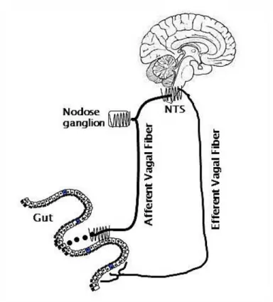
Figure 1.4 The vago-vagal loop
Schematic representation of vagally-mediated nutrient sensing along the gut-brain axis. The presence of nutrients in the lumen are sensed by enteroendocrine cells in the gut to induce the release of hormones that can then enter circulation and bind to receptors found on terminals of the vagus nerve. These signals are processed in the nodose ganglia and transduced to the NTS of the hindbrain.
1.4.1 Phenotypic Changes of VAN According to Feeding Status
It is well established that VANs exhibit plasticity according to physiological changes. Nutrient status induces neurochemical changes in VAN by regulating the switch between states that promote orexigenic and anorexigenic phenotypes (56). Anorexigenic receptor expression will decrease and orexigenic receptor expression will increase in VAN in a food-deprived condition. Conversely, anorexigenic receptor expression is upregulated and orexigenic receptor expression is downregulated postprandially. For example, the expression of anorexigenic PYY receptor (Y2) receptor expression is downregulated after a fast between 6 and 12 hours (57). Retrograde labeling revealed that the changes in fasting-induced Y2R expression in VAN occur in neurons innervating the upper GI tract. Refeeding of fasted rats will restore Y2R expression (57). Orexigenic receptors have been demonstrated to cannabinoid-1 (CB-1) and MCH-1 receptors increase significantly in a fasted state whereas Y2, a receptor associated with inhibition of food intake, is decreased (56).
CCK induces phenotypic changes of VAN by regulating the expression of peptides and receptors according to nutritional status (56). In a fasted state, when CCK levels are low, mRNA levels and protein expression of orexigenic MCH, MCH1R and CB1 receptor (CB1-R) are increased. In a fed state, when CCK levels are high, the expression of anorexigenic PYY, cocaine amphetamine transcript (CART) and Y2R are decreased (Figure 1.5). Furthermore, administrations of exogenous CCK will act on CCK1Rs to decrease expression of orexigenic MCH, MCH-R and CB1-R and increase PYY, Y2R and CART expression (57, 58). The mechanism by which CCK alters neuropeptide CART expression according to nutritional status has been shown to work through phosphorylation of CREB and translocation of EGR-1 (59). In culture, CCK-induced CART expression is decreased by ghrelin (59). Although exogenous ghrelin
30
does not increase the abundance of orexigenic peptides in refed rats, it does inhibit the decrease in expression of MCH and CB-1 in fasted then refed rats (60). CCK is the master regulator of the VAN neurochemical switch involved in regulating food intake to potentate the actions of gut-derived hormones.
Figure 1.5 Neurochemical phenotype of VAN changes according to feeding status In a fasted state, when CCK levels are low, mRNA levels and protein expression of orexigenic melanin-concentrating hormone (MCH), MCH receptor (MCH-R) and CB1 receptor (CB1-R) are increased. In a fed state, when CCK levels are high, the expression of anorexigenic PYY, CART and PYY receptor (Y2R) are decreased.
32
1.4.2 VAN phenotype in diet induced obesity
The inability to respond to satiety signals in obese models may be due to the disruption in VAN signaling. VANs DIO have decreased sensitivity to VAN stimulation. Daly et al. showed reduced VAN sensitivity to CCK, 5HT and distension in DIO mice (61). Furthermore, de Lartigue et al. have demonstrated that chronic exposure to a HF diet leads to the development of leptin resistance on VAN of obese diet-induced rats compared to lean low fat-fed rats (2). VAN sensitivity to anorexigenic peptides is significantly reduced in high fat feeding. For example, CCK fails to inhibit food intake in DIO rats compared to lean chow-fed rats. NTS neuronal activation is decreased in DIO animals in response to exogenous peripheral CCK compared to control (46).
The exact mechanism by which VAN sensitivity is attenuated is unclear. However, a possible mechanism that may play a role in development of an obese phenotype is the inability of VAN to change their expression of orexigenic and anorexigenic peptides and receptors. Rats maintained on a high fat diet for 8 weeks became hyperphagic, increased body weight and adiposity compared to rats on a control diet (2). Studies have demonstrated that orexigenic receptors such as CB1R and GHS-R are increased and anorexigenic Y2R and CART are decreased in obese models (27). Leptin resistance in VAN of DIO rats leads to a change in neurochemical phenotype of VAN and hyperphagia. Altogether, studies have indicated that the normal vagal afferent phenotypic switch in response to feeding is lost in obesity where VANs are locked in a fasting phenotype (Figure 1.6).
Figure 1.6 Chronic high fat diet leads to a dysregulation in VAN phenotype Long-term ingestion of a high fat diet renders the ability of vagal afferent neuronal phenotypic switch between fasting and fed conditions. Chronic high fat diet leads a consititutive fasting phenotype in nodose ganglia.