http://www
.bsa
v
a.c
om/
4
Journal of Small Animal Practice • Vol 51 • January 2010 • © 2010 British Small Animal Veterinary AssociationEmesis in dogs: a review
INTRODUCTION
In October 2006, after a series of
meet-ings, the authors published ‘Approach
to the Management of Emesis in Dogs’
(Devauchelle and others 2006), intended
as a clinical guide to ‘best practice’ in the
management of canine emesis. Statements
in these guidelines were developed from
published papers, consensus opinion and,
where necessary, the authors’ own expert
opinions. This review details the evidence
and emphasises the opinion from which
the guidelines were developed and, by
doing so, highlights where evidence is
lacking or contradictory.
METHODS
A systematic search of the literature was
performed on the sites Google Scholar,
Web of Science and PubMed, using the
terms ‘vomit*’ or ‘emesis’ AND ‘dog’ or
‘canine’ to identify relevant references.
Where primary sources were available
(pa-pers published in peer reviewed journals)
these are referenced. Where relevant
infor-mation did not fi t the above search terms
(e.g. secondary effects of drugs), references
were identifi ed in a standard manner. To
further quantify the strength of evidence
available to support the information
pro-vided, individual references used to
sup-port statements were classifi ed according
the scheme shown in Table 1a and assigned
an evidence level (EL). As appropriate to
support the text an overall evidence grade
(OEG) was given according to the scheme
in Table 1b. Where multiple references
were available, we attempted to ensure
those with the highest evidence level were
cited. Where peer-reviewed sources were
lacking, statements should be considered
the opinion of the authors.
THE EMETIC REFLEX
Emesis is facilitated by a sequence of
pro-grammed overlapping and coordinated
events which reduce the risks of adverse
consequences (such as aspiration of acid
stomach contents) whilst achieving
elimi-nation. The refl ex is controlled within the
brainstem by a central pattern generator,
Emesis is a common presenting sign in small animal practice.
It requires a rational approach to management that is based
upon a sound understanding of pathophysiology combined with
logical decision making. This review, which assesses the weight
of available evidence, outlines the physiology of the vomiting
refl ex, causes of emesis, the consequences of emesis and the
approach to clinical management of the vomiting dog. The
applicability of diagnostic testing modalities and the merit of
traditional approaches to management, such as dietary changes,
are discussed. The role and usefulness of both traditional
and novel anti-emetic drugs is examined, including in specifi c
circumstances such as following cytotoxic drug treatment. The
review also examines areas in which common clinical practice
is not necessarily supported by objective evidence and, as such,
highlights questions worthy of further clinical research.
C. E
LWOOD, P. D
EVAUCHELLE*, J. E
LLIOTT†, V. F
REICHE‡, A. J. G
ERMAN§, M. G
UALTIERI¶,
E. H
ALL||, E.
DENH
ERTOG**, R. N
EIGER††, D. P
EETERS‡‡, X. R
OURA§§
ANDK. S
AVARY-B
ATAILLE¶¶
Journal of Small Animal Practice (2010) 51, 4–22
DOI: 10.1111/j.1748-5827.2009.00820.x
Accepted: 9 June 2009
Davies Veterinar y Specialists, Manor Farm Business Park, Higham Gobion, Hitchin, Her tfordshire SG5 3HR
*Centre de Cancérologie Vétérinaire, ENVA, 7 Avenue du Général du Galle, 94700 Maisons Alfor t, France
†Royal Veterinar y College, Royal College Street, London NW1 0TU
‡Clinique Vétérinaire Alliance, 8 boulevard Godard, 33300 Bordeaux, France
§Depar tment of Veterinar y Clinical Sciences, University of Liverpool, Leahurst Campus, Chester High Road, Neston, Wirral CH64 7TE
¶Depar tment of Veterinar y Clinical Science, Section of Surger y, University of Milan, Via Celoria, 10, 20133 Milan, Italy
||Division of Companion Animal Studies, Depar tment of Clinical Veterinar y Science, University of Bristol, Langford House, Langford, Bristol BS40 5DU
**Dierenar ts Specialisten Amsterdam, Weesperzijde 147, 1091 ET Amsterdam, The Netherlands
††Universität Giessen, Veterinämedizinsche Fakultät, Medizinische und Gerichtliche Veterinerklinik I, Franfur terstrasse 126, 35392 Giessen, Germany
‡‡Université de Liège, Faculté de Médecine Vétérinaire, Clinique Medicale des Petits Animaux, Bat 44, Sar t-Tilman, 4000 Liège, Belgium
§§Ser vei de Medicina Interna, Hospital Clínic Veterinari, Facultat de Veterinària, Universitat Autònoma de Barcelona, Bellaterra 08193, Spain
¶¶PFIZER Animal Health, Europe & AfME ahg, 23-25, avenue du Dr Lannelongue, 75668 Paris Cedex 14, France
JSAP_820.indd 4
Journal of Small Animal Practice • Vol 51 • January 2010 • © 2010 British Small Animal Veterinary Association
5
loosely termed the ‘vomiting centre’,
located in the area dorsomedial to the
retrofacial nucleus which initiates a
co-ordinated stimulation and inhibition of
relevant motor nuclei (Fukuda and Koga
1991 [2b], Fukuda and Koga 1992 [2b],
Koga and others 1998 [2b]). Direct
pro-jections from this region to motor nuclei
such as the caudal part of the ventral
res-piratory group have been identifi ed (Koga
and Fukuda 1997 [2b]). The refl ex has
visible phases of hypersalivation,
retch-ing and expulsion (Figure 1). Efferent
pathways controlling these processes
in-clude the vagal and phrenic nerves,
para-sympathetic nerves to the salivary glands
and somatic motor nerves to abdominal
muscles [OEG B].
CAUSES OF EMESIS
Experimental studies show that many
pe-ripheral stimuli of abdominal structures
will initiate emesis in dogs (Lang and
Marvig 1989 [2b], Xu and Chen 2008
[2b]). Release of 5-hydroxytryptamine/
serotonin (5-HT) from
enterochromaf-fi n cells, which have been demonstrated
in canine gastric and duodenal mucosa,
stimulates vagal afferents via 5-HT
3receptors (Fukui and others 1992 [2b],
Fukui and others 1993a [2b]). It seems
that other pathways and local modulatory
signals are also important (Lang and
oth-ers 1988 [2b], Sanger and Andrews 2006
[3a]). Peripheral emetogenic triggers may
be abrogated by bilateral vagotomy, effects
which are enhanced when combined with
ablation of the greater splanchnic nerves,
suggesting more than one signal pathway
(Fukui and others 1993b [2b]). Vagal
afferents, carrying peripheral emetogenic
signals, enter the rostral medulla
oblon-gata and pass via the solitary tract to the
nucleus of the solitary tract (Fukuda and
Koga 1991 [2b], Koga and Fukuda 1992
[2b]). [OEG B].
The ‘chemoreceptor trigger zone’ of the
brainstem has been identifi ed as the area
postrema which is located on the dorsal
surface of the medulla oblongata adjacent
to the caudal end of the fourth ventricle
(Chernicky and others 1980 [2b]). This
region, lacking a blood-brain barrier,
is responsive to circulating emetogens.
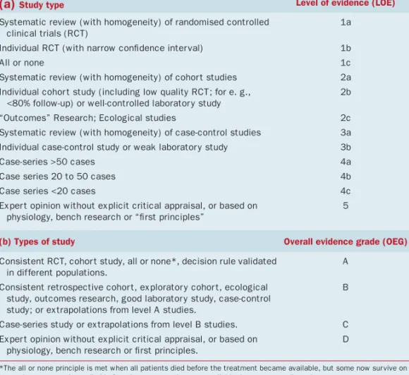
Table 1
.
Scheme used to grade (a) individual references and (b) overall level of
evidence. Adapted from 1
(a)
Study type Level of evidence (LOE)Systematic review (with homogeneity) of randomised controlled clinical trials (RCT)
1a
Individual RCT (with narrow confi dence inter val) 1b
All or none 1c
Systematic review (with homogeneity) of cohor t studies 2a Individual cohor t study (including low quality RCT; for e. g.,
<80% follow-up) or well-controlled laborator y study
2b
“Outcomes” Research; Ecological studies 2c Systematic review (with homogeneity) of case-control studies 3a Individual case-control study or weak laborator y study 3b
Case-series >50 cases 4a
Case series 20 to 50 cases 4b
Case series <20 cases 4c
Exper t opinion without explicit critical appraisal, or based on physiology, bench research or “fi rst principles”
5
(b) Types of study Overall evidence grade (OEG)
Consistent RCT, cohor t study, all or none*, decision rule validated in different populations.
A
Consistent retrospective cohor t, explorator y cohor t, ecological study, outcomes research, good laborator y study, case-control study; or extrapolations from level A studies.
B
Case-series study or extrapolations from level B studies. C Exper t opinion without explicit critical appraisal, or based on
physiology, bench research or fi rst principles.
D
*The all or none principle is met when all patients died before the treatment became available, but some now sur vive on it; or when some patients died before the treatment became available, but none now die on it
FIG 1. A diagrammatic representation of the series of events comprising the emetic refl ex in the dog. LES: Lower oesophageal sphincter. UES: Upper oesophageal sphincter. Summarised from: (Abe and others 1993 [2b], Abe and others 1994 [2b], Furukawa and Okada 1994 [2b], Koga and others 1998 [2b], Koga and Fukuda 1990 [2b], Lang and others 1986b [2b], Lang and others 1986a [2b], Lang and others 1993 [2b], Lang and others 2002 [2b], Onishi and others 2007 [2b], Sha and oth-ers 1996 [2b]). [OEG C.]
Prodromal
Retching
Explusion
Nausea and hypersalivation
Increased swallowing and relaxation of LES and proximal stomach.
Retrograde giant contractions of duodenum delivers contents to stomach Contraction of pylorus. Relaxation of fundus. Rhythmic retching. Inhibition of salivation. Mixing of gastric contents
Activation of expiratory intercostal muscles Elevation of larynx. Increased tone in UES. Protection of airways. Cervical oesophageal and pharyngeal tone increases between retches
Cervical oesophageal and pharyngeal tone decreases
Relaxation of diaphragmatic crura, reduced LES tone Rectus abdominis contraction. Diaphragm squeezes stomach Inhibition of breathing. Glottal closure. Contraction of genohyoideus
JSAP_820.indd 5
6
Journal of Small Animal Practice • Vol 51 • January 2010 • © 2010 British Small Animal Veterinary AssociationA number of receptor types have been
noted in the area postrema of the dog
in-cluding dopamine (Stafanini and
Clem-ent- Cormier 1981 [2b]), histamine
(Bhar-gava and others 1976 [2b]) and peptide
YY (Leslie and others 1988 [2b]).
Nu-merous chemicals, that can induce emesis
when administered systemically, also do so
via direct application to the area postrema.
These include apomorphine, xylazine,
prostaglandins and various hormones and
peptides (Briggs and Carpenter 1986 [3b],
Carpenter and others 1983 [3b],
Carpen-ter and others 1988 [2b], CarpenCarpen-ter and
Briggs 1986 [2b], Hikasa and others 1987
[2b]). Ablation of the area postrema
in-hibits the emetogenic effects of these
sub-stances (Carpenter and Briggs 1986 [2b]).
Emetogenic signals from the area postrema
excite neurones of the nucleus of the
soli-tary tract in the area subpostrema and,
from there, the central pattern generator
of the vomiting refl ex (Koga and Fukuda
1992 [2b]). [OEG B].
Whilst it is generally accepted that
dogs, like humans, can suffer motion
sickness which can be therapeutically
managed, mechanisms are poorly
un-derstood and little studied (Conder and
others 2008 [1b], Benchaoui and others
2007 [1b], Boyd 1953 [5]). Ablation of
the area postrema was thought to stop
emetogenic responses to motion, but later
critique suggested that a lack of specifi city
in ablation may have damaged associated
structures, possibly the nucleus of the
solitary tract, which modulated emetic
signals from the inner ear (Borison 1985
[5,] Wang and Chinn 1954 [5]). [OEG
for mechanisms D].
As well as inputs from peripheral,
vestib-ular and area postrema triggers, stimulation
from higher centres has been proposed,
presumably co-ordinated in the nucleus
tractus solitarius.
Clinical diseases associated with emesis
in dogs are summarised (Tables 2-5). In
many of these diseases emesis is triggered
peripherally and co-ordinated centrally,
although there may be concomitant
acti-vation of the chemoreceptor trigger zone
in some conditions e.g. uraemia. Whilst
generally considered a mechanism of
pro-tection, vomition of food by bitches is
believed to be part of the normal rearing
process (Korda 1972 [3b]) [OEG C].
Table 2. Gastrointestinal disease conditions associated with emesis in the dog
Process Reference (LOE) Overall evidence
grade Gastritis Eosinophilic Lymphoplasmacytic Granulomatous Acute 2 (4c) 3 (4c) 4 (4c) 5 (4c) 6 (4a) C
Associated with spiral bacteria 7 (3a) C 8 (4b)
9 (4b)
Gastric neoplasia 10 (4b) C
6 (4a) 11 (4b) Gastric ulceration Non-steroidal anti-infl ammator y
drugs (NSAIDs) 12 (4c) B 13 (3b) 14 (4c) Neoplasia 15 (4c) C 10 (4b) 11 (4b) Metabolic 16 (4b) C Hypergastrinaemia/Other APU-Domas 17 (4c) C 18 (4c) 19 (4c) Irritant D Mastocytosis 20 (4c) C Gastric/intestinal entrapment Ruptured diaphragm 21 (4b) C Gastric dilatation/volvulus D Hiatal hernia 22 (4c) C 23 (4c)
Pyloric stenosis Congenital 24 (4c) C
25 (4b) 26 (4c) Chronic hyper trophic pyloric
gastropathy
27 (4c) C
28 (4c)
Foreign body 29 (4a) C
30 (4c) 31 (4c)
Dietar y 32 (4b) C
33 (4c)
Infection/Infestation Canine par vovirus 34 (1b) A 35 (1b)
Canine distemper virus D
Canine coronavirus 36 (4c) C Salmonellosis 37 (4b) C Campylobacteriosis 38 (4c) C Mycobacterial infection 39 (4c) C Fungal infection 40 (4c) C 41 (4c) 42 (4a) Hookworms/Roundworms 43 (4c) C Infl ammator y bowel
diseases Eosinophilic 44 (4a) C Lymphoplasmacytic Granulomatous (Continued overleaf) JSAP_820.indd 6 JSAP_820.indd 6 12/23/09 10:00:17 PM12/23/09 10:00:17 PM
Journal of Small Animal Practice • Vol 51 • January 2010 • © 2010 British Small Animal Veterinary Association
7
Table 2. (continued)
Process Reference (LOE) Overall evidence
grade Intestinal neoplasia 45 (4b) 46 (4c) 47 (4b) 48 (4c) C Intussusception 49 (4b) C Intestinal volvulus 50 (4c) 51 (4c) C Intestinal entrapment 52 (4c) C
Motility disorders Dysautonomia 53 (4a) 54 (4c)
C
Localised autonomic dysfunction 55 (4c) 56 (4c) 57 (4c) 58 (4c)
C
Table 3. Non-gastrointestinal abdominal disease conditions associated with
emesis in the dog
Process Reference (LOE) Overall evidence grade
Peritoneal neoplasia 59 (4c) C Steatitis 60 (4c) C Peritonitis Septic 61 (4c) C 62 (4c) Bile 63 (4c) 64 (4b) C Urine D Idiopathic 65 (4c) C
Hepatobiliar y disease Neoplasia 66 (4c) Hepatitis/hepatopathy 67 (4a) 68 (4c) 69 (4b) C Infectious 70 (4a) 71 (4b) 72 (4b) C Immune (?) D Toxic 73 (4c) 74 (4c) 75 (4b) C Cholangiohepatitis/ Cholangitis/Cholelithiasis 76 (4c) 63 (4c) 77 (4c) C
Gall bladder torsion/rupture 78 (4c) C
Lobe Torsion 79 (4c) C
Abscess 80 (4c) C
Splenic diseases Torsion 81 (4c) C
Abscess 82 (4c) C
Infarction 83 (4c) C
Neoplasia 84 (4c) C
Pancreatic diseases Pancreatitis 85 (4a) C
Neoplasia 86 (4c) C
Phlegmon 87 (4c) C
Pseudocyst 87 (4c) C
88 (4c)
Abscess 89 (4c) C
Renal diseases Nephrolithiasis/Abscess 90 (4c) C
Neoplasia 91 (4c) C
Urogenital diseases Pyometritis 92 (4c) C
Endometritis 93 (4c) C
Urethrolithiasis 94 (4b) C
CONSEQUENCES AND
COMPLICATIONS OF EMESIS
Emesis is associated with signs of nausea
(e.g. depression, salivation, lip licking,
in-creased swallowing motions) and loss of
ap-petite. Whilst, in most clinical situations,
the consequences of the disease process
per se and of emesis cannot be completely
distinguished, persistent and severe emesis
leads to loss of gastrointestinal fl uid and
electrolytes, with consequent dehydration,
hypovolaemic shock, acid-base and
elec-trolyte disturbances (e.g. metabolic
aci-dosis/alkalosis, hypokalaemia) which can
be life-threatening (Boag and others 2005
[4a], Cornelius and Rawlings 1981 [4a]).
Aspiration pneumonia can occur
second-ary to vomiting (Kogan and others 2006
[4a]). Persistent vomiting that prevents
ef-fective oral intake of food is likely to lead to
protein-calorie malnutrition (see ‘ Dietary
management’). These concomitant
prob-lems must be assessed and treated
appro-priately as part of the clinical management
of the dog with emesis. [OEG C].
CLINICAL PRESENTATION AND
INITIAL ASSESSMENT
Initial assessment of dogs with emesis
should evaluate their general health
condi-tion (determinacondi-tion of the severity of the
disease process) which will differentiate
those in which no treatment is necessary,
those which need to be treated
sympto-matically and those which need further
examination or specifi c treatment. In
addition the initial assessment may give
clear indications of the underlying cause
of the vomiting.
The initial assessment starts with the
age, breed and gender of the dog. The
age is important because some diseases
are more common in young dogs, e.g.
ingestion of foreign bodies, foreign body
induced ileus (Capak and others 2001
[4a]), dietary indiscretion, infectious
dis-eases, intussusception, chronic intestinal
pseudo-obstruction (Johnson and others
2007 [4c]), and other diseases e.g.
gas-tric neoplasia, are more common in older
dogs (Gualtieri 1996 [4b], Gualtieri and
others 1999 [3a], Sautter and Hanlon
JSAP_820.indd 7
8
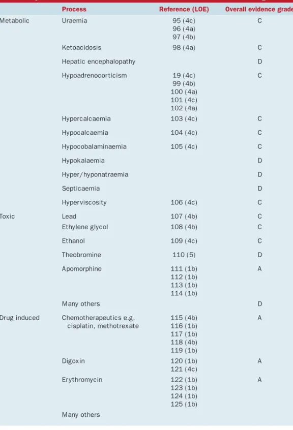
Journal of Small Animal Practice • Vol 51 • January 2010 • © 2010 British Small Animal Veterinary AssociationTable 4. Systemic disease conditions associated with emesis in the dog
Process Reference (LOE) Overall evidence grade
Metabolic Uraemia 95 (4c) 96 (4a) 97 (4b) C Ketoacidosis 98 (4a) C Hepatic encephalopathy D Hypoadrenocor ticism 19 (4c) 99 (4b) 100 (4a) 101 (4c) 102 (4a) C Hypercalcaemia 103 (4c) C Hypocalcaemia 104 (4c) C Hypocobalaminaemia 105 (4c) C Hypokalaemia D Hyper/hyponatraemia D Septicaemia D Hyper viscosity 106 (4c) C Toxic Lead 107 (4b) C Ethylene glycol 108 (4b) C Ethanol 109 (4c) C Theobromine 110 (5) D Apomorphine 111 (1b) 112 (1b) 113 (1b) 114 (1b) A Many others D
Drug induced Chemotherapeutics e.g. cisplatin, methotrexate 115 (4b) 116 (1b) 117 (1b) 118 (4b) 119 (1b) A Digoxin 120 (1b) 121 (4c) A Er ythromycin 122 (1b) 123 (1b) 124 (1b) 125 (1b) A Many others
1975 [4b]). Breed is an important
con-sideration; Belgian shepherd dogs have a
breed predisposition for gastric carcinoma
(Scanziani and others 1991 [4c]), chronic
hypertrophic pyloric gastropathy is seen
more often in certain toy breeds
(Belleng-er and oth(Belleng-ers 1990 [4c], Walt(Belleng-er and
oth-ers 1985 [4c]), and hypoadrenocorticism
in the Nova Scotia Duck tolling retriever
(Hughes and others 2007 [4b]). There are
many more breed predilections that can
be mentioned. Some diseases also have a
gender predilection e.g.
hypoadrenocor-ticism is more commonly seen in female
dogs (Kintzer and Peterson 1997 [3a]) and
some diseases exclusively affect one gender
(e.g. pyometra, prostatitis).
A full and complete history is essential
for evaluation of a vomiting dog.
Infor-mation which should be obtained is listed
( Table 6). The most important distinction
is that between vomiting and regurgitation,
because their aetiologies are very different
and this will direct specifi c diagnostic
test-ing. Regurgitation is passive, with
undi-gested food or saliva returned under gravity,
whereas vomiting is a refl ex, accompanied
by signs of nausea, hypersalivation and
ac-tivity of the abdominal musculature.
A thorough physical examination is
re-quired and should include assessment of
features shown (Table 7). From the
signal-ment, history and physical examination,
the clinician should be able to identify
cri-teria for concern which might indicate a
need for immediate diagnostics
investiga-tion and/or therapy (Table 8). [OEG C].
DIAGNOSTIC APPROACH
The misnomer ‘acute gastritis’ is
common-ly used to describe a syndrome of acute
and self limiting emesis. In almost all of
these cases, however, gastric infl ammation
is not proven by histopathology. Gastritis
is a frequently cited yet rarely confi rmed
diagnosis in cases of canine anorexia and
emesis. Dogs with simple, mild, acute self
limiting emesis do not need further
work-up, and can be treated symptomatically.
Many of these animals are not seriously
ill, and may need no treatment. A recent
study suggests 95% of dogs with emesis
do not present to the veterinary surgeon
(Hubbard and others 2007 [4a]). Even in
Table 5. Nervous system disease conditions associated with emesis in the dog
Reference (LOE) Overall evidence grade
Trauma D Hydrocephalus D Space-occupying lesion 126 (4c) 127 (4c) C Meningitis D Encephalitis D Motion sickness 128 (1b) 129 (1b) 130 (2b) B Vestibular disease D Cerebellar disease D Visceral epilepsy D Sialadenosis(?) 131 (4c) C JSAP_820.indd 8 JSAP_820.indd 8 12/23/09 10:00:18 PM12/23/09 10:00:18 PM
Journal of Small Animal Practice • Vol 51 • January 2010 • © 2010 British Small Animal Veterinary Association
9
Table 6. History taking for the dog with emesis
A thorough history should be obtained during the initial assessment, including the following:
Example demonstrating the importance of this information
Reference (LOE) Overall evidence
grade
Onset and progression of signs Sudden onset can suggest ingestion of foreign body or dietar y indiscretion
29 (4a) C
Emesis or regurgitation Regurgitation is seen in oesophageal disease 132 (4b) C Relationship to eating Vomiting > 10-12 hours after meal indicates delayed
gastric emptying (outfl ow obstruction, motility disorder)
30 (4c) C
The frequency, volume and nature of vomitus, including the presence of any fresh or digested blood
Haematemesis is sometimes seen after use of NSAIDs or acute vomiting
133 (4c) 134 (4b) 85 (4a) 135 (4c) 136 (4c) 137 (4c) 138 (4c) C
Whether or not there is any diarrhoea Diarrhoea may suggest concurrent intestinal disease, but can be seen with other conditions e.g. pancreatitis 139 (4b) 140 (2a) 85 (4a) 141 (4c) 142 (4c) 143 (4a) C
Presence and progression of weight loss Weight loss suggests chronic disease, e.g. gastro-intestinal tumour
144 (4a) C
Appetite and ability to maintain nutritional status Early enteral nutrition is impor tant in recover y 145 (3b) B Fluid intake (increased, decreased or normal) Polydipsia is seen with pyometritis 146 (4a) C Presence and nature of any abdominal pain Abdominal pain can e.g. be seen in pancreatitis 85 (4b) C Recent changes in diet or provocative changes,
including recent or ongoing drug treatment and access to toxins or foreign bodies
Emesis can be seen as a side effect of many drugs. 147 (4c) 148 (4b) 149 (4c) 150 (1b) 151 (4b)
A
Change in diet can cause vomiting D
Severe exercise can cause gastritis. 152 (4c) C Intoxication can cause vomiting e.g. ethylene glycol,
grapes, Bufo marinus
108 (4b) 97 (4b) 153 (4a)
C
Ingestion of foreign body is a cause of emesis 29 (4a) C Vaginal discharge can be seen in pyometritis 146 (4a)
154 (4a)
C
Reproductive status including recent seasons and presence of any vaginal discharge
Information on the reproductive status can suggest mucometra or closed cer vix pyometra
155 (4b) 156 (4c)
C
Co-existing neurological signs suggest neurologic disease 55 (4c) 53 (4a) 157 (4c) 126 (4b) C
Presence of neurological signs e.g. head tilt, ataxia, nystagmus, altered behaviour or consciousness
Emesis associated with motion sickness 128 (4c) B Urinar y tract disorders can be associated with emesis 158 (4b)
159 (4c)
C
Presence of other signs suggestive of a systemic disease e.g. urinar y tract signs (dysuria etc)
those that present to a veterinary surgeon,
in most cases of acute self limiting emesis,
the aetiology is never determined:
diet-related factors (dietary indiscretion),
in-fectious agents and toxins are considered
the most important causes. If the signs
resolve after 1 to 2 days, with or without
symptomatic and supportive therapy, the
tentative diagnosis of acute self limiting
emesis is considered correct.
In those cases where further
investiga-tion is considered necessary a variety of
diagnostic tests may be indicated ( Table 9).
[OEG D]
TREATMENT
A number of potential adverse effects
of persistent emesis have already been
detailed. Treatment of persistent emesis
reduces suffering and prevents
complica-tions whilst a thorough investigation is
undertaken to identify and, where
possi-ble, treat the underlying cause.
The decision to treat emesis or to wait
and see if the problem resolves will
de-pend on the circumstances in each
indi-vidual case where the risk-benefi t analysis
of using a drug to prevent further emesis
JSAP_820.indd 9
10
Journal of Small Animal Practice • Vol 51 • January 2010 • © 2010 British Small Animal Veterinary AssociationTable 7. Important considerations in the physical examination of the dog with emesis
Physical examination of the vomiting dog should include assessment of:
Example demonstrating the importance of this information
Reference (LOE) Overall evidence
grade
Cardiovascular and hydration status, including mucous membrane colour, capillar y refi ll time, hear t and pulse rate, rhythm and strength
Fluid therapy should be star ted in a dehydrated dog Bradycardia can be seen in hypoadrenocor ticism.
160 (4a) 99 (4b) 100 (4a)
C
Body temperature Fever can be an indicator of infectious or infl ammator y diseases
34 (1b) 35 (1b)
A
Peripheral lymph nodes assessment Gastrointestinal lymphoma can be associated with multicentric lymphoma
45 (4c) 161 (4c)
C
Skin examination Cutaneous mast cell tumour can cause emesis 162 (4c) C Presence of halitosis Halitosis can be an indicator for the presence of necrosis
in the oral cavity, phar ynx or oesophagus, e.g. due to a foreign body or necrosis of the salivar y gland
163 (4c) 164 (4c) 165 (4b)
C
Body condition Weight loss suggests chronic disease, e.g. small intestinal tumour
144 (4a) C
Presence and localisation of abdominal pain, masses and foreign bodies
Abdominal pain can be seen in e.g. pancreatitis An abdominal mass can be the cause of the emesis
91 (4c) 85 (4a) 166 (4c) 161 (4c)
C
Presence of free abdominal fl uid Peritonitis can cause emesis
Ascites can be seen in por tal hyper tension
167 (4c) 168 (4b)
C
Presence and nature of any vaginal discharge Vaginal discharge can be seen in bitches with pyometritis 154 (4a) C Oral cavitar y examination Presence of foreign bodies (including under the base
of the tongue)
169 (4c) C
needs to be assessed (Hubbard and others
2007 [4a]). These authors showed that
in 89% of dogs with signs of vomiting,
signs resolved in less than two days. The
clinician should judge the need for further
investigation and treatment; a suggested
approach is summarized in the algorithm
(Figure 2). Emesis may be a desirable
out-come following toxic ingestion, and
anti-emetics, especially where there is also a
pro-kinetic effect, are not indicated where
there is gastrointestinal obstruction. To
minimise the risk of anti-emetics masking
signifi cant clinical signs it is important to
initially identify those cases requiring
fur-ther investigation and to ensure effective
follow-up examinations are planned to
re-assess the progress of cases that are treated
symptomatically. A risk benefi t assessment
should be made of the likely success of a
particular drug in preventing and treating
emesis versus the likelihood of the drug
inducing adverse effects.
If the veterinarian considers initial
treat ment unnecessary or institutes
non-specifi c, symptomatic management for
suspected acute self-limiting vomiting, pet
owners should be advised that, following
initial assessment, there is no immediate
need for a more specifi c diagnosis or
treat-ment and that non-specifi c therapy is
suf-fi cient in many cases (Hubbard and others
2007 [4a]). They should be advised of the
benefi ts and effects of therapy and of what
outcome measures to monitor (see
‘Moni-toring’ below). The use of an antiemetic
drug should not delay any necessary
inves-tigation or treatment if deemed necessary
by the clinician. Supportive care of the
patient with emesis may include fl uid and
electrolyte therapy to correct or prevent
de-hydration and/or electrolyte and acid-base
therapy. Though treating the symptom
it-self will often improve patient demeanour
and comfort, it is no replacement for
mak-ing a correct diagnosis. [OEG D].
The ideal antiemetic
Antiemetics are used symptomatically to
manage a clinical manifestation of a wide
spectrum of different diseases. In many
clinical situations e.g. uraemia, emesis
may occur because of a combination of
stimuli (central and peripheral). The
rela-tive importance of the different pathways
may or may not be apparent from the
clin-ical presentation or diagnosis. The ideal
drug will, therefore, prevent both central
and peripheral stimuli of the ‘vomiting
centre’ (see section ‘Causes of vomiting’).
In addition, because persistent and/or
se-vere emesis can result in signifi cant fl uid
loss and electrolyte disturbances, the ideal
antiemetic drug should be without effect
on the cardiovascular system since actions
here can upset the delicate haemodynamic
balance in a dehydrated patient.
Further-more, a drug with a very wide therapeutic
index would be desirable, particularly as
emesis can be associated with kidney and
liver disease, two major organ systems
involved in the clearance of drugs from
the body. Drugs with narrow therapeutic
indices would be unsafe to administer to
dogs with signifi cant kidney or liver
dys-function. In addition central nervous
sys-tem side-effects, such as sedation, might
be undesirable in drugs used treat emesis
because changes in central nervous system
(CNS) function may make diagnosis of
the underlying cause of the emesis or
as-sessment of the progression of the dog’s
condition diffi cult and, potentially,
pre-dispose to adverse events such as
aspira-tion. Finally, a lack of direct effects of an
antiemetic on GI motility would be
desir-able in most cases, although a prokinetic
effect may be benefi cial in some
condi-tions such as chronic gastritis.
It should be recognised that, because
of the multiple inputs into the vomiting
centre, the involvement of co-transmitters
within a given pathway and the
facilita-tory actions of a number of
neurotrans-mitters on each pathway, the holy grail
of identifying one drug that inhibits all
JSAP_820.indd 10
Journal of Small Animal Practice • Vol 51 • January 2010 • © 2010 British Small Animal Veterinary Association
11
Table 8. Criteria for concern in vomiting dogs
A number of fi ndings on initial consultation and examination might indicate a need for further investigation including:
Example demonstrating the importance of this information
Reference (LOE) Overall evidence
grade
Ver y frequent acute emesis, vomiting large volumes (especially if food has been withheld), vomiting contents of a foetid nature
Can be a sign of ileus, needing surgical inter vention and symptomatic treatment
58 (4c) 170 (4a)
C
Chronicity (>3-4 weeks) Chronic emesis can indicate a not self limiting chronic gastrointestinal disease, which need specifi c diagnosis and treatment
46 (4c) 171 (4c) 3 (4c) 44 (4a) 45 (4b) C
Marked weight loss/failure to thrive Marked weight loss is seen in dogs with neoplasia or chronic small intestinal disease.
105 (4c) 44 (4a) 45 (4b)
C
Marked malaise Signifi cant malaise is rarely seen in trivial disease. D Marked abdominal pain Can indicate signifi cant disease e.g. peritonitis,
pancreatitis
14 (4c) 142 (4a)
C
Haematemesis and/or melaena Suggests gastro-intestinal ulceration or neoplasia 172 (4c) 134 (4a) 173 (4a)
C
Abdominal swelling/free fl uid/palpable abdominal mass
Protein losing enteropathy can lead to hypoalbuminaemia and subsequent ascites Abdominal masses always warrant fur ther
examination 174 (4b) 175 (4a) 176 (4c) 177 (4c) 178 (4c) C
Fever Might indicate peritonitis or other infl ammator y/ infectious disease
63 (4b) 179 (4b)
C
Associated polyuria/polydipsia Seen with pyometra, kidney failure, hypercalcaemia and hypoadrenocor ticism
96 (4a) 85 (4a) 103 (4c) 102 (4b)
C
Severe dehydration/hypovolaemia/shock Needs fl uid therapy 99 (4b) C Bradycardia (absolute or relative to volume status) Seen in hypoadrenocor ticism 100 (4a)
102 (4b)
C
Other abnormal physical examination fi ndings e.g., pale mucous membranes, jaundice, neurological signs, cardiac dysrhythmias etc.
Pale mucous membranes and jaundice can be signs of haemolytic anaemia
Jaundice is seen in hepatobiliar y diseases
Severe dermatologic signs together with emesis can indicate specifi c diseases
180 (4c) 77 (4c) 181 (4c) 46 (4c) 182 (4b) 183 (4c) C
Persistence of emesis despite symptomatic therapy
Needs fur ther work up 184 (4a) C
causes of emesis and nausea is never likely
to be achieved. Figure 3 outlines the
anti-emetic drug target receptor distribution in
relation to different arms of the vomiting
refl ex and Table 10 summarizes the
prop-erties of currently available antiemetic
drug classes in veterinary medicine and the
evidence for their usefulness in dogs,
help-ing the clinician to select the drug whose
profi le best suits the individual patient.
Antiemetic treatment in practice
Most of the studies used to provide
evi-dence for the statements made in table 10
are from experiments where the dog has
been used as a model and emesis has been
induced to determine the antiemetic’s
ef-fi cacy. In clinical practice across Europe, a
number of antiemetics may be used, such
as metoclopramide (orally or by infusion),
domperidone, ondansetron and
acepro-mazine. Many of these drugs are in routine
clinical use and are generally considered to
be effective and useful, but few have been
subjected to rigorous testing and there is a
dearth of clinical evidence to support the
effi cacy of many of these antiemetic drugs
under fi eld conditions. A systematic search
of the literature for ‘antiemetics AND
dogs’ (limited to clinical trials) yielded 97
papers, only 5 of which were true
clini-cal trials involving fi eld cases in veterinary
practice. Moore and others (1994 [1b])
examined the antiemetic effects of
bu-torphanol and cyproheptadine in clinical
cases of lymphoma receiving cisplatin and
demonstrated butorphanol was
moder-ately effective. Valverde and others (2004
[1b]) demonstrated the value of
pre-treat-ing with acepromazine in preventpre-treat-ing
opiate-induced emesis in dogs receiving
opiates as part of a pre-anaesthetic
proto-col. The other three papers involved the
neurokinin-1 (NK-1) receptor antagonist,
maropitant. De La Puente-Redondo and
others (2007a [1b]) conducted a clinical
trial examining the ability of antiemetic
drugs to arrest emesis due to medical
con-ditions in dogs under fi eld concon-ditions. In
both phases maropitant performed signifi
-cantly better than metoclopramide, both
in terms of a lower proportion of dogs that
vomited after administration of the
an-tiemetic and the number of emetic events
JSAP_820.indd 11
12
Journal of Small Animal Practice • Vol 51 • January 2010 • © 2010 British Small Animal Veterinary AssociationTable 9. Diagnostic tests used in the investigation of dogs with emesis
Diagnostic test Indication Information that is intended to
be obtained
Reference (LOE) Overall evidence
grade
Complete blood count Criteria of concern (table 5) Dehydration Hemoconcentration Leucopenia Polycythaemia Anaemia Microcytosis Eosinophilia 100 (4a) 185 (4c) 186 (4b) C
Total protein, albumin Diarrhoea, ascites Hypoproteinaemia 175 (4a) C Liver enzymes, bile acids Jaundice, chronic emesis Hepatobiliar y disease 168 (4b)
63 (4b) 77 (4c) 69 (4b)
C
Blood glucose Diarrhoea in toy breeds, seizures Hypoglycaemia 187 (5) D Calcium Polyuria/polydipsia Hypercalcaemia
Hypocalcaemia
100 (4a) 102 (4b) 103 (4c)
C
Pancreatic enzymes, cPLI Abdominal pain Pancreatitis 188 (4b) C Adrenocor ticotrophic hormone
(ACTH) stimulation test
Bradycardia, hyperkalemia, dehydration, polyuria, weakness, lack of stress leukogram, hypocholesterolaemia
Hypoadrenocor ticism 100 (4a) 102 (4b) 189 (4b) 19 (4c)
C
Coombs’ test Pale mucous membranes, jaundice Immune-mediated haemolytic anaemia
186 (4b) C
Lipid profi le Par voviral enteritis Prognostic factor 190 (3b) B Electrolytes Dehydration, dysrhythmias, bradycardia,
fl uid therapy
Electrolyte disturbances that need correction by fl uid therapy Changes of hypoadrenocor ticism
100 (4a) 102 (4b) 160 (4a)
C
Culture of bile Liver enzyme activity increases, abnormal gall bladder or gall bladder content on ultrasound
Bacterial cholecystitis 183 (4c) 77 (4c)
C
Ultrasonography Abdominal mass, increases in liver enzyme activity, free fl uid in abdomen
Hepatobiliar y disease Foreign bodies Neoplasia,
Urinar y tract disorders Muco-/pyometra Pancreatitis 77 (4c) 156 (4c) 158 (4c) 85 (4a) 79 (4c), 159 (4c), 31 (4c) 180 (4c), 191 (4b) C
Radiography Ver y frequent acute vomiting, vomiting large volumes (especially if food has been withheld), vomiting contents of a foetid nature
Foreign body
Gastric position and size Peritonitis Ileus Intestinal entrapment 192 (4c) 52 (4c) 85 (4a) 193 (4c) 31 (4c) 191 (4b) C
Electrocardiography Dysrhythmias, bradycardia Hyperkalaemia 100 (4a) 102 (4b)
C
Computed tomography Abdominal organomegaly, focal pain Evaluation of abdominal organs 194 (4c) 87 (4c)
C
Magnetic resonance imaging Abdominal organomegaly, focal pain Neurological signs
Evaluation of abdominal organs Evaluation of CNS disease
195 (4b) 196 (4c) 197 (4c)
C
Liver biopsy Increases in liver enzyme activity and/or bile acid concentration, abnormal ap-pearance of liver on ultrasound
Hepatobiliar y diseases 177 (4c) 69 (4b)
C
Endoscopy Ingestion of foreign body Chronic emesis and/or diarrhoea
Visualisation of mucosa Gastric and intestinal biopsies
29 (4a) 198 (4b)
44 (4a)
C
Faecal examination Diarrhoea Parasitic disease 199 (4a) C
Urinalysis Signs of urinar y tract disease (dysuria, haematuria)
Urolithiasis, urinar y tract infl ammation and/or infection
158 (4c) 159 (4c)
C
Par vovirus antigen test Diarrhoea, haematochezia Par voviral enteritis 200 (1b) A
JSAP_820.indd 12
Journal of Small Animal Practice • Vol 51 • January 2010 • © 2010 British Small Animal Veterinary Association
13
FIG 2. An algorithm to guide the approach to the management of emesis in the dog. If the patientis initially treated symptomatically, re-examinations should be scheduled and the patient re-as-sessed for criteria of concern (see table 8) that might prompt further investigation. [OEG D]
Initial assessment:
Treatment needed?
If no intoxication or obstruction suspected consider
anti-emetic therapy for 24 hours.
Fluid support?
Dietary therapy?
Yes
Yes
Yes
No
No
No
Further investigation as
appropriate
Monitoring of vomition, de-
meanour, appetite and other
signs
Wean back to normal diet
over next 48 hours
Oral fluid support
Low fat diet
Management as indicated
Veterinary Re-assessment:
criteria for concern?
Deterioration at any point or continued
illness at 48 hours?
Criteria for concern [table 8]?
recorded in those dogs that vomited. Vail
and others (2007 [1b]) examined the
ef-fect of maropitant on frequency of
em-esis in clinical patients receiving cisplatin,
given at a higher dose than in the study
of Moore and others (1994 [1b]), and
demonstrated that maropitant prevented
emesis and also suggested effi cacy to treat
and prevent nausea induced by
chemo-therapy, as evaluated by a visual analogue
scale. The fi nal clinical trial also involved
maropitant and examined its ability to
prevent motion sickness in dogs prone to
this problem (Benchaoui and others 2007
[1b]). This was a large multicentre placebo
controlled clinical trial which
demonstrat-ed that maropitant rdemonstrat-educdemonstrat-ed the number of
dogs vomiting on the journey when
com-pared to placebo.
DIETARY MANAGEMENT
There are limited data to advise small
animal clinicians on the optimal feeding
strategy for vomiting patients. Two main
scenarios should be considered, and will
be approached separately. The fi rst is a
severely affected vomiting patient where
hospitalisation is required; the second is a
patient where vomiting is less severe and
can be handled as an out-patient.
Vomiting patients requiring
hospitalisation
In humans, there is a wealth of
informa-tion supporting the use of enteral
meth-ods of feeding over parenteral nutrition.
In a critical review by Zaloga (2006 [1b]),
compared with parenteral nutrition (PN),
the use of enteral nutrition (EN)
im-proved survival, decreased infection rate,
decreased bacterial translocation, enabled
earlier discharge from hospital, and was
more cost effective. However, a
meta-analysis examining the benefi ts of either
enteral nutrition or volitional nutritional
support over nil per os strategies is more
controversial, suggesting that, asides from
using volitional nutritional support in
ger-iatric patients, most studies did not
dem-onstrate a clear benefi t (Koretz and others
2007 [1a]).
Numerous studies are available which
detail the methodology, applications,
benefi ts and complications of both EN
(Abood and Buffi ngton 1992 [4a]; Michel
and Higgins 2006 [4b]) and PN
(Lip-pert and others 1993 [4a]; Chandler and
Payne-James 2006 [4c]) in dogs. A
com-plete discussion of this information is
out-side the scope of the current review, but
broadly speaking, both techniques can
provide benefi t to hospitalised in-patients,
but are associated with various
complica-tions. Most notably vomiting and other
alimentary tract signs are a common
com-plication of the enteral method of feeding
(Abood and Buffi ngton 1992 [4a]). Thus
one potential benefi t of controlling
nau-sea and vomiting in companion animals
is that it may enable enteral nutrition to
be administered at an earlier opportunity
and with lower associated morbidity. Data
from clinical studies directly comparing
EN and PN in dogs with alimentary tract
disease are, however, extremely limited.
Experimental studies in dogs
Two experimental studies have assessed
the effects of early EN on pancreatic
path-ological features and gut barrier function
in dogs with experimental acute
pancrea-titis (Qin and others 2002 [2b], Xu and
others 2006 [2b]). The conclusion from
these studies was that EN is preferred over
PN for cases of acute pancreatitis. Clinical
studies are, however, recommended to
de-termine applicability in this setting.
Clinical studies in dogs
One randomised, unblinded, clinical
study has compared the effect of early
en-teral nutrition (EEN), versus food
with-holding, in cases with parvoviral enteritis
whose signs included emesis (Mohr and
others 2003 [2b]). The EEN group were
fed with a standard critical care diet, via
naso-oesophageal tube, commencing
af-ter 12 hours of hospitalisation; in
con-trast, food was withheld in the ‘nil per
os’ (NPO) group until emesis had ceased.
There was a trend towards improved
sur-vival in the EEN group, given that all
EEN dogs survived whilst 13/15 NPO
did. The EEN group also showed earlier
JSAP_820.indd 13
14
Journal of Small Animal Practice • Vol 51 • January 2010 • © 2010 British Small Animal Veterinary AssociationFIG 3. Schematic diagram of the vomiting refl ex indicating probable sites of action of antiemetic drugs. Peripheral emetogens (e.g. ipecac) stimulate the pharynx or stomach where Neurokinin-1 receptors are involved in local sensory nerve activation and 5-HT3 receptors are involved in modulating the activity of visceral afferent nerves which convey the sensory information to the CNS. Stimuli which cause vomiting can also reach the CNS through the blood stream (e.g. toxins – central emetogens; e.g. apomorphine) to stimulate the chemoreceptor trigger zone. Within the central nervous system, inputs are integrated and the relayed to the ‘vomiting centre’ a collection of neurones in the brain stem which are thought to be the origin of the efferent arm of the vomiting refl ex. Vomiting is elicited when the integrated inputs exceed the threshold and the motor output from these neurones leads to the co-ordinated action of vomiting
Pain
Repulsive sights
smells
Motion sickness
Endogenous toxins
drugs
Stimuli from pharynx
& stomach
NK-1 receptors in the stomachStimuli
Input
Higher centre
Integration
Output
Sensory afferents and CNS pathways H1 receptors AChM receptors blood Release of emeto-genic agents (5-HT, free radicals,prosta-glandins)Visceral
Afferents
(5-HT3 receptors)Vestibular nuclei
Nucleus of
solitary tract AChM receptors H1 receptorsVOMITING
CENTRE
AChM receptors NK-1 receptorsChemoreceptor
Trigger zone
D2 receptors 5-HT3 receptors NK-1 receptorsclinical improvement, with more rapid
(by 1 day) improvement in demeanour,
appetite, vomiting and diarrhoea. Further,
signifi cant weight gain occurred in this
group, but did not in the NPO group,
whilst improved intestinal barrier
func-tion was seen.
A similar clinical study has assessed the
benefi ts of combined parenteral and oral
nutrition compared with parenteral
nu-trition alone, in young dogs with
haem-orrhagic gastroenteritis (Will and others
2005 [2b]). Dogs were alternately
allocat-ed to each group, and treatallocat-ed for at least
4 days. In the enteral nutrition group, a
‘hydrolysed’ cottage cheese based diet,
pre-incubated with pancreatic enzymes,
was administered by syringe from day 2
onwards. Most dogs in the enteral
nutri-tion group vomited within half an hour of
starting feeding on the second day, but were
able to tolerate food better on subsequent
days. There was no signifi cant difference in
general health status and
clinicopathologi-cal parameters between groups. However,
all 10 dogs in the early enteral nutrition
group survived, compared with 7 out of 9
of those in the parenteral nutrition group.
Although the study is small, this latter
fi nding in conjunction with the survival
data from Mohr and others (2003 [2b]),
may suggest a benefi t of early enteral
feed-ing in patients with severe gastrointestinal
disease. More work would, however, be
re-quired to support such an approach.
The remaining publications are either
review articles or pertain to single case
re-ports or small case series where nutritional
support is employed as a component of
therapy for patients with severe
gastroin-testinal signs (Aroch and others 1997
[4c], Holland 1996 [4c], Young and
oth-ers 2007 [4c]). Such information suggests
that early enteral nutrition is of benefi t,
but should be interpreted with caution.
[OEG C]
Vomiting patients handled as
out-patients
In dogs with acute emesis that are
system-ically well, the most common approach
is to withhold food for a period (usually
~24 hours). In contrast, the trend in
hu-man gastroenterology is to continue to
feed in the face of gastrointestinal signs,
and there is now clear evidence that
con-tinuation of feeding during
gastroenteri-tis has advantages (Sandhu 2001 [2b]).
There are no equivalent published studies
assessing the relative merits of food
with-holding and early feeding in veterinary
medicine. Given that the majority of dogs
with acute gastrointestinal signs are likely
to have self-limiting disease, it is unlikely
that there would be a dramatic benefi t
or detriment for either method. Finally,
published data examining what dietary
characteristics are most appropriate for
acutely vomiting dogs are lacking. In the
absence of such information, a
highly-digestible diet seems most appropriate.
[OEG D].
MONITORING
When symptomatic management is
in-stituted, only the prescribed medications
and diet recommended by
veterinar-ian should be administered and an
ini-tial limit of 24 hours of any anti-emetic
treatment is suggested. Owners should be
made aware of the potential of any
anti-emetic therapy to mask emesis and should
be advised to observe the pet closely and
to contact the veterinary surgeon as soon
as possible if there are any signs of
deterio-ration and/or the patient is getting worse,
with a view to arranging a re-examination.
They should be advised to re-present the
patient after a maximum of 48 hours if
there is continued emesis or if there is no
improvement in other outcome measures.
Alternative outcome measures include
ap-petite (which may refl ect associated
nau-sea), general demeanour, and other
associ-ated clinical signs e.g. diarrhoea. It should
be stressed that the owners should return
more quickly if they are concerned. At
re-assessment, the clinician should repeat the
initial consultation and re-consider
crite-ria for further treatment and/or
investiga-tion as above. [OEG D].
JSAP_820.indd 14
Journal of Small Animal Practice • Vol 51 • January 2010 • © 2010 British Small Animal Veterinary Association
15
Table 10. Antiemetic drugs in dogs
Drug group and drug doses
Receptor pharma-cology
Pathways inhibited Other actions
(including adverse effects)
Side-effects and contra-indications Reference (LOE) Overall evidence grade (anti-emetic action) Phenothiazines Acepromazine: 0.01-0-05 mg/kg i.m., s.c. 1-3 mg/ kg p.o. Chlorpromazine: 0.5 mg/kg i.m., s.c. q6-8h D2 and H1 receptor antagonist. Anti-cholinergic and anti-serotinergic actions (weaker) Central emetogens (anti-D2); motion sickness (anti-H1) Alpha1 receptor antagonist Pre-licensing safety studies not per formed. Decreases blood
pres-sure in dehydrated animals Anti-cholinergic side effects Movement disorders Sedative actions 201 (3b) 202 (2c) 203 (1b) B Butyrophenones Domperidone: 2-5 mg per animal q8h
D2 antagonist Central emetogenic pathway
Alpha1 receptor antagonists
Pre-licensing safety stud-ies not per formed. Decreases blood
pres-sure in dehydrated animals Sedative actions 117 (2b) 112 (2b) C Metoclopramide: 0.2-05 mg/kg i.m., s.c., p.o. q6-8h or 1-2 mg/kg i.v. over 24 hours as slow constant rate infusion. D2 antagonist 5-HT3 antagonist (weak) H1 antagonist (weak) Central emetogenic pathway (D2 antago-nism); some action versus peripheral emetogens
Some effect vs. motion sickness (weak) At high doses reduces
gastro-oesophageal refl ux associated with anaesthesia and dopamine-induced inhibition of lower oesophageal sphincter tone Variable proki-netic effect ( peripheral) which may contribute to antiemetic action in some, but not all cases
Pre-licensing safety studies not per formed. Increases detrusor
muscle contractil-ity reducing bladder capacity Movement disorders Extrapyramidal signs 204 (2b) 205 (1b) 206 (1b) 207 (3b) 117 (2b) 208 (1b) 209 (1b) 210 (2b) 114 (1b) 211 (2b) 212 (2b) B Ondansetron: 0.5 mg/kg i.v.
load-ing dose followed by 0.5 mg/kg/h infusion for 6 hours or 0.5-1 mg/kg p.o. q12-24 hours
5-HT3 selective antgonists
Works best versus acute peripheral emetogens (e.g. chemical irritants to the gut - cisplatin causing degranulation of enterochromaffi n cells and 5-HT release). Also effective vs. radiation induced emesis. Represented a major breakthrough in pre-venting acute (but not delayed) emesis as-sociated with cancer chemotherapy. Relatively ineffective
versus central emetic stimuli 5-HT3 receptors are involved in regulating GI mo-tility so blockade could disrupt these physiologi-cal functions 5-HT3 receptors involved in sleep-induced apnoea; ondansetron inhibits this phenomenon
Pre-licensing safety stud-ies not per formed. Dose escalation studies
in for human toxicity suggests safe at 100 times normal dose
213 (2b) 214 (2b) 215 (2b) 216 (1b) 217 (1b) 218 (2b) 114 (1b) 219 (2b) 220 (3b) 221 (1b) 222 (2b) B Maropitant: standard emesis 1mg/kg s.c. q24h. For prevention of motion sickness up to 8mg/kg p.o. q24h for maximum of 2 days NK1 receptor antagonists (highly selective)
Work well versus both peripheral and central emetogens.
Higher dose required to prevent motion sickness
Anti-nausea effect more diffi cult to measure and assess clinically
Binds to voltage dependent calcium channels at ver y high concentrations; signifi cant inhibi-tion only seen at concentrations 77 times peak plasma concen-trations when dosed at 8mg/kg (bradycardia, decrease in BP)
Use with caution in cardiac disease, hepatic disease, hypoproteinaemia and when administering other highly protein bound drugs.* 128 (1b) 129 (1b) 206 (1b) 119 (1b) 114 (1b) 223 (1b) 224 (2b) A
*Safety of maropitant in lactating/pregnant bitches and in dogs <16 weeks old is not established. US Food and Drug Administration licence contraindicates use in these groups and where GI obstruction or toxin ingestion suspected. European Medicines Agency (EMEA) Summar y of Product Characteristics advises risk/benefi t assessment by veterinarian in these situations.
JSAP_820.indd 15
16
Journal of Small Animal Practice • Vol 51 • January 2010 • © 2010 British Small Animal Veterinary AssociationTable 11: Anticipated severity of
vomiting according to the cytotoxic
drug used. Adapted from 225 [3a].
[OEG C.]
Type of vomiting Cytotoxic drug
Severe Cisplatin Dacarbazine Streptozotocin Actinomycin Moderate Cyclophosphamide Doxorubicin Methotrexate Carboplatin Mitoxantrone Ilosfamide Mild Asparaginase Docetaxel 5 Fluorouracil Thiotepa Paclitaxel Vinblastine Vinorelbine Minimal Bleomycin Busulfan Chlorambucil Hydroxyurea Vincristine Inter feron (α, β, γ)
CANCER CHEMOTHERAPY
AND EMESIS
Nausea and vomiting are among the most
feared complications of chemotherapy
and the owner of an animal with cancer is
often more concerned about the
well-be-ing of the patient than about the success
of a treatment. Nausea and vomiting in
an animal with cancer can be explained by
three main mechanisms:
i. The localisation of the tumour:
gastro-intestinal tract (oesophagus, stomach,
intestine), liver or pancreas (Sullivan
and others 1987 [4b], Wang and
oth-ers 2002 [4c]).
ii.
Associated paraneoplastic syndromes
(e.g. mastocytoma, APUDoma)
(O’Keefe and others 1987 [4c], Zerbe
and others 1989 [4c]).
iii. The treatment (surgery, radiotherapy
and, most importantly, chemotherapy)
(Gylys and others 1979 [2b]).
Here, we will only discuss nausea and
vomiting originating from chemotherapy
in dogs. Treatment has changed over time
because of better understanding of the
pathophysiology, more insight in the
rela-tionship between the different drugs used
in cancer chemotherapy and, fi nally, the
development of new drugs. [OEG C].
Pathophysiology and origin of
vomiting
Three types of vomiting due to cancer
chemotherapy can be distinguished:
i.
Anticipated vomiting, which is
fre-quently seen in human medicine but
is very rare in our domestic animals.
It corresponds to a Pavlov-like type of
refl ex and is dependent on the
mem-ory (e.g. visual stimuli, stimuli by
odour related to the clinic, the
hospi-talisation or personnel). In this type of
vomiting it is important to treat with
an antiemetic before chemotherapy to
avoid activation of the refl ex. Whilst
there is evidence of nausea and emesis
as a conditioned response in humans
receiving chemotherapy, there is no
published evidence for these
mecha-nisms in dogs.
ii.
Acute vomiting, which can
mani-fest during the fi rst 24 hours after
chemotherapy and can caused by
ei-ther
central (chemoreceptor trigger
zone) or peripheral stimulation. This
is the predominant mode of action of
cytotoxic drugs.
iii. Delayed vomiting which starts between
1 and 5 days after treatment (Fukui
and Yamamoto 1999 [1b]). Its
mecha-nism is complex and multi-factorial.
It may be attributed to a reduction in
intestinal motility or to alteration of
the intestinal mucosa and its release of
hormones (serotonin, norepinephrine)
or to a reduction of urinary cortisol
excretion. It can also be the result of
accumulation of metabolites of
cyto-toxic agents (especially those derived
from platinum).
[OEG C].
Classifi cation of the risks of
emesis
The risk of and severity of emesis seen
var-ies according to the cytotoxic agent used
and may be classifi ed as heavy, moderate,
weak and minimal (Jordan and others
2005 [3a]) (Table 11).
Prevention and treatment of
vomiting in the dog
Whilst, in human medicine, there are
ac-cepted protocols for the management of
chemotherapy induced nausea and
vom-iting, these have yet to be established in
veterinary medicine. Until recently the
antiemetics commonly used to treat
em-esis associated with chemotherapy in
ca-nine medicine were metoclopramide and
ondansetron. Controlled clinical trials of
these drugs are, however, scarce, and no
studies have demonstrated their
effective-ness in placebo controlled trials in tumour
bearing dogs. Other drugs have been
in-vestigated; butorphanol was moderately
effective in clinical cases of lymphoma
re-ceiving cisplatin and ginger extracts have
also been tested against cisplatin-induced
emesis in dogs, with suggested benefi t
(Moore and others 1994 [1b], Sharma
and others 1997 [2b]). In the 1990s, the
discovery of the anti-serotoninergics (e.g.
ondansetron) led to improved prevention
of acute emesis but delayed vomiting was
still frequently seen (Sagrada and others
1991 [1b]). A small experimental trial of
cisplatin-induced emesis in dogs showed
that pre-treatment with both ondansetron
and granisetron could signifi cantly inhibit
vomiting (Topal and others 2005 [3b]).
Recently, the NK-1 antagonist,
maropi-tant, has proven its effi cacy in the
preven-tion of emesis after treatment with
cispla-tin in the clinic (De La Puente-Redondo
and others 2007b [1b], Vail and others
2007 [1b]). [OEG A].
CONCLUSION
Undertaking this extensive review of the
literature relating to the aetiology,
diag-nosis and management of emesis in dogs
has emphasised how much of accepted
practice is based on little peer-reviewed
evidence, with much extrapolation from
human medicine and application of
ex-pert experience and opinion. This review
highlights those important clinical
ques-tions of genuine uncertainty and should
provide a basis to direct future clinical
studies.
JSAP_820.indd 16