HAL Id: tel-00972057
https://tel.archives-ouvertes.fr/tel-00972057
Submitted on 3 Apr 2014
HAL is a multi-disciplinary open access
archive for the deposit and dissemination of sci-entific research documents, whether they are pub-lished or not. The documents may come from teaching and research institutions in France or abroad, or from public or private research centers.
L’archive ouverte pluridisciplinaire HAL, est destinée au dépôt et à la diffusion de documents scientifiques de niveau recherche, publiés ou non, émanant des établissements d’enseignement et de recherche français ou étrangers, des laboratoires publics ou privés.
Novel in vitro models for pathogen detection based on
organic transistors integrated with living cells.
Scherrine Tria
To cite this version:
Scherrine Tria. Novel in vitro models for pathogen detection based on organic transistors integrated with living cells.. Other. Ecole Nationale Supérieure des Mines de Saint-Etienne, 2013. English. �NNT : 2013EMSE0712�. �tel-00972057�
1 NNT : 2013 EMSE 0712
THÈSE
présentée par
Scherrine Tria
pour obtenir le grade de
Docteur de l’École Nationale Supérieure des Mines de Saint-Étienne
Spécialité: Bioelectronics
Novel in vitro models for pathogen detection based on
organic transistors integrated with living cells.
soutenue à Gardanne, le 18 Octobre 2013
Membres du jury
Président : Lena ALEXOPOULOU DR1, Centre d’Immunologie de Marseille-Luminy Rapporteurs : Fréderic LUTON CR1 Institut de Pharmacologie Moléculaire et
Cellulaire, Valbonne Co encadrants : Roisin OWENS
Romaric DEYDIER
Professeur Assistant, ENSMSE, Gardanne PDG, CDL Pharma, Marseille
Directeur de thèse :
2
Spécialités doctorales : SCIENCES ET
GENIE DES MATERIAUX MECANIQUE ET INGENIERIE GENIE DES PROCEDES SCIENCES DE LA TERRE SCIENCES ET GENIE DE L’ENVIRONNEMENT
MATHEMATIQUES APPLIQUEES INFORMATIQUE IMAGE, VISION, SIGNAL
GENIE INDUSTRIEL MICROELECTRONIQUE
Responsables :
K. Wolski Directeur de recherche S. Drapier, professeur F. Gruy, Maître de recherche B. Guy, Directeur de recherche D. Graillot, Directeur de recherche O.
Roustant, Maître-assistant O. Boissier, Professeur
JC. Pinoli, Professeur A. Dolgui, Professeur
EMSE : Enseignants-chercheurs et chercheurs autorisés à diriger des thèses de doctorat (titulaires d’un doctorat d’État ou d’une HDR)
AVRIL Stéphane PR2 Mécanique et ingénierie CIS BATTON-HUBERT Mireille PR2 Sciences et génie de l'environnement FAYOL
BENABEN Patrick PR1 Sciences et génie des matériaux CMP BERNACHE-ASSOLLANT Didier PR0 Génie des Procédés CIS BIGOT Jean Pierre MR(DR2) Génie des Procédés SPIN BILAL Essaid DR Sciences de la Terre SPIN BOISSIER Olivier PR1 Informatique FAYOL BORBELY Andras MR(DR2) SMS BOUCHER Xavier PR2 Génie Industriel FAYOL BRODHAG Christian DR Sciences et génie de l'environnement FAYOL BURLAT Patrick PR2 Génie Industriel FAYOL COLLOT Philippe PR0 Microélectronique CMP COURNIL Michel PR0 Génie des Procédés DIR DARRIEULAT Michel IGM Sciences et génie des matériaux SMS DAUZERE-PERES Stéphane PR1 Génie Industriel CMP DEBAYLE Johan CR Image Vision Signal CIS DELAFOSSE David PR1 Sciences et génie des matériaux SMS DESRAYAUD Christophe PR2 Mécanique et ingénierie SMS DOLGUI Alexandre PR0 Génie Industriel FAYOL DRAPIER Sylvain PR1 Mécanique et ingénierie SMS
FEILLET Dominique PR2 Génie Industriel CMP FOREST Bernard PR1 Sciences et génie des matériaux CIS FORMISYN Pascal PR0 Sciences et génie de l'environnement DIR FRACZKIEWICZ Anna DR Sciences et génie des matériaux SMS GARCIA Daniel MR(DR2) Génie des Procédés SPIN GIRARDOT Jean-jacques MR(DR2) Informatique FAYOL GOEURIOT Dominique DR Sciences et génie des matériaux SMS GRAILLOT Didier DR Sciences et génie de l'environnement SPIN GROSSEAU Philippe DR Génie des Procédés SPIN GRUY Frédéric PR1 Génie des Procédés SPIN GUY Bernard DR Sciences de la Terre SPIN GUYONNET René DR Génie des Procédés SPIN
HAN Woo-Suck CR SMS
HERRI Jean Michel PR1 Génie des Procédés SPIN INAL Karim PR2 Microélectronique CMP KLOCKER Helmut DR Sciences et génie des matériaux SMS LAFOREST Valérie MR(DR2) Sciences et génie de l'environnement FAYOL
LERICHE Rodolphe CR Mécanique et ingénierie FAYOL LI Jean Michel Microélectronique CMP MALLIARAS Georges PR1 Microélectronique CMP MOLIMARD Jérôme PR2 Mécanique et ingénierie CIS MONTHEILLET Franck DR Sciences et génie des matériaux SMS PERIER-CAMBY Laurent PR2 Génie des Procédés DFG PIJOLAT Christophe PR0 Génie des Procédés SPIN PIJOLAT Michèle PR1 Génie des Procédés SPIN PINOLI Jean Charles PR0 Image Vision Signal CIS ROUSTANT Olivier MA(MDC) FAYOL
STOLARZ Jacques CR Sciences et génie des matériaux SMS SZAFNICKI Konrad MR(DR2) Sciences et génie de l'environnement CMP TRIA Assia Microélectronique CMP VALDIVIESO François MA(MDC) Sciences et génie des matériaux SMS VIRICELLE Jean Paul MR(DR2) Génie des Procédés SPIN WOLSKI Krzystof DR Sciences et génie des matériaux SMS XIE Xiaolan PR1 Informatique CIS
ENISE : Enseignants-chercheurs et chercheurs autorisés à diriger des thèses de doctorat (titulaires d’un doctorat d’État ou d’une HDR)
FORTUNIER Roland PR Sciences et Génie des matériaux ENISE BERGHEAU Jean-Michel PU Mécanique et Ingénierie ENISE DUBUJET Philippe PU Mécanique et Ingénierie ENISE LYONNET Patrick PU Mécanique et Ingénierie ENISE SMUROV Igor PU Mécanique et Ingénierie ENISE ZAHOUANI Hassan PU Mécanique et Ingénierie ENISE BERTRAND Philippe MCF Génie des procédés ENISE HAMDI Hédi MCF Mécanique et Ingénierie ENISE KERMOUCHE Guillaume MCF Mécanique et Ingénierie ENISE RECH Joël MCF Mécanique et Ingénierie ENISE TOSCANO Rosario MCF Mécanique et Ingénierie ENISE GUSSAROV Andrey Andrey Enseignant contractuel Génie des procédés ENISE PR 0 Professeur classe exceptionnelle Ing. Ingénieur SMS Sciences des Matériaux et des Structures PR 1 Professeur 1ère classe MCF Maître de conférences SPIN Sciences des Processus Industriels et Naturels PR 2 Professeur 2ème classe MR (D R2) Maître de recherche FAYOL Institut Henri Fayol
PU Professeur des Universités CR Chargé de recherche CMP Centre de Microélectronique de Provence MA (MDC) Maître assistant EC Enseignant-chercheur CIS Centre Ingénierie et Santé DR Directeur de recherche IGM Ingénieur général des mines
3 Content ... 3 Acknowledgement ... 6 Abbreviations: ... 8 Abstract ... 11 Motivation ... 13 Chapter 1... 14 Introduction ... 14 1.1 Barrier tissue ... 15
1.2 Structure and function of the tight junction ... 16
1.2.1 Structure of the tight junction ... 18
1.2.2 Function of the tight junction ... 21
1.3 Importance of measuring barrier function ... 22
1.4 Methods to assess barrier tissues integrity ... 23
1.4.1 Biological methods to assess barrier tissue integrity ... 24
1.4.2 Electronic methods to monitor cells ... 25
1.5 References ... 35
Chapter 2 ... 45
Validation of the Organic Electrochemical Transistor ... 45
2.1 Introduction ... 47
2.2 Material and methods ... 49
2.2.1 Cell Culture : ... 49
2.2.2 Permeability Assays : ... 50
2.2.3 Device Fabrication : ... 50
4
2.3 Results ... 51
2.4 Conclusions ... 62
2.5 References ... 64
Chapter 3: ... 68
Optimization of sensor towards high-throughput screening ... 68
3.1 Introduction ... 70
3.2 Material and Methods ... 71
3.2.1 Cell Culture. ... 71 3.2.2 Immunofluorescence. ... 72 3.2.3 Permeability Assays. ... 72 3.2.4 CellZscope Measurements. ... 72 3.2.5 OECT Fabrication. ... 72 3.2.6 OECT Measurements. ... 73
3.3 Result and discussion ... 74
3.3.1 OECT Measurement of EGTA Mediated Barrier Tissue Disruption 74 3.3.2 Validation of EGTA Effect Using Immunofluorescence Staining of Junctional Proteins ... 76
3.3.3 Validation of EGTA Effect Using CellZscope Measurement of TER and Permeability Assays ... 78
3.4 Conclusions ... 80
3.5 References ... 81
3.6 Appendix ... 85
Chapter 4: ... 86
Dynamic monitoring of Salmonella typhimurium infection of polarised epithelia using organic transistors ... 86
4.1 Introduction ... 88
4.2 Experimental Section ... 89
4.2.1 OECT Fabrication... 89
5
4.2.3 Data analysis ... 90
4.2.4 Cell Culture. ... 91
4.2.5 Bacterial Growth. ... 91
4.2.6 Bacterial quantitation. ... 92
4.2.7 Infection of polarised epithelia with S. typhimurium. ... 92
4.2.8 Immunofluorescence. ... 92
4.2.9 CellZscope measurements. ... 92
4.3 Results ...93
4.3.1 Multiplexed OECTs for long-term monitoring of integrity of polarised epithelia ...93
4.3.2 Kinetics of Salmonella typhimurium infection of polarised epithelial cells. ... 95
4.3.3 Initial kinetics of Salmonella typhimurium infection of polarized epithelial monolayers ... 97
4.3.4 Kinetics of Salmonella typhimurium infection in milk ... 99
4.4 Discussion ... 101 4.5 Conclusions ... 102 4.6 Supplemental datas ... 103 4.7 References ... 107 Conclusions ... 112 Appendix A: Publications ... 114
6
First of all I would like to say that these 3 years of PhD was a great chance for me, I really appreciate this experience. I came away stronger scientifically and technically but also culturally. I spent beautiful years in a lab where I met remarkable and amazing people who have enriched me and that made this unforgettable adventure.
For the honor they have done me to participate in my thesis committee and for their analysis this thesis, I also express my thanks to Lena Alexopoulou who was rapporteur then after a happy event experienced by Guisseppe Scarpa, she was president of my jury as well as Frédéric Luton.
I would like first to thanks the Region PACA and Romaric Deydier from CDL Pharma, without which this project could not be financed. This thesis took place in the Microelectronics Center of Provence, Site Georges Charpak Ecole Nationale Supérieure des Mines de Saint-Etienne. I thank Philippe Collot and Stéphane Dauzères-Pérez, director of the Center, for their hospitality. I also want to express my gratitude to George Malliras and Róisín Owens
I also address my thanks to the informatics and infrastructure service without which the center will not beat. In particular, a big thanks you to Veronique who pampers PhD as no one else, Barbara and Michelle for their assistance in orders and missions and Sabine for the most part literary. Gracien, thank you for always had been there for express repairs, without forgetting Manon, Stephane and Jonathan.
I also thank all the scientific staff of the CMP for all the good times past. I also think of all those who helped me during these three years, Jessica for your help and your explanations for the SEM, Romain Etienne C and for their advice and special procedures PhD. In addition to that, I thank them for their good humor and laughter filled lunch break with the help of Thierry and Brice.
I also want to thank my aunt, Assia, who allowed me to meet Roisin and George during his HDR
My greatest of thanks go firstly to Roisin Owens for choosing me and helped make my goal in his team. Over the years it has transmitted me his expertise and experience. It was an extraordinary bordering, who guided me and make me change to reach the end of this adventure. I also thank George Malliaras who accept in my laboratory, a laboratory in his exceptional scientific and cultural wealth.
Then I also want to thank my fellow adventurers, especially what with all that to start, and this at any point, we started our project together but also
7
create the beginning this lab. I think of Dion, Pierre, Thomas, Esma, Moshe, Jin, Eleni, Erica, Jacqueline Tong, Sylvain and Sebastian. Dion (Kido), you are the only PhD student in BEL when I arrived, I remember our seeming discussion when ) didn t talk English and you didn t speak French, but we found a way to communicate. Esma, you were the only French speaking non-permanent at the beginning, you help me a lot as much for English as science. It was during this first year that I met my teammate on my project, Leslie Jimison. By your perseverance and your organization, you showed me how to conduct good research and should look like and very good scientist. You taught me a lot about areas that I was still very abstract. Then I met Adel with whom I shared a lot and whose advice in these areas of expertise have been an important asset. Pierre, we share the same office and a lot of adventure between our conference and travel, I also appreciate all of our discussion and your explanation about electronics and other subject. We had amazing funny time all together with our Wednesday in Aix and Thursday at Ti Bar. Others then arrived to enrich this fabulous team: Xenofon, Jonathan, Michele, Marc R and Marc F, Miriam, Manuelle, Cassandra, Liza, Duc and Dimitris.
Finally I would like to thank all of my friends, my family, my father Mehrez, my Mother Catherine and my brother Alexandre. Thank you for putting up with me. Thank you for making me smile, laugh and relax. Special thanks to my boyfriend Romain for his undying support, encouragement and love.
8 A: Area
Ag/AgCl: Silver/Silver chloride AJ: Adherens Junction
BSA: Bovine Serum Albumin
C0: initial concentration of Lucifer yellow on the apical side
Caco-2: heterogeneous human epithelial colorectal adenocarcinoma cells line
Ccell: Capacitance of the cell layer, CCP: Capacitance conducting polymer
Cfilter: Capacitance of the porous filter
CFU: Colony-Forming Unit CP: Conducting Polymer CPE: Constant Phase Elements DAPI: 4',6-diamidino-2-phenylindole DBSA : DodecylBenzeneSulfonic Acid
DMEM : Advanced Dulbecco s Modified Eagle Medium DNA: DeoxyriboNucleic Acid
ECIS system : Electric Cell-substrate Impedance Sensing
EGTA : Ethylene Glycol-bis(2-aminoethyl-ether)-N, N, N , N ,-tetraacetic acid
EIS: Electronic Impedance Spectroscopy ELISA: Enzyme-Linked ImmunoSorbent Assay EtOH; Ethanol
EVOM: Epithelial VoltOhmMeter FBS: Fetal Bovine Serum
9
FRAP: Fluorescence Recovery After Photobleaching GOPS: 3-glycidoxypropyltrimethoxysilane
GUK: GUanylate Kinase H2O2: Hydrogen peroxide
HCl: Hydrogen Chloride HRP: HorseRadish Peroxidase IBD: Inflammatory Bowel Disease ID: drain current
IG: gate current
IgG: Immunoglobulin G
Io: drain current when VG is off
JAM: Junctional Adhesion Molecule LB: Luria Broth
LDH: Lactate DeHydrogenase LY: Lucifer Yellow
MAGUK: Membrane-Associated GUanylate Kinase MDCK: Madin-Darby Canine Kidney
MEA: MicroElectrodes Arrays MOI: Multiplicity Of Infection NH4Cl: Ammonium chloride
NI: Non-Invasive
NR: Normalized Response OD: Optical Density
OECT: Organic ElectroChemical Transistor
P app: apparent permeability
PBS: Phosphate Buffered Saline
PBST: Phosphate Buffered Saline with Tween PCR: Polymerase Chain Reaction
10
PDMS: PolyDiMethylSiloxane
PEDOT: PSS: (poly(3,4-ethylenedioxythiophene)-poly(styrenesulfonate) Pen-strep: penicillin–streptomycin
P-gp: Permeability glycoprotein
Ppy:PSS: polypyrrole doped with polystyrene sulfonate
Rfilter : resistance of the porous filter
Rmed : resistance of the media
ROS: Reactive Oxygen Species Ser: Serine
Ʈ: Tau
TER: TransEpithelial Resistance Thr: Threonine
TJ: Tight Junction Tyr: Tyrosine VG: gate voltage
WT: WildType
ZO: Zonula Occludens
ZONAB : (ZO-1)-associated nucleic acid binding protein
ΔID : drain current modulation in response to the application of the gate
11 In vitro cell models for barrier tissue abound, and depending on the
tissue type, and the cell type, are more or less successful at mimicking in vivo conditions. The validity of the model also depends on the method used to assess it. The main parameters to assess barrier tissue integrity are permeability and transepithelial resistance. These parameters generally correlate as an intact barrier tissue exhibit high electrical resistance and a low permeability. Different methods to measure these parameters were developed. Typically, the permeability is measured by the passage of a radio labeled or fluorescent compound across the epithelial barrier. For the measurement of the transepithelial resistance (TER), different devices have been developed. Some, such as the handheld volt Ohm meter are relatively inexpensive but are not compatible with high throughput screening, other such as impedance spectroscopy, are more automatized and reproducible but remain expensive.
The advent of organic electronics has created a unique opportunity to interface the worlds of electronics and biology, using devices such as the organic electrochemical transistor (OECT). This device provides a very sensitive way to detect minute ionic currents in an electrolyte, as the transistor amplifies the gate current. These devices have unprecedented sensitivity, in a format that can be mass produced at low-cost. So, the OECT represent a new exciting method for assessing barrier tissue cell layers.
The aim of this study is to integrate barrier tissue layers with OECTs to yield devices that can detect minute disruptions in barrier function, as seen by a decrease in transepithelial resistance. A disruption in the barrier (eg. caused by a pathogen) will be detected electrically through a measurement of the drain current. The resulting integration will yield devices that can detect minute disruptions in barrier function, with unprecedented sensitivity, by means of a simple electrical measurement, and in a format that can be mass produced using low-cost manufacturing techniques.
Firstly, gastro-intestinal cell monolayers that form a barrier tissue layer were characterized using established techniques such as immunofluorescence, permeability assays with Lucifer Yellow and measurement of the transepithelial resistance using a commercially available EIS system. Second, gastro-intestinal cell were integrated with the OECT. The technique was then validated using toxic compounds (Ethanol and hydrogen peroxide) and a calcium chelator. Finally the system was transitioned to an in situ measurement setup for longer term measurements under physiological conditions and used to detect
12
In summary, a novel device which can be fabricated at low cost, which is capable of label-free monitoring of barrier tissue, has been developed. This device shows greater sensitivity and higher temporal resolution than existing methods. The integration of in vitro models with devices such as the OECT can be a great alternative to animal testing for drug discovery and toxicology.
13
Electrical monitoring of cell health has been broadly used in basic research but such methods are urgently needed for high throughput screening. The main part of the European REACH regulation is to develop new strategies to limit the use of animals to test the toxicity of new products. This regulation has much more interest than the more than 30,000 chemical substances that are imported into Europe. This regulation can be applied only by the development of a competent validation method with an accurate in vitro model to produce data needed to ensure a high level of protection of human health or the environment.
Different methods available but most of them used labels. Labelling adds cost, time, steps and can generate artefacts. Therefore it is important to create a device without labels to measure barrier tissue integrity which is low cost, sensitive, rapid and adaptable to high throughput screening.
Due to the transistor geometry an inherent amplification takes place resulting in much more sensitive OECTs. OECT require very simple electronics to read out), making the devices easily portable. They are fully compatible to roll-to-roll techniques so they can be fabricated at a low cost. Coupling OECT with live epithelial cell layer will create a device that is very sensitive and tailored to detect all manner of pathogens and toxins.
15
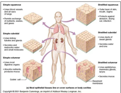
In most multicellular organisms, all cells are not identical. There are important differences in their morphology and function and those due to the process of differentiation. Differentiation leads to changes in many aspects of cell physiology: its size, shape, polarity, metabolic activity, sensitivity to signals and gene expression can all be modified during differentiation. Differentiation of stem cells is a mechanism that allows humans to renew its cells. The basal portion of the skin is comprised of stem cells, which are differentiated so as asymmetric: a stem cell gives an epithelial cell and a skin stem cell. Epithelial cells are commonly found throughout the body of multicellular organisms, and constitute the border between the internal and external environments. Epithelia cells can have varied morphology depending on their location (figure 1.1). Due to their localization, some epithelial cells become stratified because they are exposed to chemical and mechanical stress. Elsewhere in the body, the epithelia form monolayers. These barrier layers can be columnar in the intestine, squamous in the lungs, or tubular in the renal tubule.
Figure 1.1: different types of epithelia
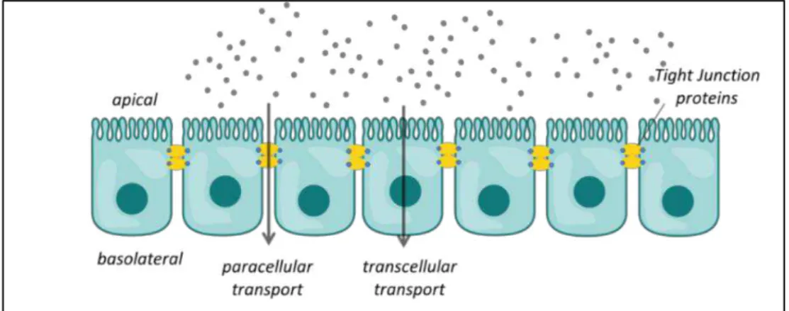
Epithelial and endothelial cell layers serve as functional barriers in many different parts of the body. These cell layers form selectively permeable interfaces that control not only diffusive permeation of solutes along paracellular routes between adjacent cells, but can also actively transport substances along transcellular pathways. Individual epithelial and endothelial cells are joined to each other by specialized complexes, including the adherens
16
junction and the Zonula occludens, commonly referred to as tight junction [1] (figure 1.2).
Figure 1.2: Transport across barrier tissues
Tight junctions are composed of several proteins, such as the transmembrane proteins occludin and claudin and the intracellular proteins ZO-1 and ZO-2. The tight junctions seal the intercellular space between adjacent cells to create a barrier, thereby restricting paracellular diffusion [2]. These tight junctions are of particular relevance for the active barrier functionality of the cell layer. They regulate the passage of molecules across the barrier as they selectively open and close in response to various signals from the inside and outside of the cells. There are many different types of barrier tissue in the body.
Tight junction was first described as Zonula occludens in a study from Schneeberger et al. [3] where horseradish peroxidase (HRP) was injected into mice to determine alveolar-capillary membrane permeability. Upon fixation, tissues sections were examined under electron microscopy. They found that HRP pass endothelial junction but cannot reach alveolar space because HRP was stop by junction between epithelial cells. Similar results were observed when HRP were instilled intranasally. These finding demonstrate that these junction form a barrier to the passage of HRP.
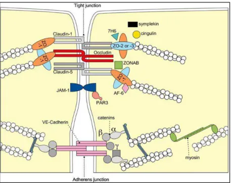
17 Figure 1.3: Molecular composition of tight junctions. The transmembrane proteins occludin, the claudin(s) and junctional adhesion molecule-1 (JAM-1) constitute
the barrier formed by TJs, sealing the paracellular space. From Förster, 2008 [4].
By electron microscopy TJ appear as kissing points at the outer leaflets of the plasma membrane of adjacent cells, which seal the intercellular space [5]. When TJs are observed by immunofluorescence, they are found at the cell border [6]. Following a freeze fracture technique they are observed to be a network of fibrils which circle the apical plasma membrane [7]. From this observation, TJ have been described as a scaffold of both integral and peripheral proteins (figure 1.3). The former play a role in the establishment of cell-cell contact while the latter act as a bridge to link the integral protein to the cytoskeleton.
18
The integral proteins responsible for occluding of intercellular space are mainly occludin, claudin, and junctional adhesion molecule (JAM) protein.
Figure 1.4. Integral membrane TJ proteins from Schneeberger et al, [8]
Occludin was the first TJ transmembrane protein identified [9]. The four transmembrane domains of occludin (figure 1.4) are separated by hydrophilic extracellular loops rich in tyrosine and serine. In epithelial cell lines, highly phosphorylated occludins are found at the TJ, so the action of phosphatase/kinase can modulate the assembly of the filament at the TJ [10]. The carboxyl terminal of the protein can directly interact with actin [11] or bind to the peripheral protein ZO-1 [12].Experiments using synthetic peptides homologous to the region of the first extracellular loop suggest that extracellular loops are critical in the formation of the paracellular barrier [13]. Truncated protein experiments also demonstrated that the carboxyl terminal of occludin is necessary for the function of TJs [14]. Although occludin has been shown to be a constituent of TJ filaments, its role remains unclear as occludin knock-out mice are viable with well-defined TJs [15].
Claudin proteins, like occludin, form four transmembrane domains and two extracellular loops (figure 1.4). By using different size of polyethylene glycol, it has been demonstrated that the permeability has two components,: One pathway is formed by claudin pore and is charge selective, where only molecules less than 4Å in diameter can pass through the pore. Molecule larger than 4Å in diameter can also pass across a intact TJ, but require a dynamic and temporary opening of the TJ [16]. The first extracellular loop, rich in charged amino acids, creates the ionic selectivity. Through the presence of disulfide bonds, the stability of the extracellular loop in claudins is enhanced and thus facilitates the pore formation [17]. The transepithelial resistance (TER) reflects
19
the passage of ion and charge molecules through the TJ. The pore form by claudins is the main pathway for ions, so pores are the key determinants in ion permeability. The second extracellular loop takes part in the interaction with claudin in the neighboring cell plasma membrane and within the same. The expression of claudin proteins in fibroblasts demonstrates that claudin constitutes the backbone of a TJ strand [18]. It has been shown that claudin is the key regulator of the TJ formation [19,20]. By co-cultivating fibroblasts which express different claudin [21], it has been found that claudins form selective interactions and that this interaction determines the barrier properties [22].
JAM proteins are single transmembrane proteins (figure 1.4), and members of the immunoglobulin superfamily [23]. These proteins have been shown to interact with peripheral TJ proteins such as ZO-1 via their carboxy terminus [24], but are not able to from TJ strands in fibroblasts [25]. They are concentrated at the TJ and appear to be associated laterally to claudin proteins [26]. Thus, JAM proteins are involved in the formation and assembly of TJ in epithelial cells but are not involved in the barrier function.
A number of proteins are located in the submembraneous region of TJ. Most of these proteins function as molecular scaffolds by containing multiple protein-protein binding sites.
Zonula occludens -1 (ZO-1) was the first TJ associated protein identified [27]. Later, two homologues, ZO-2 and ZO-3 were discovered [28,29]. These proteins are members of the membrane-associated guanylate kinase (MAGUK). Due to its protein-protein binding site, ZO-1 plays a central role in TJ scaffolding (Figure 1.5).
20 Figure 1.5: Schematic diagram of interactions of ZO-1 with transmembrane,
cytosolic and cytoskeletal proteins, from Kosińska, et al [30].
Via its PDZ domain, ZO-1 interacts with claudin [31], other ZO proteins [11] and JAM [32], with occludin via guanylate kinase (GUK) homology domain and with F-actin through its C-terminal domain. Through its interaction with signaling proteins, ZO-1 can also regulate gene expression, cell proliferation and paracellular permeability [33].
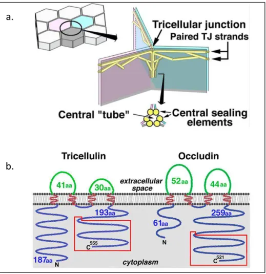
It should be noted that for many years, TJ has been described as cell-cell sealing proteins between two adjacent cells. However, at the meeting point of three epithelial cells, junctions are also present and contribute to the sealing of the paracellular pathway. These structures are named tricellular TJ [34] (figure 1.6). Tricellulin is the protein involved in tricellular junction, which has been demonstrated using two different experiments. A delay in barrier formation has been observed in tricellulin knock down [35]. Moreover, a mutation in this protein induces a decrease in transepithelial resistance and an increase in permeability. Interestingly, the loss of tricellulin also destabilized bicellular TJ by disturbing occludin strands [34]. Furthermore, occludin knock-down leads to a mislocation of tricellulin to bicellular TJ [35]. Together these results demonstrate that tricellulin may compensate for the role of occludin proteins. It has been shown that the region homolog to occludin, in the carboxy terminal domain, bind to ZO-1 [36]. Krug et al also demonstrate that tricellulin
21
selectively restricts the passage of macromolecules without affecting the passage of ions [37]. Through these studies, the action of tricellulin in TJ has been demonstrated, but the mechanisms by which these proteins interact with other proteins remain unclear.
Figure 1.6: (A) Schematic drawing of the organization of tTJs. One tricellular contact (left drawing) is enlarged in the right drawing. (B). Structure and expression of
mouse tricellulin. Adapted from Ikenouchi et al [34].
Tight junctions are complexes of protein that contain cytoplasmic protein associated with transmembrane protein. These cytoplasmic proteins can have different roles to the tight junction.
When epithelial cells adhere one to the other, tight junction protein JAM and ZO-1 are present in high concentration. TJ proteins will interact with polarity complex [38]. Based on recent data [39], the interaction of polarity complex and TJ protein is crucial for the apical basal polarity and maturation of apical junction. The presences of apical and basal domains confer two roles to the tight junction: a gate and a fence function [40,41].
22
The regulation of the passage of ions and molecules across barrier tissues is determined by the opening of TJs. This action refers to the gate function of TJ and can be measured by the TER of barrier tissues. The value of TER results from the sum of the transcellular and paracellular resistance [4]. The transcellular resistance, which is defined by the resistance of the apical and basal membrane, remains stable. Therefore, the fluctuation of the TER value is due to a modification of the paracellular flux resulting from the opening of the tight junction.
The fence function corresponds to the ability of lipids to move within the membrane from the apical to the basal domain. The polarity of the membrane is maintained by this function and can be measured by the free diffusion of fluorescent lipids across the barrier [42].
Tight junction proteins are also involved in signaling event from and to the environment of the cell. Through its interaction with different kinase, occludin protein can induce the proliferation of cells [43-45]. Recent data also suggests that transition from epithelial to endothelial transition imply the targeting of TGF- by occludin [46]. Other studies demonstrated that ZO-1 localizes in the nuclear region to regulate proliferation during maturation of cells by interacting with ZONAB [47]. Through this interaction, ZO-1 protein plays a role in signaling. Protein involved in TJ have also play a role in tumor genesis [48] membrane trafficking [49] and transcriptional regulation [50].
Measuring barrier tissue integrity is important as the degree of intactness can be an indicator for disease state [2,51] and also of the suitability of a particular in vitro model for use in toxicology and drug screening.
Pathogens have devised multiple mechanisms to destroy the integrity of the intestinal epithelial barrier, for example. The effect of individual pathogens/toxins on the intestinal epithelium has been well characterized: these pathogens disrupt barrier tissue in a variety of ways, such as by targeting tight junction proteins. Enteropathogenic and Enterohaemorrhagic E. coli are known to target occludin and ZO-1 respectively [52,53], while rotaviruses appear to disrupt the extracellular domains of tight junctions [54]. Once the barrier tissue is destroyed, the normal absorption of water in the intestine is severely compromised and diarrheal disease usually results [55]. In Europe, 5609 food-borne outbreaks were reported in 2007, affecting 39,727 (you can round to , or say nearly , people, resulting in round hospitalizations, and causing 19 deaths [56]. The global incidence of food-borne disease is difficult to estimate, but it was reported that in 2005 alone 1.8 million people died from diarrheal diseases. A great proportion of these cases can be attributed to contamination of food and drinking water. Additionally, diarrhea is a major cause of malnutrition in infants and young children [57]. The action
23
mechanism of toxins or pathogens targeting other barrier tissues are less well known, possibly because in vitro models are less well developed.
Thousands of new chemical products are tested every year to determine their organ toxicity following a long-term exposure to chemicals, their carcinogenic potential, toxicity to reproductive functions and to the developing fetus, and to their long-term toxicity to the aquatic environment. The uses of in
vitro models based on barrier tissues are one of the first steps of toxicity tests.
To induce toxicity, a chemical product should be able to cross the human epithelial barrier. Pharmaceutical industries, beside the toxicity aspect, want to know if the use of excipient modifies the barrier properties and if this modification is reversible or induce adverse effects.
Several methods exist to assess barrier tissue integrity. Many molecular biology techniques require chromophores or fluorophores. These methods require the realization of different steps, which increase the time and the cost of the experiment. They also require specific kits and sophisticated instruments to measure the read-out. Measurements of transepithelial resistance have been demonstrated to be an indicator of barrier tissue integrity. Since then, different methods have emerged to measure the TER or cell permeability. These include the use of transwell devices as a permeability assay for high throughput screening, which is extensively used in the pharmaceutical industry. Cells are seeded on a porous membrane, compounds are added upon confluency, and samples are subsequently harvested from the chamber below and analyzed by mass spectrometry. Permeability is often measured using radiolabeled compounds or molecules such as Lucifer yellow which are normally impermeable to barrier tissue, although the degree of permeability needs to be defined depending on the tissue type. For measuring TER there are a variety of tools available: traditionally TER has been measured using an epithelial voltohmmeter, which measures resistance across the cell monolayer. This technique, however, requires the use of a bulky probe which must be physically placed into a tissue culture plate and causes large scale disruptions to the cell layers. The CellZscope is an automated device for measuring the impedance of cell layers on permeable membranes (www.nanoanalytics.com), thus combining the ability to measure permeability and TER. New technology has evolved to allow the growth of epithelial cells directly on planar capacitors using the ECIS system (Electric Cell-substrate Impedance Sensing; Applied Biophysics). However, this system is rather costly, and the sensor heads are difficult to fabricate and require a very specific geometry to accommodate the electrodes.
24
Immunofluorescence uses the specificity of antibodies to target a specific molecule by an antibodies/antigen reaction. This technique needs instruments such as a fluorescent microscope or a confocal microscope as the antibodies are coupled with fluorescent probes.
There are two types of immunofluorescence techniques, direct or indirect.
In the case of direct or primary immunofluorescence, a single antibody which is coupled with a fluorophore is used to target the protein of interest and label it. The advantage of using single antibodies is the reduction of the steps and background by decreasing the risk of cross reactivity or non-specificity. But since the number of fluorescent probe linked to the antibodies is limited, the signal is lower than in indirect techniques.
Secondary, or indirect, immunofluorescence uses two antibodies. The first antibody targets the molecule of interest while the second antibody which is coupled with the fluorophore targets the first antibody. As many secondary antibodies can recognize the primary antibody, the sensitivity of the signal is increased. This technique is more time consuming but offers more flexibility in the choice of antibody.
Limitations of immunofluorescence common with all techniques that use fluorescence are photobleaching and the fixation process. The former can be avoided by reducing the intensity and exposure time or by employing robust fluorophores such as AlexaFluor. For the latter, the used of fixative agent can induce a cross linking of cell proteins leading to non-specific bounding.
To avoid the use of antibodies, recombinant proteins can be a good alternative as they can also be used in living cells to follow, for example, trafficking proteins. Even if this technique seems to be elegant, they require transfection or transduction assays.
Beyond these constraints, the protocol should be adjusted for each type of experiment, as the fixative agent, the blocking step and the incubation temperature can have dramatic effects on the staining. Also, the use of monoclonal or polyclonal antibodies can affect the sensitivity of the binding.
25
The permeability of a barrier reflects the ability of a molecule to pass across the barrier. This technique requires the use of radiolabeled or fluorescent compounds which are normally impermeable to the barrier. A barrier tissue is size and charge selective. As described above, claudins proteins are the major player in selective permeability. As each cell line express different claudin, the compound of interest should be chosen according to the cell line used.
Radioactive mannitol has been widely used in fundamental studies and pharmaceutical industry as it can diffuse across the intestinal epithelium with a specific rate according to the stomach segment [58]. The uses of radioactive labels require the specific equipment, room, and safety equipment to confine the radioactivity to one area. The safety procedures induce high cost and risks for laboratories, which now try to exclude technics based on radioactivity.
An alternative is to use of Dextran or polyethylene glycol, which can be available in different size depending on synthetic control. Thus, the permeability to different sizes can be tested with the same compound. They also offer a wide possibility of branching sites which allow different detection technique colorimetric, fluorescence… [59,60].
Another permeability marker commonly used is Lucifer Yellow. It was first used as an intracellular dye for nerve cells [61]. Later, this dye was used to test the permeability of barrier tissues, in particular for the Caco-2 cell line. This dye can be used to follow the differentiation of the monolayer forming barrier tissue as well as the effect of toxic compounds on the barrier tissue and the absorption of drugs.
As biological techniques uses labels, the generation of artifact is possible. The emergence of electronic methods for live cell sensing permits the data acquisition of a wide variety of measurements. Furthermore, these techniques have the advantage of providing dynamic and real time measurements since they are label free and non-invasive.
Pharmaceutical companies could be the main entities interested in electronic methods as they need to test at the same time the ability of the drug to reach their target and the toxicity of the drug. For the moment they use permeability measurements which are cell line and molecule dependent, so different marker (size and charge) must be used to determine the toxicity and the efficiency of drug. The diagnostic industry also is also interested in the use of such techniques to detect pathogens. Monitoring cell health with electronic
26
methods has applications in food and water safety as well as medical diagnostic and environmental protection.
Integrity of epithelia has been assessed by the measurement of ion flow across epithelial tissue generally termed the transepithelial resistance (TER). As describe before in this chapter the ion flux across the epithelium is controlled by TJ and the tightness of these junctions depend of the proteins complex composition, particularly the claudins [62,63]. Steady-state ion flux via the paracellular pathway goes either through the pore pathway formed by transmembrane tight junction proteins, or via the non-pore pathway, postulated to be caused by dynamic breaking and resealing of tight junction strands [63]. The distinction between these pathways is often not considered. As permeability measurement use molecule that pass through the non-pore pathway, it cannot be used as an accurate measurement to assess the function of the pore pathway [64]. Electrical measurement will provide a direct assessment of ions flux by taking account of both pathway, and so an accurate method to assess barrier tissue integrity.
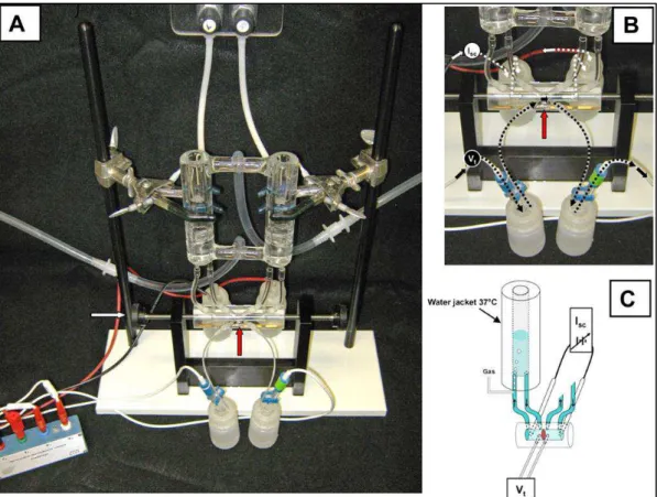
The first in vitro model for measuring electrical parameters of cells was based on using chambers (figure 1.7), where it was demonstrated that cell forming barrier tissue develop the same transport and permeability characteristics as in vivo. Later on, Cereijido and co-workers grew monolayers of cells on filters and placed it between two chambers (filled with the same electrolyte) and measured the electrical potential difference. They demonstrate that cells provide an electrical resistance known as Trans epithelial resistance (TER or TEER). The value is calculated as the inverse of the conductance, expressed in Ω.cm2 and was found to increase over time and settle, remaining
stable for a few weeks [65]. Later on, Cereijido and co-workers grew monolayers of cells on filters and placed it between two chambers (filled with the same electrolyte) and measure the electrical potential difference. As researchers were interested in understanding what is influencing the variation of ions flux among different cell lines, different studies were performed. One of them lead to the development of a microelectrode array to study and measure electrical activity of cell culture in vitro [66].
27 Figure 1.7: A classical Ussing chamber. A: assembled apparatus with water-jacketed reservoirs, Ussing chamber (intestinal preparation is mounted vertically; red
arrow) secured by thumb wheel screws (white arrow), and electrodes attached to voltage clamp head stage. B: close-up view of voltage (Vt)-measuring and short-circuit current (Isc)-passing pathways. Calomel half-cell electrodes used for Vt measurements are connected by 3 M KCl salt bridges at each side of intestinal preparation (red arrow). Ag-AgCl electrodes used for Isc passing across the intestinal preparation are connected
to the chamber by Krebs bicarbonate Ringer (KBR) salt bridges at each end of the chamber. C: schematic cut-away diagram of Vt and Isc circuits of the Ussing chamber.
Short circuiting (Isc) is provided by an automatic voltage clamp (symbol). Note superfusion circulation of KBR is driven by gas lift using 95%O2-5% CO2. Intestinal preparation (red disc) separates the mucosal and serosal baths. From Clarke, L.L [67].
The opposition that a circuit presents to an alternative current defines the electrical impedance. Electrical impedance is usually measured in a range of frequencies and corresponds to the ratio of voltage and current. This technique uses low voltage [68] and is, therefore, compatible with biological systems. The first device using this technology was monitoring cell attachment and spreading on a gold electrode [69]. This method, named electrical cell substrate impedance sensing (ECIS), serves as a new method to monitor arrays of cells in
28
ECIS is now a commercially available product (www.biophysics.com) and has been used for applications such as kinetics of cell spreading [70] analysis of adherent cells after electroporation [71]. This system is composed of an interdigitated electrode, or two gold electrodes immersed in cell culture media. When cells are cultured on top of the working electrode, the flow is restricted and the impedance is modified. This technique has the advantage of monitoring the TER change in real time but is not compatible with cell culture insert.
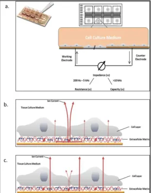
Figure 1.8: The ECIS system. a. Schematic drawing of an ECIS array and principle of the electric cell-substrate impedance sensing (ECIS) method. Cell layers are grown to confluence on integrated gold-film electrodes. An applied AC current flows between
small working electrodes and the larger counter electrode using normal culture medium as an electrolyte. By a variation of the frequency ω, a spectrum can be obtained. Applying higher frequencies the current flow is dominated by the capacity of
the total system, at mid-range frequencies the ohmic resistance of the total system is mirrored b. The current pathway at low frequencies on a cerebral endothelial cell monolayer (ECIS method, 400 Hz). At low frequencies the current predominantly flows
paracellular (through extracellular matrix proteins) and between adjacent cells (through tight junctions) and the electrolyte (medium), see bold arrows. c. By applying
high frequencies (ECIS method, > 40 kHz), the capacitive amount of measured impedance is especially sensitive for adhered cells. The current passes through the insulating cell monolayer, especially through cell membranes. Adapted from Benson et
29
A number of different cell lines need to be cultured in cell culture insert to become differentiated. Indeed, some cells line need to be fed by the basal and apical side to become polarized and exhibit a fully differentiation. This cells lines are often use for transport assays. The integrity prior and during experiment is measured by the electrical properties of the cell monolayer. The development of chopstick electrodes allows measurement the resistance across the monolayer based on the current recorded between electrodes. However, studies demonstrate that the use of DC current can damage cell layers [73,74].
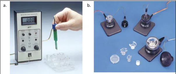
To avoid this problem, the EVOM setup (Epithelial Voltohmmeter; world precision instruments) was developed (Figure 1.9). It can measure the resistance of a cell layer at low frequency with a square current pulse. However, this setup still presents disadvantages. The main issue is that the measurement is operator dependent, and, depending on where the electrodes are placed, the TER values significantly fluctuate. Also, the measurements are carried out outside the incubator which leads to a perturbation of physiologic parameters and thus to a variation of TER values. Despite this drawback, the EVOM system was used in several studies, from basic research to the effect of pathogenic organisms infecting cell layers [75-79]. Based on the same technology, an automated system was developed to allow the measurement on 24 and 96 well plates. To improve the EVOM system, industry developed the Endohm system in which the electrodes are circular, to improve measurement heterogeneity.
Figure 1.9: Hand voltometers. a. EVOM device; b. Endohm system. Adapted from wpiinc.com.
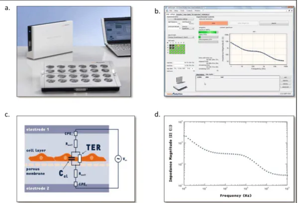
In 2004, Wegener et al. [80] developed a new device which solves two of the main problems of the devices presented above, maintaining physiological conditions by measuring the TER in an incubator, and homogenous results by using round fix electrodes. This device is named the CellZscope and is commercially available (NanoAnalytics GmbH) (figure 1.10). The experiment is controlled with a PC interface and the electrical measurements are performed
30
with a data acquisition board. 6 to 24 well formats can be used; each filter can be controlled individually. The cell culture insert is placed between two electrodes. The bottom electrode is common for all wells, while the top one is small and dipped in each well. A small AC current is applied between the two electrodes; the impedance frequency on a range from 1 to 105 Hz is recorded. The TER and the capacitance are extracted from an equivalent circuit model. The capacitance is also an important parameter as it can be indicative of membrane folding, an increase in capacitance reflects more folding or ruffling of the membrane [81]. The equivalent circuit and the model used allow the extraction of the resistance due to the filter and/or media, as the addition of compound or the change of media can induce variation of the TER result. To permit the comparison of the result with other techniques and experiments, the device gives the result of TER as Ω.cm2.
Figure 1.10: The CellZscope a. Overview of CellZscope device, b. Overview of CellZscope data acquisition window, c. Equivalent circuit for epithelial cell layer grown on porous filter, d. Typical Impedance frequency scan. Adapted from technical bulletin,
Nanoanalytics.com.
The ECIS system cannot be used with cell culture insert, because cells must be grown directly on top of the electrodes. To overcome this disadvantage, Sun et al [82] developed transwell filters that are enclosed in a polycarbonate holder and separated from the gold plated dot ring electrode by an insulating epoxy layer (figure 1.11). The electrode are fabricated by electrochemical deposition of a conducting polymer (polypyrrole doped with polystyrene sulfonate (Ppy: PSS)) on the gold plated electrodes. This deposition was realized to solve the effect of the electrical double layer, which usually
31
appear at the interface between the surface and an ionic fluid. This layer corresponds to the formation of a first layer of ion adsorbed onto the surface and a second layer composed of ion attracted to the first charge layer. The deposition of this conducting layer increases the sensitivity of the measurement at low frequency, which is known to be dominated by the paracellular resistance. The model used to extract TER is very similar to the one used for the CellZscope. The differences reside in the epithelial cells impedance; they shown that the electrical field which enters in the cell layer has a longitudinal and orthogonal component. The impedance was therefore divided in two parts. They also demonstrate that the Ppy electrode have an impedance lower than gold electrode by one order in magnitude. To validate this method, they authors used Triton X 100 and the calcium chelator EGTA (ethylene glycol-bis(2-aminoethyl-ether)-N, N, N , N ,-tetraacetic acid) to induce a destruction of a barrier tissue.
Figure 1.11: Conducting polymer coated electrodes for impedance scanning. A. Diagram shows the 3D assembly structure and 2D cross-section view of the bio-impedance chip. B. Image showing the experimental system instrumentation. From Sun
et al [82].
Organic electronics refers to devices using carbon based polymers and small molecules as the electrically active component. Organic electronics offer a number of unique advantages including their ease of processing, ionic and electronic conductivity, and their flexible mechanical properties. The soft nature of these materials and their compatibility with soft substrates makes them ideal for flexible displays and solid state lighting, and also for interfacing with biological systems.
32
Organic bioelectronics, first named by Berggren and Richter-Dahlfors [83], couple device based on organic semiconductors with biological elements. This coupling is bi-directional: a signal from a biological event can be transduce to the electronic device or the device can stimulate a biological element to induce a reaction or an event into this biological element (figure 1.12).
Figure 1.12: Integrated systems of biomaterials and electronic elements for bioelectronics applications. From Itamar Willner and Eugenii Katz, 2005
A numbers of biological elements are capable of recognition, including enzymes, oligonucleotide and DNA. Coupling these elements with electronic device to build biosensors has been of great interest. The major challenge with this type of sensor is the optimization of the information transfer between the two parts of the device. The information sent by the biomaterial should be translated to the recognition element such that the output signal could be differentiated from the background signal. Glucose sensors were developed in 1960 to monitor glucose level in blood diabetic patents. As OECT provide an inherent amplification, detection of glucose in human saliva can be achieve using an all polymer sensor [84]. Using the same approach, OECTs were integrated with microfluidic to detect multi-analytes [85]. Another type of transistor (organic field effect transistor) was developed with microfluidic to sense in aqueous solution without recognition elements [86]. In this device, detection of glucose is attributed to an interaction of the glucose with the conducting polymer.
Organic electronic materials can also be used to control biological elements. Investigation of the influence of the redox state of conducting
33
polymers on cells proliferation and differentiation was the first application [87]. Applications in the biomedical field come out with the functionalization of conducting polymer with biomolecules [88] [89]. Further, Wan et al, developed a device in which a redox gradient is patterned [90]; this gradient is critical to understanding cellular mechanisms in normal and pathologic tissues; similar work was also done by Berggen et al. [91]. To move toward more relevant cell-cell interactions, a 3D scaffold was developed; nanofibers coated with conductive polymer were used to induce calcium signaling on nerve cells [92]. Finally, to control release of biomolecules, organic electronic ion pumps were develop for in vitro [93] and in vivo [94] used.
In the last area, organic electronic can stimulate biological structure and in return sense or record a response to the stimulus. This field focuses on developing tools for neuroscience. The soft conducting polymer offer great opportunities to replace silicon based probes or electrodes. The first advances in this field demonstrate higher performance of these new electrodes compared to the state of the art devices [95]. These improvement were combined with hydrogels and then biomolecules were incorporated [96-98]. The combination of conducting polymers, biomolecules and hydrogel improve the biocompatibility of the device and also permit the exploration of electrical stimulation. Based on this work, microelectrodes were used as sensors of neurotransmitter release [99]. Evolution of this work and advantages of the conducting polymers device were discussed in a review by Kim et al. [88]. Electrodes were then fabricated in a network to create microelectrodes arrays (MEA). The first generation of these MEA were too thick and would limit their conformability [100]. Recently, a new way of fabrication demonstrated the use of dense arrays of electrode for in vivo recording [101]. These polymers electrodes were shown to have better performance than gold electrodes.
Impedance spectroscopy provides significant contributions in the field of measurement of capacitance and resistance of a cell layer. This technique is more automated and reproducible than other technique but still presents limitation in temporal resolution and sensitivity as well as cost. Transistors provide a good alternative to conventional electrodes for in vitro and in vivo monitoring of cells. Organic electrochemical transistors (OECT) have been recently introduced for biomedical applications. Unlike field effect transistors and inorganic transistors, the electrolyte plays an important role in the device operating mechanism. OECT based on the conducting polymer PEDOT: PSS (poly(3,4-ethylenedioxythiophene)-poly(styrenesulfonate) were shown to be stable in media in physiological condition [102-104].Organic materials are compatible with roll to roll, large scale processing for low cost processing of disposable devices. The key advantage of OECT is that it converts ionic signals
34
to electronic signal efficiently and can operate at low voltage which is fully compatible with biological environments.
The first study based on this technology was the observation of cells detachment upon treatment with trypsin on a cell layer [104]. Authors determined that the change detected by the OECT was due to an electrostatic effect, whereas it is instead an effect of ion penetrating the conducting polymer [105].
Previously, bilayer lipid membrane were used to control the gating of an OECT [106]. When an intact bilayer lipid membrane was placed on top of an OECT, the gating was suppressed. Whereas, the addition of ion channel into the bilayer lipid membrane restore the gating. This work demonstrates that OECT can be used as biosensors as they convert ion to electron and that movement of ions across a membrane can be detected by an OECT. In this project, we use this property to create a new device to detect pathogens. Gastrointestinal cells will be grown as monolayer in cell culture insert to form an epithelial barrier and integrated with an OECT.
35
1. Anderson, J.M.; Van Itallie, C.M., Physiology and function of the tight junction. Cold Spring Harb Perspect Biol 2009, 1, a002584.
2. Balkovetz, D.F.; Katz, J., Bacterial invasion by a paracellular route: Divide and conquer. Microbes and infection / Institut Pasteur 2003, 5, 613-619. 3. Schneeberger-Keeley, E.E.; Karnovsky, M.J., The ultrastructural basis of
alveolar-capillary membrane permeability to peroxidase used as a tracer.
The Journal of cell biology 1968, 37, 781-793.
4. Forster, C., Tight junctions and the modulation of barrier function in disease. Histochemistry and cell biology 2008, 130, 55-70.
5. Furuse, M., Molecular basis of the core structure of tight junctions. Cold
Spring Harb Perspect Biol 2010, 2.
6. Li, N.; Lewis, P.; Samuelson, D.; Liboni, K.; Neu, J., Glutamine regulates caco-2 cell tight junction proteins. American journal of physiology.
Gastrointestinal and liver physiology 2004, 287, G726-733.
7. Gonzalez-Mariscal, L.; Betanzos, A.; Nava, P.; Jaramillo, B.E., Tight junction proteins. Progress in biophysics and molecular biology 2003, 81, 1-44.
8. Schneeberger, E.E.; Lynch, R.D., The tight junction: A multifunctional complex. American journal of physiology. Cell physiology 2004, 286, C1213-1228.
9. Furuse, M.; Hirase, T.; Itoh, M.; Nagafuchi, A.; Yonemura, S.; Tsukita, S., Occludin - a novel integral membrane-protein localizing at tight junctions. Journal of Cell Biology 1993, 123, 1777-1788.
10. Anderson, J.M.; Van Itallie, C.M., Tight junctions and the molecular basis for regulation of paracellular permeability. The American journal of
physiology 1995, 269, G467-475.
11. Wittchen, E.S.; Haskins, J.; Stevenson, B.R., Protein interactions at the tight junction. Actin has multiple binding partners, and zo-1 forms independent complexes with zo-2 and zo-3. The Journal of biological
chemistry 1999, 274, 35179-35185.
12. Furuse, M.; Itoh, M.; Hirase, T.; Nagafuchi, A.; Yonemura, S.; Tsukita, S., Direct association of occludin with zo-1 and its possible involvement in the localization of occludin at tight junctions. The Journal of cell biology
1994, 127, 1617-1626.
13. Van Itallie, C.M.; Anderson, J.M., Occludin confers adhesiveness when expressed in fibroblasts. Journal of cell science 1997, 110 ( Pt 9), 1113-1121.
36
14. Balda, M.S.; Whitney, J.A.; Flores, C.; Gonzalez, S.; Cereijido, M.; Matter, K., Functional dissociation of paracellular permeability and transepithelial electrical resistance and disruption of the apical-basolateral intramembrane diffusion barrier by expression of a mutant tight junction membrane protein. The Journal of cell biology 1996, 134, 1031-1049.
15. Saitou, M.; Furuse, M.; Sasaki, H.; Schulzke, J.D.; Fromm, M.; Takano, H.; Noda, T.; Tsukita, S., Complex phenotype of mice lacking occludin, a component of tight junction strands. Molecular biology of the cell 2000,
11, 4131-4142.
16. Knipp, G.T.; Ho, N.F.H.; Barsuhn, C.L.; Borchardt, R.T., Paracellular diffusion in caco-2 cell monolayers: Effect of perturbation on the transport of hydrophilic compounds that vary in charge and size. Journal
of pharmaceutical sciences 1997, 86, 1105-1110.
17. Li, J.; Angelow, S.; Linge, A.; Zhuo, M.; Yu, A.S., Claudin-2 pore function requires an intramolecular disulfide bond between two conserved extracellular cysteines. American journal of physiology. Cell physiology
2013, 305, C190-196.
18. Furuse, M.; Fujita, K.; Hiiragi, T.; Fujimoto, K.; Tsukita, S., Claudin-1 and -2: Novel integral membrane proteins localizing at tight junctions with no sequence similarity to occludin. The Journal of cell biology 1998, 141, 1539-1550.
19. Furuse, M.; Sasaki, H.; Fujimoto, K.; Tsukita, S., A single gene product, claudin-1 or -2, reconstitutes tight junction strands and recruits occludin in fibroblasts. The Journal of cell biology 1998, 143, 391-401.
20. Furuse, M.; Hata, M.; Furuse, K.; Yoshida, Y.; Haratake, A.; Sugitani, Y.; Noda, T.; Kubo, A.; Tsukita, S., Claudin-based tight junctions are crucial for the mammalian epidermal barrier: A lesson from claudin-1-deficient mice. The Journal of cell biology 2002, 156, 1099-1111.
21. Furuse, M.; Sasaki, H.; Tsukita, S., Manner of interaction of heterogeneous claudin species within and between tight junction strands. The Journal of cell biology 1999, 147, 891-903.
22. Furuse, M.; Furuse, K.; Sasaki, H.; Tsukita, S., Conversion of zonulae occludentes from tight to leaky strand type by introducing claudin-2 into madin-darby canine kidney i cells. The Journal of cell biology 2001,
153, 263-272.
23. Martin-Padura, I.; Lostaglio, S.; Schneemann, M.; Williams, L.; Romano, M.; Fruscella, P.; Panzeri, C.; Stoppacciaro, A.; Ruco, L.; Villa, A., et al., Junctional adhesion molecule, a novel member of the immunoglobulin superfamily that distributes at intercellular junctions and modulates monocyte transmigration. The Journal of cell biology 1998, 142, 117-127.
37
24. Itoh, M.; Sasaki, H.; Furuse, M.; Ozaki, H.; Kita, T.; Tsukita, S., Junctional adhesion molecule (jam) binds to par-3: A possible mechanism for the recruitment of par-3 to tight junctions. The Journal of cell biology 2001,
154, 491-497.
25. Tsukita, S.; Furuse, M., [identification of two distinct types of four-transmembrane domain proteins, occludin and claudins: Towards new physiology in paracellular pathway]. Seikagaku. The Journal of Japanese
Biochemical Society 2000, 72, 155-162.
26. Tsukita, S.; Furuse, M.; Itoh, M., Multifunctional strands in tight junctions. Nature reviews. Molecular cell biology 2001, 2, 285-293.
27. Stevenson, B.R.; Siliciano, J.D.; Mooseker, M.S.; Goodenough, D.A., Identification of zo-1: A high molecular weight polypeptide associated with the tight junction (zonula occludens) in a variety of epithelia. The
Journal of cell biology 1986, 103, 755-766.
28. Haskins, J.; Gu, L.; Wittchen, E.S.; Hibbard, J.; Stevenson, B.R., Zo-3, a novel member of the maguk protein family found at the tight junction, interacts with zo-1 and occludin. The Journal of cell biology 1998, 141, 199-208.
29. Jesaitis, L.A.; Goodenough, D.A., Molecular characterization and tissue distribution of zo-2, a tight junction protein homologous to zo-1 and the drosophila discs-large tumor suppressor protein. The Journal of cell
biology 1994, 124, 949-961.
30. Kosińska, A.; Andlauer, W., Modulation of tight junction integrity by food components. Food Research International 2013.
31. Itoh, M.; Furuse, M.; Morita, K.; Kubota, K.; Saitou, M.; Tsukita, S., Direct binding of three tight junction-associated maguks, zo-1, zo-2 and zo-3, with the cooh termini of claudins. Journal of Cell Biology 1999, 147, 1351-1363.
32. Ebnet, K.; Schulz, C.U.; Meyer Zu Brickwedde, M.K.; Pendl, G.G.; Vestweber, D., Junctional adhesion molecule interacts with the pdz domain-containing proteins af-6 and zo-1. The Journal of biological
chemistry 2000, 275, 27979-27988.
33. Anderson, J.M.; Van Itallie, C.M., Tight junctions: Closing in on the seal.
Current biology : CB 1999, 9, R922-924.
34. Ikenouchi, J.; Furuse, M.; Furuse, K.; Sasaki, H.; Tsukita, S., Tricellulin constitutes a novel barrier at tricellular contacts of epithelial cells. The
Journal of cell biology 2005, 171, 939-945.
35. Raleigh, D.R.; Marchiando, A.M.; Zhang, Y.; Shen, L.; Sasaki, H.; Wang, Y.; Long, M.; Turner, J.R., Tight junction-associated marvel proteins
![Figure 1.5: Schematic diagram of interactions of ZO-1 with transmembrane, cytosolic and cytoskeletal proteins, from Kosińska, et al [30]](https://thumb-eu.123doks.com/thumbv2/123doknet/11580403.298065/21.892.257.637.104.510/figure-schematic-interactions-transmembrane-cytosolic-cytoskeletal-proteins-kosińska.webp)