i
Université de Montréal
---
INVESTIGATING THE
THYMOPOIESIS-STIMULATING PROPERTY OF INTERLEUKIN-21 IN
AGING MICE
--- par Edouard Al-Chami Département de Pharmacologie Faculté de MédecineMémoire présenté à la Faculté des Études Supérieures en vue de l’obtention du grade de Maître ès sciences (M.Sc.) en Pharmacologie
Août 2015
ii
Université de Montréal Faculté des Études Supérieures
Ce mémoire intitulé :
---
INVESTIGATING THE
THYMOPOIESIS-STIMULATING PROPERTY OF INTERLEUKIN-21 IN
AGING MICE
--- a été évalué par un jury composé des personnes suivantes :
Jean-François GAUCHAT , Ph.D. --- Président-rapporteur Moutih RAFEI, Ph.D. --- Directeur de Recherche Martin G. SIROIS, Ph.D. --- Membre du jury
iii RÉSUMÉ
La vaccination est largement utilisée pour la génération de lymphocytes T spécifiques contre les tumeurs. Malheureusement, cette stratégie n'est pas adaptée aux personnes âgées car leur thymus régresse avec l'âge conduisant ainsi à une baisse dans la production de cellules T et à l'accumulation de cellules immunitaires âgées ayant des défauts liés à leurs stimulations. Comme il a été démontré auparavant que L’IL-21 est capable d’induire des fonctions thymiques, nous avons émis l’hypothèse que l’injection d’IL-21 à des souris âgées stimulera la thymopoïèse. Nos résultats montrent que l’administration de l’IL-21 augmente le nombre absolu de thymocytes chez les souris âgées et augmente la migration de ces cellules vers la périphérie ou ils contribuent à la diversité du TCR. De plus les cellules T en périphérie expriment un niveau plus élevé de miR181-a, et par conséquent moins de phosphatase comme SHP2, DUSP5/6 qui inhibent le TCR. En vaccinant des souris âgées avec le peptide Trp2, les souris traitées avec l’IL-21 montrent un retard dans la croissance des cellules B16 tumorales. Cette étude montre que l’IL-21 pourrait être utilisé comme stratégie pour le rétablissement du systeme immunitaire chez les personnes âgées.
iv ABSTRACT
Vaccines have been largely sought for the generation of protective tumor-specific T cells. Unfortunately however, this strategy is not suitable for the elderly as their thymus regresses with age leading to a decline in T-cell production and accumulation of aged lymphocytes endowed with stimulation-related defects. Interleukin (IL)-21 has been shown to display thymo-stimulatory properties. Therefore, we hypothesized that IL-21 administration to ageing host may boost T-cell production and restore a competent peripheral T-cell compartment. Our study shows that IL-21 injection to aged mice lead to an increase in the thymocytes absolute number and an increase in thymocytes egress to the periphery where they enhance the T-cell receptor (TCR) diversity. Furthermore T-cell in the periphery express higher level of miR-181a and thus less TCR-inhibiting phosphatases SHP-2 and DUSP5/6 enable them to be more responsive upon stimulation. Consequently, aged rIL-21-treated mice vaccinated using a tyrosinase-related protein 2 (Trp2)-derived peptide exhibited a substantial delay in B16 tumor growth and improved survival. The results of this study highlight the immunorestorative function of rIL-21 paving its use as a strategy for the re-establishment of effective immunity in the elderly.
v
ACKNOWLEDGEMENTS
First of all, I would like to thank my research supervisor Dr. Moutih Rafei for giving me the opportunity to work in his laboratory. The training I acquired during the last two years was full of challenges, which turned into victories thanks to Dr. Rafei's patience, intelligence and guidance. I was very lucky to work under his supervision. He invested so much in training me and I hope I was at his level of expectations. He helped me writing my scholarship applications, corrected my Power Point presentations and even assisted me in some of my graduate courses. Dr. Rafei is a young investigator full of energy and I wish him the best of luck for the rest of his career. I learned a lot from him and I hope he learned something from me too.
I would also like to thank Dr. Aurélie Tormo for her patience and help. Aurélie was always around for help. Her long expertise in research made my master degree much easier. Thank you for showing and teaching me all the techniques that I used and all the small tricks that allowed me to save time. Many thanks for correcting my terrible French in all of my applications and e-mails. You are such a wonderful lab manager, wish you all the best!
I would also like to thank Dr. Jean-Francois Gauchat. Besides being an expert in Immunology and Molecular Biology, he helped me a lot through his wise feedbacks. What fascinates me the most in Dr. Gauchat is his humbleness. Whenever you cross him in the hallway, he will always smile and greet you. Thank you for charging my days with positive energy! May the force be always with you!
vi
"Impossible is a word to be found only in the dictionary of fools."
vii
PREFACE
lmost 50% of cancer cases are diagnosed in patients aged 60 years and older. As more than 25% of the total U.S. population will reach the age of 60 by 2050, the number of patients suffering from cancer is expected to rise. Since living standards and overall healthcare are improving worldwide, a higher life expectancy requires the adoption of novel strategies aimed at keeping the aging population healthy. Although most cancer types can be removed by surgery, followed by radiation and/or chemotherapy, these multimodality therapies are ineffective against spread metastases. Cancer vaccination represents therefore an appealing alternative due to its limited toxicity and ability to stimulate the host's own immune system to seek and destroy metastatic cancer cells. Unfortunately however, vaccines are less effective at older age due to impaired T-cell responsiveness due to immunosenescence; a degeneration of the immune system caused by thymic atrophy. As a result, the output of newly developed naïve T cells endowed with the capacity of effectively responding to new antigenic challenges dramatically decreases pocking major holes in patients’ immunocompetency. Thus, there is an urgent need for the development or improvement of immunotherapies to suit the elderly population. The study described herein will highlight the utility and potency of interleukin (IL)-21 at stimulating de novo thymopoiesis to rejuvenate the immune system of aged subjects. Optimising and adopting such concept will lead to innovative IL-21-based strategies directed against two key elderly-related health problems: i) providing superior responsiveness to cancer vaccines, and ii) reducing the emergence and/or cancer relapse in this vulnerable population. Therefore, the favourable outcome of this study will have significant ramifications on the future use of IL-21 in the context of preventive/therapeutic cancer vaccination measures aimed at enhancing anti-tumoral immunity of the aging population.
viii TABLE OF CONTENTS Résumé ...iii Abstract...iv Acknowledgments...v Preface...vii Table of contents……….……….…..viii List of abbreviations...x List of figures...xiv
CHAPTER 1: General Introduction 1.0 - Aging and Thymopoiesis ...2
1.2 - Thymus Structure...3
1.2 - Intrathymic T-cell Development………… ...3
1.3 - Thymic Involution from an Evolutionary Perspective...6
1.4 - Factors Causing Thymic Involution...8
1.5- Strategies to Reconstitute the Thymus...………...…..10
¾ 1.5.1- Sex Steroid Ablation………...10
¾ 1.5.2- Keratinocyte Growth Factor...…...………...13
¾ 1.5.3- Ghrelin...…………...…………...14
¾ 1.5.4- Interleukin-7...………....………..…14
¾ 1.5.5-Interleukin-22………...……...…...………...15
¾ 1.5.6-Interleukin-21…………..…..…………...………...……16
ix
1.6.1 - Objective 1…………...…...………...……19
1.6.2 - Objective 2………...…...…….……19
1.6.3 - Objective 3………...………...………20
CHAPTER 2: Interleukin-21 Administration to Aged Mice Rejuvenates their Peripheral T-cell Pool by Triggering De Novo Thymopoiesis 2.0 - Summary...22 2.1 - Introduction………...…...23 2.2 - Results...25 2.3 - Discussion………...…….………...45 2.4 - Experimental Procedure...49 2.5 -Acknowledgments...………..53 2.6- Author Contribution……….54 2.7- References………55 2.8- Supporting Informations……….……….61
CHAPTER 3: General Conclusion 3.0- Conclusion………...74
Bibliography...78
x
LIST OF ABBREVIATIONS
AP-1: Activator Protein-1 AR: Androgen Receptor
Bcl6: B-Cell Lymphoma 6 Protein Bim: Bcl-2-Like Protein 11
BM: Bone Marrow
BMT: Bone Marrow Transplantation
CCL: C-C Motif Ligand CCR: C-C Motif Receptor
CD: Cluster of Differentiation CLP: Common Lymphoid Progenitor
cTECs: Cortical Thymic Epithelial Cells
CTP: Circulating Bone-Marrow Derived T-Lineage Progenitor DC: Dendritic Cells
DEX: Dexamethasone DL4: Delta-Like 4 DN: Double-Negative DP: Double-Positive
DUSP5/6: Dual Specificity Phosphatase 5/6 ELISA: Enzyme-Linked Immunosorbent Assay ERK: Extracellular Signal-Regulated Kinases ETPs: Early Thymic Progenitors
xi FOXN1: Forkhead Box N1 FOXP3: Forkhead Box P3
FSH: Follicle Stimulating Hormone
GATA3: Trans-Acting T-Cell-Specific Transcription Factor
GFP: Green Fluorescent Protein GH: Growth Hormone
GM-CSF: Granulocyte Macrophage-Colony Stimulating Factor GRL: Ghrelin
GRLR: Ghrelin Receptor
GVHD: Graft Versus Host Disease Gzm: Granzyme
HSC: Hematopoietic Stem Cells IFNγ: Interferon-Gamma
IL: Interleukin
IL-21R: Interleukin-21 Receptor IP: Intraperitoneal
KGF: Keratinocyte Growth Factor
KGFR: Keratinocyte Growth Factor Receptor
Lck: Lymphocyte-Specific Protein Tyrosine Kinase LH: Luteinizing Hormone
LHRH: Luteinizing Hormone Releasing Hormone LPS: Lipopolysaccharide
xii LT-HSC: Long Term-HSC
MFI: Mean Fluorescent Intensity MPP: Multi-Potent Progenitor
mTECs: Medullary Thymic Epithelial Cells NFAT: Nuclear Factor of Activated T-cells NK: Natural Killer OVA: Ovalbumin
PBS: Phosphate Buffer Saline Prdm1: PRD Zinc Finger Protein 1
PTPN22: Protein Tyrosine Phosphatase Non Receptor Type 22 qPCR: Quantitative Polymerase Chain Reaction
RAG: Recombination-Activating Genes RNA: Ribonucleic Acid
RORC: RAR-Related Orphan Receptor C
RORγt: RAR-Related Orphan Receptor Gamma
RTE: Recent Thymic Emigrant SLO: Secondary Lymphoid Organ
SP: Single-Positive
STAT: Signal Transducer and Activator of Transcription ST-HSC: Short Term-HSC
SOCS: Suppressor of Cytokine Signaling
SSA: Sex Steroid Ablation T-bet: T-box transcription factor
xiii TCR: T-Cell Receptor
TES: Thymic Epithelial Space TFH: T-Follicular Helper TREC: TCR Excision Circles
TRP-2: Tyrosinase-Related Protein 2 TSP: Thymic Seeding Progenitors WT: Wild-Type
XSCID: X-linked Severe Combined Immunodeficiency ZAP-70: Zeta-Chain-Associated Protein kinase 70
xiv LIST OF FIGURES FIGURE 1...4 FIGURE 2...9 FIGURE 3...12 FIGURE 4...18 FIGURE 5...27 FIGURE 6...30 FIGURE 7...33 FIGURE 8...36 FIGURE 9...40 FIGURE 10...43 FIGURE S5...63 FIGURE S6...65 FIGURE S7...67 FIGURE S8...69 FIGURE S9...71 FIGURE S10...73 FIGURE 11...76
xv CHAPTRE 1
2 1.0 - Aging and Thymopoiesis
One of the most important attribute of adaptive immunity is its capacity to discriminate between self and non-self-antigens (1). In order to achieve that, its main components, B and T lymphocytes, must undergo restricted processes during their development (2). In jawed vertebrates including humans, B and T cells develop at distinct anatomical sites, the former in the bone marrow (BM) or fetal liver and the latter in the thymus (2). From an evolutionary point of view, we can assume that the thymus emerged as a dedicated environment to facilitate and support the development of self-tolerant T cells expressing a diverse repertoire of T-cell receptor (TCR) (3). Failure to build or maintain a proper thymus can lead to defects ranging from immunodeficiency to autoimmunity (4).
The main thymus-associated limitation is the progressive decrease of its function with age; a phenomenon known as thymic involution (5). Age-associated thymic involution occurs in all species, indicating that this process is evolutionary ancient and conserved. It is characterized by a reduction in mass, cellularity as well as loss of normal architecture consequently leading to a decrease in T-cell development (thymopoiesis) (6). As a result, diminished production of naïve T cells and attrition of TCR repertoire in aging hosts is amenable to increased susceptibility to infectious diseases, cancers and autoimmunity (6). From a general perspective, thymic involution can be considered as a natural leading global health problem. Thus, a tremendous amount of effort is put to prevent or reverse age-associated thymic involution as a mean to ensure the well-being of the aging society.
3 1.1 - Thymus structure
The thymus (FIGURE 1) consists of two distinct lobes connected by connective tissues and surrounded by capsular tissues (7). Histologically, each lobe can be subdivided into four major compartments each having a distinct role in thymopoiesis. The four compartments include the subcapsular zone, the cortex, the medulla, and the cortico-medullary junction (4). The subcapsular zone contains mainly cortical thymic epithelial cells (cTECs), whereas the cortex contains a mix of cTECs, fibroblasts and macrophages. The medulla is comprised of medullary thymic epithelial cells (mTECs) and dendritic cells (DC). The cortico-medullary junction is the vascular region where thymic arteries enter the organ. The stroma is considered as the non-hematopoietic component of the thymus and contains keratin positive and negative cells. Keratin positive cells represent TECs and it is divided into keratin5- keratin8+ (K5-k8+ for cTECs) and
keratin5+ keratin8- (k5+k8- for mTECs) cells. Keratin negative cells are a mixture of fibroblasts,
non-fibroblastics mesenchymal cells and endothelial cells (4). The thymus stroma is essential to the regulation of T-cell development and selection. For example, TECs secrete chemokine and growth factors and express cell surface molecules all involved in supporting migration and development of progenitor cells (8). In addition, TECs present distinct peptide repertoires essential for central tolerance (8). During thymic involution, the thymus stroma degenerates and is subsequently replaced by adipose tissue, which negatively interferes with thymopoiesis.
1.2 - Intrathymic T-cell development
Thymopoiesis involves several differentiation and proliferation events during which cross-talks between stromal and lymphoid cells take place (10) (FIGURE 1). Lymphoid progenitor cells
4
FIGURE 1: THYMIC STRUCTURE AND T-CELL DEVELOPMENT.
A simplified overview of the thymus defined regions and stages of T-cell development. (Blackburn CC et al. Nature Reviews Immunology 4, 278-289 (2004) (9).
5
migrate from the BM, enter the thymus via the cortico-medullary junction, and undergo four stages of maturation as they pass from the subcapsular zone through the cortex and to the medulla before reaching finally peripheral circulation as immature naïve T cells (7). For that to happen, T-cell migration to and through the thymus is guided by chemokines produced by thymic stromal cells in distinct regions or microenvironments, and developing thymocytes seem to find their way by sequentially expressing different chemokine receptors specific for the transition between each stage of maturation (11). Developing thymocytes are sub-divided into three major groups based on the expression of CD4 and CD8 on their cell surface. They start as double-negative (DN) cells that do not express CD4 or CD8 and after different maturation processes they become double positive (DP) cells expressing both CD4 and CD8. They finally become single-positive (SP) T cells expressing either the CD4 or CD8 co-receptor (11). DN thymocytes can be further sub-divided into four sub-populations according to their expression of CD44 and CD25: DN1 (CD44+CD25-), DN2 (CD44+CD25+), DN3 (CD44-CD25+), and DN4
(CD44-CD25-). The first step in T-cell development is the migration of lymphoid progenitor cells or thymic seeding progenitors (TSP) from the BM to the thymus. This step is controlled by the adhesive interaction between platelet P-selectin glycoprotein ligand 1, which is expressed by circulating TSP, and P-selectin found on the surface of thymic endothelium. They enter the thymus via the vasculature at the cortico-medullary region and become early thymic progenitors (ETPs) (the most important sub-population of the DN1 fraction). ETPs still retain the potential to give rise to DC, natural killer (NK), and macrophages, but they lose their multipotency as they differentiate and become restricted to the T-cell lineage (12). The first checkpoint in the developmental process of T cells is the T lineage commitment at the DN1/DN2 stages. In order to become committed to the T-cell lineage, DN1 cells interact with the delta-like 4 (DL4) ligand
6
expressed on cTECs through their Notch1 receptor (11). In addition, cTECs secrete interleukin (IL)-7 which supports their differentiation process. DN2 differentiate to DN3 in the subcapsular zone and during this transition the αβ versus γδ T cells fate is specified. The DN3 cells follow another checkpoint known as the β-selection where only DN3 who have successfully rearranged the β-chain of the TCR survive by receiving a signal through a functional pre-TCR complex (11). Thymocytes that have been selected proceed to the DN4 stage, initiate CD4 and CD8 expression and become DP thymocytes in the cortex. At that point, DP cells initiate the TCRα gene rearrangement resulting in the expression of TCRαβ complexes. DP cells in the cortex will then undergo positive selection where only thymocytes with a functional TCR capable of recognizing self-peptide-MHC complexes presented by cTECs receive a survival signal and differentiate into SP cells (13). Surviving DPs will then express the C-C chemokine receptor type (CCR) 7, which guides them to the medulla in response to CCR7 ligands (C-C motif ligand (CCL)19 and CCL21). In the medulla, mTECs and DCs will express and present tissue-specific antigens to SP cells to negatively select any TCR with self-reactive potential in order to avoid generating reactive T cells capable of causing autoimmunity (13). SP thymocytes will spend ~12 days in the medulla whereby they will express the sphingosine-1-phosphate receptor 1. At that time point, CD4 or CD8 T cells egress from the thymus and continue their maturation in the periphery for an additional period of three weeks (13).
1.3 - Thymic Involution from an Evolutionary Perspective
Thymic involution is characterized by the expansion of perivascular space due to adipocytes accumulation with age shifting the ratio of thymic epithelial space (TES) to perivascular space (5). Consequently, the TES represents less than 10% of total thymic tissue by the age of 70 (5).
7
Such thymic regression results in constriction of peripheral T-cell diversity, alterations in their phenotype and function, and corrosion of telomeres causing replicative senescence (6). Age-associated thymic involution has been suggested to occur in all species, indicating that this process is evolutionary ancient and conserved (14). If so, are there any benefits to thymic involution? The human life span has extended drastically over the last centuries due to decline in infectious disease-caused mortality owing to improved sanitation, medical care as well as development of vaccines and antibiotics (15). This means that the immune system, which was once "designed to serve for almost 40 years" has to provide continuous protection for the decades to come (16). Of all evolutionary hypotheses, the "disposable soma theory", which suggests that the body must budget its available amount of energy between locomotion, thermogenesis, growth, reproduction and repair appears to be highly compatible with thymic involution as the thymus is an "energy expensive organ" due to the fact that more than 90% of all developed thymocytes die daily (17). Under such context, thymic involution is divided in two main stages and may be regulated by different mechanisms (18). The first stage occurs early in life mostly at 6 weeks of age in mice and at one year after birth in humans (19). This early thymic involution is believed to happen as newborns have essentially an ''empty'' peripheral immune compartment (20). Thus, the thymus increases its T-cell output capacity for the purpose of populating the peripheral immune-based compartment (18). Once this task fulfilled, thymic size and output are reduced (18). The second thymic involution wave is gradual throughout life and starts after puberty (24). Briefly, the TES begins to involute at a rate of 3% per year until middle age (35-45 years) and continues afterwards to decrease at a rate of 1% per year throughout the rest of life (5). Little is known about the precise mechanisms that lead to the second stage of thymic involution, but a substantial amount of evidence suggests that both extrinsic and intrinsic factors
8
may be involved (5). In fact, acute thymic involution caused by extrinsic stimuli continuously occur especially when energy and resources are scarce in certain conditions including starvation, malnutrition and pregnancy (18). In support of this notion, the exogenous administration of the energy intake regulating hormone leptin has been shown to prevent thymic atrophy (21).
1.4 - Factors Causing Thymic Involution
It is well established nowadays that thymic involution is based on two essential factors: i) a defect in the survival/proliferation/differentiation abilities of the pre-thymic hematopoietic progenitor pool, and ii) the precious loss of TECs (22). In support of the former point, several studies have shown that transplanting BM derived from aged mice into irradiated recipient younger hosts is less efficient at reconstituting the hematopoietic system (23). This is explained by the fact that aging hematopoietic stem cells (HSCs) are biased towards myeloid differentiation (24). Interestingly however, when young BM is transferred to aged recipient mice, a decrease in thymic reconstitution was also observed indicating that alterations in thymic microenvironment can also negatively impact thymopoiesis (25). Such alterations can be caused by stress factors capable of disrupting the homeostatic balance of the immune system (FIGURE
2). For example, physiological stress such as malnutrition, infections, irradiation, and pregnancy
trigger transient but reversible acute thymic involution (6). This is different from chronic age-associated thymic involution in that acute atrophy is characterized by enhanced death of thymocytes followed by a recovery phase after the insult has been removed (18). So far, no approved treatments are available to protect against acute and/or chronic thymic atrophy, thereby leaving the immune system compromised under these conditions.
9
FIGURE 2: EXTRINSIC FACTORS AFFECTING THYMUS HOMEOSTASIS.
Besides age-related chronic involution, several extrinsic factors can cause acute thymic atrophy including viral or bacterial infections, strong inflammation, malnutrition or pre-conditioning treatments such as chemo- or radiotherapies given prior to some medical interventions.
10
1.5 - Experimental Strategies to Reconstitute the Thymus
There is a growing body of evidence that the thymic tissue is plastic and that the involution process might be therapeutically halted or reversed. Therefore, understanding the processes triggering declines in thymic function during aging would help in the development of strategies that can reverse thymic atrophy and improve the overall outcome of immunocompromised patients. As restoration of immune competence is critically dependent on residual thymic function, most attempts to regenerate the thymus involve factors targeting the stromal compartment. Of all strategies developed so far, we will only provide a brief overview on the most promising ones.
1.5.1 - Sex Steroid Ablation (SSA)
The decline in size and function of the thymus is more pronounced from the onset of puberty, which fits the increase in circulating sex steroids at that time (27). Therefore, increased sex steroids secretion during puberty has been proposed to contribute to the process of thymic involution. Several studies have validated this hypothesis either by administering sex steroids (androgens or estrogens) in young pre-pubertal mice to promote thymic involution or through their neutralization as a mean to inhibit/reverse thymic involution (27). Mechanistically, steroid hormones mediate their biological effects by interacting with their cognate receptors. Androgen and estrogen receptors are expressed in male and female thymi in both the hematopoietic and stromal compartments (28). In most instances, after binding its respective sex steroid, the receptor translocate to the nucleus where it directly mediates changes in the expression of target genes. As a result, male sex steroids induce CD4+CD8+ DP thymocytes apoptosis though the
11
ETPs and inhibiting the proliferation of thymocytes at the β-selection stage (30). Based on these observations, SSA was proposed as a rational strategy to boost thymic function and promote thymic rejuvenation in young and adult subjects (FIGURE 3). Although surgical castration displays beneficial effects on thymic cellularity, architecture, organization, and enhances thymopoiesis in both young and adult animals, its therapeutic translation cannot be ensured due to its irreversible effects and ethical considerations (except for prostate cancer patients). However, a transient and reversible approach using compounds targeting the upstream signaling events such as the luteinizing hormone releasing hormone (LHRH) or directly blocking sex steroid receptors originally developed for prostate and breast cancer patients can be used (31). For instance, mice treated with the LHRH-agonist Lupron (aka ASC-J9) showed an increase in the number of lymphoid and myeloid progenitors in the BM and increased thymic and splenic recovery after BM transplantation (BMT) (32). In addition, clinical trials of SSA have shown that the use of LHRH-agonist in a small cohort of prostate cancer patients between 60 and 77 years enhances thymic function (increased levels of naïve CD4+ and CD8+ T cells and NK counts
4 months post-treatment) (24). Analysis of thymic function by measuring recent thymic emigrants by TCR excision circles (TREC) revealed that 8 of the 10 patients showed an increase in the total TREC+ cells/ml of blood compared to placebo (24). Similarly, a non-randomized
pilot study involving patients undergoing BMT showed a significant increase in naïve TREC+ T
cells with the use of the LHRH-Agonist Goserelin (33). These clinical studies clearly demonstrate that SSA represents a valid strategy to enhance thymic function, not just in immunocompromised patients, but also during aging. However, the systemic effects of SSA in the long term run is not yet clear nor are the potential systemic side effects. Thus, more in depth investigations are required to assess the safety of SSA-based compounds.
12
FIGURE 3: OVERVIEW OF THE HYPOTHALAMIC–PITUITARY–GONADAL AXIS AND ITS IMPLICATION IN REGULATING THYMIC FUNCTION.
SSA using LHRH-R inhibitors or anti-androgen receptors (AR), blocks the negative effects of sex steroids on BM and the thymus and promotes their rejuvenation in steady-state conditions as well as following immune insults. LHRH, Luteinizing Hormone Releasing Hormone; LH, Luteinizing Hormone; FSH, Follicle Stimulating Hormone; GVHD, Graft Versus Host Disease;
13
CTP, Circulating Bone Marrow-Derived T-lineage Progenitor. (Velardi E. et al. Bone Marrow
Transplant 2015. 50, S77–S81) (32).
1.5.2 - Keratinocyte Growth Factor (KGF): the Stalwart of Thymic Epithelium Protection
In the thymus, KGF is produced by thymic stromal cells and thymocytes, but its receptor (KGFR) is only expressed by TECs (35). KGFR-deficient mice have arrested thymic development (35). In contrast, an increase in the proportion of naïve T cells is observed following KGF administration to mice undergoing allogeneic BMT (36). Because KGFR is expressed by many organs targeted by alloreactive T cells, several research groups studied the effects of KGF in the setting of acute graft-versus-host disease (GVHD). Administration of KGF under such context led to: i) facilitated allo-engraftment, ii) alleviated GVHD, iii) protected and repaired epithelial cell in the gut mucosa, iv) reduced inflammatory cytokine release, and v) diminished allogeneic T-cell responses (36). Furthermore, KGF was recently reported to inhibit
Ink4a expression; a tumor-suppressor gene repressing proliferation of T-cell progenitors (37).
This was unexpected as KGFR is exclusively expressed by TECs. To explain this contradictory finding, Berent-Maoz et al. suggested that KGF triggers TECs to secrete soluble factors, which act on T-cell progenitors and down-regulate Ink4a expression (37). As a result, the proliferative potential of ETP and DNs is increased leading to enhanced T-cell progenitors output. KGF may therefore protect TECs while stimulating thymopoiesis to levels sufficient to boost the peripheral pool of naïve T cells and improve impaired immune functions. To validate these observations clinically, two separate phase I/II trials were conducted on patients undergoing allogeneic BMT (38). Although treated patients displayed ameliorated mucositis, an inflammation and ulceration of the mucous membranes lining the digestive tract, no significant improvements were observed
14
with respect to the incidence and severity of acute GVHD, T-lymphopoiesis, infections, overall survival, or cancer relapse rates (39). Altogether, KGF ameliorates mucotoxicity following allogeneic BMT without any favourable impact on immune recovery in patients.
1.5.3 - Ghrelin (GRL)
GRL is a 28 amino acid mature polypeptide released from the stomach into the circulation where it stimulates the feeding center of the hypothalamus to induce hunger (41). The GRL receptor (GRLR), on the other hand, is highly expressed by the pituitary gland, the central nervous system and on various immune cells (40). GRL administration has been shown to inhibit adipogenesis and inflammation in various preclinical models whereas its deficiency leads to reduced thymopoiesis and increased thymic adiposity (40). As thymic involution is naturally accompanied by adipocytes accumulation and increased production of pro-inflammatory cytokines, a role for GRL in reversing age-related thymic involution was proposed (40). To this end, Dixit et al. demonstrated that GRL administration to 14-22 months aged mice resulted in: i) increased thymic size and cellularity, ii) loss of thymic adipocytes, iii) increased number of both c- and mTECs, ETPs, recent thymic emigrants (RTEs), and finally iv) improved TCR diversity (40). Although GRL shows impressive pleiotropic thymopoietic-stimulating effects, its use as a potential therapy may be limited by the progressive loss of thymic GRLR expression with aging (40).
1.5.4 - IL-7: The First Thymopoietin
Initially discovered in 1988 as a murine B-cell precursor growth factor, IL-7 was quickly found to enhance: i) expansion of naïve peripheral CD4 and CD8 SP T cells, ii) antiviral/antitumour
15
activity of cytotoxic T cells, and iii) survival/proliferation of CD8 memory T cells (42). Based on these properties, IL-7 was extensively tested as an immunotherapeutic in the context of infections, cancer therapies and for T-cell reconstitution following BMT (42). Although these preclinical studies defined IL-7 as the cornerstone of T-lymphopoiesis, studies conducted in higher species contradicted this notion. For example, an autologous CD34+ cell transplantation
study aimed at reconstituting peripheral T cells in non-human primates was inconsistent with observations made in rodent models. IL-7 administration to transplanted baboons had profound proliferative effects on peripheral T cells with marginal de novo thymopoiesis (43). Furthermore, two IL-7 phase I clinical trials conducted on cancer patients failed to provide evidence for thymopoiesis. In the first trial, IL-7 administration to 16 subjects with refractory malignancy (age ranging from 20-71 years old) led to preferential expansion of RTEs and memory T cells (44). The second trial, on the other hand, investigated the impact of IL-7 administration on gp100/MART-1-based vaccination of 12 patients aged 20-67 with metastatic melanoma/sarcoma. In addition to the absence of enhanced anti-tumoral effects, no significant increase in naïve T-cell frequency was observed (45). Even though the lack of evidence for IL-7 in triggering thymopoiesis in higher species is yet to be clarified, IL-7 may still be clinically relevant as a potent adjuvant to stimulate T-cell effector functions.
1.5.5 – Exploiting the IL-22/IL-22R Axis to Rebuild the Thymus
Since its discovery in 2000, knowledge of IL-22 biology has evolved rapidly. Part of the IL-10 cytokine family, IL-22 is produced by Th17 cells, γδ T cells, NKT cells, as well as innate lymphoid cells (46). Beside regulating host defenses and playing major roles in autoimmune pathological conditions, IL-22 has been shown to promote tissue regeneration and proliferation
16
of epithelial and stromal cells in numerous tissues including the thymus (46). Although not required for normal thymus organogenesis or maintenance, as no defects were detected in
Il22−/− mice, constitutive expression of IL-22 (at a circulating range of 35-95 ng/ml of blood) at
steady-state leads to thymic involution (48). Under thymic injury however, IL-22 is critical for the endogenous regeneration of thymic tissue and restoration of T-cell development as it promotes TEC proliferation and survival (48). A recent report even suggested that IL-22 might regulate Foxn1 (47), a forkhead box transcription factor critical for TEC development, maintenance, and regeneration (47). Although promising based on the sole available study conducted so far, further investigations are warranted to confirm and highlight the immuno-regenerative potency of this new cytokine.
1.5.6 - IL-21: the most recent thymopoiesis licensee
21 is the latest member of the cytokine family sharing the common γ-chain that comprises IL-2, IL-4, IL-7, IL-9, and IL-15 (49). The γ-chain family is central to the establishment and maintenance of the immune system as humans with a mutation in this receptor develop a disease known as X-linked severe combined immunodeficiency (XSCID), which is characterized by the absence of T cells and NK cells and the presence of non-functional B cells (50). The early event triggered by IL-21 engagement is the activation of the Janus Kinase 1 and 3 that phosphorylate tyrosine residues of the intracellular regions of the receptor chains (49). These phosphorylated regions serve as docking sites for the SH2 domains of specific signal transducers and activators of transcription (STAT) proteins, including STAT1, STAT3, and to a lesser extent STAT5 (49). Among IL-21 sensitive genes, Gzma and Gzmb encode for granzymes involved in the activity of cytotoxic T cells and natural killer (49). Other genes such as Bcl6, Bim and Prdm1 are major
17
regulator of B-cell survival and differentiation, while SOCS1/3 encode for negative feedback regulator of cytokine receptor signaling (49). Previous studies have shown that IL-21 has a critical role in generating Th17 and T-follicular helper (TFH) cells (51). Furthermore, IL-21 is able to increase interferon-γ (IFN-γ) production and cytotoxic activity of TCR-engaged CD8+ T
cells (52). Other studies have shown that IL-21 induces the differentiation of B cells into plasma cells and in increasing IgG production (53).
In addition to its pleotropic effects on immune cells, a novel function has been recently described for IL-21 in T-cell development and thymus regeneration (54-56). More specifically, in an attempt to study the effect of the γ-chain cytokine family on thymocytes differentiation in vitro, the group of Claude Perrault found that the expression of the IL-21 receptor (IL-21R) was strongly up-regulated on DP3 thymocytes (CD4+CD8+TCRβhiCD5med) undergoing positive
selection (56). This up-regulation did not stimulate their differentiation (due to absent activation of extracellular signal-regulated kinases (ERK) by IL-21) of DP to the SP stage but was essential for DP3 thymocyte expansion (56). The combination of IL-21 with other differentiation-inducing cytokines such as IL-4, IL-7, or IL-13 generated a 3-fold increase in the number of SP CD8 T cells indicating that IL-21 can synergize with other cytokines for increased production of SP CD8 T cells (FIGURE 4). In a subsequent study by the same group, administration of IL-21 to mice suffering from acute thymic atrophy triggered by the synthetic glucocorticoïd dexamethasone (DEX) accelerated thymic recovery (54). This therapeutic effect was caused by the upregulation of the IL-21R on DP cells after DEX treatment which led to their proliferation. The study also showed that ETP, DN2, and DN3 thymocytes but not TECs express the IL-21R in steady-state conditions and that treatment of isolated DN cells in vitro with rIL-21 induced their
18
FIGURE 4: IN VITRO DIFFERENTIATION OF POST-SELECTED THYMOCYTES BY γC-CYTOKINE SIGNALING.
IL-21 signaling induces expansion of post-selected thymocytes without CD8 lineage commitment, while IL-4, IL-7 and IL-13 promoted CD8 T-cell differentiation. All other tested γc cytokines had no effect on post-selected thymocytes (Luckey MA et al. Blood 2013) (52).
19
proliferation (54). The overall significance of these findings epitomizes rIL-21 as a potent pharmacological agent capable of accelerating thymic recovery following iatrogenic-induced thymocyte depletion or natural age-related atrophy.
1.6 - Project Hypothesis
Since thymic involution has a negative impact on the health of aged subjects, efforts were put to understand the causes triggering thymic involution in order to prevent or reverse it. Based on recent findings indicating that ETP, DN, and a subset of DP thymocytes express the IL-21R and expand in response to IL-21 treatment both in vitro and in vivo, we hypothesized that the treatment of aged mice with IL-21 will stimulate thymopoiesis leading to enhanced thymic function and subsequent increase in naïve T-cell output and peripheral TCR diversity. We also speculate that such increase in naïve T cells will render aged mice resistant to cancer due to improved T-cell responses following tumor-associated antigen vaccination.
1.6.1 - Objective 1
TO DISSECT THE NATURE OF T-CELL POOLS FOLLOWING IL-21 TREATMENT.
The effect of IL-21 on de novo T-lymphopoiesis will be analyzed in three-folds. First, thymic subsets and generation of RTEs will be assessed in PBS- versus IL-21-treated aged mice. Second, flow-cytometry screening of TCRvβ-chains will be conducted to evaluate TCR repertoire diversity. Finally, peripheral T cells will be analyzed to obtain a global estimation of naïve versus memory T-cell pools.
20 1.6.2 - Objective 2
TO EVALUATE THE SENSITIVITY AND SIGNALING ALTERATIONS FOLLOWING TCR STIMULATION OF ENDOGENOUS VERSUS NEWLY DEVELOPED T CELLS.
We presume that injection of IL-21 will increase the frequency of naïve T cells with enhanced TCR sensitivity. To test this hypothesis, T cells derived from treated aged mice will be analyzed for their: i) expression profile of the phosphatases SHP-2, protein tyrosine phosphatase non-receptor type 22 (PTPN22), and dual specificity phosphatase (DUSP5/6) known to regulate signaling downstream of the TCR, ii) RNA transcripts of mir-181a; the intrinsic modulator of TCR sensitivity, and iii) ERK1/2, lymphocyte-specific protein tyrosine kinase (Lck) and zeta-chain-associated protein kinase 70 (ZAP-70) phosphorylation. T-cell functionalities in response to TCR stimulation will be assessed as well.
1.6.3 - Objective 3
TO EVALUATE THE THERAPEUTIC VALUE OF IL-21 PRE-CONDITIONING THERAPY ON ANTITUMORAL RESPONSES FOLLOWING VACCINATION.
The impact of IL-21 pre-conditioning on cancer vaccination will be tested using the Trp-2 peptide expressed on the surface of B16 melanoma cells. Briefly, aged wild-type C57Bl/6 mice pre-treated with PBS versus IL-21 will undergo peptide-pulsed dendritic cell-based vaccination. The generation of functional effector/memory T cells as well as protective responses to cancer challenge will be analyzed.
In the present project, we propose that biological stimulation of de novo intrathymic T-lymphopoiesis using rIL-21 will provide meaningful leads in triggering potent T-cell responses
21
in the elderly. Furthermore, this unforeseen approach will greatly tailor cancer vaccination to suit aged patients.
22 CHAPTRE 2
INTERLEUKIN-21 ADMINISTRATION TO AGED MICE REJUVENATES THEIR PERIPHERAL T-CELL POOL BY TRIGGERING DE NOVO THYMOPOIESIS
23 2.0 - SUMMARY
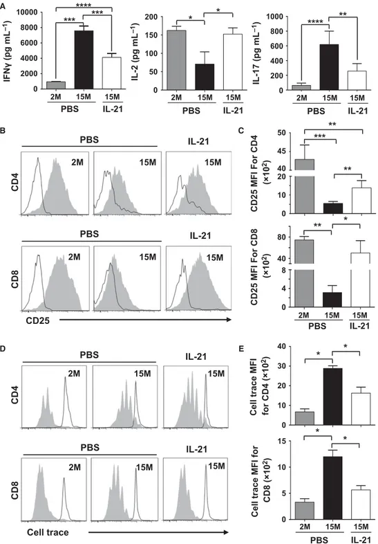
The vaccination efficacy in the elderly is significantly reduced compared to younger populations due to thymic involution and aged-related intrinsic changes affecting their naïve T-cell compartment. Interleukin (IL)-21 was recently shown to display thymostimulatory properties. Therefore, we hypothesised that its administration to aging hosts may improve T-cell output and thus, restore a competent peripheral T-cell compartment. Indeed, an increase in the production of recent thymic emigrants (RTEs) attributable to intrathymic expansion of early thymic progenitors (ETPs), double-negative (DN)-, and double-positive (DP) thymocytes as well as thymic epithelial cell (TEC) was observed in recombinant (r)IL-21-treated aged mice. In sharp contrast, no alterations in the frequency of bone marrow (BM)-derived progenitors were detected following rIL-21 administration. Enhanced production of naïve T cells improved the T-cell receptor (TCR) repertoire diversity and re-established a pool of T cells exhibiting higher levels of miR-181a and diminished amounts of the TCR-inhibiting phosphatases SHP-2 and DUSP5/6. As a result, stimulation of T cells derived from rIL-21-treated aged mice displayed enhanced activation of Lck, ZAP-70 and ERK, which ultimately boosted their IL-2 production, CD25 expression, and proliferation capabilities in comparison to T cells derived from control aged mice. Consequently, aged rIL-21-treated mice vaccinated using a tyrosinase-related protein 2 (Trp2)-derived peptide exhibited a substantial delay in B16 tumor growth and improved survival. The results of this study highlight the immunorestorative function of rIL-21 paving its use as a strategy for the re-establishment of effective immunity in the elderly.
24 2.1 - INTRODUCTION
In addition for being the key site of T-lymphopoiesis in jawed vertebrates, the thymus maintains a competent peripheral T-cell pool with a broad spectrum of TCR specificities (Lynch et al. 2009). It is however well established that immunity declines with aging owing to two key factors impeding thymic function: a defect in the survival and proliferation ability of the pre-thymic hematopoietic progenitor pool coupled to the precocious loss of TECs (Boehm & Swann 2013). These age-related changes, collectively known as thymic involution, represent major driving forces for homeostatic expansion of pre-existing peripheral T cells (Lynch et al. 2009). The net outcome culminates in TCR repertoire skewing with a noticeable increase in the number of effector/memory T cells (Zanni et al. 2003). Notably, a growing body of literature established that while the size of the peripheral T-cell compartment remains unchanged throughout aging, an increase in post-thymic life span of T cells takes place consequently leading to the emergence of T-cell intrinsic defects (Haynes & Swain 2006; Maue et al. 2009; Tsukamoto et al. 2009). For instance, naïve CD4+ T cells derived from aged mice display defects in TCR threshold
calibration, do not readily form immunological synapses and have a marked reduction in the recruitment of TCR-associated signaling molecules when compared to younger mice (Garcia & Miller 1997; Tamir et al. 2000; Garcia & Miller 2001; Garcia & Miller 2002). Furthermore, increased expression of inhibitory receptors such as PD1, LAG3, 2B4 and CD160 were observed on the surface of aging CD8+ T cells (Decman et al. 2012) while both IL-2 secretion and
proliferation potential are limited in naïve CD4+ and CD8+ T cells derived from aged mice
(Eaton et al. 2004). Thus, stifled thymopoiesis combined to global qualitative changes affecting the aging peripheral T-cell pool limits the host’s ability to mount effective responses against new antigenic challenges and accounts for the eroded immunity commonly observed in the elderly.
25
Primarily produced by activated CD4+ T cells, IL-21 is a prominent member of the common
γ-chain family of cytokines (Spolski & Leonard 2014). In addition to its wide ranging effects on immune cells, IL-21 overexpression in vivo triggers potent expansion of BM-derived progenitors (Ozaki et al. 2006). Furthermore, we recently reported a novel mitogenic function for IL-21 on peptide-mediated TCR-engaged DP thymocytes using a newly developed in vitro co-culture system designed for T-cell differentiation (Rafei et al. 2013b). Likewise, rIL-21 administration to mice with glucocorticoïd-induced thymic atrophy dramatically accelerates thymic function recovery by stimulating the proliferation of ETPs, DN and positively selected DP thymocytes (Rafei et al. 2013a). Such unprecedented thymopoiesis-supporting function suggests that rIL-21 is indeed a promising therapeutic tool endowed with the capacity of improving T-cell output in aged hosts owing to the expression of the IL-21 receptor (IL-21R) on both BM and thymic progenitors (Ozaki et al. 2006; Rafei et al. 2013a; Rafei et al. 2013b). We wished therefore to scrutinize whether rIL-21 administration to aged mice can rejuvenate their T-cell immunity by targeting de novo thymopoiesis as a mean to enhance their anti-tumoral response following vaccination.
26 2.2 - RESULTS
Administration of rIL-21 enhances thymopoiesis in aged mice.
To ensure maximal thymopoiesis-stimulating effects in vivo, we first conducted a dose response study in young (2 months - 2M) versus old (15 months - 15M) RAG2p-GFP mice by intraperitoneally (IP) administering rIL-21 (Fig. S5A). The use of the RAG2p-GFP model allows to easily assess de novo thymopoiesis as expression of the GFP transgene, marking newly developed T cells, is controlled by the Rag2 promoter activity (Monroe et al. 1999; Yu et al. 1999; Rafei et al. 2011). Thymic analysis one week following the last injection revealed a progressive increase in total thymocyte absolute count in 15M but not 2M old rIL-21-treated mice with an optimal response rate achieved at a dose of 50ug/Kg (Fig. S5B). Similarly, only aged mice receiving rIL-21 exhibited an increase in the counts of all thymic subsets (DN, DP and single-positive) (Fig. 5A) including ETPs (Fig. 5B-C). Even though the percentage of GFP+
thymocytes remained unchanged in all studied groups (Fig. S5C), the increased thymic count observed in the rIL-21-treated aged mice was sustained over a three weeks period post-cytokine administration (Fig S5D). To determine whether rIL-21-enhanced thymopoiesis involves the expansion of BM progenitors, which could have increased their migration rate to the thymus, we next monitored the frequency of LSK+ cells (Lin-Sca1+c-Kit+) and its sub-populations including
the long-term (LT; Lin-Sca1+c-Kit+CD34-CD135-) and short-term (ST; Lin-Sca1+
c-Kit+CD34+CD135-)-hematopoietic stem cells (HSCs), as well as multi-potent progenitors (MPPs;
Lin-Sca1+c-Kit+CD34+CD135+) following rIL-21 treatment. Despite IL-21R expression on the
surface of wild-type (WT) LT-, ST-HSCs and MPPs (Fig. S6A), the overall proportion of LSK+
27
FIGURE 5. ADMINISTRATION OF rIL-21 PROMOTES DE NOVO THYMOPOIESIS IN AGED BUT NOT YOUNG MICE.
A) Counts of thymocyte sub-populations at 50ug/kg of rIL-21. B) Representative flow-cytometry analysis of ETPs. C) Absolute count of ETPs derived from 2M (PBS ), 15M (PBS ) or 15 21 ). D-E) Percentages of total, cTECs and mTECs in 2M (PBS ), 15M (PBS ) or 15M (rIL-21 ). F) All rIL-(rIL-21-treated aged mice display enhanced counts of total, cTECs and mTECs in comparison to PBS-treated aged mice. All data are representative of three independent experiments (n = 5/group with *p˂0.05, **p˂0.01, ***p˂0.001, and ****p˂0.0001).
28
A
B
F
D
2M DN DP CD4 CD8 50 10 5 0 100 150 15M DN DP CD4 CD8 60 0 80 100 * * * * 4 8 T h ym o c yt e A b so lu te N u m b er s ( X 10 6) Thymic SubsetsE
TE C s A b s o lu te Nu m b e r (X 1 0 3) 0 20 15 10 5 25 PBS 15M PBS rIL-21 2M *** **** *** m T E C s A b so lu te Nu m b e r (X 1 0 3 ) 0 15 10 5 20 PBS 15M PBS rIL-21 2M ******* *** cT E C s A b so lu te N u m b er ( X 10 3) 0 6 4 2 PBS 15M PBS rIL-21 2M **** ****TECs mTECs cTECs
P e rcen ta g e s 25 50 75 100 0 0.1 0.2 *** *** CD45 Ep C A M Ly51 UA E -1 0.13 0.11 0.11 2M (PBS) 15M (PBS) 15M (rIL-21) 74 23 48 52 51 49 CD25 c-K it PBS 15M PBS rIL-21 2M 0.17 0.08 0.20 P e rcen ta g es o f ET Ps 0 0.10 0.20 0.15 0.25 0.05 ET P A b s o lu te N u m b er (x10 3) 0 4 8 6 2 2M (PBS) 15M (PBS) 15M (rIL-21)
C
*** * *** ** **29
Likewise, no increase in the number of LSK+ sub-populations (Fig. S6D) nor in the more
differentiated CLP (Lin-IL-7R+Sca1+c-Kit+) population (Fig. S6E) was observed.
We recently reported that TECs are devoid of IL-21R (Rafei et al. 2013a). Therefore, we presumed that the thymic effects observed following rIL-21 infusion strictly affect hematopoietic cells. Indeed, when given to aged mice, rIL-21 does not fluctuate the frequency of total TECs (EpCAM+CD45-), nor it did affect the ratio of cortical (c)TEC (EpCAM+CD45-UAE-1-Ly51+)
relative to medullary (m)TECs (EpCAM+CD45-UAE-1+Ly51-/lo) populations (Fig. 5D-E).
Conversely, absolute counts analysis showed marked improvements in the stromal compartment as total, cTEC and mTEC populations were significantly higher in rIL-21-treated aged mice compared to those derived from the PBS group (Fig. 5F). Higher production of IL-7 /thymus was also noticed in the rIL-21-treated aged mice compared to the control group (Fig. S5E). These data suggest that rIL-21 administration is beneficial to aged mice by directly targeting thymopoiesis in situ without triggering the expansion of BM-derived LSK+ cells.
Physiological levels of RTE are restored in aged mice following rIL-21 treatment.
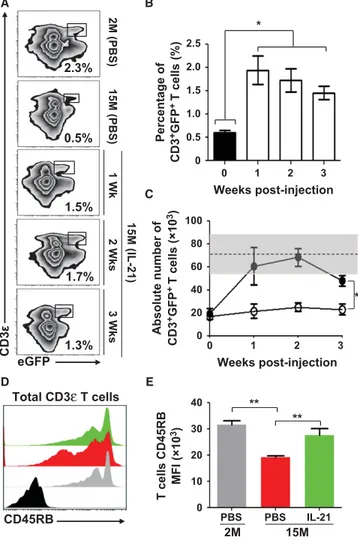
To interrogate the functional relevance of rIL-21-enhanced thymopoiesis on the peripheral T-cell pool of aged RAG2p-GFP mice, we next assessed the percentage of circulating RTEs mirrored by the level of peripheral GFP+CD3ε+ T cells. In contrast to 2M old animals, where RTEs
represent roughly 2.3% of total circulating lymphocytes, lower percentages (~0.5%) are detected in the peripheral blood of 15M PBS-treated aged mice (Fig. 6A). Following rIL-21 treatment, the percentage of GFP+CD3ε+ T cells reached a range of 1.3-1.7% over a period of three weeks post-
30
FIGURE 6. INCREASED LEVELS OF CIRCULATING RTES IN rIL-21-TREATED AGED MICE.
A) Representative flow-cytometry analysis of RTEs (CD3+GFP+ T cells) in peripheral blood of
young (2M) or aged (15M) mice 1, 2, or 3 weeks post-rIL-21 administration. Young mice (2M) treated with PBS were used as comparative positive controls. B-C) Analysis of overall percentages (B) and counts (C) of RTEs in the weeks following rIL-21 administration. The gray zone represents the RTE level calculated using 2M young mice (n=10) and displayed as the average RTE number + two S.D. Filled circles represent rIL-21-treated aged mice. D) Representative flow-cytometry analysis of CD45RB on the surface of all CD3+ T cells derived
from 2M (PBS ), 15M (PBS ) and 15M (rIL-21 ). CD45RB isotype is displayed in black. E) Compiled MFIs for CD45RB expression in treated mice. All data are representative of three independent experiments (n = 5/group with *p˂0.05 and **p˂0.01).
31
A
D
B
C
0
1
2
3
Weeks Post-Injection
0
0.5
1.0
1.5
2.0
2.5
P
e
rcent
a
g
e of
CD3
+GF
P
+T
c
e
lls
(%
)
*
eGFP
CD3
ε
0.5%
2.3%
1.5%
1.7%
1.3%
2M ( P B S ) 15M ( P B S ) 15M ( IL -21) 1W k 2W ks 3W ks0
20
40
60
80
100
*
0
1
2
3
Weeks Post-Injection
Abs
o
lu
te
Nu
m
b
e
r of
CD3
+GF
P
+T cel
ls
(x10
3)
CD45RB
Total CD3ɛ T cells
E
2M
15M
PBS
PBS
IL-21
0
10
20
30
40
T
c
e
lls
CD4
5
RB
M
F
I (
x10
3)
**
**
32
cytokine treatment (Fig. 6A-B) with absolute numbers attaining physiological levels according to RTE counts calculated using blood derived from young mice (Fig. 6C).
With increased encountered antigens and declined RTE levels, qualitative changes in the phenotype of peripheral T-cell composition occur with aging (Boursalian et al. 2004). More specifically, the expression of various cell surface makers including the glycoprotein CD45RB is down-regulated as T cells become activated and progress from a naïve to a memory phenotype (Tough & Sprent 1994). We therefore hypothesized that enhancing RTE generation in aged mice would increase the overall expression pattern of CD45RB on peripheral T-cell pool and found that it was indeed the case in aged mice treated with rIL-21 as depicted by histogram overlaps (Fig. 6D) and compiled mean fluorescent intensities (MFIs) (Fig. 6E). We therefore conclude that rIL-21 treatment enhances the de novo generation of RTEs, which incorporate the peripheral T-cell pool of aged mice.
The nature of aging T-cell pool is greatly affected by rIL-21 administration.
Following thymic egress, RTEs continue their maturation in the periphery to eventually become fully competent mature naïve T cells (Boursalian et al. 2004). To do so, they require access to secondary lymphoid organs (SLO) as a mean to encounter other cell types and cytokines required for their maturation (Houston et al. 2008). Although the percentage of GFP+ T cells increased
significantly in the spleen of rIL-21-treated aged mice in the three weeks following cytokine treatment (Fig. 7A), the overall number of splenocytes remained steady (Fig 7B). Further in depth analysis revealed a progressive time-dependent increase in the absolute counts of GFP+CD4+ and GFP+CD8+ T cells in the spleens of rIL-21-treated aged mice (Fig. 7C)
33
FIGURE 7. AGED MICE TREATED WITH rIL-21 DISPLAY INCREASED PROPORTION OF NAÏVE T CELLS WITH ENHANCED TCR DIVERSITY.
A) Percentages of GFP+ events in the spleen of 2M (PBS ), 15M (PBS ), and 15M (rIL-21 )
aged mice. B) Splenocytes counts in all experimental groups. C) Absolute counts of CD4+GFP+
and CD8+GFP+ T cells in 2M (PBS ), 15M (PBS ) and 15M (rIL-21 ) treated mice. D) A
representative flow-cytometry analysis of naïve (CD62Lhi, CD44lo), memory (CD62LhiCD44hi)
and effector (CD62LloCD44hi) T cells in all experimental groups. E) Compiled percentages of all
three sub-populations in CD4+ (top panel) and CD8+ (lower panel) T cells. F) Flow-cytometry
analysis of 15 TCRVβ-chains using peripheral CD4+ (top panel) or CD8+ (lower panel) T cells.
All data are representative of three independent experiments (n = 5/group with *p˂0.05, **p˂0.01, ***p˂0.001, and ****p˂0.0001).
34
A
B
D
C
A b so lu te N u m b er s ( X 10 6 ) PBS IL-21 1 2 3 1 CD8+GFP+ 0 1.5 1.0 0.5 2.0 * ** CD4+GFP+ 0 1.5 1.0 0.5 2.0 2.5 ** * * PBS IL-21 1 2 3 1E
0 20 40 60 80 100 P erce n ta ge of CD 4 + T ce lls ** ** *** ** ****Naïve Memory Effector 0 20 40 60 80 100 P erce n ta ge o f CD 8 + T ce lls ** *** **** **** *
Naïve Memory Effector
F
Spl enoc yte s Num b er s (X 10 7 ) PBS IL-21 1 2 3 1 0 15 10 5 n.s. P e rc en ta g e o f GF P + Ev e n ts PBS IL-21 1 2 3 1 0 15 10 5 20 25 ******* * CD44 C D 62L CD4 T Cells 2M 15M 41% 6% 42% 15M 17% 9% 72% 75% 3% 6% 16% 34% 47% 68% 7% 25% 45% 11% 28% CD8 T Cells 2M 15M 15M J E U N E P B S I L - 2 1 20 15 10 5 0 2 3 4 5.1 5.2 6 7 8.1 8.2 8.3 9 10 11 12 13 14 17 Vβ Chains ** **** * ** * * ** **** J e u n e P B S I L - 2 1 25 20 15 10 5 0 ** * ** **** * ****** ** **** * P erce n ta ge s35
suggesting that SLO-resident aged T cells were displaced by newly migrating RTEs. We next examined by flow-cytometry the differentiation stages of spleen-derived CD4+ and CD8+ T cells
and found increased abundance of T cells with a naïve phenotype (CD44loCD62Lhi) in
rIL-21-treated aged mice bolstering the notion of rIL-21-mediated enhanced T-cell output (Fig. 7D-E).
Given that thymic involution compromises the TCR repertoire (Yager et al. 2008; Ahmed et al. 2009), we continued our analysis by investigating the effect of rIL-21 administration on TCR diversity. To address this question, the expression profile of 15 TCRVβ-chains was analyzed on the surface of spleen-derived CD4+ and CD8+ T cells collected from treated mice. Significant
improvements were observed in the proportion of Vβ2-, 6-, 8.1/8.2-, 11-, 12- and 14-expressing CD4+ T cells as well as Vβ2-, 5.1/5.2-, 6-, 8.1/8.2-, 8.3-, 11- and 12-expressing CD8+ T cells
following rIL-21 administration to aging mice (Fig. 7F). These results indicate that rIL-21-mediated de novo RTE generation is associated with major qualitative changes in the peripheral T-cell pool of aged mice including improved TCR diversity.
Characterizing the biochemical responses of T cells.
Thymic involution cannot solely account for impaired immune responses as additional T-cell intrinsic defects appear in aging naïve T cells due to prolonged post-thymic life span (Haynes & Swain 2006; Maue et al. 2009; Tsukamoto et al. 2009). More specifically, TCR activation is blunted with aging due to increased cytoplasmic concentration of phosphatases known for inhibiting TCR signaling (Li et al. 2012). Given such fluctuation in TCR threshold calibration, we next explored whether the previously observed changes in the nature of the peripheral T-cell pool induced by rIL-21 affect the expression levels of the TCR-targeting phosphatases SHP-2,
36
FIGURE 8. THE PERIPHERAL POOL OF T CELLS IN rIL-21-TREATED AGED MICE DISPLAY IMPROVED TCR SIGNALING RESPONSES.
A) Representative western-blot analyses of phosphatases at 1, 2 or 3 weeks post-treatments of 2M PBS (1), 15M PBS (2) and 15M rIL-21 (3) aged mice. β-actin was used as internal loading control. B) Compiled densitometry analysis of SHP-2, PTPN-22 and DUSP5/6 phosphatase expression levels. The displayed groups are: 2M (PBS ); 15M (PBS ); and 15M (rIL-21 ) aged mice. C) qPCR analysis of miR-181a in freshly isolated T cells from 2M (PBS ); 15M (PBS ); and 15M (rIL-21 ) treated mice. D) Representative intracellular flow-cytometry staining of pLck, pZAP-70 and pERK in 2M (PBS ); 15M (PBS ); and 15M (rIL-21 ) aged mice. Non-stimulated T cells derived from young 2M mice are displayed by black histograms. All data are representative of three independent experiments (n = 5/group with *p˂0.05, and **p˂0.01).
37
C
A
D
B
2M 15M 15M
PBS
IL-21
*
*
*
0
0.5
1.0
1.5
m
iR
-181a R
e
la
ti
ve
E
x
pr
ess
io
n
Fol d I n cr ease O ver B asal Level s 1 2 3 4 0 ** * * 1 2 3 0 1 Wk 2 Wks 3 Wks 1 2 3 0 ** * *SHP-2
DUSP5
DUSP6
* *PTPN-22
0.5 1.0 1.5 0 4CD8
pLck
CD4
pZAP-70
pERK
SHP-2
1 2
1 Wk
3
2 Wks
3 Wks
1
2
3
1 2
3
β-actin
PTPN-22
1 2
3
1 Wk
2 Wks
3 Wks
1
2 3
1 2
3
β-actin
DUSP6
1 2
1 Wk
3
1 2 3
2 Wks
1 2
3 Wks
3
β-actin
DUSP5
1 2
1 Wk
3
1 2
2 Wks
3
1 2
3 Wks
3
β-actin
1
2 3
n
1 2
3
1 2
3
1
2
3
38
PTPN-22 and DUSP5/6. Delineation of phosphatase levels by western-blotting (Fig. 8A) and densitometry-based quantification (Fig. 8B) revealed diminished SHP-2 and DUSP5/6 levels in T cells derived from rIL-21-treated aged mice at all tested time points. Only PTPN-22 remained unchanged in all groups throughout treatments (Fig. 8A-B). Previous studies conducted in mice and humans demonstrated that SHP-2 and DUSP5/6 were among several phosphatases controlled by miR-181a; a ~22nt microRNA molecule capable of repressing the translation of over 40 phosphatases (Li et al. 2007). Consistently, the progressive decline in miR-181a levels observed in aging human naïve CD4+ T cells dovetails the decreased T-cell responsiveness following TCR
stimulation (Li et al. 2012). To examine the possibility that rIL-21 could have reversed such defect through enhanced generation of RTEs expressing normal levels of miR-181a, quantitative (q)PCR studies were conducted using T cells sorted from spleens of treated mice. Our analysis revealed a 2-3 fold increase in miR-181a levels in T cells derived from rIL-21-treated aged mice (Fig. 8C), which is consistent with the diminished SHP-2 and DUSP5/6 levels observed earlier (Fig 8A-B). We next tested the responses of T cells derived from treated mice by probing early signaling events triggered by TCR stimulation. Although not efficient as younger lymphocytes, CD4+ or CD8+ T cells derived from rIL-21-treated aged mice displayed higher Lck, ZAP-70 and
ERK phosphorylation compared to control PBS aged mice as shown by phospho-flow analysis (Fig. 8D). Taken collectively, these data indicate that enhanced TCR responses in the pool of T cells derived from rIL-21-treated aged mice is attributable to the increase in the proportion of naïve T cells displaying lower SHP-2 and DUSP5/6 phosphatase levels owing to improved miR-181a expression.