HAL Id: tel-01127180
https://tel.archives-ouvertes.fr/tel-01127180
Submitted on 7 Mar 2015HAL is a multi-disciplinary open access archive for the deposit and dissemination of sci-entific research documents, whether they are pub-lished or not. The documents may come from teaching and research institutions in France or abroad, or from public or private research centers.
L’archive ouverte pluridisciplinaire HAL, est destinée au dépôt et à la diffusion de documents scientifiques de niveau recherche, publiés ou non, émanant des établissements d’enseignement et de recherche français ou étrangers, des laboratoires publics ou privés.
Role of EMMPRIN and MMPs in tooth development,
dental caries and pulp-dentin regeneration
Mayssam Khaddam
To cite this version:
Mayssam Khaddam. Role of EMMPRIN and MMPs in tooth development, dental caries and pulp-dentin regeneration. Genetics. Université René Descartes - Paris V, 2014. English. �NNT : 2014PA05T046�. �tel-01127180�
UNIVERSITE PARIS DESCARTES
Ecole doctorale Génétique Cellule Immunologie Infectiologie
Développement
Laboratoire EA2496
Pathologies, Imagerie, et Biotherapies Orofaciales
Role of EMMPRIN and MMPs in tooth
development, dental caries and pulp-dentin
regeneration
Par Mayssam KHADDAM
THESE
Pour obtenir le grade de
DOCTEUR
Spécialité : Sciences de la Vie et de la Santé
Dirigée par Professeur Catherine CHAUSSAIN
Présentée et soutenue publiquement le 24 novembre 2014 Devant un jury composé de :
Pr. BERDAL Ariane, Université Paris Diderot Président Pr. CHAUSSAIN Catherine, Université Paris Descartes Directeur Pr. MOURAH Samia, Université Paris Diderot Rapporteur Pr. MANZANARES Maria Cristina, Université de Barcelone Rapporteur Dr. MENASHI Suzanne, Université Paris est Créteil Examinateur Dr. ROCHEFORT Gael, Université Paris Descartes Examinateur Dr. HUET Eric, Université Paris est Créteil invité
Acknowledgment:
At the end of my thesis I would like to thank all those people who made this thesis possible and an unforgettable experience for me.
First of all, I would like to express my deepest sense of gratitude to my supervisor Prof.
Catherine Chaussain who offered her continuous advices and encouragement throughout
the course of this thesis. I thank her for the systematic guidance and great effort she put into training me in the scientific field.
I am very grateful to Prof. Samia Mourah, from Paris Diderot University and Prof. Maria
Cristina Manzanares, from Barcelona University for being part of this PhD thesis
committee and for accepting being the reviewers of this PhD thesis.
I would like to thank Prof. Ariane Berdal, from Paris Diderot University, Dr. Suzanne Menashi from Paris Est Créteil University, Dr. Gael Rochefort from Paris Descartes
University, and Dr.Eric Huet from Paris Est Créteil University for accepting to be members of this PhD thesis committee. I am grateful and honored by their participation.
I would like to thank Dominique Le-Denmat, Dominique Septier, and Brigitte Baroukh; I learned a lot of things from your scientific and general experiences.
Thanks to Tchilalo Boukpessi; TP at 8:00 Am was a good chance for me to catch some of your experience in teaching, and i am happy to work with you in GSE article.
Thanks to Elvire Le Norcy; 6 months in the orthodontics department at Charles Foix (Ivry-sur-Seine) hospital was very useful for me especially with your experience.
I would like to thank Anne Polliard, Claire Bardet, Sandy Ribes, Julie Lesieur, Benjamin Salmon, Jeremy Sadoine, Benoît Vallée, Sibylle Vital, Céline Gaucher, Marjolaine Gosset, Bernard Pellat, Jean-Louis Saffar, Marie-Laure Colombier, and Philippe Bouchard.
Cedric Mauprivez and Caroline Gorin, we are at the end of our thesis so I hope that everything will be OK.
Thanks to Elisabeth Jimenez for her help in all my administrative problems.
I am thankful to all in EA 2496: Anne-Margaux Collignon, Benjamin Coyac, Maxime Ghighi, Frederic Chamieh, Laurent Detzen, Jean-Baptiste Souron, Annie Llorens, Aurélien
Vernon, Cyril Willig, Maheva Garcia, Anita Novais, Sonia Pezet, Tania Selbonne, and Alexandra Benoist.
Special thanks to Jiyar Naji, Xuan Vinh Tran, Soledad Acuña Mendosa, Francesca Mangione, Yong Wu, and Bassam Hassan; you are completely different and I am so happy to work with you.
Thanks to Jotyar Khlaf Arif for his help.
I am thankful to Syrian government who gives me the grant to continue my study in France. These acknowledgments would not be complete without thanking my family for their constant support and care. Very special thanks to my parents, despite they can’t read these words but they know how i appreciate their efforts and their times spent for me.
Finally, I would like to mention two other people who are very important in my life: My wife, Khawla and my little daughter, Nada. I thank Khawla for her encouragement and support, especially in the hardest time. I thank my little daughter for making me so happy with her cute smile which gives me the hope at the most difficult moments.
1
Table of Contents
Table of figures ... 2
List of abbreviations ... 6
General introduction and specific objectives ... 8
1 Introduction ... 11
1.1 Tooth description ... 11
1.2 Tooth development ... 12
1.2.1 Stages of tooth development ... 13
1.2.2 Basement membrane ... 17 1.2.3 Dentin ... 19 1.2.4 Enamel ... 23 1.3 Matrix MetalloProteinases MMPs ... 30 1.3.1 MMPs and teeth ... 31 1.4 EMMPRIN (Basigin,CD147)... 32 1.4.1 Historic ... 32 1.4.2 Structure ... 32
1.4.3 Phenotypes of EMMPRIN knock out (KO) mice ... 35
1.4.4 EMMPRIN interactions ... 35
5.4.1 EMMPRIN functions ... 37
6 . 5.4 EMMPRIN and tooth ... 43
2 MMPs and dentin matrix degradation ... 48
3 Results ... 57
3.1 Role of EMMPRIN in tooth formation ... 57
3.1.1 Supplementary data ... 69
3.1.2 Supplementary results ... 72
3.2 Role of EMMPRIN in pulp-dentin regeneration ... 79
3.2.1 Background and project aim ... 79
3.2.2 Materials and methods ... 79
3.2.3 Results ... 80
3.3 Inhibition of MMP-3 and dentin matrix degradation ... 82
4 Discussion ... 91
5 References ... 95
2
Table of figures
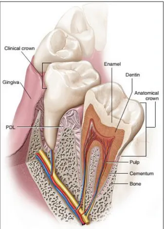
Figure 1 : The tooth and its supporting structure. Adapted from (Antonio Nanci and Cate
2013) ... 11
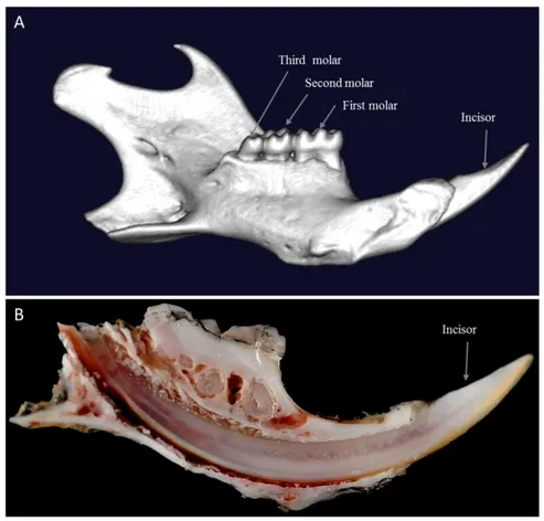
Figure 2 : Adult mouse mandible (own data). ... 12
Figure 3 : Dental lamina of tooth development. Adapted from (Antonio Nanci and Cate
2013) ... 13
Figure 4 : Bud stage of tooth development. Adapted from (Antonio Nanci and Cate 2013) . 14
Figure 5 : Cap stage tooth germ showing the position of the enamel knot. Adapted from
(Antonio Nanci and Cate 2013) ... 15
Figure 6 : Cap stage, beginning of cellular differentiation within the enamel organ. Central
cells form the stellate reticulum. Adapted from (Antonio Nanci and Cate 2013) ... 16
Figure 7 : Early bell stage of tooth development (own data) ... 17
Figure 8 : Characteristics of dentin formation. Odontoblasts secrete an ECM composed of
type I collagen and NCPs. Within the predentin type I collagen molecules are assembled as fibrils. Mineralization occurs at the mineralization front by growth and fusion of
calcospherites formed by hydroxyapatite (HAP) crystals. This mineralization process is controlled by NCPs and by mineral ion availability. Cell processes remain entrapped within dentin whereas cell bodies remain at the periphery of the pulp. Adapted from (Vital et al. 2012) ... 20
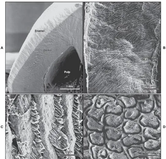
Figure 9 : Human dentin by scanning electronic microscopy (SEM). A. cutting line is
parallel to dentin tubules, B. cutting line is perpendicular to dentin tubules. PtD: peritubular dentin, ItD: intertubular dentin. (Own data) ... 21
Figure 10 : Scanning electron microscope views of (A) the enamel layer covering coronal
dentin, (B) the complex distribution of enamel rods across the layer, (C and D) and
perspectives of the rod-interrod relationship when rods are exposed longitudinally (C) or in cross section (D). Interrod enamel surrounds each rod. DEJ: Dentinoenamel junction; IR: interrod; R,rod. Adapted from (Antonio Nanci and Cate 2013) ... 24
Figure 11 : Semi-thin (0.5 µm) sections from glutaraldehyde-fixed, decalcified, and plastic
embedded mandibular incisors of wild-type mice stained with toluidine blue to illustrate the appearance of enamel and enamel organ cells at mid-secretory stage (A) and near-mid-maturation stage (B) of enamel development. Abbreviations: E, enamel; Am, ameloblast; Si, stratum intermedium; pd, predentin; D, dentin; ae, apical end; be, basal end; bv, blood vessel; as, artifact space; b, bone; c, cementum. Adapted from (J. D. Bartlett and Smith 2013) ... 29
Figure 12 : Schema present incisor enamel and denin formation. p-Am: pre-ameloblast; pOd:
pre-odontoblast; s-Am: secreting Ameloblasts; od: odontoblasts; pos-Am: post-secretory ameloblasts; pD: predentin; D: dentin; pm-E: premature enamel; E: enamel. Adapted from (Khaddam et al. 2014)... 30
Figure 13 : Schematic representation of MMP activity during the dentin carious process.
Cariogenic bacteria present in the caries cavity release acids such as lactic acid that reduce the local pH. The resulting acidic environment demineralizes the dentin matrix and induces the activation of host MMPs derived from dentin or saliva (which bathes the caries cavity). Once the local pH is neutralized by salivary buffer systems, activated MMPs degrade the demineralized dentin matrix. Adapted from (Chaussain et al. 2013) ... 32
3
Figure 14: Basigin isoforms. Characteristic features of isoforms are mentioned within
blanket. Carbohydrates are shown by light grey color. Adapted from (Takashi Muramatsu 2012) ... 33
Figure 15: Scheme of EMMPRIN structure. EMMPRIN contains an extracellular domain
composed of two Ig loops with three Asn-linked oligosaccharides and short single
transmembrane domain (TM) and a cytoplasmic domain (Cyt). The first Ig domain is required for counter-receptor activity, involved in MMP induction. Adapted from (Gabison et al. 2009). ... 34
Figure 16: Possible EMMPRIN-mediated interactions stimulating MMP production. (A)
Homophilic cis interaction between EMMPRIN molecules within the plasma membrane of a tumor cell. (B) Homophilic trans interaction between EMMPRIN molecules on apposing tumor cells. (C) Heterophilic interactions between EMMPRIN on a tumor cell and a putative EMMPRIN receptor on a fibroblast. Adapted from (Toole 2003)... 35
Figure 17 : Tumor-cell induced activation of adjacent fibroblasts by homophilic EMMPRIN
signaling. Adapted from (Joghetaei et al. 2013) ... 40
Figure 18 : Immunoreactivity (IR) for EMMPRIN. a Cells of the inner enamel epithelium
(cap stage of the enamel organ) show intense EMMPRIN IR (Alexa-coupled). b Ameloblasts as well as odontoblasts (bell stage of the enamel organ) exhibit strong EMMPRIN IR. Note the IR in the borderline between ameloblasts and the stratum intermedium. Mesenchymal cells of the dental papilla are only weakly immunoreactive. Abbreviations: A ameloblast; DL dental lamina; EEE external enamel epithelium; IEE internal enamel epithelium; EO enamel organ; Od odontoblast; SI stratum intermedium; SR stellate reticulum. Adapted from
(Schwab et al. 2007) ... 43
Figure 19 : Transcription level of EMMPRIN in different stages of tooth development. a
EMMPRIN mRNA was higher in E13.0 mandible than that in E11.0. b The expression of EMMPRIN mRNA was higher in P1 tooth germ than that in E14.0. Adapted from (Xie et al. 2010) ... 44
Figure 20 : Examination the role of EMMRIN in early tooth germ development using
EMMPRIN siRNA in the cultured mandible at E11.0. a After being cultured for 6 days, the tooth germ was found to have developed into the cap stage in mandibles cultured with scramble siRNA. b Dental epithelial bud was observed in the mandible treated with
EMMPRIN siRNA after 6 days of culture. c A cap-like mature enamel organ was observed in the mandibles with scrambled siRNA supplement at 8-day culture. d EMMPRIN
siRNAtreated mandible explants also showed a bud-like tooth germ at 8-day culture. EMMPRIN siRNA had a specific effect on the morphogenesis of tooth germ. DE dental epithelium, DM dental mesenchyme, DP dental papilla, EO enamel organ, OE oral
epithelium, PEK primary enamel knot. Adapted from (Xie et al. 2010) ... 45
Figure 21 : Temporal expression and localization of EMMPRIN in the gingival epithelium
during ligature-induced periodontitis in the first mandibular molar of rats. (A) On day 0 (health), the immunoreactivity was strong in the basal cells, with a decrease toward the upper layers in the attached gingival epithelium (star in a1). (B) On day 7, immunoreactivity was greatly enhanced in the attached gingiva (star in b1). (C) On day 15, immunoreactivity was
4
similar to that seen in the healthy state in the attached gingival epithelium (star in c1).
Adapted from (L. Liu et al. 2010) ... 46
Figure 22 : EMMPRIN expression in the developing incisor of 3 month-old mice Immunostaining with EMMPRIN antibody on sagittal section of the mandible shows that the secretory ameloblasts, the stratum intermedium and odontoblasts are positive for EMMPRIN (A and B). By contrast, no staining is observed in the post-secretory ameloblast (C). Am: ameloblast; s-Am: secretory ameloblast; pos-Am: post-secretory ameloblast; Od: odontoblast; D: dentin; pD: predentin; pm-E: premature enamel; Si: stratum intermedium; fm: forming matrix. From (Khaddam et al. 2014) ... 69
Figure 23 : KLK-4 expression in tooth germs of EMMPRIN KO mice when compared with WT. For mRNA expression, a 33 % increase is observed by qRT-PCR in KO mice. KLK-4 activity is hardly detectable by casein zymography (with 20 mM EDTA in the incubation buffer to inhibit MMP activity). No activity is seen for recombinant MMP-20. From (Khaddam et al. 2014)... 70
Figure 24 : SEM observation of 3 month-old mouse mandible sections. At M1 level, no difference in the morphology of either the bone or the teeth is detected between WT and KO mice (A, B). Both dentin (E, F) and enamel appear normal and the enamel prisms are normally constituted (C, D). From (Khaddam et al. 2014). ... 71
Figure 25: TEM analysis was performed on M1 and M2 germs of new born mice. In the KO M2 germs, a cell polarization delay is observed in both pre-ameloblasts and pre-odontoblasts localized at the tip of the cusps (b). In the WT, well-organized ameloblast and odontoblast palisades are seen, with a basal localization of the nuclei and long cell processes (arrow-heads) (a), whereas in the KO, cells are seen proliferating with centrally localized nucleus (b). At higher magnification, the basement membrane (black arrows) is partially degraded in WT (white arrows) (c), but appears still intact in the KO (d). In M1germ, the basement membrane which can no longer be detected in the WT (e) is partially degraded in the KO (arrow) (f). Dentin matrix (black arrow-heads) is secreted in both mice models (e-f-g-h) but at a higher rate in the WT (e) where a greater amount of fibrillated collagen is seen associated with hydroxyapatite crystals (white arrow heads). In addition, mineralizing enamel matrix can already be observed at the secreting pole of WT ameloblasts localized at the tip of the cusp (g) but is not detectable in the KO (h). pam: pre-ameloblast; pod: pre-odontoblast; am: ameloblast; od: odontoblast; fde: forming dentin; fen: forming enamel. (own data). ... 73
Figure 26: EMMPRIN expression in the first molar of mouse embryo. immunoreactivity (IR) for EMMPRIN in paraffin sections of mouse embryo tooth germ tissue at 16 day and 17 day (cap stage). Inner enamel epithelium cells show EMMPRIN IR, this IR in the buccal side is stronger than in the lingual side of the molar germ. Iee: innerenamel epithelium; dp: dental pulp; bs: buccal side; ls: lingual side. (own data) ... 74
Figure 27: Alveolar bone density. ... 76
Figure 28: Percent of bone volume in VOI.... 76
Figure 29: Trabecular bone thickness in VOI. ... 77
Figure 30: Trabecular number in VOI. ... 77
5
Figure 32: Mouse first upper molar after 7 and 28 days of capping with Biodentine. 7 days
post operatively, dentin formation was detected in +/+ and -/- EMMPRIN mice (A,C), but it was more in -/- (brown arrow C) than in +/+ (brown arrow A). 28 days post operatively, dentine bridge was visible, but it was more continuous in -/- (arrow in D) than in +/+ (arrow in B) where it was not continued. e: enamel; d: dentin; GIC: glass ionomer cement; red *: Biodentine. ... 80
Figure 33: Percent of dentin volume in volume of interest VOI. Significant increase in dentin
density was detected in -/- EMMPRIN mice when compared with +/+ mice at 7 days post operatively... 81
Figure 34: Recapitulative schema proposing the role of EMMPRIN in tooth formation. At
early bell stage, EMMPRIN is expressed by pre-ameloblast (p-Am) and may orchestrate basement membrane degradation (black line) to allow direct contact with pre-odontoblast (p-Od), which is mandatory for the final cell differentiation. At secretory stage, both secreting ameloblasts (s-Am) and odontoblasts (Od) highly express EMMPRIN. This expression may enhance MMP-20 synthesis by ameloblasts allowing for early enamel maturation. At the enamel maturation stage, post-secretory ameloblasts (pos-Am) lose their EMMPRIN
expression. The arrows indicate EMMPRIN expression by cells. The red line schematizes the time window where a direct effect of EMMPRIN is allowed by a direct cell contact. D: dentin; pD: predentin; pm-E: premature enamel; E: enamel. ... 92
6
List of abbreviations
AI: Amelogenesis imperfecta Ambn : Ameloblastin AMTN : Amelotin Asn : Asparagine
ATK: serine/threonine kinase and is known as protein kinase B (PKB) or RAC-PK (‘related
to A and C protein kinase’)
ASARM: acidic serine and aspartate-rich motif BM: Basement membrane
BSP : Bone sialoprotein BV : Bone volume CyPA : Cyclophilin A
DI : Dentinogenesis imperfecta DMP-1 : Dentin matrix protein 1 DSPP : Dentin sialophosphoprotein DEJ: Dental enamel junction ECM : Extra cellular matrix Enam : Enamelin
EMMPRIN : Extra cellular matrix metalloproteinase inducer GAG: Glycoaminoglycans
GIC: Glass ionomer cement HAP : Hydroxyapatite crystals HIF-1α : hypoxia-inducible factor 1α KLK-4 : Kallikrine-4
KO: Knock out
MAPK: Mitogen-activated protein MCT : Monocarboxylate transporter
MEPE: Matrix extracellular phosphoglycoprotein MMP : Matrix metalloproteinase MMP-1 : Collagenase 1 MMP-2 : Gelatinase A MMP-3 : Stromelysin 1 MMP-9 : Gelatinase B MMP-20 : Enamelysin
7 MVs : Membrane vesicules NCPs: Non-collagenous proteins OPN : Osteopontin PDL : Periodontal ligament PFA: Para-formaldehyde PG: Proteoglycans
PI3K: Phosphoinositide 3-kinase
RGD: Arginine–glycine–aspartate cell adhesion domain RA: Rheumatoid arthritis
SEM: Scanning electron microscopy
SIBLINGs : Small integrin-binding ligand N-linked glycoprotein SLRPs: Small leucine-rich proteoglycans
Tb: Trabecular bone
TEM: Transmission electron microscopy TIMP: Tissue inhibitor of MMPs
TV: Tissue volume
VEGF: Vascular endothelial growth factor VOI : Volume of interest
8
General introduction and specific objectives
Tooth development results from reciprocal inductive interactions between the ectomesenchyme and oral epithelium and proceeds through a series of well-defined stages including the initiation, bud, cap and bell stages (Ruch, Karcher-Djuricic, and Gerber 1973; Slavkin 1974; Catón and Tucker 2009; Miletich and Sharpe 2003; I Thesleff and Hurmerinta 1981; Mitsiadis and Luder 2011). At the bell stage which is the last step of tooth crown formation, signals from the dental epithelium (i.e., inner enamel epithelium) instruct dental mesenchymal cells to differentiate into odontoblasts. Differentiated odontoblasts signal back to inner enamel epithelial cells and induce their differentiation into ameloblasts, which are responsible for enamel matrix synthesis. Ameloblast terminal differentiation necessitates the presence of an extracellular matrix that is secreted by odontoblasts and forms the predentin (Zeichner-David et al. 1995). The degradation of the basement membrane (BM) separating the dental epithelium from the mesenchyme is a key step in this process that allows direct contact of ameloblasts with both odontoblasts and the unmineralized dentin matrix (Catón and Tucker 2009; Olive and Ruch 1982). Matrix metalloproteinases (MMPs) are involved in all stages of tooth formation (Bourd-Boittin et al. 2005; Chaussain-Miller et al. 2006). At the bell stage, MMPs have a major role in BM degradation (Heikinheimo and Salo 1995; Sahlberg et al. 1992a), thus allowing direct cross-talk between odontoblasts and ameloblasts (Heikinheimo and Salo 1995; Sahlberg et al. 1999). It has been shown that at more advanced stages MMPs also regulate the processing of dental extracellular matrix (ECM) proteins prior to mineralization. Indeed, it has been demonstrated that MMPs regulate amelogenin (AMEL) cleavage by enamelysin (MMP-20) during early enamel maturation (Bourd-Boittin et al. 2005; Bourd-Boittin et al. 2004; Lu et al. 2008; Nagano et al. 2009; Turk et al. 2006; Simmer and Hu 2002; J. D. Bartlett and Simmer 1999). The notion of direct epithelial-mesenchymal (or epithelio-stromal) interactions was first introduced in the cancer field when EMMPRIN, a membrane glycoprotein also known as CD147, was identified as a MMP inducer present at the cell surface of tumor cells which can activate stromal cells through direct contact and signal them to increase MMP production (Toole 2003). Recently accumulating data also advocate a role for EMMPRIN in modulating MMP expression during non-tumorigenic pathological conditions as well as in physiological situations such as tissue remodeling and cytodifferentiation events (Gabison, Hoang-Xuan, et al. 2005; Huet, Gabison, et al. 2008; Mohamed et al. 2011; Kato et al. 2011; Nabeshima et al. 2006; L. Liu et al. 2010; Gabison et
9
al. 2009; Zhu et al. 2014) . The expression of EMMPRIN in the developing tooth germs was previously described (Schwab et al. 2007; Xie et al. 2010). EMMPRIN expression was shown to increase gradually in the forming molar germ in mice from E14 to P1(Xie et al. 2010). However, the in vivo role of EMMPRIN in tooth development and homeostasis is still unknown. In this PhD, our first specific objective was to investigate EMMPRIN
functions in tooth formation using EMMPRIN KO mice by exploring the modifications
occurring in their dental phenotype and the consequences on EMMPRIN’s molecular targets, in particular on MMPs.
In parallel, EMMPRIN has been shown to be involved in the repair process of different injured tissues. Indeed, the role of EMMPRIN in wound healing through MMP induction and increase in myofibroblast contractile activity has been established (Gabison, Mourah, et al. 2005; Huet, Vallée, et al. 2008). As our team has developed several pulp injury models to follow-up the repair process, and as we had access to EMMPRIN KO mice it was tempting to study the repair process in this model. Therefore, our second specific objective
was to investigate for a potential role of EMMPRIN in the pulp dentin repair process by comparing the healing of injured pulps of EMMPRIN KO and WT mice.
MMPs were shown to be expressed during tooth development and to be necessary for normal dentin formation (Bourd-Boittin et al. 2005). After dentin mineralization, they remain trapped in the calcified matrix either under active or proenzyme forms (Palosaari et al. 2003), which may explain their persistent presence within the dentin of adult teeth (A Mazzoni 2007; Tjäderhane et al. 1998). The role of these trapped MMPs in the progression of the carious process within dentin matrix has been proposed by several studies (Tjäderhane et al. 1998; Sulkala et al. 2001). Indeed, MMPs have been proposed to have an important role in the dentin organic matrix degradation following demineralization by bacterial acids (Tjäderhane et al. 1998; Chaussain-Miller et al. 2006). Cariogenic bacteria are essential to initiate the carious process but they cannot degrade the dentin organic matrix. After dissolution of the mineral part, the organic part of dentin becomes exposed to degradation by host-derived enzymes, including salivary and dentinal MMPs, and cysteine cathepsins (Nascimento et al. 2011; van Strijp et al. 2003). Because MMPs have been suggested to contribute to dentin caries progression, the hypothesis that MMP inhibition would affect dentin caries progression is appealing. This hypothesis was supported by in vivo studies in rat caries models where dentin caries progression was delayed by intra-oral administration of chemical MMP inhibitors, modified tetracylines and zoledronate (Sulkala et al. 2001;
10
Tjäderhane et al. 1999). Several natural molecules have been previously reported to have MMP inhibitory properties. Grape-seed extracts (GSE) have been shown to suppress lipopolysaccharide-induced MMP secretion by macrophages and to inhibit MMP-1 and MMP-9 activities in periodontitis (La et al. 2009). The MMP-inhibitory effects of these natural substances suggest, therefore, that they could be effective in inhibiting dentin caries progression. Recently, a new daily mouthrinse composed of grape-seed extracts and amine fluoride has been developed. As grape-seed extracts are known to be natural inhibitors of
MMPs, our last specific objective was to evaluate the capacity of these natural agents to prevent the degradation of demineralized dentin matrix by MMP-3.
11
1 Introduction
1.1 Tooth description
Tooth is the hardest organ of the mammalian body and it provides several functions such as mastication, and phonation.
Anatomically, tooth structure can be distinguished in a visible part (crown) and a hidden part embedded in the alveolar bone of the jaw (root) (Figure 1). Instead of a considerably different shape and size (e.g., an incisor compared with a molar), teeth are histologically similar.
Figure 1 : The tooth and its supporting structure. Adapted from (Antonio Nanci and Cate 2013)
Tooth consists of several layers: enamel, dentine, cement, and dental pulp. The enamel is a hard, and acellular structure formed by epithelial cells and supported by dentin. This less mineralized, more resilient, and vital hard connective tissue, is formed and supported by the dental pulp, a soft connective tissue (Figure 1).
12
In mammals, teeth are attached to the bones of the jaw by the periodontium, consisting of the cementum, periodontal ligament (PDL) and alveolar bone, which provide an attachment with enough flexibility to withstand the forces of mastication.
Human and most of the mammals have two generations of teeth, primary and permanent; since the size of teeth cannot increase after formation, the primary dentition becomes inadequate and must be replaced by more and larger teeth (permanent dentition).
Otherwise, mice have only one generation highly reduced dentition having one incisor, separated from three molars by an edentulous region in each semi-maxilla (Figure 2.A). Incisor growth is continuous throughout the animal’s life (Figure 2.B).
Figure 2 : Adult mouse mandible (own data).
1.2 Tooth development
Since toothed vertebrate have conserved tooth development process stages, data obtained from rodents studies may provide a lot of information about dental development in diverse species, including humans (Streelman et al. 2003).
Organogenesis results from three fundamental processes: I) initiation, within positional information are provided and interpreted to initiate organ formation at the right place; II)
13
morphogenesis, in which cells build up a rudimental organ; finally, III) differentiation where cells form organ-specific structures.
As also showed in mouse tooth development model (Irma Thesleff and Nieminen 1996), teeth are vertebrate-specific structures which, like many other organs, develop through a series of reciprocal interactions between two adjacent tissues, an epithelium and a mesenchyme (I Thesleff, Vaahtokari, and Partanen 1995). Tissue-recombination experiments have shown that the oral epithelium isolated from the mandibular arch of a mouse embryo, between embryonic day 9.0 and 11.5 (E9.0–E11.5), can stimulate a non-dental neural crest-derived mesenchyme to form a tooth. After E11.5, the odontogenic potential subsequently shifts from the epithelium to the mesenchyme, which can induce dental formation when combined with a non-dental epithelium, whereas the dental epithelium has lost this ability(Mina and Kollar 1987; Lumsden 1988).
1.2.1 Stages of tooth development
Tooth development takes place through a series of well-defined stages: epithelial thickening of the dental lamina, bud, cap and bell.
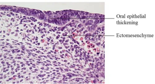
1.2.1.1 Dental lamina Stage
The thickening of the mouse oral epithelium is first visible at around E11.5 (Figure 3). The epithelial thickening forms the dental and vestibular lamina on the lingual and buccal aspect, respectively. The vestibular lamina forms a sulcus between the cheek and the teeth, and the dental lamina gives rise to the teeth. During this stage, dental lamina expresses several important signaling molecules such as (Sonic Hedgehog) Shh that increases cell proliferation at the tooth development site (Hardcastle et al. 1998).
Figure 3 : Dental lamina of tooth development. Adapted from (Antonio Nanci and Cate 2013)
14 1.2.1.2 Bud stage
After the dental lamina stage, an epithelial structure that has a bud shape results from proliferating and invagination of the epithelium within the underlying ectomesenchyme. The bud is clearly formed at E13.5 and it consists in several layers: the dental follicle made by condensed mesenchymal cells, oriented in a radial pattern and encasing the dental papilla and the enamel organ; enamel organ, in which the internal epithelial cells meets the external epithelial cells and form a structure called the cervical loop; finally, dental papilla, which is a ball of densely packed ectomesenchyme (Figure 4).
Figure 4 : Bud stage of tooth development. Adapted from (Antonio Nanci and Cate 2013)
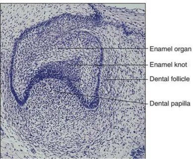
1.2.1.3 Cap stage
Around E14.5, the condensing mesenchyme signals back to the enamel organ and induces the formation of a specific group of signaling epithelial cells known as the enamel knot which takes control of odontogenesis processes (Irma Thesleff, Keranen, and Jernvall 2001). The enamel knot is visible as a bulge in the center of the inner enamel epithelium at the cap stage (Figure 5). Enamel knot expresses a host of signaling molecules, such as Shh, Fgf4, Bmp4 and Wnt10b (Vaahtokari et al. 1996; Sarkar and Sharpe 1999).
Then, in multi-cusped teeth, secondary enamel knots guides the differentiation at each cusp tip, during the bell and crown formation stages (Irma Thesleff, Keranen, and Jernvall 2001; Matalova et al. 2005).
15
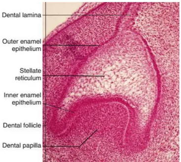
Figure 5 : Cap stage tooth germ showing the position of the enamel knot. Adapted from (Antonio Nanci and Cate 2013)
By E 15, the differentiation of enamel organ central cells forms the stellate reticulum cells (Figure 6) having a star shape with large intercellular spaces potentially playing a role in enamel-secreting ameloblasts nutrition. Another layer of cells known as “stratum
intermedium”, at E16.0 in the incisor and E17.0 in the molar, becomes recognizable from the
internal dental epithelial cells as flattened epithelial cells, between the stellate reticuIum and the internal dental epithelium whose cells, progressively, lengthened to become preameloblasts.
16
Figure 6 : Cap stage, beginning of cellular differentiation within the enamel organ.
Central cells form the stellate reticulum. Adapted from (Antonio Nanci and Cate 2013)
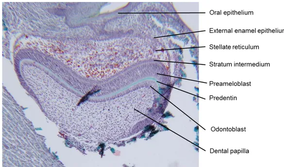
1.2.1.4 Bell stage
During this stage (EI7.0 for incisor and by E17.5-18.0 for molars), dental papilla cells differentiate into odontoblasts, beginning in the most anterior mesenchymal cells (Figure 7). The external dental epithelial cells thickness decreases and becomes a one or two cuboidal cell layer.
The preameloblasts about double in height and differentiate into ameloblasts and their nuclei peripherally placed, this differentiation firstly occurs in the most anterior regions. The lingual side of the incisors does not become coated with enamel because that the internal dental epithelial cells do not differentiate into ameloblasts on this side. At El7.0 these non-differentiating internal dental epithelial cells, diminish and become cuboidal in shape in subsequent stages of development, then merge with adjacent connective tissue cells.
By EI8 in the incisors and El9 in molars, odontoblasts begin to secrete predentin (Figure 7). After 24 hours of development, the predentin starts mineralizing and enamel matrix will be secreted by ameloblasts. Mineralization of the enamel matrix is postnatal and the incisors and the first molar erupt by day 20 after birth (P20). Tooth shape will be established when mineralization of dentin and enamel starts.
17
Figure 7 : Early bell stage of tooth development (own data)
1.2.1.5 Second and Third Molar Development
When the jaws elongate enough, the second and third molars start developing. Second molar development starts with the dental lamina, which can be seen at E15.5 forming as an outgrowth of the first molar tooth germ epithelium. By E18.5 the second molar is at the cap stage and erupts approximately at P25. The lamina of the third molar appears at P4, reaches the cap stage by P7-9 and the bell stage by P10, the third molar erupts by P35 (Rossant and Tam 2002).
1.2.2 Basement membrane
The basement membranes (BM) are the first extracellular matrices to appear and they are critical for organ formation and tissue repair (Martin and Timpl 1987; Kleinman et al. 1986). They act like scaffolds for cells and play an essential role in morphogenesis that affects cell adhesion, migration, proliferation, and differentiation.
The structure and components of BMs vary among tissues, resulting in tissue-specific structures and functions. BMs consist of supramolecular structure which is formed by reciprocal interaction of collagen IV, laminin, perlecan, nidogen/entactin, and other molecules (Martin and Timpl 1987; Kleinman et al. 1986).
18
BM components play an important role in tooth development. They control proliferation, polarity, attachment and determine tooth germ size and morphology (I. Thesleff et al. 1981; Fukumoto and Yamada 2005; Fukumoto et al. 2006).
For example, laminin α5 (Lama5), is a component of the major laminin chain in tooth basement membranes. Absence of Lama5 in KO mice lead to a small tooth germ with no cusps, in which the inner dental epithelium is not polarized and enamel knot formation is defective (Fukumoto et al. 2006).
Another laminins such as laminin α2 (Lama2) are expressed in odontoblasts during the late stage of germ development (Yuasa et al. 2004; Salmivirta, Sorokin, and Ekblom 1997). Its deficiency in mice manifests in thin dentin and defective dentinal tube structure (Yuasa et al. 2004). These phenotypes are similar to dentinogenesis imperfecta (DI) in humans. It was found that laminin-2, increases dentin sialoprotein expression in odontoblasts in cell culture, but its deficiency in mutant mice, reduces dentin sialoprotein expression in odontoblasts, suggesting that Lama2 is required for odontoblast differentiation.
Perlecan (HSPG2) is a major heparan sulfate proteoglycan in BMs. Its expression in developing teeth, was detected in BMs, intercellular spaces of the enamel organ, and the dental papilla including odontoblasts (Ida-Yonemochi et al. 2005). Overexpression of perlecan in transgenic mice results in abnormal tooth morphology and deregulation of growth factors such as TGF-b1 and bFGF (Ida-Yonemochi et al. 2011).
19 1.2.3 Dentin
1.2.3.1 Dentin structure
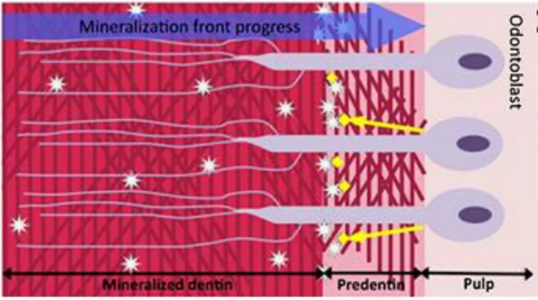
Dentin has a complex structure, similar to bone for mineralization ratio of about 70% mineral. In contrast with bone, dentin is not vascularized, and has not remodeling process. During the secretory stage, odontoblasts polarize, elongate and start to display two distinct parts: a cell body and a process. During the next step of evolution, the cell bodies stay outside the mineralized dentin, along the border of the mineralization front and the processes occupy the lumen of dentin tubules. Tubule diameter varies between 2 and 4 micrometers and its number is about 18 000 and 21 000 tubules per mm2 (Schilke et al. 2000). They are more numerous in the inner third layer than the outer third layer of the dentin.
1.2.3.1.1 Outer mantel dentin layer
Outer mantel dentin is a thin atubular layer with thickness of 15–30µm, at the periphery of coronal region. It is less mineralized than the rest of dentin and consequently the resilient mantle dentin allow dentin to dissipate pressures which otherwise would induce enamel fissures and detachment of the fragmented enamel from the dentin-enamel junction(R. Z. Wang and Weiner 1998).
1.2.3.1.2 Circumpulpal dentins
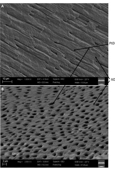
The circumpulpal dentin appears as a thin layer at initial stages of dentinogenesis, its thickness continuously increases at the expense of the pulp and then it becomes the largest part of the dentin layer. The circumpulpal dentin is formed by circles of peritubular dentin around the lumen of the tubules separated by the intertubular dentin. The ratio between inter and peritubular dentin is species dependent, it is about 50% in horses and about 10-20% in humans, and in the continuously growing rodent incisors no peritubular dentin can be found. Several differences in the structure and composition of these two types of dentin are found. In the intertubular dentin, the major protein is type I collagen (90%), whereas in the peritubular dentin no collagen is observed. The differences in the composition of noncollagenous proteins (NCPs) of the two types of dentin have been reported. (M. Goldberg, Molon Noblot, and Septier 1980; Weiner et al. 1999; Gotliv, Robach, and Veis 2006; Gotliv and Veis 2007). Intertubular dentin (Figure 9) results from transformation of predentin into dentin (Figure 8). It is compound of dense network of collagen fibrils, coated by NCPs, where needle like-crystallites locate at the collagen fibrils parallel to their axes and other like-crystallites fill inter-fibrillar spaces (M. Goldberg and Boskey 1996).
20
Figure 8 : Characteristics of dentin formation. Odontoblasts secrete an ECM composed
of type I collagen and NCPs. Within the predentin type I collagen molecules are assembled as fibrils. Mineralization occurs at the mineralization front by growth and fusion of calcospherites formed by hydroxyapatite (HAP) crystals. This mineralization process is controlled by NCPs and by mineral ion availability. Cell processes remain entrapped within dentin whereas cell bodies remain at the periphery of the pulp. Adapted from (Vital et al. 2012)
Peritubular dentin result from a passive deposit of serum-derived molecules along the tubule walls and the crystals form a ring around the tubules lumen (Figure 9). In this type of dentin no collagen fibrils are detectable, but a thin network of non-collagenous proteins and phospholipids are visible (M. Goldberg, Molon Noblot, and Septier 1980; Gotliv and Veis 2007; M. Goldberg and Boskey 1996).
21
Figure 9 : Human dentin by scanning electronic microscopy (SEM). A. cutting line is
parallel to dentin tubules, B. cutting line is perpendicular to dentin tubules. PtD: peritubular dentin, ItD: intertubular dentin. (Own data)
1.2.3.2 Dentin proteins 1.2.3.2.1 Collagens
In the dentin ECM, collagens form a 3D scaffold which is very important in dentinogenesis. Type I collagen is the major type in dentin matrix collagens (90%), other types of collagen were identified but at lower levels (1-3%) like types III and V collagens (Michel Goldberg and Smith 2004; Vital et al. 2012).
Collagen I formed by gathering of two α1 (I) chains and one α2 (I) chain. These chains entwine to form a triple helix of coiled coil framework (Rest and Garrone 1991). The
22
odontoblasts secrete thin collagen fibril subunits at their apical pole. Lateral fibril subunits assembly leads to fibrillar growth and then straight integration leads to the collagen lengthening.
1.2.3.2.2 Noncollagenous proteins (NCPs)
Noncollagenous proteins (NCPs) constitute the remaining 10% of the ECM scaffold and play an essential role in the regulation of bone and dentin mineralization. NCPs are divided into phosphorylated and nonphosphorylated NCPs.
1.2.3.2.2.1 Phosphorylated NCPs
SIBLINGs (Small Integrin Binding LIgand N-linked Glycoproteins), are a phosphoprotein family in which mutations are associated with abnormal phenotypes in the mineralization of bone and/or dentin (Qin, Baba, and Butler 2004; Vital et al. 2012). This family includes dentin sialophosphoprotein (DSPP), dentin matrix protein 1 (DMP1), bone sialoprotein (BSP), matrix extracellular phosphorylated glycoprotein (MEPE), and osteopontin (OPN). All SIBLINGs were identified in dentin and bone ECM, but a high rate of DSPP expression was shown to be specific to dentin. The SIBLING members carry an arginine– glycine– aspartate cell adhesion domain (RGD) and a highly conserved acidic serine and aspartate-rich motif (ASARM) (P. S. Rowe et al. 2000; Fisher and Fedarko 2003). Noteworthy, the function of ASARM domain in bone and teeth mineralization (apatite crystals nucleator or inhibitor) is at present debated by the scientific community, in particular its implication in pathological processes such as inherited rickets (Addison and McKee 2010; David and Quarles 2010; P. S. N. Rowe 2012). It is of interest that, in addition to binding integrins SIBLINGs, may also specifically bind and activate several MMPs in the ECM suggesting that they could be involved in dentin matrix degradation (Fedarko et al. 2004).
1.2.3.2.2.2 Nonphosphorylated NCPs
The second group of NCPs is nonphosphorylated proteins, such as osteonectin (SPARC protein or BM40) and proteins with gamma-carboxylated glutamates (Gla) residues (osteocalcin and matrix Gla protein-MGP-). While osteonectin may contribute to the mineralization process, osteocalcin and MGP have been suggested to regulate HAP crystal nucleation (Bronckers et al. 1998; Onishi et al. 2005; Kaipatur, Murshed, and McKee 2008). The small leucine-rich proteoglycans (SLRPs), such as decorin, biglycan, fibromodulin, lumican, and osteoadherin, have also been identified in predentin and dentin (M. Goldberg, Septier, and Escaig-Haye 1987; M. Goldberg et al. 2003). They are thought to be involved in the transport of collagen fibrils through the predentin and in collagen fibrillogenesis (M.
23
Goldberg et al. 2003). Predentin is also rich in dermatan and chondroitin sulphate-containing (PG). It is of interest that adjacent to the mineralization front, predentin contains a large quantity of keratan sulphate-containing PG associated with a dramatic decrease in dermatan and chondroitin sulphate-containing PG. This switch in the proteoglycan type was attributed to MMP-3, which is closely related to a control of the dentin mineralization process (Hall et al. 1999).
1.2.3.3 Dentinogenesis
At the early stage of tooth development, the dental mesenchyme originates from the neural crest-derived mesenchyme migrate to the oral cavity under the oral epithelium and contribute to the tooth bud formation. During the last mitosis of the proliferate mesenchymal cells the cell located in contact with the basement membrane become preodontoblasts, whereas the daughter cells away from the basement membrane form the Hoehl’s layer which constitutes a reservoir for replacing the damaged odontoblasts. After the differentiation odontoblasts become polarized and start to secret the extra cellular matrix components which will be the scaffold for hydroxyapatite (HAP) crystals deposition to form at the end the dentin.
Another classification showed that there are four dentins: Primary dentin, which is formed by odontoblasts which secret this dentin until the tooth becomes functional. Secondary dentin, is secreted by odontoblasts immediately after the end of primary dentin secretion (when the contact of antagonistic cusps is established), and continues throughout life. The major difference between primary and secondary dentins is morphological; in the secondary dentin the S-curve of the tubules is more accentuated. Tertiary reactionary dentin, is synthesized by odontoblasts or, if these cells are destroyed, by the subjacent cells of the Höehl‘s layer, as a reaction to carious decay, to abrasion or as a response to the release of some components of dental material fillings. Depending the severity and speed of the carious lesion, the age of the patient and the progression of the reaction, it appears as a layer of the osteodentin type, or as a tubular or atubular orthodentin. Tertiary reparative dentin is formed by pulp progenitors, implicated in the formation of a bone-like or in structure-less mineralization (pulp diffuse mineralization or pulp stones). These structures are closer to bone (osteodentin) rather than to dentin (Michel Goldberg et al. 2011).
1.2.4 Enamel
Enamel is the hardest and outer layer of tooth crown that protects the mammalian tooth from external chemical and physical effects. Enamel properties are associated with its special structural organization and connection with underlying dentin.(Janet Moradian-Oldak 2012).
24
Mature enamel consists of approximately 4% water and organic material and 96% inorganic materials (Table 1). Enamel inorganic content is a crystalline calcium phosphate (hydroxyapatite) which also is found in dentin, cementum, bone, and calcified cartilage (Antonio Nanci and Cate 2013).
Table 1 : Percentage Wet Weight Composition of Rat Incisor Enamel. From (Antonio Nanci and Cate 2013)
The principal structural units of enamel are the rods (prisms) and interrod enamel (interprismatic substance) (Figure 10).
Figure 10 : Scanning electron microscope views of (A) the enamel layer covering
25
and perspectives of the rod-interrod relationship when rods are exposed longitudinally (C) or in cross section (D). Interrod enamel surrounds each rod. DEJ: Dentinoenamel junction; IR: interrod; R,rod. Adapted from (Antonio Nanci and Cate 2013)
1.2.4.1 Enamel proteins
Enamel proteins are synthesized by ameloblasts. During tooth development, the ameloblasts control the synthesis and secretion of the organic extracellular matrix (ECM) and then the biomineralization of this ECM. Enamel proteins are hydrophobic proteins known such as amelogenins and nonamelogenin proteins including ameloblastin, enamelin, tuftelin, tuft proteins, sulfated proteins and enamel proteases such as enamelysin (MMP-20) and KLK-4.
1.2.4.1.1 Amelogenin
Amelogenin gene exists only on the X chromosome in rodents (Snead et al. 1983; Chapman et
al. 1991), while it exists on both X and Y chromosomes In human and cow (Lau et al. 1989). Amelogenin constitutes more than 90% of the enamel protein content. It is secreted as a variety of isoforms because of alternative splicing of the amelogenin gene and processing of the parent molecules (C. W. Gibson et al. 1991; Lau et al. 1992), the major isoform is about 25 kDa. Amelogenin has a bipolar nature: it contains highly hydrophobic domains and hydrophilic N- and C-terminal sequences and this bipolar nature allows amelogenin monomers by self-assembly to form supramolecular resulting in the formation of nanospheres which regulate crystal spacing (Fincham et al. 1994; Fincham and Simmer 1997). The N-terminal A-domain is involved in the formation of nanospheres, whereas the carboxy-terminal B-domain prevents their fusion to larger assemblies (J. Moradian-Oldak et al. 2000). Amelogenin has signaling activities (Carolyn W. Gibson 2008; Veis 2003), especially the small isoform; leucine-rich amelogenin peptide (LRAP) (Warotayanont et al. 2008). Because of its potential to promote cell differentiation and its interaction with bone cells, it has been used in periodontal regenerative therapies.
Amelogenin is not essential for the initiation of mineralization, but is essential for the elongation of enamel crystals and the achievement of proper enamel formation, because in spite of its absence in KO mice, a thin layer of mineralized enamel is formed.
1.2.4.1.2 Ameloblastin
Ameloblastin constitutes about 5% of enamel protein. Its expression significantly decrees during enamel maturation. The isolation of this protein is so difficult for several reasons, the
26
limitations in expression and hydrolysis by enamel proteinase MMP-20 as soon as secreted (Iwata et al. 2007; Yasuo Yamakoshi, Hu, Zhang, et al. 2006). In the ameloblastin KO mice, ameloblasts detach from the surface of the developing teeth, suggesting a potential role for ameloblastin in ameloblasts adhesion to the forming enamel (Fukumoto et al. 2004).
1.2.4.1.3 Enamelin
Enamelin is the largest enamel protein and constitutes about (3–5%) of enamel proteins. It is a phosphorylated, glycosylated protein and is rapidly cleaved following its secretion. The intact protein is only observed at the mineralization front, so it proposed to be implicated in crystal elongation (C. C. Hu et al. 1997; C. C. Hu et al. 2000).
Enam gene mutations cause an autosomal dominant forms of amelogenesis imperfecta AI
(Hart et al. 2003) and no true enamel layer is formed in the Enam KO mice(J. C.-C. Hu et al. 2008).
Recently, it was reported that a large increase or decrease in enamelin expression impairs the production of enamel crystals and the prism structure (J. C.-C. Hu et al. 2014).
Enamelin and ameloblastin appear to have similar roles like crystallite initiation and elongation, whereas amelogenin appears to form a framework to allow the continued elongation of the already initiated crystallites (John D. Bartlett 2013).
1.2.4.1.4 Tuftelin
Tuftelin is expressed early at the bud stage of tooth development (several days befor the onset of mineralisation) so it is suggested to play a nucleator role during crystals formation. Its expression is also detected in several organs kidney, lung, liver, and testis (Zeichner-David et al. 1997; MacDougall et al. 1998).
1.2.4.1.5 Sulfated enamel proteins
Sulfated enamel proteins constitute an acidic nature family of proteins with unknown roles. They are difficult to be detected because of their presence in a small amount (C. E. Smith et al. 1995).
1.2.4.1.6 Amelotin
Amelotin is a glycoprotein recently discovered, its role is not yet clear (Iwasaki et al. 2005). It is expressed during the secretory stage of enamel development (Gao et al. 2010). Alternatively spliced variants lead to several isoform of amelotin.
27 1.2.4.1.7 Biglycan
Biglycan expression is in the dentin and the enamel (Septier et al. 2001). It is expressed by ameloblasts during tooth development, (M. Goldberg, Septier, Rapoport, et al. 2002), where it acts as an amelogenin expression repressor (M. Goldberg, Septier, Rapoport, et al. 2002; M. Goldberg et al. 2005b).
1.2.4.2 Enamel proteinases
Enamel proteinases are so important for the digestion of enamel proteins and enamel maturation. It was found that some of these proteinases have an ameloblast differentiation- dependent expression (Lu et al. 2008).
1.2.4.2.1 Matrix metalloproteinase 20 (MMP-20)
Enamelysin (MMP-20) is expressed by ameloblasts and odontoblasts (J. D. Bartlett et al. 1996; Fukae and Tanabe 1998), it is expressed from the beginning of secretion stage through the beginning of maturation stage of enamel and cleaves amelogenin, enamelin, and ameloblastin into stable intermediate products (Lacruz et al. 2011). In vitro studies showed that Mmp-20 stimulates the formation of nanorod structures formed by co-assembly of the parent amelogenin with its proteolytic products (X. Yang et al. 2011). Such assembly alteration was proposed to be related with the elongated growth of apatite crystals. It has been proposed that Mmp-20 activity produces protein intermediate products that will stimulate phase transformation of amorphous calcium phosphate nanoparticles into mineralized hydroxyapatite (Kwak et al. 2009).
1.2.4.2.2 Kallikrein-4 (KLK4)
Klk-4 is expressed from the end of secretory stage and throughout the maturation stage of enamel (Lacruz et al. 2011). Its function is to digest the intermediate products of amelogenin, enamelin and ameloblastin resulting from the MMP-20 action and facilitates enamel proteins removal which is necessary for enamel maturation and hardening (O. Ryu et al. 2002).
KLK-4 digests the 32-kDa enamelin fragment which is resistant to Mmp-20 action, (Yasuo Yamakoshi, Hu, Fukae, et al. 2006) and its activity is not affected like MMP-20 by the presence of apatite crystals in vitro (Z. Sun et al. 2010).
1.2.4.2.3 Other proteinases 1.2.4.2.3.1 Caldecrin (Ctrc)
Caldecrin Ctrc expression pattern in enamel is similar to Klk4, but lower, and it is predominantly expressed in the maturation stage of amelogenesis (Lacruz et al. 2011).
28 1.2.4.2.3.2 MMP-2
It has been demonstrated that recombinant MMP-2 cleave amelogenin into several fragments in vitro (Caron et al. 2001). MMP-2 also degraded most forms of amelogenin, suggesting that MMP-2 can participate, with MMP-20, to achieve complete amelogenin processing (Bourd-Boittin et al. 2005).
1.2.4.2.3.3 MMP-9
Recently, it was proposed that MMP-9 involved in enamel formation and controlling the processing of amelogenin (Feng et al. 2012)
1.2.4.3 Enamel formation 1.2.4.3.1 Pre-secretory stage
At this stage, ameloblasts start expressing very small amounts of enamel proteins even before the basement membrane break up and send cytoplasmic projections through the gaps directly after basement membrane disintegrate. With the disappearance of the basement membrane, dentin starts to mineralize and the apical surfaces of ameloblasts connect with the superficial collagen fibrils of the mantle dentin (Meckel, Griebstein, and Neal 1965; Cevc et al. 1980) (Figure 12).
1.2.4.3.2 Secretory Stage
At the beginning, ameloblasts secrete enamel proteins on top of and around existing dentin crystals initially and then around enamel crystals and into the space of disappeared basement membrane (Figure 11.A). With the continued secretion of enamel matrix, ameloblasts move back to create the necessary space for continuous deposition of enamel end this moment we can distinguish the initial enamel layer which is aprismatic (not separated into rod and interrod enamel)
Secretory ameloblasts develop a novel cell extension called Tomes’ process at their apical (secretory) ends. This extension which has secretory and nonsecretory regions provides the architectural basis for organizing enamel crystals into rod and interrod enamel, (Meckel, Griebstein, and Neal 1965; Cevc et al. 1980).
Secretory ameloblasts secrete enamel proteins which concentrate along the ameloblast secretory membrane and form a mineralization front (there is no pre-enamel like the predentin in dentin or osteoid in bone). The mineralization front retreats with the Tomes’ process as the enamel crystals grow in length (4µm/day) (Risnes 1986), and the ameloblasts
29
continue their secretion of enamel proteins (Figure 12). During this stage enamel crystals grow primarily in length and the enamel layer thickens.
Figure 11 : Semi-thin (0.5 µm) sections from glutaraldehyde-fixed, decalcified, and
plastic embedded mandibular incisors of wild-type mice stained with toluidine blue to illustrate the appearance of enamel and enamel organ cells at mid-secretory stage (A) and near-mid-maturation stage (B) of enamel development. Abbreviations: E, enamel; Am, ameloblast; Si, stratum intermedium; pd, predentin; D, dentin; ae, apical end; be, basal end; bv, blood vessel; as, artifact space; b, bone; c, cementum. Adapted from (J. D. Bartlett and Smith 2013)
1.2.4.3.3 Maturation Stage
At the end of secretory stage, enamel layer has its final thickness and ameloblasts reduce their secretion of enamel proteins (Figure 11.B), and start the secretion of KLK-4 which finishes the degradation of the organic matrix. The degradation and removal of growth-inhibiting enamel proteins terminate the growth of enamel crystallites in length, and accelerate their growth in width and thickness by the ion deposition on the thin crystals sides until they press against one another (C. E. Smith 1998). This process is necessary to have a harde and mature
30
enamel layer, and is directed by modulating ameloblasts that cycle through smooth and ruffle-ended phases.
During maturation stage a basal lamina is secreted at the base of the ameloblasts (Figure
12). Recently amelotin ( AMTN ) has been identified as one of the components of this basal
lamina (Iwasaki et al. 2005; Moffatt et al. 2006).
Figure 12 : Schema present incisor enamel and denin formation. p-Am: pre-ameloblast;
pOd: pre-odontoblast; s-Am: secreting Ameloblasts; od: odontoblasts; pos-Am: post-secretory ameloblasts; pD: predentin; D: dentin; pm-E: premature enamel; E: enamel. Adapted from (Khaddam et al. 2014)
1.3 Matrix MetalloProteinases MMPs
MMPs is subdivided into soluble and membrane-type MMPs (MT-MMPs). The soluble MMPs are expressed as pro-enzymes that will be activated in the extracellular environment. MT-MMPs are intracellularly activated and identified as activators of soluble MMPs and were shown to be able to degrade extracellular matrix proteins ECM (Hamacher, Matern, and Roeb 2004).
31
In addition to the inhibition by endogenous inhibitors (tissue inhibitor of MMPs TIMP) or to the proteolytic activation of pro-MMPs, MMPs are regulated by cytokines or growth factors transcriptionally (Tsuruda, Costello-Boerrigter, and Burnett 2004)
MMPs are implicated in inflammation by regulating the availability and the activity of cytokines, chemokines, and growth factors, as well as integrity of tissue barriers. MMPs are also involved in tumors (Nissinen and Kähäri 2014).
1.3.1 MMPs and teeth
1.3.1.1 In physiological processes (development)
Several MMPs have been detected in developing tooth tissues (Michel Goldberg et al. 2003). They play a central role in the disruption of basement membrane. MMPs are also implicated in the functional regulation of growth factors and their receptors, cytokines and chemokines, adhesion receptors and cell surface proteoglycans, and a variety of enzymes (H. Li et al. 2002). MMPs participate in the remodeling of the ECM during tooth development to facilitate the migration of cells and the mesenchymal condensation (Chin and Werb 1997) and participate in the regulation of the mineralization process of dental hard tissues by cleaving the matrix proteins of the dentin and the enamel matrix (Simmer and Hu 2002; Fanchon et al. 2004).
MMP-1, -2, -3, -9 and MT1-MMP have been detected during tooth development, indicating that these MMPs have roles in tooth morphogenesis and eruption (Chin and Werb 1997; Sahlberg et al. 1992b; Caron, Xue, and Bartlett 1998; Randall and Hall 2002; Yoshiba et al. 2003).
1.3.1.2 In pathological processes 1.3.1.2.1 Periodontitis
High MMPs levels were detected in the periodontitis and apical periodontitis leading to accelerated matrix degradation, (de Paula e Silva et al. 2009; Paula-Silva, da Silva, and Kapila 2010). Collagenases (MMP-1, MMP-8, and MMP-13) and gelatinases (MMP-2 and MMP-9) are implicated in the digestion of collagen in the bone and periodontal ligament (Andonovska, Dimova, and Panov 2008; Corotti et al. 2009).
1.3.1.2.2 In the caries process
We developed this point (Figure 13) in Chaussain, Boukpessi, Khaddam et al, 2013 (end of introduction).
32
Figure 13 : Schematic representation of MMP activity during the dentin carious
process. Cariogenic bacteria present in the caries cavity release acids such as lactic acid that reduce the local pH. The resulting acidic environment demineralizes the dentin matrix and induces the activation of host MMPs derived from dentin or saliva (which bathes the caries cavity). Once the local pH is neutralized by salivary buffer systems, activated MMPs degrade the demineralized dentin matrix. Adapted from (Chaussain et al. 2013)
1.4 EMMPRIN (Basigin,CD147)
1.4.1 Historic
Extra cellular matrix metalloproteinase inducer EMMPRIN (CD147), a member of the immunoglobulin superfamily, was described for the first time on the surface of solid tumor cells as an inducer of a various (MMPs in adjacent fibroblasts (Biswas 1982). Based on these latter properties it was named extracellular matrix metalloproteinase inducer EMMPRIN (Biswas et al. 1995). Previously EMMPRIN had different names including tumor cell-derived collagenase stimulatory factor (TCSF), Basigin, CD147, gp42, HT7, neurothelin, 5A11, OX-47 and M6 (T. Muramatsu and Miyauchi 2003).
1.4.2 Structure
1.4.2.1 Transmembrane form
EMMPRIN (Basigin) has four isoforms (basigin-1 to -4), caused by alternative transcription initiation and variation in splicing (Figure 14)(Belton et al. 2008) and the major isoform is basigin-2. EMMPRIN is highly glycosylated, Its protein portion is 27 kDa, and its
33
glycosylated form is 43 to 66 kDa (Miyauchi et al. 1990) and the nonglycosylated form has the ability to induce MMP expression in fibroblasts as the glycosylated form(Belton et al. 2008)
Figure 14: Basigin isoforms. Characteristic features of isoforms are mentioned within
blanket. Carbohydrates are shown by light grey color. Adapted from (Takashi Muramatsu 2012)
EMMPRIN is largely composed of three domains, extracellular immunoglobulin domain, a transmembrane domain and a cytoplasmic domain.
The extracellular domain has two immunoglobulin domains (a N-terminally located D1 domain and a more C-terminally located D2 domain) (Figure 15) and three potential Asparagine (Asn)-glycosylation sites; one in D1 domain and two in D2 domain (Miyauchi et al. 1990; Miyauchi, Masuzawa, and Muramatsu 1991).
The transmembrane domain has glutamic acid in its center, and is completely conserved between human, mouse and chicken (Miyauchi, Masuzawa, and Muramatsu 1991), this domain is important for interactions with other proteins in the same membrane.
34
Figure 15: Scheme of EMMPRIN structure. EMMPRIN contains an extracellular
domain composed of two Ig loops with three Asn-linked oligosaccharides and short single transmembrane domain (TM) and a cytoplasmic domain (Cyt). The first Ig domain is required for counter-receptor activity, involved in MMP induction. Adapted from (Gabison et al. 2009).
1.4.2.2 Soluble form
The soluble form of CD147 has been detected in conditioned media as:
full-length protein (Taylor et al. 2002)
or as part of shed microvesicles (Sidhu et al. 2004)
as well as in forms lacking the transmembrane and cytoplasmic domain derived from MMP mediated cleavage of CD147 from the cell surface (Haug et al. 2004; Y. Tang et al. 2006; Egawa et al. 2006)
35 1.4.3 Phenotypes of EMMPRIN knock out (KO) mice
EMMPRIN KO mice have a low reproduction level which is at a much lower frequency than that expected by Mendelian segregation, KO embryos develop normally during blastocyst stage but at the time of implantation, about 75% of the null embryos are lost (Igakura et al. 1998) and half of the surviving mice had interstitial pneumonia and died within 4 weeks after birth (Igakura et al. 1998). EMMPRIN KO mice have a defect in the capability of implantation of the uterus (female), arrested spermatogenesis (male) (Igakura et al. 1998; Kuno et al. 1998), abnormal behavior (Naruhashi et al. 1997), deficits in vision (Hori et al. 2000) and a decreased response to odor (Igakura et al. 1996).
1.4.4 EMMPRIN interactions
Three possible EMMPRIN interactions were descriped (Figure 16):
- Homophilic cis interaction between EMMPRIN molecules within the plasma membrane of the same tumor cell (Yoshida et al. 2000).
- Homophilic trans interaction between EMMPRIN molecules on tumoral cells.(J. Sun and Hemler 2001)
- Heterophilic interactions between EMMPRIN molecule on a tumor cell and a putative EMMPRIN receptor on a fibroblast.
Figure 16: Possible EMMPRIN-mediated interactions stimulating MMP production.
(A) Homophilic cis interaction between EMMPRIN molecules within the plasma membrane of a tumor cell. (B) Homophilic trans interaction between EMMPRIN molecules on apposing tumor cells. (C) Heterophilic interactions between EMMPRIN