UNIVERSITÉ DE MONTRÉAL
PRODUCTION OF CHITOSAN MICRO AND NANOSPHERES FOR THE FORMULATION OF ANTIBACTERIAL FOOD PACKAGING MATERIALS
NURY HAYDÉE ARDILA GUALDRÓN DÉPARTEMENT DE GÉNIE CHIMIQUE ÉCOLE POLYTECHNIQUE DE MONTRÉAL
THÈSE PRÉSENTÉE EN VUE DE L’OBTENTION DU DIPLÔME DE PHILOSOPHIAE DOCTOR
(GÉNIE CHIMIQUE) MAI 2017
UNIVERSITÉ DE MONTRÉAL
ÉCOLE POLYTECHNIQUE DE MONTRÉAL
Cette thèse intitulée:
PRODUCTION OF CHITOSAN MICRO AND NANOSPHERES FOR THE FORMULATION OF ANTIBACTERIAL FOOD PACKAGING MATERIALS
présentée par : ARDILA GUALDRÓN Nury Haydée en vue de l’obtention du diplôme de : Philosophiae Doctor a été dûment acceptée par le jury d’examen constitué de :
Mme DESCHÊNES Louise, Ph. D., présidente
Mme HEUZEY Marie-Claude, Ph. D., membre et directrice de recherche M. AJJI Abdellah, Ph. D., membre et codirecteur de recherche
M. CARREAU Pierre, Ph. D., membre M. ARUL Joseph, Ph. D., membre externe
DEDICATION
ACKNOWLEDGEMENTS
I would like to express my gratitude to all who have being part of my new life in Canada. Colleagues that became friends and friends that became like a family. All of you have made that even if family is far, life is easier and enjoyable.
I would like also to express my deep gratitude to my advisor, Professor Marie-Claude Heuzey, and co-advisor, Professor Abdellah Ajji, who gave me not only the knowledge, support and encouragement for the successful completion of this project, but also their patience and motivation in each activity I was participating. It was really pleasant and it was a very enriching experience to work with them.
Besides my supervisors, I would like to thank the rest of my thesis committee, namely Professor Louise Deschênes, Professor Pierre Carreau and Professor Joseph Arul for accepting to evaluate my thesis.
I am so thankful to Professor France Daigle at Université de Montréal for her training and continuous support in the antibacterial tests.
I acknowledge the help and hard work of Zineb Ajji and Nelson Medina in the realisation of electrospraying and electrospinning tests during their internship and Master project, respectively. I also want to thank Professor Jason Tavares and Nick Virgilio for their advice, coaching sessions and encouraging me in different academic activities during my studies.
Also special thanks to Professor Basil Favis for letting me join to students CREPEC committee in 2014. It was a very enriching experience for my professional and personal life. In addition, I thank Diane, Alyson and the student committee of that year for all the experience we share.
Many thanks to Ahmad Zohrevand who introduced me to the SPE committee. Special thanks to Marie-France Sosa, Professor Nicole Demarquette and Doru Davidescu for letting me to be part of the activities and their committee since 2013.
A deep thank to Aziz Guellouz for the coaching sessions, advices and continuous support during the Technopreneur program. Also special thanks to the ChitoPack team Mounia Arkoun, Valéry Kovalenko and Riham Hammouda.
I would also like to extend my thanks to the technical and administrative staff of the Chemical Engineering Department at Polytechnique de Montréal for their help and kind cooperation during my PhD, especially to Claire Cerclé, Mélina Hamdine, Martine Lamarche and Gino Robin. I want to thank my dear friend Mounia Arkoun for her kind help in translating the abstract of this thesis to French and for her continued support not only for the long and continuous discussions about the project but also in my personal life. I was lucky to work with her as a partner in the different academic activities during my studies.
A sincere appreciation to all people who have been part of the rheology and electrospinning group meetings for their continuous feedback. A special thanks to my dear friends and colleagues Hoda, Hajer, Hanan, Fatemeh, Gilles, Andrea, Marie, Changsheng, Xiaoyan, Davood, Mariam, Helia, Qinghua, Thibault, Amir, Sandra, Jaime, Shirley, Richard, Didac, Juan, Mar, Pau, Ricardo, Lina, Jesus and Manuel.
A big appreciation to the dedicated team of Les Petits génies, specially to Martine, Alcina, Marie-Danielle, Fatima, Amina, Farida, Lizbeth and Rosa.
Finally, all my accomplishments and deep gratitude go to my parents, my brother, my sister, especially to my husband and daughter Violette who give me the support in all my life.
RÉSUMÉ
L'emballage alimentaire actif est un sujet de recherche en cours. La fabrication de nouveaux matériaux d'emballage pour améliorer la sécurité alimentaire et la préservation des aliments est un sujet d'intérêt continu. Le chitosane est un polysaccharide d’origine marine, non toxique ayant un grand potentiel pour être utilisé comme biomatériau antimicrobien, compte tenu de son activité antibactérienne. En outre, le chitosane est utilisé comme un additif alimentaire et a d’ailleurs reçu le statut de «Generally Recognized As Safe» (GRAS) par la Food and Drug Administration (FDA) des États-Unis. Par conséquent, le chitosane peut être considéré comme un candidat idéal dans des applications liées à l’industrie alimentaire.
Le présent travail de recherche porte sur la production de nanosphères de chitosane pour la formulation de matériaux d’emballage alimentaire antibactériens. Le projet de recherche a été réalisé en trois étapes. La première étape traite de l'étude de l'influence de différents facteurs environnementaux, microbiens et caractéristiques du chitosane sur son activité antibactérienne, lorsqu'il est utilisé sous une forme solide discontinue, comme la poudre et les flocons. L'activité antimicrobienne a été évaluée contre une souche Gram-négatif (Escherichia coli) et deux souches Gram-positif (Listeria innocua et Staphylococcus aureus), qui sont généralement responsables de la détérioration des aliments. Les résultats ont montré que le chitosane nécessite une solubilisation partielle pour l'exercice de son activité antibactérienne. En outre, des conditions de température adéquates, la force ionique (salinité) et la présence d'un support physique solide peuvent favoriser l'effet antibactérien. La souche E. coli s’est révélée plus affectée par le chitosane, suivie de L.
innocua et S. aureus. D'autre part, l'action antibactérienne a augmenté avec la concentration en
chitosane jusqu'à un point critique au-dessus duquel cet effet a diminué. Cet effet pourrait être la conséquence des protéines restantes dans le chitosane et qui peuvent servir de nutriments pour les bactéries, limitant ainsi l'activité antibactérienne. Ces résultats sont prometteurs pour l'utilisation directe de la poudre et des flocons de chitosane en tant qu'agents antimicrobiens pour des applications dans l'emballage alimentaire.
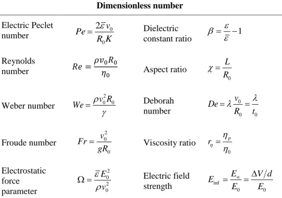
La deuxième étape a consisté en la production de micro et nanosphères de chitosane par électropulvérisation. Les effets des paramètres de solution et de procédé sur la morphologie, la collecte des particules et la stabilité du procédé ont été étudiés. La cartographie de la stabilité du procédé a été établie selon les nombres adimensionnels suivants: Reynolds (Re), Peclet électrique
(Pe), Weber (We), Froude (Fr) et un paramètre de force électrostatique (Ω) qui relie les principales variables du procédé. La stabilité des solutions de chitosane par electropulvérisation nécessitait des valeurs relativement faibles pour les nombres Re, Fr et Ω, mais des valeurs relativement élevées pour les nombres Pe et We. Le procédé d'électropulvérisation constitue une nouvelle façon d'incorporer des nanosphères de chitosane dans les emballages alimentaires existants afin de leur fournir des propriétés antimicrobiennes et d'aider à prolonger la durée de conservation des produits alimentaires.
La troisième étape étudie l'effet de la forme physique du chitosane, à savoir les solutions, les poudres et les nanosphères, ainsi que la taille des particules sur l’activité antibactérienne contre deux souches pathogènes, Staphylococcus aureus et Salmonella enterica serovar Typhimurium, des bactéries généralement associées à des infections d'origine alimentaire. Les nanosphères de chitosane ont présenté des performances antibactériennes supérieures à celles de la poudre de chitosane. Ceci a été expliqué par la petite taille et la plus grande surface de contact des nanosphères avec les parois cellulaires bactériennes. Les nanosphères ont également affiché une activité antibactérienne plus élevée que celle du chitosane en solution, ce qui peut être le résultat d'une charge superficielle plus élevée et d'une petite taille solide. Il est considéré que si le chitosane en solution interagit avec la paroi cellulaire, il restera sous forme libre dans le milieu plutôt que d’adhérer aux cellules de façon permanente. D'autre part, les souches de S. aureus étaient plus sensibles à l'action des nanosphères et étaient moins sensibles aux variations des conditions de pH et de température du milieu. Cette étude est d'une grande importance en ce qui concerne les nombreuses applications possibles des nanosphères de chitosane dans divers domaines dont l’emballage alimentaire et le biomédical.
ABSTRACT
Active food packaging is an ongoing research topic. The fabrication of packaging materials to improve food safety and food preservation is a subject of continuous interest. Chitosan is a nontoxic polysaccharide that has great potential to be used as an antimicrobial biomaterial, given its antibacterial activity. Besides, chitosan is considered as “Generally recognized as safe” (GRAS) food additive by the US Food and Drug Administration (FDA), wherewith can be considered as candidate for food related applications.
The current research work focuses on the production of chitosan nanospheres for the formulation of antibacterial food packaging materials. The research project was conducted in three phases. The first phase deals with the evaluation of different environmental, microbial and characteristics of chitosan on its antibacterial activity, when used in a discontinuous solid form, such as neat chitosan powder and flakes. The microbial activity was evaluated against one Gram-negative (Escherichia
coli) and two Gram-positive strains (Listeria innocua and Staphylococcus aureus), which are
commonly found in food spoilage. Results showed that chitosan requires a partial solubilisation for the exertion of the antibacterial activity. In addition, adequate temperature conditions, low ionic strength and the presence of a solid physical support may promote the antibacterial effect. E. coli strains were found to be more sensitive to chitosan, followed by L. innocua and S. aureus. On the other hand, antibacterial action increased with concentration up to a critical point above which this effect decreased. This effect may be due to remaining proteins in chitosan, which may serve as nutrients for the bacteria, limiting the antibacterial activity. These results are promising for the direct use of chitosan powder and flakes as antimicrobial agents for food packaging applications. The second phase consisted in the production of chitosan micro and nanospheres via electrospraying. Solution and processing parameters effects on the morphology, particle collection and processing stability were investigated. Mapping of the processing stability was established according to the following dimensionless numbers: Reynolds (Re), electric Peclet (Pe), Weber (We), Froude (Fr) and an electrostatic force parameter (Ω) which related the main variables of the process. The stability in the electrospraying of chitosan solutions required relatively low values for
Re, Fr and Ω but relatively high values for Pe and We numbers. The electrospraying process may
provide a novel way for incorporating chitosan nanospheres into existing food packaging to provide antimicrobial properties, and helping to extend the shelf-life of food products.
The third phase investigated the effect of chitosan physical form and particle size, namely solution, powder and nanospheres on the antibacterial activity, against two pathogen strains, Staphylococcus
aureus and Salmonella enterica serovar Typhimurium, commonly associated with foodborne
infection. Chitosan nanospheres displayed superior antibacterial performance than chitosan powder, explained by their small size and the larger surface area of contact of nanospheres with bacteria cell wall. Nanospheres also displayed higher antibacterial activity than chitosan in solution, which may be the result of the higher surface charge and solid and small size. It is believed that even though chitosan in solution interacts with the cell wall, it will remain as a free form in the medium rather than adhering into cells permanently. On the other hand, S. aureus strains were more sensitive to the action of nanospheres and were less influenced by the pH and temperature conditions of the medium. Given the remarkable antibacterial activity observed for chitosan nanospheres, this study is of great importance with respect to the many possible applications, such as in food packaging and in the biomedical field.
TABLE OF CONTENTS
DEDICATION ... III ACKNOWLEDGEMENTS ... IV RÉSUMÉ ... VI ABSTRACT ... VIII TABLE OF CONTENTS ... X LIST OF TABLES ... XIV LIST OF FIGURES ... XV LIST OF ABBREVIATIONS ... XX LIST OF APPENDICES ... XXIIICHAPTER 1: INTRODUCTION ... 1
CHAPTER 2: LITERATURE REVIEW ... 4
2.1 Chitosan ... 4 2.1.1 Elemental analysis ... 5 2.1.2 Chemical structure ... 6 2.1.3 Solution properties ... 7 2.1.4 Antimicrobial properties ... 8 2.1.5 Processing ... 11 2.1.6 Applications ... 12 2.2 Electrospraying process ... 13
2.2.1 Parameters of the process ... 14
2.2.2 Governing equations of the process ... 19
2.2.3 Electrospraying of chitosan ... 20
CHAPTER 3: OBJECTIVES ... 22
CHAPTER 4: ORGANIZATION OF THE ARTICLES ... 23
CHAPTER 5: ARTICLE 1: ANTIBACTERIAL ACTIVITY OF NEAT CHITOSAN POWDER AND FLAKES ... 25
5.1 Abstract ... 25
5.2 Introduction ... 26
5.3 Results and Discussion ... 27
5.3.1 SEM ... 29
5.3.2 Antibacterial Assays ... 30
5.4 Materials and Methods ... 44
5.4.1 Materials ... 44
5.4.2 Bacteria Strains and Culture ... 44
5.4.3 Methods ... 45
5.5 Conclusions ... 49
5.6 Acknowledgements ... 50
5.7 References ... 50
CHAPTER 6: ARTICLE 2: CHITOSAN ELECTROSPRAYING: MAPPING OF PROCESS STABILITY AND DROPLET FORMATION ... 57
6.1 Abstract ... 57
6.2 Introduction ... 57
6.3 Materials and Methods ... 62
6.3.1 Materials ... 62
6.3.2 Methods ... 63
6.4 Results and Discussion ... 65
6.4.2 Effect of solution parameters in electrospraying ... 69
6.4.3 Mapping of the electrospraying ability of chitosan as function of dimensionless numbers ... 78
6.5 Conclusions ... 81
6.6 Acknowledgments ... 81
6.7 Supporting information ... 81
6.8 References ... 86
CHAPTER 7: ARTICLE 3: EFFECT OF CHITOSAN PHYSICAL FORM ON ITS ANTIBACTERIAL ACTIVITY AGAINST PATHOGENIC BACTERIA ... 91
7.1 Abstract ... 91
7.2 Introduction ... 92
7.3 Materials and Methods ... 94
7.3.1 Materials ... 94
7.3.2 Methods ... 94
7.4 Results and Discussion ... 97
7.4.1 Morphology ... 97
7.4.2 Zeta potential and solubility of CS nanospheres ... 98
7.4.3 Effect of physical form on the antibacterial activity of CS ... 100
7.4.4 Effect of pH, temperature and bacterium species ... 102
7.5 Conclusions ... 107
7.6 Acknowledgments ... 108
7.7 References ... 108
CHAPTER 8: GENERAL DISCUSSION ... 113
CHAPTER 9: CONCLUSIONS AND RECOMMENDATIONS... 118
9.2 Recommendations ... 119 BIBLIOGRAPHY ... 121 APPENDICES ... 137
LIST OF TABLES
Table 2.1: Elemental analysis (%) of chitosan and chitin; N-acetyl-glucosamine reference [34].
Adapted from [22]. ... 6
Table 2.2: Trace metal content of chitosan (ppm) [35]. Adapted from [22]. ... 6
Table 2.3: Main applications for chitosan ... 12
Table 2.4: Parameters for the processing via electrospraying ... 15
Table 5.1: Chitosan grades. ... 28
Table 5.2: Elemental analysis via EDS-SEM in chitosan grades. ... 29
Table 5.3: Recovery of viable bacteria on BHI agar after exposure to 0.4 wt/v % chitosan for 4 h at 37 °C. ... 41
Table 6.1: Dimensionless numbers ruling the electrospraying process ... 62
Table 6.2: Chitosan grades ... 63
Table 6.3: Process parameters of electrospraying and range of analysis ... 64
Table 6.4: Chain overlap (C*) and critical entanglement concentration (Ce) for chitosan at 70 and 90 v/v % AcOH content ... 75
Table 6.5: Optimum parameters to produce chitosan nanospheres with an average size of 128 ± 69 nm via electrospraying ... 79
Table 7.1: Zeta potential (mV) and mean particle size (Z-average) of 0.01% chitosan ... 98
Table A.1: Properties of the electrospun solutions ... 157
LIST OF FIGURES
Figure 2.1: Industrial extraction of chitin and chitosan. ... 5 Figure 2.2: Structure of chitin and chitosan [25]. ... 7 Figure 2.3: Electrospraying process ... 14 Figure 2.4: Physical representation at the molecular level of various entanglement regimes obtained for different polymer concentrations. C*: critical chain overlap concentration, Ce: critical
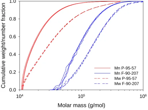
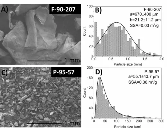
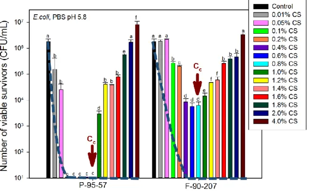
entanglement concentration. Adapted from [138] ... 17 Figure 5.1: Cumulative weight (Mw)/number (Mn) fraction as a function of molar mass for chitosan powder and flakes. ... 28 Figure 5.2: Chitosan in flakes and powder form (A, C) and their particle size distribution (B, normal distribution and D, log-normal distribution). The symbols a, b and SSA (in Figures B and D) represent the average particle size, thickness and the specific surface area, respectively. This figure has been modified with respect to the original article. ... 30 Figure 5.3: Effect of chitosan concentration in PBS on the number of viable survivors. Cc is the
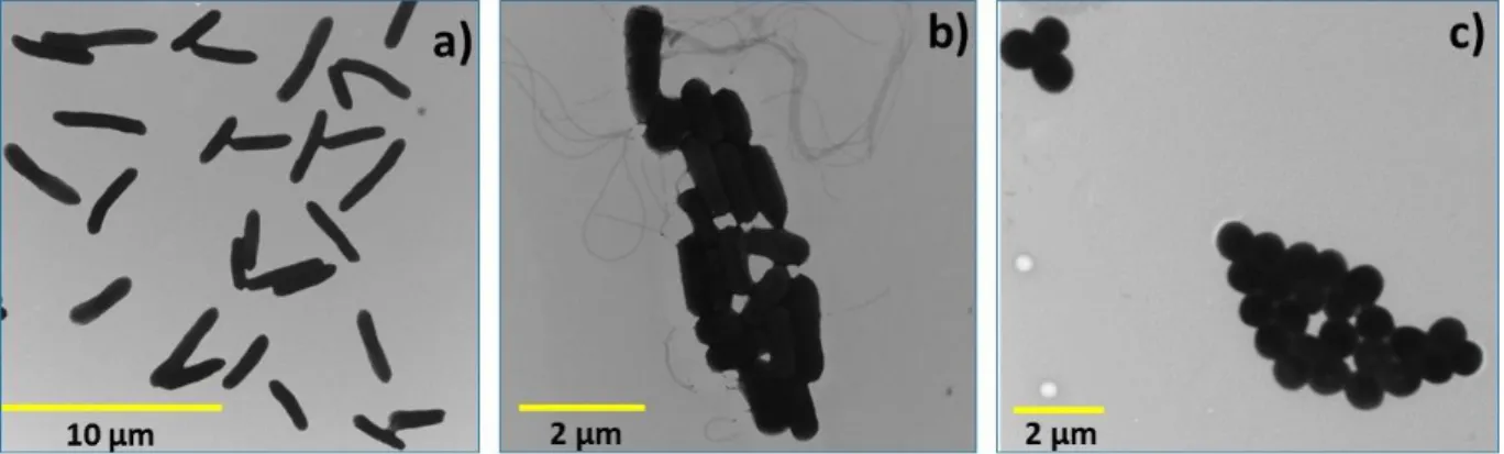
critical concentration above which the AB activity of chitosan decreases. Dashed lines represent the reduction in bacterial concentration after deproteinization. Samples are P-95-57 (powder) and F-90-207 (flakes). The number of viable organisms was the same after 18 and 48 h incubation on the agar plates, suggesting that recovery from sub-lethal injury had not taken place. For each chitosan grade, means that do not share a letter are significantly different with a confidence level of 95% by Tukey Pairwise Comparisons. ... 31 Figure 5.4: FTIR spectra: Peaks at 1345, 1420, 1560, 1655 and 3290 cm−1 confirm the solubility of chitosan powder and flakes in the suspensions during the AB tests. ... 32 Figure 5.5: Recovery of viable bacteria after exposure of chitosan and filtrate from chitosan suspensions to E. coli. The number of viable organisms was the same after 18 and 48 h incubation on the agar plates, suggesting that recovery from sub-lethal injury had not taken place. Means that do not share a letter are significantly different with a confidence level of 95% by Tukey pairwise comparisons. ... 33
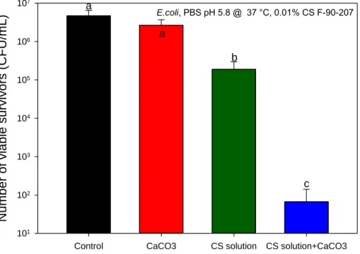
Figure 5.6: Recovery of viable bacteria after exposure of CaCO3 and CS solution (F-90-207) to E.
coli. The number of viable organisms was the same after 18 and 48 h incubation on the agar
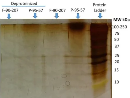
plates, suggesting that recovery from sub-lethal injury had not taken place. Means that do not share a letter are significantly different with a confidence level of 95% by Tukey pairwise comparisons. ... 34 Figure 5.7: Identification of proteins before and after deproteinization of chitosan. Samples are
P-95-57 (powder) and F-90-207 (flakes). ... 36 Figure 5.8: Effect of temperature on the antibacterial activity of chitosan (number of viable survivors). Bars with different letter are significantly different (p < 0.05). Samples are F-90-207 (flakes) and P-95-57 (powder). The number of viable organisms was the same after 18 and 48 h incubation on the agar plates, suggesting that recovery from sub-lethal injury had not taken place. Means that do not share a letter are significantly different with a confidence level of 95% by Tukey pairwise comparisons. ... 38 Figure 5.9: Effect of salt concentration and ionic strength (I) on the antibacterial activity of chitosan. Bars with different letters are significantly different (p < 0.05). Sample is F-90-207 (flakes). The number of viable organisms was the same after 18 and 48 h incubation on the agar plates, suggesting that recovery from sub-lethal injury had not taken place. Means that do not share a letter are significantly different with a confidence level of 95% by Tukey pairwise comparisons. ... 40 Figure 5.10: Morphology of intact: (a) E. coli; (b) L. innocua; (c) S. aureus cells. Images were kindly provided by Mounia Arkoun from the Chemical Engineering Department, Polytechnique Montréal. ... 42 Figure 6.1: Schematic of the electrospraying process. ... 61 Figure 6.2: SEM images showing the effect of flow rate and needle size, in terms of shear rate, on the electrospraying of 1 wt/v % (sample B1) solution in 50 v/v % AcOH, at 25 kV/10 cm. 67 Figure 6.3: SEM images showing the effect of distance and voltage, in terms of electric field on the electrospraying of 1 wt/v % (sample B1) in 50 v/v % AcOH, at 0.2 mL/h and a 22G needle. ... 69
Figure 6.4: SEM images showing the effect of distance and voltage, in terms of electric field on the electrospraying of 2 wt/v % (sample B1) in 70 v/v % AcOH, at 0.2 mL/h and a 22G size. ... 69 Figure 6.5: SEM images showing the effect of chitosan (wt/v %) and acetic acid concentration (v/v %) on the electrospraying of chitosan (sample B1), at 3 kV/cm, 0.2 mL/h and needle size 22. ... 72 Figure 6.6: SEM images showing the effect of chitosan MW and DDA on the electrospraying of chitosan, at 3 kV/cm, 0.2 mL/h and needle size 22G. The shear viscosity of the respective solutions was evaluated at the maximum apparent shear rate encountered at the needle wall (8.2 s-1) and is indicated in each case. The scale bar is the same (1 µm) for all the SEM images. ... 76 Figure 6.7: SEM images showing the effect of solvent type on the electrospraying of chitosan (sample B1): a) 1 wt/v % CS in 70 v/v % AcOH; b) 1 wt/v % CS in TFA; c) 1 wt/v % CS in LA. Process conditions: needle 22G, 3 kV/cm and 0.2 mL/h. ... 77 Figure 6.8: Dimensionless numbers establishing the process conditions in the electrospraying of CS/AcOH solutions: a) Froud number (Fr) as function of flow rate (Q), and b) Electrostatic force parameter (Ω) as function of electric field strength (E). Ω was calculated when considering R0 from needle size 22G. ... 80
Figure 6.9: Dimensionless numbers representing the solution parameters in the electrospraying of CS/AcOH solutions: Re, Pe and We as function of CS and AcOH content. Dotted lines (in green color) indicate that We is independent of CS concentration. ... 80 Figure 6.10: Conductivity of CS/AcOH solutions with different AcOH contents for various chitosan grades. ... 83 Figure 6.11: Surface tension of CS/AcOH solutions with different AcOH contents for various chitosan grades. ... 84 Figure 6.12: Viscosity of CS/AcOH solutions as a function of shear rate for chitosan grade B1. The doted lines represent the maximum shear rate at the needle wall calculated from the needle size and flow rate. ... 84
Figure 6.13: SEM images of different regions in the collected area after electrospraying 1 % CSB1
in 50 %AcOH at 0.2 mL/h and a 22G needle. ... 85 Figure 6.14: Viscosity as a function of shear rate for solutions containing chitosan with a 90 % DDA: B1 (183 kDa), B2 (207 kDa) and B3 (344 kDa). ... 85 Figure 7.1: Morphology and particle size distribution (log-normal) of chitosan a) in powder form and b) nanospheres. This figure has been modified with respect to the original article. ... 97 Figure 7.2: Antibacterial activity of chitosan under different physical forms. The number of viable organisms was the same after 18 and 48 h incubation on the agar plates, suggesting that recovery from sub-lethal injury had not taken place. For each strain, means that do not share a letter are significantly different with a confidence level of 95% by Tukey Pairwise Comparisons. Statistical analysis was done separately for S. Typhimurium and S. aureus (samples with and without *). ... 100 Figure 7.3: Influence of pH, temperature and bacterium species on the antibacterial activity of chitosan nanospheres. The number of viable organisms was the same after 18 and 48 h incubation on the agar plates, suggesting that recovery from sub-lethal injury had not taken place. For S. Typhimurium: pH and temperature significatively affects the antibacterial activity (p=0.001 and 0.001, respectively) at a confidence level of 95%. For S. aureus: pH and temperature significatively affects the antibacterial activity (p=0.001) at a 95% confidence level. ... 105 Figure A.1: Different approaches used in this work for the production of nanofibrous structures: a) direct spinning of the basic materials, b) parallel and c) coaxial electrospinning set up. .... 145 Figure A.2: SEM micrographs and their size distribution: a) BNC in film form; and mats obtained from direct electrospinning of: b) CS/PEO @ 0.5 mL/h and 2.0 kV/cm; c) BNC @ 0.3 mL/h and 3.0 kV/cm; d) BNC/PLA (1:1) @ 0.3 mL/h and 2.3 kV/cm; e) BNC/PLA (1:3) @ 0.3 mL/h and 2.3 kV/cm and f) BNC/PLA (1:5) @ 0.3 mL/h, 2.3 kV/cm and 60 ºC... 148 Figure A.3: Fiber size distribution of the mats from Figure A.2: a) CS/PEO @ 0.5 mL/h and 2.0 kV/cm; b) BNC/PLA (1:3) @ 0.3 mL/h and 2.3 kV/cm; and c) BNC/PLA (1:5) @ 0.3 mL/h, 2.3 kV/cm and 60 ºC. ... 148
Figure A.4: CS/BNC structures from parallel electrospinning and their fiber size distribution. Randomly mixed fibers of: a) CS/PEO and BNC (22 °C); b) CS/PEO and BNC/PLA (22 °C); c) CS/PEO and BNC/PLA (60 ºC). CS/PEO was electrospun@ 0.5 mL/h and 3.0 kV/cm whilst BNC and BNC/PLA blends @ 0.3 mL/h and 3.0 kV/cm... 150 Figure A.5: EDS analysis of the electrospun mats: a) CS/PEO; b) CS/PEO and BNC; c) CS/PEO and BNC/PLA. Sample a) was electrospun directly whilst samples b) and c) represent the randomly mixed fibers from the parallel electrospinning. ... 151 Figure A.6: CS/PEO-BNC core-shell structures: a) CS/PEO in the shell and BNC in the core; b) CS/PEO in the core and BNC in the shell. Core @ 0.5 mL/h, shell @ 0.3 mL/h and 2.3 kV/cm. ... 155 Figure A.7: Viscosity vs. shear rate for CS/PEO and BNC solutions. ... 156 Figure A.8: Viscoelastic properties of the CS/PEO and BNC solutions: Complex viscosity vs. frequency and tan delta vs. frequency. ... 156 Figure A.9: TEM and EDS analysis (by SEM) of the CS/PEO-BNC core-shell structure. Sample was analyzed from a TEM copper grid support. ... 157 Figure A.10: SEM image of CS/PEO-BNC mats after treatment with 50% acetic acid solution: a) CS/PEO in the core, BNC in the shell; and b) CS/PEO in the shell, BNC in the core (different magnifications). ... 158 Figure A.11: Inhibitory effects of different CS/PEO/BNC structures toward E. coli. P-parallel and C-coaxial electrospinning. The pH of the PBS solution was adjusted to 5.8 in order to protonate chitosan and activate its antibacterial activity ... 160 Figure B.1: Different stages in the electrospraying process. ... 167 Figure B.2: Momentum balance on a short section of the jet [4] ... 173 Figure B.3: Gaussian surfaces: S1 lies just inside the interface and S2 lies just outside the interface
LIST OF ABBREVIATIONS
AB Antibacterial
AcOH Acetic acid
ANOVA Analysis of variance
ATR Attenuated Total Reflectance Spectroscopy BHI Brain Heart Infusion broth
C* Critical chain overlap concentration
Cc Critical concentration
Ce Critical entanglement concentration CFU/mL Colony forming units per milliliter
CS Chitosan
CA Citric acid DCM Dichloromethane DDA Degree of deacetylation DMF N,N-dimethyl formamide
DLS Dynamic light scattering
EDS Energy Dispersive X-ray spectroscopy EDTA Ethylenediaminetetraacetic acid EVA Ethylene vinyl acetate
Fr Froude number
FTIR Fourier transform infrared spectroscopy GPC Gel Permeation Chromatography GuHCl Guanidinium chloride
HCl Hydrochloric acid
κ−1 Debye-length
KBr Potassium bromide KCl Potassium chloride KOH Potassium hydroxide
LA Lactic acid
LB Luria Bertani
MgCl2 Magnesium chloride
MW Molecular weight
NaCl Sodium chloride NaOH Sodium hydroxide
NIR Near-infrared spectroscopy
P Powder
PAGE Polyacrylamide gel electrophoresis PDI Polydispersity index
PBS Phosphate buffer saline PCL Poly (caprolactone)
Pe Electric Peclet number PLGA Poly (lactic-co-glycolic acid)
Re Reynolds number
RH Relative humidity
Ω Electrostatic force parameter SDS Sodium lauryl sulfate
SEC Size-exclusion chromatography SEM Scanning electron microscope SSA Specific surface area
TEM Transmission electron microscopy TFA Trifluoroacetic acid
TGA Thermogravimetric analysis
LIST OF APPENDICES
APPENDIX A - ARTICLE 4: CHITOSAN-BACTERIAL NANOCELLULOSE NANOFIBROUS STRUCTURES FOR POTENTIAL WOUND DRESSING APPLICATIONS ... 137 APPENDIX B - GOVERNING EQUATIONS IN THE ELECTROSPRAYING PROCESS .... 166
CHAPTER 1:
INTRODUCTION
For years, foodborne diseases have been of great concern to public health. Two factors have contributed to the appearance of new diseases as well as the re-emergence of old ones, representing a significant risk for food safety. The first one is the industrialization that has led to rapid-growing mass production and hence to an inadequate harvesting, storage, handling and transportation of food; and the second one is the globalization that has allowed international trade, facilitating the entry of pathogens that previously were not a threat for health [1, 2].
Canada is one of the countries whose population has been affected by the outbreak of pathogens resulting from contaminated food. Bacteria including Escherichia coli, Salmonella, Listeria, and
Staphylococcus have been reported in spoiled products such as raw meat and pork, tainted cold
cuts, oysters, cantaloupes, spinach and cheese, causing several pathogenic diseases [3, 4]. Foodborne infections are known to affect not only industrialized, but also non-industrialized countries [1]. The latter ones are particularly affected due to a wide range of diseases and the lack of governmental regulations regarding food inspection and handling.
On the other hand, there is an increasing public, industrial and governmental concern about food waste. About 30 to 40% of the world’s food production is wasted before consumption either because of inappropriate handling and transportation which could cause contamination with bacteria, or because the food has reached its expiry date. North American and European countries are the most affected. Just in Canada, the losses in wasted food reach $ 31 billion each year [5]. Different methods including the use of volatile gas indicators, oxygen absorbers, carbon dioxide scavengers, preservatives, moisture absorbers, among others, have been employed to extend the shelf life, maintain or improve the conditions of the packaged food and therefore to decrease food waste. Some of them, although appropriate for keeping freshness, are insufficient for keeping the food free of microorganisms. Therefore, technological solutions are being targeted to provide better public protection, decrease the economic losses for industry and reduce food spoilage. The fabrication of novel food packaging materials having antibacterial properties can be part of the solution to inhibit/eradicate pathogen growth, improving food safety and extending shelf life of food products. In this regard, chitosan is a highly promising candidate to be employed as biomaterial in food related applications, mainly due to its availability, nontoxicity and antimicrobial properties.
The present research aims towards the production of chitosan nanospheres for the formulation of antibacterial food packaging materials. To date, different studies have focused in evaluating the antimicrobial activity of chitosan solutions, gels, fibers and films [6-18]. However, micro and nanosize morphologies that allow increasing chitosan/cell interactions are on the target in recent research [19-21]. The first part of the current study focuses on the evaluation of the antibacterial activity of chitosan in a discontinuous solid form such as neat chitosan powder and flakes. Different environmental, microbial and characteristics of chitosan including pH, temperature, ionic strength, the presence of a solid physical support, bacterial species, as well as the effect of chitosan concentration and purity are investigated. The second part comprises the study of producing chitosan micro and nanospheres via electrospraying. Both processing and solution parameters are subjected to consideration. The third part compares the effect of chitosan physical form and particle size, namely solution, powders and nanospheres on the antibacterial activity, against two pathogen strains (S. aureus and S. Typhimurium) commonly found associated with foodborne infection. In our first study, chitosan in powder and flake forms reduced totally E. coli contamination, but displayed a limited antibacterial activity against the pathogen strain S. aureus. Therefore, pathogen strains, which are considered more resistant to antibacterial agents were considered relevant for testing the antibacterial activity of chitosan nanospheres. Environmental conditions such as pH and temperature are also considered. Given the remarkable antibacterial activity observed for chitosan nanospheres, the electrospraying process may provide a novel way for making chitosan-based food packaging materials. The incorporation of chitosan nanospheres into existing food packaging may be an alternative to further increase chitosan antimicrobial properties and lead to the formulation of new packaging materials. This novel packaging can impact the food industry and consumers by providing a way to inhibit bacterial growth and increase the shelf life of food products, such as chicken, beef and pork.
In addition, the results of a research project aimed at the development of new chitosan-based materials for wound healing, is presented in Appendix A. This subject was part of an NSERC-Engage project that was conducted during the accomplishment of this PhD. The antibacterial properties of chitosan can be combined with the regenerative properties of bacterial nanocellulose for potential biomedical applications. However, given that the two biopolymers cannot be solubilized in the same solvent, their processing become challenging and coaxial electrospinning was considered, as a new approach.
The main contributions of this research work are found in three scientific articles; the first one has been published in the journal Molecules, the second one has been submitted to the Journal of
Aerosol Science, and the third one has been published in the Journal of Food Science. In addition,
a fourth article is included as an appendix (Appendix A) in the thesis and reports the findings of a work performed in an NSERC-Engage project and comprises the results regarding the coaxial electrospinning of chitosan and bacterial nanocellulose for potential biomedical applications. This article has been published in the journal Cellulose.
Organization of the thesis
This thesis consists of the following chapters: • Chapter 1: Introduction
• Chapter 2: Literature review • Chapter 3: Objectives
• Chapter 4: Organization of the articles
• Chapters 5 to 7: The three articles reporting the main results of this research project • Chapter 8: General discussion
• Chapter 9: Conclusions and recommendations for future work
• Appendix A: The fourth article reporting the main results of the NSERC-Engage Project • Appendix B: Governing equations in the electrospraying process
CHAPTER 2:
LITERATURE REVIEW
2.1 Chitosan
Chitosan is a natural cationic polysaccharide derived from chitin, the second polysaccharide most abundant in nature after cellulose [22]. Chitin is a biopolymer extracted from the exoskeleton of marine crustaceans such as crab, shrimp and lobsters; cuticle of insects and cell walls of fungi, mainly [23, 24]. The shells of marine crustaceans are the more convenient sources given the availability from the wastes of the fishing industry [25]. Generally, shells contain between 15 to 40 % of chitin, 20 to 40 % of proteins and 20 to 50 % of calcium carbonate, besides pigments and metal salts as minor components [24]. Due to its low solubility in organic solvents and low chemical reactivity, chitin is usually transformed into chitosan [22, 26].
Industrially, chitosan is obtained after several processing steps as shown in Figure 2.1. First, crustacean shells are subjected to a demineralization step under acid treatment with HCl to dissolve calcium carbonate, calcium phosphate and other mineral salts. Second, an alkaline treatment with NaOH or KOH follows, to solubilize the proteins. Third, a discolouration step with NaOH and ethanol is often done to remove pigments. Fourth, after a washing and drying step, chitin is obtained. Finally, a deacetylation step under concentrated alkaline conditions (concentrated NaOH) produces chitosan, which is the most important chitin derivative in terms of applications [23, 24]. The quality and properties of chitosan products such as purity, molecular weight (MW) and degree of deacetylation (DDA) depend on the conditions and treatment used for its preparation, including the concentration of the chemicals used and the sequence and time of the treatments for deproteination, decalcification and deacetylation [26, 27]. Most commercial chitosan grades are mainly produced by alkaline deacetylation of chitin. Recently, enzymatic hydrolysis in the presence of chitin deacetylase have been used to produce chitosan [26]. However it is not yet available for industrial scale [27, 28]. When the degree of deacetylation (DDA) is higher than 50 % chitin is named chitosan [23, 29]. The DDA usually ranges from 66 to 96 %, depending on the method used for its production, whilst the molecular weight ranges from 4 kDa to 2000 kDa that is much lower than chitin, which is usually larger than 1000 kDa [26, 30-32].
Figure 2.1: Industrial extraction of chitin and chitosan.
2.1.1 Elemental analysis
Depending on the extent of deacetylation, the nitrogen content of chitin and chitosan varies from 5 to 8% [25, 33]. Table 2.1 and Table 2.2 present the elemental analysis and trace metal content, respectively, in a chitosan sample [22, 34]. In addition to carbon, nitrogen and hydrogen, elements such as aluminum, calcium, silicon and iron are present in the material itself whilst chromium and copper come from the stainless steel reactor and distilled water respectively during processing [22].
Table 2.1: Elemental analysis (%) of chitosan and chitin; N-acetyl-glucosamine reference [34]. Adapted from [22]. Polymer C H N Chitosan 39.99 6.80 7.40 Chitin 43.53 6.12 6.26 N-acetyl glucosamine 43.53 7.15 6.26
Table 2.2: Trace metal content of chitosan (ppm) [35]. Adapted from [22].
Aluminum 5-50 Calcium 10-150 Copper 2 Iron 10-100 Lead <10 Manganese <1 Silicon 10-100 Titanium <1 Silver <1 Chromium 2.2 Cobalt 0.2 Zinc 0.3
2.1.2 Chemical structure
Structurally, chitin consists of 2-acetamido-2-deoxy-β-D-glucose through a β (1→4) linkage. Chitosan is the N-deacetylated derivative of chitin, however, this N-deacetylation is almost never complete [33]. Both the sequence and the content of the N-acetyl-D-glucosamine (acetylated unit) and D-glucosamine (deacetylated unit) in chitosan determine its properties and reactivity [32]. The structures of chitin and chitosan are presented in Figure 2.2.
Figure 2.2: Structure of chitin and chitosan [25].
2.1.3 Solution properties
Due to the extensive intra- and intermolecular hydrogen bonding, and hydrophobic inter-chain interactions, chitosan degrades before melting, which makes it necessary to be dissolved in appropriate solvents [23, 25]. Chitosan is insoluble in water at neutral or basic pH values [22]. However, when the pH of the medium is lower than chitosan’s pKa (6.2-6.5) [36, 37], amino groups undergo protonation making the polymer soluble in water [23, 26]. Chitosan is soluble in organic acids including formic, acetic, citric, pyruvic and lactic acid; and in inorganic acids such as nitric, hydrochloric, sulphuric and perchloric acids [22, 24, 38].
Chitosan solubility is mainly controlled by its DDA and MW. In general, the solubility increases at high DDA and low MW values. However, other parameters such as ionization degree, ionic strength, pH, type of solvent, concentration, chain flexibility, crystallinity, distribution of acetyl glucosamine units and time and temperature of the chemical deacetylation reaction may influence chitosan solution properties [23, 38-40]. In HCl and acetic acid, chitosan is soluble for a degree of protonation around 0.5 [39]. Chitosan solubility is reported for DDA>50% [41].
The viscosity of chitosan aqueous solutions is influenced by factors such as DDA (deacetylation time), MW, concentration, degree of protonation, ionic strength, pH and temperature [26]. In general, chitosan solution viscosity decreases with temperature and ionic strength; and it increases with chitosan concentration and MW [22, 38, 42-44]. However, a pH change in the polymer
solution may give different results depending on the types of acids employed [26]. With acetic acid, the viscosity of chitosan tends to increase with decreasing pH, whereas with HCl the viscosity decreases when pH is lowered. This relates to the degree of protonation of chitosan [26, 38, 45, 46]. In the first case, at low pH, intra and electrostatic repulsions due to protonation increase, hence promoting higher solubility, chain expansion and larger hydrodynamic volumes, which increase the viscosity. In the second case, also at low pH, the presence of large amounts of ions from a strong acid cause electrostatic charge screening (of the NH3+ groups), enhancing chain flexibility
and hence decreasing viscosity [38, 46].
2.1.4 Antimicrobial properties
Chitosan exhibits suitable functional and biological properties, including antimicrobial activity [47]. Several authors have reported antimicrobial properties for chitosan under different physical forms including chitosan in solutions [6-10, 15, 16], gels [48], films [11-14, 17, 18], fibers [49-52] and micro and nanoparticles [19-21, 53-56], on different microorganism species [12, 18, 57, 58]. Literature reports chitosan antimicrobial effectiveness not only on inhibiting the growth of Gram-positive and Gram-negative [6, 59] bacteria but also yeast and molds [60]. The main difference between Gram-positive and Gram-negative strains is their membrane structure. Gram-positive strains are composed of one phospholipid bilayer and a thick peptidoglycan, whilst Gram-negative consist of two phospholipid bilayers and a thin peptidoglycan between them [51]. The antimicrobial properties of chitosan are affected by different factors: 1) microbial species and cell age [6, 61-63]; 2) environmental conditions such as pH, ionic strength, temperature and time [61, 63, 64]; 3) chitosan characteristics including source, MW, DDA, concentration and physical form [6, 16, 61, 62, 64-69]; among others.
The antimicrobial properties of chitosan have been used for food preservation. Ouattara et al. [14] demonstrated delayed growth or the complete inhibition of different kind of bacteria (including Enterobacteriaceae and Serratia liquefaciens) in processed meats covered with chitosan films and stored at 4°C up to 21 days. Bhale et al. [65] showed that albumin quality and eggs conservation increased by three weeks at 25 ºC in the case of eggs covered by chitosan coating solutions (pH 5.6). Coated eggs were overall acceptable to the consumer and did not differ from the control and commercial eggs regarding the organoleptic properties. Yingyuad et al. [60] dipped grilled pork samples in chitosan solutions with different chitosan concentrations and vacuum packaged. Their
results showed that the shelf life increased by at least two weeks compared to the samples that were not dipped. Throughout the storage period samples were found to be organoleptically acceptable and without significant changes in odour and colour.
Different pathogenic organisms including Escherichia coli, Listeria and Salmonella have been reported as being inhibited by chitosan, in contaminated beef, ham, turkey, sausages, pork and fish products [16, 60-62, 70, 71]. Chitosan also protected mice against infection by Listeria
monocytogenes, suggesting that the antimicrobial action may also occurs in humans [72].
Muzzarelli et al. [59] reported the antibacterial effect of carboxybutyl chitosan on 298 pathogen strains including Gram-positive and Gram-negative bacteria and observed morphological damage of the cell wall and the internal cell structures. The bactericidal activity between the tested strains was different which suggested that different mechanisms of action of chitosan might be operating. However, the experiments were carried out with only a chitosan derivative, and results might differ in case of using unmodified chitosan.
Chitosan is also reported to be effective against yeast and molds. Roller and Covill [73] studied the antifungal properties of chitosan against 15 microorganisms (8 yeast and 7 filamentous fungi) associated with food spoilage, at different chitosan concentrations, pH and temperature conditions. Concentrations of 0.1, 1 and 5 g/l of chitosan were necessary to reduce the growth of
Zygosaccharomyces bailii, Mucor racemosus and Byssochlamys, respectively. El Ghaouth et al.
[74] reported the antifungal activity of chitosan coating on strawberry fruits inoculated with
Botrytis cinerea and Rhizopus stolonifer. Although a complete inhibition of fungal growth was not
achieved, chitosan coating reduced significantly spore germination and radial growth, and indicated that chitosan was fungistatic rather than fungicidal. These results assess the potential use of chitosan as natural food preservative.
Although the exact mechanism of the antimicrobial action of chitosan and its derivatives is still unknown, different mechanisms of growth inhibition have been proposed in literature [64, 75]: 1) Electrostatic interactions: at pH<pKa, chitosan amino groups are protonated, creating electrostatic interactions with the anionic cell wall components (lipopolysaccharides and proteins) of the microorganism.
2) The hydrophobic and chelating effects occurring at pH>pKa. N-acetylated chitosan and the presence of a long aliphatic chain in chitosan can favor the absorption onto cell walls via
hydrophobic interactions with cell wall proteins. As chelating agent, chitosan can selectively bind metals ions of the cell wall or from the medium, affecting bacteria stability and growth, respectively.
3) Inhibition of the synthesis of mRNA and DNA transcription, produced when small particle sizes and low molecular weight chitosan penetrate the cell wall of bacteria and combines with DNA. 4) Alteration of permeability, when high molecular weight chitosan or large size particles interact with cell surface blocking the entrance of essential nutrients to the cell or by forming an impermeable layer.
These mechanisms will eventually result in biological changes and the leakage of intracellular components, leading to cell death [32, 64].
As mentioned in 3) and 4), chitosan molecular weight can determine the mechanism of antimicrobial action, but also, the mechanism depends on bacterial type. Chitosan with molecular weight greater than 10 kDa have been found to inhibit bacterial growth regardless of its type [13, 66, 76]. Different studies [6, 10, 13, 67, 77] have demonstrated that, in general, chitosan antimicrobial activity increases with molecular weight when tested against Gram-positive bacteria, while for Gram-negative this activity is more efficient at low molecular weights.
With respect to molecular weight, two different mechanisms for the antimicrobial activity have been suggested: in Gram-positive bacteria, chitosan on the surface of the cell can form a polymer membrane, which inhibits nutrients from entering the cell. In Gram-negative ones, chitosan of low molecular weight might enter the cell through penetration, disturbing the physicochemical activities of bacteria cells until cell death [10, 78].
In addition, some authors reported higher effectiveness of chitosan against Gram-positive than Gram-negative bacteria, presumably due to the presence of lipid outer membrane in the latter that can be a barrier to chitosan. Others consider that Gram-negative bacteria are more sensitive to chitosan bioactivity because the negative charge on the cell surface of this bacteria is stronger than in Gram-positive, leading to more chitosan adsorbed and hence to a higher inhibitory activity [6, 7, 61, 63, 67, 75, 79].
Recently, Arkoun et al. [51] investigated the antibacterial mechanism of action for chitosan nanofibers. The results showed that chitosan nanofibers were both bacterial membrane
permeability disrupter and perforator. A series of four steps were proposed as the main mode of action for chitosan nanofibers. First, the adsorption of chitosan nanofibers onto bacterial surface; second, the perforation of the membrane; third, the leakage of cytosolic compounds including enzymes, proteins and DNA; and fourth, cell lysis and disintegration.
2.1.5 Processing
Chitosan is generally purchased in the form of fine powder, flakes or in solution, and can be processed into different physical forms through solubilisation in acidic media or by using an adequate solvent. Forms such as films [71, 80-83], fibers [84-89], micro and nanobeads [20, 31, 90-101], hydrogels [102-104], membranes [105] and sponge (foams) [106] of chitosan alone or in blends [105, 107-109] are produced by several technological processes for applications in different fields [110]. For instance, chitosan film is the most studied physical form with potential use as packaging material, given its promising results regarding the antibacterial activity [13, 18]. However, chitosan films have low mechanical and barrier properties, and therefore need to be blended with another polymer for the improvement of these properties [111, 112]. Chitosan films are generally produced by the solution casting/solvent evaporation method [60, 113], or by blending with a thermoplastic polymer via twin-screw extrusion [114-116].
Chitosan beads in the form of micro and nanospheres are of great interest in the biomedical field and are increasingly studied for drug delivery applications because of their small size and the large surface to volume ratio. Although the size of a nanoparticle is not clearly defined in literature, ASTM E2456-06 defines a nanoparticle as an ultrafine particle with at least one dimension in the range between 1 and 100 nm [117].
Several methods have been employed to produce micro and nanospheres of chitosan. The most used ones are emulsion cross-linking, coacervation/ precipitation, spray-drying, emulsion droplet coalescence, ionic gelation and electrospraying [30]. Others like reverse micellar [118] and sieving methods are less used [31]. The selection of any method depends upon factors such as particle size requirement, chemical and thermal stability and residual toxicity associated with the final product [31].
2.1.6 Applications
The unique properties of chitosan, including its nontoxicity 1 [26, 119], natural origin, biodegradability [26, 57], availability as well as the wide range of physical forms in which it could be transformed, are the main reason for its multiple applications. In particular, its antimicrobial properties [10, 16, 18, 63, 64, 120-122] and biocompatibility [6] make it of great interest in food packaging and biomedical applications, respectively. Chitosan has been approved as a food ingredient in Japan, Korea, Finland and Italy. In North America and Europe, it has received the “Generally recognized as safe” (GRAS) status by the US Food and Drug Administration (FDA), and has been used as food additive, nutritional supplement, natural health product and in drugs [32, 123, 124] . Other main applications of chitosan-based materials are cited in Table 2.3.
Table 2.3: Main applications for chitosan
Field Application References
Food sector Food additive and preservative, antioxidant, emulsifying, thickener, cholesterol reductor, antibacterial coating, food packaging, dietary supplements.
[23, 26, 51, 61, 68, 71, 125, 126]
Biomedical Anticoagulant, hemostatic, wound healing, wound dressing, encapsulation, antitumor, bone and cell culture, gene therapy, controlled drug delivery, engineering scaffolds.
[6, 23, 89, 106, 127-130]
Agriculture Pesticide, fungicide, soil enrichment, seed coating. [23, 71, 122, 131, 132] Water and waste
treatment
Membranes for water filtration (removal of metal ions such as Hg, Cd, Ni, Zn).
[23, 133, 134]
1 On accounting of the low lethal dose (LD
50) value, which is in the same order of magnitude as the LD50 for sugar (16
2.2 Electrospraying process
Electrospraying is a method of electrodynamic atomization. Its principle is based on the theory of deformation of charged droplets developed by Lord Rayleigh in 1882, Zeleny in 1917 and Sir Taylor in 1964 [135, 136]. The schematic of a typical electrospraying setup is illustrated in Figure 2.3. The device consists of three main parts: a syringe pump that control the flow rate of the polymeric solution, a voltage generator (0 to 50 kV) and a metallic collector plate.
In electrospraying, a high voltage electric field is applied to a polymeric solution that is pumped through a syringe. The electric charge generated on the exiting liquid makes the droplet deforms into the shape of a Taylor cone from which a jet is ejected and elongated [92, 137, 138]. When the electrostatic forces in the fluid overcome the surface tension, the jet breaks into nano and micro droplets that eventually travel to a collector plate [30, 92, 139-141]. Different spraying modes can occur during electrospraying, the single cone-jet mode is the most desired because of its stability and reproducibility. Once the droplets are separated, the solvent is evaporated. Due to Coulomb repulsion of the charges, the droplets are well dispersed during the process [138].
Electrospraying is a relatively novel technique for the production of micro and nanospheres and its understanding and optimization are still under development [138]. There are four advantages in the use of electrospraying with respect to other conventional methods for the production of micro and nanoparticles: 1) one step processing which usually does not require extra drying; 2) no need for external dispersion/emulsion phase containing undesirable materials (generally toxic) for biomedical and food applications; 3) no high temperatures as in spray drying; and 4) better control over particle size, particle size distribution and morphology than in other methods. In the case of chitosan, which has the advantage of being soluble in aqueous acidic solutions, it avoids the use of hazardous organic solvents for the fabrication of micro and nanoparticles [92, 137].
Figure 2.3: Electrospraying process
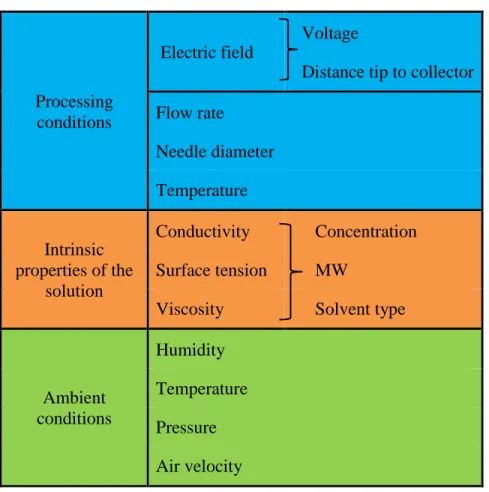
2.2.1 Parameters of the process
Table 2.4 shows the electrospraying processing parameters. Important parameters are not only polymer and solution properties such as molecular weight, concentration, viscosity, conductivity and surface tension, but also specific parameters of the electrospraying machine such as applied electric voltage, flow rate, tip-to-collector distance, temperature, needle gauge and the environmental conditions. All these parameters may affect the processing, size, morphology and reproducibility of the produced micro and nanospheres [86, 138, 142-144].
Table 2.4: Parameters for the processing via electrospraying
Processing conditions
Electric field
Voltage
Distance tip to collector Flow rate Needle diameter Temperature Intrinsic properties of the solution Conductivity Concentration Surface tension MW
Viscosity Solvent type
Ambient conditions Humidity Temperature Pressure Air velocity 2.2.1.1 Processing conditions
Electric field strength. The electric field strength (kV/cm) generally varies between 1 and 5 kV/cm.
The currents that flow during the electrospraying process range from a few hundred nanoamperes to microamperes [145]. Voltages between 15 to 30 kV and distances between 5 to 25 cm are usually used. Short distances between needle and plate collector can generate a wet deposition of particles, causing coalescence of droplets. The shortest distance is determined as the distance that produces a shortcut. The optimum distance is the one that assures the complete evaporation of the solvent, the collection of the particles and a morphology with a narrow polydispersity [92, 137, 138].
Flow rate. This parameter along with solution parameters (polymer concentration and molecular
weight, solvent type and conductivity) can control polymer entanglements and particle formation. A high flow rate can cause the formation of secondary and satellite droplets and produce particles with high polydispersity. An optimal value for the flow rate is essential in order to achieve higher
evaporation of the solvent and to avoid droplets clustering. [138]. Literature reports a wide range of values from 0.003 to 30 mL/h, for the electrospraying of different materials [146].
Needle gauge. Needle diameter is inversely proportional to its gauge. This is an important
parameter in order to avoid coalescence of droplets and to have a reasonable collection and better particle formation. While lower needle gauge produces sputtering and broader size distributions, higher gauges allow narrowing the particle size range. Common needle gauges lie between 16 and 26 (1.194 and 0.260 mm, internal diameter, respectively) [137], although lower sizes in the order of 100 µm of internal diameter have also been reported [145].
Temperature. In general, the electrospraying process is conducted at ambient temperature. An
increase in solution temperature can be favorable to the process because it decreases the surface tension and the viscosity of the solutions. An increase in temperature can also lead to a faster evaporation of the solvent, affecting particle size and morphology [147].
2.2.1.2 Intrinsic properties of the solutions
In general, variables such as polymer and solvent concentration, molecular weight, DDA (in the case of chitosan) affect the viscosity, surface tension, conductivity and chain entanglement, and influence the electrospraying process [143, 144].
Polymer concentration. As opposed to electrospinning where higher polymer concentrations are
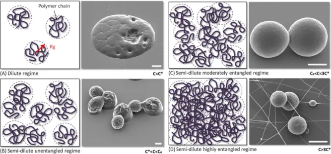
employed to form continuous fibers, the electrospraying process requires relatively low polymer concentrations to generate micro and nanoparticles [137]. Two concentrations, the one associated with critical chain overlap, C*, and that for critical entanglement, Ce, determine the production of
fibers or spheres [143] (see Figure 2.4). A dilute regime is obtained when polymer concentration is less than C*, where there is no entanglement formation (Figure 2.4a). At higher polymer concentrations, the semi dilute unentangled regime is achieved and some entanglements are observed, but they are not sufficient to maintain the morphology of the droplets (Figure 2.4b). Well-defined droplets are obtained at the semi-dilute moderately entangled regime (Figure 2.4c), at polymer concentrations in the range Ce<C<3C*; beyond this concentration, fiber emergence is attained [84, 92, 138, 143]. Stable fiber formation occurs at more than 2.5 entanglements per chain, or as C>>C* [143, 148] and increases with chitosan concentration [142].
Figure 2.4: Physical representation at the molecular level of various entanglement regimes obtained for different polymer concentrations. C*: critical chain overlap concentration, C
e: critical
entanglement concentration. Adapted from [138]
Polymer molecular weight. In the same manner than concentration, molecular weight affects the
value of the critical concentration at which beads and fibers are obtained from the process; however, this concentration should be below the one of process limiting viscosity [86]. Both molecular weight and concentration influence particle size and morphology owing to viscosity [149, 150]. Beads and fibers are generally formed at low and high molecular weights, respectively [86]. Molecular weight has also a significant effect on the rheological and electrical properties such as surface tension, viscosity and conductivity.
Solvent. Solvents with high vapor pressure (low boiling temperatures), which are highly volatile,
are preferred in electrospraying. However, a fast evaporation of the solvent could hinder the diffusion of the polymer in the electrosprayed droplets and may lead to the formation of pores and hollow particles. The solvents more frequently used in electrospraying are acetone, acetic acid, acetonitrile, chloroform, 1,2-dichloroethane, dichloromethane (DCM), ethanol and N,N-dimethyl formamide (DMF), which can be used alone or combined [138]. Solvent concentration affects the morphology of electrosprayed droplets. Deposited particles with irregular shape can be obtained due to the solvent that is not evaporated before droplets reach the collector plate [138]. However, low concentrations of solvent may produce a phenomenon called sputtering, which affects process
stability and droplets deposition [137]. Remaining solvent content must be within the limits of safety standards. Solvent has also an important role in determining the conductivity of the electrosprayed solutions, a parameter that must be taken into account for the optimization of the process [138].
Viscosity. Viscosity along with conductivity are the dominant solution characteristics for the
electrospraying process [142]. Spinning or spraying can be achieved depending on this value [144]. In general, low viscosity polymer solutions allows for droplets formation. However, relative low concentrations and hence low viscosities could limit the formation of the spherical shape and hinder the shrinkage of droplets during solvent evaporation [138, 151]. High viscosity solutions shift the cone-jet mode to higher voltages. Particle size can be controlled by the solution viscosity and operation parameters. A decrease in viscosity or an increase in conductivity decreases the particle size [138].
Surface tension. Surface tension is mainly affected by the solvent type and concentration, while
being less influenced by polymer concentration [86]. A low surface tension of the solution along with a low conductivity is needed to obtain a stable process and the single cone-jet mode [138, 142]. Droplet and fiber formation can be determined by means of this parameter [86].
Electrical conductivity. Low electrical conductivity values are preferred in order to obtain the
single cone-jet mode and a stable process [138, 142]; however, too low values can be unfavorable for the process [149] and a value of at least 0.01 µS/m is required for the current to flow [138]. When small particle sizes are required, an increase in the conductivity can lead to their formation due to enhanced Coulomb repulsion, but sufficient viscosity is needed to ensure that entanglement forces remains higher than the Coulomb ones to maintain the stability of the process [138, 142]. The solvent is the main parameter influencing the conductivity of the solutions [138].
2.2.1.3 Ambient conditions
Parameters such as field strength, conductivity and flow rate of the polymer solution affect the stability of the jet cone. Morphology and size of the droplets are mainly affected by concentration and molecular weight of the polymer, flow rate, vapor pressure of the solvent, electrospraying distance and environmental conditions of the chamber, including humidity, temperature, atmospheric pressure and air velocity [138].
High temperature conditions promote the rapid removal of the solvent affecting the particle size and shape. The rate of solvent evaporation and the solidification of the electrosprayed particles can also be influenced by atmospheric pressure, humidity and air velocity [152].
2.2.2 Governing equations of the process
The steady jet in electrospraying is governed by four steady state equations representing the conservation of mass and electric charges, the linear momentum balance and the Coulomb’s law for the electric field [153, 154]. The complete development of the equations is shown in Appendix B.
• Mass conservation:
𝑣𝜋𝑅
2= 𝑄
• Electric charge conservation:
𝜋𝑅
2𝐾𝐸 + 2𝜋𝑅𝑣𝜎 = 𝐼
• Momentum conservation:𝜌𝑣𝑣
′=
𝑇′
𝜋𝑅
2+
𝜎𝜎′
𝜀̅
+ (𝜀 − 𝜀̅)𝐸𝐸
′+
𝛾
𝑅
2𝑅
′+ 𝜌
g+ 2
𝜎𝐸
𝑅
• Coulomb’s law for electric field:𝐸(𝑧) = 𝐸
∞− ln (
𝐿
𝑅
0) [
4𝜋
𝜀̅
𝑑(𝜎𝑅)
𝑑𝑧
−
1
2
(
𝜀
𝜀̅
− 1)
𝑑
2(𝐸𝑅
2)
𝑑𝑧
2]
where (in order of appearance) 𝑣 is the axial velocity of the jet, 𝑅 is the radius of the jet, 𝑄 is the flow rate, 𝐾 is the conductivity of the solution, 𝐸 is the axial component of the electric field inside the jet evaluated at the surface, 𝜎 is the surface charge density, 𝐼 is the current carried by the jet, 𝜌 is the solution density, 𝑇 is the tensile force in the jet, 𝜀̅ is the dielectric constant of the air, 𝜀 is the dielectric constant of the jet, 𝛾 is the surface tension, g is the gravity, 𝐸∞is the external electric field, 𝐿 is the length of the jet, 𝑅0 is the initial radius of the jet (needle), 𝑧 is the flow direction, and
2.2.3 Electrospraying of chitosan
Many studies have successfully synthesized uniform particles of polymers such as poly (lactic-co-glycolic acid) (PLGA), poly (caprolactone) (PCL) and ethylene vinyl acetate (EVA) via the electrospraying process [92, 139-141]. However, relatively few studies have considered the electrospraying of chitosan solutions [137, 142, 155], despite the extensive investigations published regarding its electrospinnability and fiber formation [86, 89, 156-159].
The electrospraying of chitosan can be considered as a promising technique for the synthesis of defined size micro and nanoparticles. One of the main advantages is that chitosan is soluble in acetic acid, which is a nontoxic solvent with a low vapor pressure, which is desirable for the processing. Other advantages regarding the processing and final morphology obtained were exposed in Section 2.2. In addition of being a one-step process that does not require extra drying steps nor high temperature conditions, electrospraying allows to have better control over particle size, particle size distribution and morphology than with other conventional methods for micro and nanoparticle formation. However, the number of parameters for its optimization can be a complex task, and is a subject of ongoing research.
To date, studies relating the electrospraying ability of chitosan have been mainly focused on the determination of the processing parameters for particle formation [137, 142, 155]. The study of the intrinsic properties of chitosan solutions for the optimization and the complete understanding of this process started to be addressed recently. Kuo et al. [155], Arya et al. [137] and Zhang et al. [142] evaluated the effect of different processing or solution parameters to control particle size and particle size distribution. Their studies comprised the analysis of one chitosan grade characterized by a given MW and DDA. Chitosan particles with sizes in the range of 124 to 940 nm in diameter were reported. Recently, Gómez-Mascaraque et al. [160] described the effect of chitosan molecular weight on the electrospraying of chitosan microspheres, and its correlation with electrical conductivity, viscosity and surface tension of the solutions. No processing parameters were studied and chitosan with only one DDA was considered in their study. In addition, the viscosity of the solutions was studied at a particular shear rate of 200 s-1, which is far from the one calculated,
given the flow rate and needle size, in real electrospraying conditions (would be around 0.6 s-1
![Table 2.2: Trace metal content of chitosan (ppm) [35]. Adapted from [22].](https://thumb-eu.123doks.com/thumbv2/123doknet/2323223.29580/29.918.328.588.390.753/table-trace-metal-content-chitosan-ppm-adapted.webp)