C
e
ntre
int
e
runi
ve
rsit
aire
de rec
h
erche sur la scien
c
e
et
la
te
c
hn
o
lo
g
ie
Note
de recherche
Signs, Markers, Profiles, and Signatures:
Clinical Haematology Meets the
New Genetics (1980-2000)
Peter Keating
Alberto Cambrosio
Pour se procurer des copies de cette note de recherche communiquer avec les chercheurs:
Adresse postale: CIRST UQAM
C.P. 8888, Succursale Centre-ville
Montréal, Québec
Canada, H3C 3P8
Adresse civique: CIRST UQAM
Pavillon Thérèse-Casgrain , 3e étage
455, boul. René-Lévesque Est, Bureau W-3042
Montréal, (Québec) Canada
H2L 4Y2
Téléphone (secrétariat du CIRST): (514) 987-4018 Télécopieur (secrétariat du CIRST): (514) 987-7726 Courrier électronique: CIRST@uqam.ca
Abstract
We propose that a fruitful way to understand the extension of molecular biology into clinical practice is through the notion of biomedical platform. In this article, we analyze the development of the new genetics in clinical haematology since the mid-1980s focusing on two recent instantiations of the molecular biology platform: RT-PCR and DNA microarrays. We show how clinical research is more closely entwined with ‘fundamental’ biology than is often imagined and simultaneously caution that the use of techniques in clinical research does not automatically entail their use in routine clinical practice. The article calls attention to the work of articulation and regulation as constitutive aspects of a platform that enters standard clinical use.
Signs, Markers, Profiles, and Signatures: Clinical Haematology Meets
the New Genetics (1980-2000)
The phrase “Someday simple molecular tools will provide an objective and quantitative diagnosis for Pap smears and/or other cytologic specimens” is getting old. ... NOTE ADDED IN PROOF: It has now occurred that a molecular method correctly classified tumors without morphological information. (Rimm, 2000, pp. 1 and 9)
Introduction
This article analyzes the operational dynamics of new genetic technologies at the interface between laboratory and clinical work. In a very general sense, it focuses on the relations between technology and medicine. There are, however, many different ways of pursuing such a broadly defined analytical agenda. Consider, for example, Wailoo’s (1997) discussion of how medical technologies modified disease definitions in 20th century and, in particular, of how the technologies implicated in the development of haematology participated in the construction and the transformation of the various diseases (e.g. sickle cell anaemia, leukaemia, and so on) constitutive of the specialty, the patients targeted by diagnosis and therapy, and, finally, the practitioners themselves. Wailoo intends to show that these technologies acquired as much of their meaning and power from the cultural context of their use as from their use per se, or, in other words, that the technologies were shaped by social, political and cultural forces.
Our approach is less encompassing: we are not so much interested in technology as a bridge between society, culture and medicine, as in the material culture of medicine itself. Moreover, in contrast to long-range historical surveys (e.g. Reiser, 1978), we seek to investigate how, in recent decades, specific kinds of technologies have participated in the articulation of biological and clinical practices and thus in the establishment of the novel material, epistemic and institutional configurations that, taken together, characterize the post-WWII domain of practice known as biomedicine.
Let us briefly consider the context created by biomedicine. Whereas in the early 20th century several areas of medical diagnostics became routinely based on technologies derived from chemistry and physics (Howell, 1995), in the second half of the century, and despite the contributions of nuclear medicine and biophysics (Blume, 1992; Lenoir & Hays, 2000), biomedical technologies have become relatively independent of physics and chemistry. Indeed, the post-war alliance of biology and medicine has made possible a new kind of medical technology that is truly biomedical (Thomas, 1972): generated within biology and medicine, these biotechnologies use fragments of living systems such as cells, enzymes, antibodies and gene segments (Rheinberger, 2000; de Chadarevian & Kamminga, 1998). One of the consequences, but also one of the causes of the use of biological tools and variables in the diagnosis of diseases, and thus of the renewed alignment of the biological and the medical, has been that the technologies for the investigation of the normal and the pathological have become increasingly the same (Keating & Cambrosio, 2003).
Historical studies have shown, for example, that an experimental system instituted for the study of cancer can easily become a system for the study of protein synthesis (Rheinberger, 1997). As medical problems thus find their solution through biological innovation, the general perception is that the transfer of concepts and techniques from fundamental biology to medical practice has been vastly accelerated, a perception embodied in the notion of translational research (e.g. Chabner, Boral & Multani 1998). Conversely, observers have also noted that medical problems have prompted many fundamental biological investigations (Sinding, 1991). Finally, clinical trials, the hallmark of so-called evidence-based medicine, are no longer restricted to the testing of therapies. Since the 1960s, an expanded notion of clinical trial now includes the study of biological (genetic) markers and etiological research. No longer solely a determination of the efficiency of treatment, clinical trials are simultaneously an exploration of human biology (Löwy, 1996; Keating & Cambrosio, 2002; forthcoming).
In order to capture the new dynamics of biomedical innovation, sociologists of science have advanced a number of analytical categories. Consider, first, Fujimura’s (1992; 1996) notion of standardized packages, which she defines as ‘theory-methods packages’ whose description shows “how tools, practices, and theories circulate through and across worlds of practice … and both change and are changed by their circulation”. In practice, Fujimura’s application of her concept results in the recapitulation of the actors’ narratives, insofar as it tells the story of how biomedical innovations (in her case, oncogene theory and related techniques) emerged from within a few elite biology laboratories and spread, as a pre-packaged technique, to lower-level clinical laboratories (for a similar criticism, see Gaudillière, 1993; Morange, 1997). In other words, Fujimura’s “theory-methods package”, because it adheres so closely to the actors’ accounts, becomes little more than a description of a linear process of innovation leading from the laboratory to the clinic. Such a narrative omits all mention of clinical work in the innovation process itself. In this sense, standardized packages do not so much circulate as drive the teleological accounts offered by participants.
Rheinberger’s (1997) notion of ‘experimental system’ avoids this pitfall insofar as it takes into account the previously mentioned fact that, for instance, an experimental system instituted for the study of cancer can easily become a system for the study of protein synthesis. In this sense, it crosses the biology-medicine divide. Experimental systems also cross the science/technology divide, insofar as the difference between science (epistemic things) and technology is generated within an experimental system. While experimental systems enter into the description of biomedical research, they do not, however, cover the whole range of biomedical practices that stretch from the clinic to research. In particular, they do not account for the regulation of the epistemic things they generate once they have been translated into routine practices.
In order to overcome these problems, we recently introduced the notion of biomedical platforms as a means to describe the constitution of medical innovations and routines in the post-WWII era (Keating & Cambrosio, 2000; 2003).
Biomedical Platforms
Briefly put, we claim that, since WWII, biology and medicine have come together, both institutionally and intellectually, in a hybrid practice that is neither syncretic nor synthetic. We argue, in other words, that a new way of conducting research in biology and medicine has emerged. The new, in this instance, is characterized by a pathology that is aligned but not fused with the normal and a problematic space of scientific representation (Rheinberger, 1997, pp. 102-13) that is the result of this realignment. Within this space, truly biomedical entities—e.g., cell surface markers, oncogenes, DNA microarray signatures, etc.—exist as both normal biological entities and as pathological signs, i.e., as biomedical substances with regards to their origins, their uses and their meanings. In this context, we define biomedical platforms as material and discursive arrangements that act as the bench upon which conventions concerning the biological or normal are connected with conventions concerning the medical or pathological. We use this category to describe a range of activities in contemporary biomedicine running from laboratory research to clinical trials and routine diagnosis.
As an example of what we mean by biomedical platforms, consider Figure 1, a representation of a hierarchical set of procedures used to diagnose lymphoid tumours first published in 1991. Confronted with a patient presenting telltale clinical signs, a clinician will request a biopsy. The resulting tissue sample will first be examined by a trained pathologist who, with the help of a microscope and histochemical dyes, will investigate the architectural arrangement and the shape and size of the cells in the sample (Foster, 1961). This morphological analysis can lead directly to a final diagnosis. But morphological analysis is often—and in difficult cases routinely— supplemented by immunophenotypic analysis. For this investigation, panels of standardized antibodies are combined with the use of computerized, laser-based equipment to detect the presence or absence of distinctive cell surface molecules indicative of the diagnostic sub-group of the disease. An additional more recent diagnostic step—that will be the focus of this article— is provided by molecular genetics. Suspicious cells are examined for culpable DNA fragments and genes using techniques such as, in their chronological order of appearance, Southern Blot, RT-PCR and DNA microarrays. Figure 2 depicts the same process as Figure 1 about 10 years later, with additional specifications concerning molecular genetics.
FIGURE 1
A hierarchical representation of sequential steps in the diagnosis of lymphoid tumours in 1991. Reprinted from Whittaker & Willman (1991, p.124), © 1991, with permission from the United States and Canadian Academy of Pathology.
FIGURE 2
Sequential steps in the diagnosis of lymphoid tumours in 2000, with a focus on molecular genetic techniques. Reprinted from Arber (2000, p. 186), © 2000, with permission from the Association for Molecular Pathology and the American Society for Investigative Pathology.
The three scenarios are each characterized by a distinctive configuration of techniques, instruments, reagents, diagnostic and laboratory skills, organic entities (cell morphologies, cell markers, genes), spaces of representations, diagnostic, prognostic and therapeutic indications and related etiological accounts. Each of these configurations constitutes a separate biomedical platform, so that it is possible to speak of a morphological platform, an immunophenotypic platform and a molecular genetic platform. Although platforms emerge in chronological order, the development of a new platform does not result in the replacement of the previous ones. Rather, new platforms are articulated and aligned in complex ways with the pre-existing ones, and thus integrated into an expanding set of clinical-biological strategies.
Platforms cannot be reduced to mere technology or infrastructure (Star & Ruhleder, 1996), insofar as they generate and reproduce specific etiological accounts and biomedical entities. Consider, for instance, the immunophenotyping (IPT) platform. The term IPT, as previously mentioned, refers to the investigation of distinctive markers or antigens on the surface of cells using antibodies that selectively bind to these substances, fluorescent dyes conjugated to the antibodies to visualize the bound antibodies, and sophisticated equipment to do the visualizing and counting, in the simplest case a fluorescence microscope, but in most cases a computerized apparatus able to count thousands of fluorescent cells per second. IPT, however, began in the field of leukaemia as a search for unique cell surface markers that were, in other words, distinctive diagnostic signs for cancer cells. Soon, however, IPT was translated into a new etiological account of leukaemia, according to which cancer cells are not mature cells that have undergone some process of degeneration (a stance attributed to morphological pathology), but are, in fact, normal cells whose development has been arrested at a stage prior to final maturation. The markers detected by IPT are thus ‘normal’ markers of cells frozen at a given developmental stage. One would thus no longer speak of leukaemia-specific markers but, rather, of leukaemia-associated markers. In other words, IPT aligned biological and pathological variables so that it became possible to speak, in a strong sense, of a biology of leukaemia. Figure 3 is a visual representation of this etiological account: each cell differentiation stage (pre-B-cell, early-B-cell, etc.) is characterized by a list of markers expressed at that stage; the black ribbon running above the cells features the names of the specific kinds of leukaemia (pre-B-ALL, B-ALL where ALL means Acute Lymphoblastic Leukaemia, etc.) that correspond to a maturation arrest at a given stage. A single differentiation path thus accounts for both normal and pathological developments.
FIGURE 3
Immunophenotypic representation of normal and pathological lymphoid differentiation. See text for explanations. Reprinted from Van Dongen & Adriaansen. (1996, p. 91), © 1996, with permission from Elsevier Science.
Given that the alignment of the normal and the pathological is a central feature of biomedical platforms, it is not surprising to find striking parallels between the accounts generated by the IPT platform and by the succeeding molecular genetic platform. Consider, for example, a recent article published in the Health & Science section of a daily newspaper describing advances in the molecular genetic analysis of tumours (Pollack, 2002). Readers are told that while tumours were traditionally classified primarily according to their anatomical site of origin (e.g., lung, colon, breast, blood cancers), genetic analysis shows that tumours developing in different anatomical locations may share the same genetic characteristics (or ‘signature’) and thus be, in fact, the same disease. The underlying story here is that an old method or platform (pathological anatomy) is about to be replaced by a new platform (molecular genetic analysis). Similar claims emerged when the IPT platform was initially developed: then, researchers suggested that morphologically distinct (sub)types of cancer might in fact belong to the same category as that defined by the use of immunological techniques targeting cell surface markers. Both of these revolutionary claims (insofar as they threatened to relegate time-honoured pathological classifications and techniques to the dustbin of medical history) confronted, however, powerful counterclaims according to which the clinical considerations entrenched in morphological and anatomical distinctions remained critical components of the biomedical enterprise. In concrete terms:
the pathologists quoted in the 2002 newspaper article pointed out that even if tumours belonged to the same genetic category, they could very easily grow differently in a different anatomical environment, and would thus have different clinical courses. Indeed, as shown by Figures 1 and 2, the new platforms were hierarchically subordinated to the previous ones, according to an “algorithm” that gave diagnostic precedence to morphology over IPT and molecular genetics, in a context where these platforms could not always be aligned.
These remarks suggest that the alignment of the clinical and the biological is an open-ended problem and that, rather than a distinctly defined domain, biomedicine is an ongoing project. In this paper we would like to explore this project further by examining one of the latest biomedical platforms to emerge, molecular genetics. Two general restrictions apply to our present paper. First, we have chosen to concentrate on the constitution of the molecular genetics platform in relation to the immune and blood cell cancers that lay at the centre of the development of the IPT platform. Secondly, we have already discussed elsewhere the contributions of clinical medicine to the development of the “new genetics” in the 1970s and early 1980s. We have shown, in particular, that rather than a direct technology transfer from biology to medicine, recent developments are more appropriately described in terms of an interaction within the framework of a larger activity termed biomedicine (Keating & Cambrosio, 2001). These restrictions thus limit our analysis to the development of the new genetics and the new molecular biology techniques within the field of clinical haematology since the mid-1980s and the two most prominent instantiations of the molecular biology platform: RT-PCR and DNA microarrays.
Restriction Enzymes, Southern Blot, and Northern Blot (1979-1988)
The first genetic engineering techniques used for clinical purposes, namely those involving restriction enzymes, coincided with the laboratory production of the first human restriction fragment length polymorphisms (RFLPs), a technique used to screen human DNA for the presence of potentially deleterious genes.1 First encountered by Kan & Dozy (1978) as part of a clinical research project on sickle cell anaemia, the original human RFLPs were thus not a product of biological investigations. The RFLPs were put to clinical use shortly thereafter when the DNA fragments obtained through the use of restriction enzymes were identified using a number of hybridization techniques, the earliest and most widespread of which was the Southern Blot developed in 1975 (Southern, 2000).2 Similar techniques for the hybridization of RNA fragments such as the Northern blot were soon produced.
1 Restriction enzymes cut DNA at specific points producing a collection of DNA fragments of defined length that can be separated by electrophoresis. Polymorphisms are inherited differences found among the individuals in a population. Difference in DNA sequence between individuals affects the recognition sequence for restriction enzymes and when DNA is digested by a particular enzyme the fragment sizes will differ depending on the presence or absence of the proper recognition sequence for the enzyme. For any given gene it is often possible to test different restriction enzymes until one is found which gives a pattern difference (a RFLP) between two individuals.
2 In a Southern Blot, after the various DNA fragments have been separated by gel electrophoresis, the DNA is rendered single-stranded, attached to a sheet of nitrocellulose paper, and hybridized with radioactive
Sickle cell specialists were not, of course, alone in the detection of faulty genes for diagnostic purposes. Within the field of the leukaemias and the lymphomas, significant work accumulated in the late 1970s and early 1980s relating genetic events such as gene rearrangements3 to specific diseases. Carried out within the framework set up by the IPT platform, this work set out to refine categories based on cell surface markers at the DNA level. Two brief examples of the kind of work involved will suffice to show the degree to which the molecular work was indeed dependent upon and articulated with previous histopathology and IPT.
First, consider a study carried out by Philip Leder and his colleagues at the NIH in the early 1980s. Working with a specific kind of leukaemia, and using restriction enzymes and related molecular hybridization techniques, they identified a series of gene rearrangements affecting a type of protein known as immunoglobulins that are characteristically produced by B cells. Aligning the normal with the pathological, they hypothesized that in the normal process of B-cell differentiation, the genes would be activated in a specific order. Genes that failed to activate would fail to make protein thus creating the conditions where standard IPT methods involving the detection of immunoglobulins would fail. They found, surprisingly they say, that most of the cases that had been classified on the basis of the absence of protein markers, did, in fact, have genes for immunoglobulins. This meant that the cells in question were B cell precursors but that gene rearrangement had presumably rendered them inapt to produce immunoglobulin (Korsmeyer et al.,1981). Leder and his associates then went on to propose a sub-classification of the (pathological) leukaemic immunoglobulin gene disorders according to the normal order of gene activation as shown in Figure 4. In spite of the fact that the molecular genetic approach pointed to the presence of events missed by IPT analysis, there is a striking resemblance between the molecular genetic diagram and the IPT diagram shown in Figure 3: both diagrams articulate the normal with the pathological within the same space of representation. As mentioned at the end of the previous section, the fact that this common scheme runs through different platforms is what makes them distinctively biomedical.
genetic probes (DNA sequences of known composition) that will show up on a film, thus allowing researchers to identify the occurrence and frequency of particular genetic patterns.
3 The term gene rearrangement refers to the process involving the breakage and re-sealing of chromosomes and thus the movement of a gene or group of genes to a different location within the same chromosome or to a different chromosome.
FIGURE 4
Hypothetical differentiation scheme showing gene rearrangements in pre-B-cell leukaemia. The illustration aligns normal and pathological entities and events. Reprinted from Korsmeyer et al. (1981, p. 7099). with permission from the authors.
Our second example concerns the lymphomas. Following Leder’s example, Sklar and his colleagues at the Stanford University Department of Pathology undertook technically similar work with the lymphomas (Cleary et al., 1984). Starting with B-cell lymphomas, they confronted the same problem as that encountered in the leukaemias. Specifically, at the time, tumours were generally considered of B-cell origin if the immunoglobulin surface markers detected through IPT signified B-cells. Alternatively, if no markers were present but immunoglobulin could be detected inside the cells, then, once again, diagnosticians concluded that the tumour originated in a derangement of the B cells. However, as claimed by the authors, some lymphomas that did not display B cell markers either at the surface or inside the cells, did show immunoglobulin gene rearrangements thus demonstrating a B-cell (or at least pre-B-cell) origin to the tumour. Comparing their findings to the information provided by the morphology platform, and shifting to clinical considerations, the authors, however, added:
Clonal immunoglobulin gene rearrangements were found in every histologic subtype of B-cell lymphoma tested. Analysis of immunoglobulin gene arrangements fails to distinguish between these subtypes and, therefore, provides no information about expected biologic behavior (e.g. clinical aggressiveness), as can be obtained from the morphologic characteristics of the tumor. (Cleary et al., 1984, p. 597)
There are several features of these two examples worth noticing. As previously mentioned, while of diagnostic value, detection of the genes for the surface marker refined without necessarily replacing or displacing the grounds of the morphological and IPT classifications already in place: at the very least, the issue of aligning information
provided by these different platforms remained a central one.4 Second, and partly for similar reasons, neither of these two contributions made much headway in terms of routine clinical analysis. A variety of factors, which form part of a larger pattern, conspired to confine genetic analysis to the clinical research laboratory. Generally speaking, the various forms of molecular genetic analysis such as Southern and Northern blotting offered little more clinical information than conventional immunohistochemistry (Whittaker & Willman, 1991). When they did, it was as ancillary techniques. As the course notes to the 1990 Special Course in Diagnostic Molecular Pathology initiated by the United States and Canada Academy of Pathology a year earlier tell us: “Molecular genetic techniques are usually employed [for lymphoid malignancies] when clinical, morphological and immunophenotypic studies are inconclusive” (Whittaker & Willman, 1991, p. 124). The ancillary nature of molecular genetics techniques is emphasized in one of the course overheads that we have already seen as Figure 1. A decade later, as we saw in Figure 2, the diagnostic algorithm had not changed despite the enormous development in PCR methods (see next section) and the resultant conflicts between methods. As in 1990, so in 2000: “The vast majority of leukemias and lymphomas can be diagnosed without the use of molecular genetic or cytogenetic studies” (Arber, 2000, p. 178). In sum, while the use of restriction enzymes and hybridization techniques generated information hitherto unavailable to pathologists, the diagnostic routines that used them remained time-consuming, expensive and slow.
Nonetheless, professional boards have duly recognized the inroads made by molecular analysis at the research level. In 1999, the Assembly of the American Board of Medical Specialties approved the joint application made by the American Board of Pathology and the American Board of Medical Genetics to create a subspecialty entitled Molecular Genetic Pathology (Byers, 1999). At the same time, the Association for Molecular Pathology, formed in 1994, joined forces with the American Society of Investigative Pathology to create a Part B for The American Journal of Pathology entitled The Journal of Molecular Diagnostics (Fausto & Kaul, 1999). Let us thus examine the more recent instantiations of the molecular genetic platform.
The PCR Revolution (1985-1995)
The development of the Polymerase Chain Reaction (PCR) is one of the success stories of biotechnology and has duly attracted the attention of social scientists (Rabinow, 1996). The technique allows for the amplification of specific sequence of DNA up to one billion times: it thus permits the analysis of any short sequence of DNA without having to go through the time-consuming process of cloning. In principle, the PCR revolution has considerably accelerated the spread of molecular genetics techniques in the clinical laboratory. Although often hailed as a classic case of serendipitous, pure research within a biotech company, PCR moved into the clinical world so instantaneously that it is difficult to speak of application and, consequently, any notion of purity remains entirely relative. Even as Kary Mullis, the Cetus researcher who received the Nobel prize for the development of the PCR technique, wrote up the paper explaining the basic PCR
4 For pathologists: “In situ hybridisation has failed to make an impact because until now it has offered little that conventional immunohistochemistry cannot provide” (McCarthy, 1997).
protocol, a second group at Cetus Corporation, lead by Randall Saiki, simultaneously worked on the first ‘application’ of the technique developing a diagnostic test for sickle cell anaemia. Moreover, the application appeared in print prior to publication of the technique.5 Only later, in 1989, did Science hail PCR as its first ‘molecule of the year’(Cook-Deegan, 1994, pp. 72-4).
Saiki’s group had not chosen sickle cell anaemia at random as a target PCR application. Following the previously mentioned work by Kan & Dozy, which they duly acknowledged, routine diagnostic tests had emerged to directly detect sickle cell DNA in foetal blood using Southern blotting (e.g. Orkin, Markham & Kazazian, 1983). Sickle cell anaemia and other pathologies affecting the haemoglobin molecule, in other words, constituted the model system for the routine diagnostic detection of diseased genes in humans and the standard against which competing methods could be tested (Feldman & Tauber, 1997). We thus understand why the first paper published on PCR claims: “We have developed a procedure for the detection of the sickle cell mutation that is very rapid and is at least two orders of magnitude more sensitive than standard Southern blotting” (Saiki et al., 1984, p. 1350, our emphasis).6
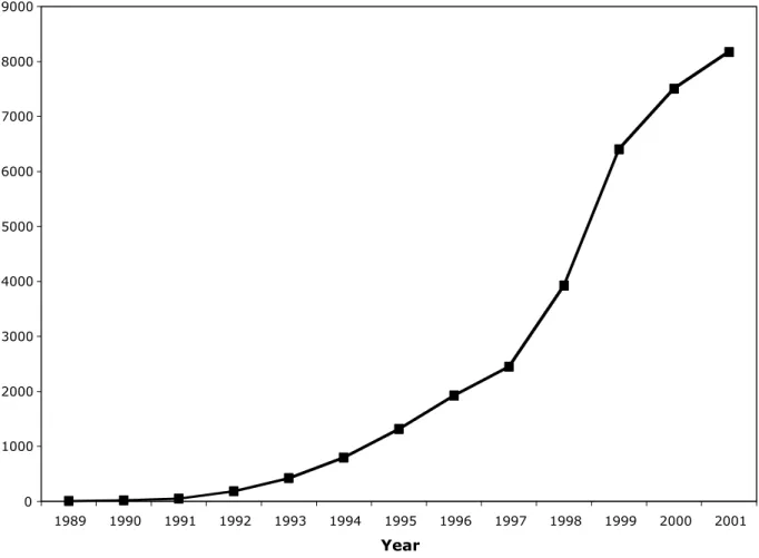
One of the most successful applications of the PCR technique in clinical immunology has been in the form of reverse transcriptase PCR (RT-PCR).7 This variant of PCR was developed in 1988 and by the end of 2001 over 30,000 articles using the term RT-PCR in the title or abstract of the article had been indexed in Medline (see Figure 5). Once again, Cetus played a central role and the biological and the clinical forms appeared more or less simultaneously.8 In the clinical case, RT-PCR proved to be less an application of biology to medicine than a biological solution to a problem raised in the clinic. Specifically, the presence of aberrations, namely translocations, affecting cell chromosomes in lymphoid tumours had long been observed. For instance, the Philadelphia chromosome,9 present in 95% of cases of a subspecies of leukaemia known as chronic myelogenous leukaemia (CML) (Chopra, Pu & Elefanty,1999), became associated with the disease in 1959 and had already been used as a disease marker in a number of clinical studies throughout the 1960s and 1970s. At the beginning of the 1980s, researchers pointed to the presence, at
5 Saiki’s paper appeared in December of 1984 (Saiki et al., 1984). Still in preparation at the time of the Saiki publication, Mullis’ paper was subsequently refused by both Nature and Science and did not appear until 1987 (Mullis & Faloona, 1987).
6 Saiki et al. (1984, p. 1353), also pointed out that compared to the standard prenatal diagnosis of sickle cell anaemia, their test required 10 hours rather than several days and that “all of the reactions can be done in two small microcentrifuge tubes and could readily be automated”.
7 RT-PCR uses messenger RNA (mRNA) instead of DNA as its starting point. When genes are active, their DNA is transcribed into mRNA and subsequently into proteins. In RT-PCR, using an enzyme called reverse transcriptase, the mRNA is translated back into DNA (more precisely: complementary DNA or cDNA) and the latter is then amplified using PCR.
8 The simultaneous nature of the invention is attested to by the authors of the clinical paper who noted in their final sentence: “while this manuscript was in preparation, two studies were reported in which amplification of RNA sequences was used (i) to determine the mechanism for tissue-specific processing of an apolipoprotein and (ii) to facilitate cloning of the HLA-DQ sequences” (Kawasaki et al., 1988, p. 5702). 9 A translocation is one type of gene rearrangement (see note 3). In the case of the Philadelphia chromosome, the latter is formed when a portion of chromosome 22 is translocated to chromosome 9. For a recollection of events related to the Philadelphia chromosome, see Rowley (2001).
the translocation breakpoint, of a human homologue of a murine leukaemia virus (de Klein et al., 1982), and CML became a model disease providing a molecular or genetic reading of the translocation event.
FIGURE 5
9000 8000 7000 6000 5000 4000 3000 2000 1000 0 1989 1990 1991 1992 1993 1994 1995 1996 1997 1998 1999 2000 2001 YearGrowth of the number of papers indexed in PubMed with the term RT-PCR.
When researchers at Cetus began work, there were several ways of spotting the molecular trouble at the base of the disease. The translocation had initially been characterized using the Southern blot and restriction enzymes (Kurzrock, Gutterman & Talpaz, 1988). The DNA sequence associated with the translocation was thus an obvious target for PCR amplification. Due, however, to the presence of large non-coding sequences of DNA within the gene, inter-patient variation and, above all, the fact that breaks occurred anywhere within a large region of the chromosome, standard PCR protocols could not be used. In collaboration with clinical researchers at the Department of Pediatrics at the Stanford Children’s Hospital and the Department of Hematology at the University of California Center for the Health Sciences, Cetus researchers set out to overcome these impediments. Using reverse transcriptase they were able to isolate much shorter and homogenous fragments of DNA and subsequently apply PCR techniques (Kawasaki et
clinical trial group used the RT-PCR technique to detect presence of residual cancer cells in CML patients that had undergone therapy (Lee et al., 1989). By the mid-1990s, the analysis of translocation breakpoints entered the era of optimization and quantification (Cross et al., 1994; Malcomson et al., 1995) and techniques developed in one domain were quickly transferred to the analysis of other domains (Lynas et al., 1995).10
So, to sum up, while the PCR episode is often presented as an instance of the veni, vidi, vici dynamics whereby biology techniques have swept the clinical domain, a closer look reveals the existence of mutual regulatory relations between the clinical and genetic research, similar to those already found in the case of the early molecular genetic techniques such as Southern Blot.
Gene Expression Profiling: From Markers to Signatures (1995-2000)
The light at the end of the reductionist tunnel has turned out to be the headlight of a locomotive called genomics. (Shaffer et al., 2001, p. 375)
The latest articulation of the molecular biology platform to enter the clinical haematology arena is the DNA microarray or DNA chip. A microarray consists of a small solid surface (generally glass) on which individual DNA samples (up to thousands of them) are arranged in a grid.11 In a typical run, a technician will investigate the differential expression of genes using, say, a breast cancer specimen and a normal tissue specimen by hybridizing them with the DNA sequences present on the microarray.12 To quote one of the developers of the technique, “the fluorescent signal at the spot in the array representing each individual gene provides a quantitative readout of the level of expression of that gene in the sample. This simple procedure offers a systematic way to
10 The journal issue in which the Lynas et al. (1995) appeared was the first issue of a subedition of the
Journal of Clinical Pathology. The reasons for the creation of the new Journal—to familiarize clinicians with results and techniques that do not have immediate application but that may in future prove useful—are explained in Anon. (1995).
11 There are, in fact, two main variants or formats of microarrays. In the first case, DNA spots are deposited by a robot onto a coated glass slide; this approach allows for the use of two different fluorescent colours (and thus the simultaneous analysis of, say, a control and a diseased tissue sample) on the same microarray. In the second kind, developed and sold by Affymetrix under the trademark GeneChip, photolithographic methods are used to synthesize hundreds of thousand of oligonucleotides on a small glass surface. For a description and comparison of these two formats, see, e.g., Gerhold, Rushmore & Caskey (1999). For an illustrated description of microarray equipment see Cheung et al. (1999). For simplicity’s sake, and because most of the early important contributions in the clinical field have used the “spot” microarray format, our discussion will focus on this latter format.
12 The term gene expression refers to the process by which the information coded in a gene is converted into the structures present and operating in the cell, i.e., proteins or RNAs. The genetic material extracted from the two specimens is labelled with different fluorescent dyes (e.g. green and red) and incubated with the microarray. Gene active or expressed in the sample tissues will hybridize with their counterpart fixed to the microarray and the result of this hybridization process will become visible as a fluorescent spot at a specific grid location, therefore showing what genes are busy in what disease (Steinberg, 2000; Southern, 2001).
monitor expression of tens of thousands of genes simultaneously, in thousands of samples per year”.13
The growth of the technique, as measured through publications has been quite substantial (see Figure 6). Other indicators, such as an estimated annual compounded market growth rate of 63% between 1999 and 2004, similarly support the claim that “the genomics research community has embraced … microarrays” (Constans, 2003). Unsurprisingly, microarray technology and gene expression profiling have elicited quite a reaction. We will see some in what follows but let us begin with a historian’s reaction.
FIGURE 6
F
F
F
F
F
F
F
F
F
1993 1994 1995 1996 1997 1998 1999 2000 2001 0 100 200 300 400 500 600 700 800 900Number of "microarray" or "DNA-chip" articles
Growth of the number of papers indexed in PubMed with the terms “microarray” or “DNA chip.”
Some commentators have suggested that the emergence of DNA microarrays represents more than a simple quantitative movement from the analysis of single genes transcripts (RT-PCR) to many gene transcripts (microarrays). It is a question of quality insofar as microarrays represent the transition from the singular to the global. It is also epochal in the sense that it is suggested that RT-PCR represents the failure of the oncogene paradigm whereas microarrays express a newer and more profound understanding of the
13 Howard Hughes Medical Institute Website; Description of Patrick Brown’s research concerning microarrays: http://www.hhmi.org/research/investigators/brown.html.
cancer process. Describing the fading hopes of a complete description of the cancer process through a description of oncogenes, Morange (2000, p. 1152) tells us:
In 1982, when the first genes were discovered that, when mutated, led to cell transformation and tumorigenesis (the ‘oncogenes’), it might have seemed reasonable to expect to isolate and characterize a small group of genes responsible for all forms of cancer. In 1986, when the human genome sequencing project was first proposed, one of its justifications was the hope of isolating not the genes involved in the first steps of oncogenesis, which are relatively well known, but the genes responsible for the later steps of tumor evolution such as metastasis.
This hope is now considered to be an illusion. The existence of oncogenes, which when mutated are involved in cancer, has been amply confirmed. The illusion is that oncogenes constitute a distinct group of genes which can be easily described. Hopes are now turned toward a global description of transformed cells and their characteristic patterns of gene expression, in comparison with the normal untransformed cell.14
Is it truly the case that we have moved from the local to the global? While conceding that, technically speaking, a microarray test, when compared to previous molecular genetic approaches, involves the simultaneous analysis of many hundreds or thousands of genes, as opposed to single ones, could we not maintain that, in both cases, the analysis operates at a similar level of representation? What does it mean, for instance, to claim, as researcher do in the case of cancer diagnosis, that we have moved from individual markers not so much to the global, but, more specifically, to patterns, profiles or signatures? We will return to this question but first, following once again our analytical strategy, we need to relate the development of microarrays to their clinical use.
A review of microarray technology that appeared in the journal Nature offers a rational reconstruction of the origins of the innovation suggesting that the technology emerged from a biological demand. According to Liotta & Petricoin (2000, p. 48), the intrinsic complexity of biologic systems and the recent completion of the human genome have prompted biomedical researchers to demand “more sophisticated platforms for studying the activity of many genes or proteins in parallel”. Hence, DNA microarrays. Be that as it may, on a more empirical level, DNA microarrays as presently used in clinical immunology emerged out of collaboration between the NCI and Stanford University. Alerted by the fact that two papers published by the Stanford-NCI group in the same issue of Nature Genetics (Ross et al., 2000; Scherf et al., 2000) had attained the status of ‘hot papers’, The Scientist sent a reporter to inquire into the origins of the collaboration (Kling, 2002b).
According to The Scientist, it seems that in the late 1990s, John Weinstein, a senior investigator at NCI’s Division of Basic Sciences (Laboratory of Molecular
14 Morange (2000, p. 1152) adds: “It could be objected that the failure of this project was foreseeable. Oncogenes are not a natural, physiological category. Cancer is not a physiological process, but a de-regulation of normal processes, and the category of oncogenes exists only in the minds of biologists and physicians. But developmental biologists have the same difficulty in categorizing genes”.
Pharmacology) presented his work on the NCI cancer-drug screening program at Stanford University. Patrick Brown, one of the pioneers of microarray technology and a Howard Hughes Medical Institute investigator at Stanford attended the talk. Until then, we are told, Brown had worked on gene expression in yeast cell lines and Weinstein had been following one gene at a time in an attempt to reconstruct oncogene pathways (Brown & Botstein, 1999). The two decided to combine their efforts and the results, a profile of about 8,000 genes followed through 60 cancer cell lines, appeared in the aforementioned Nature Genetics articles.
While the article in The Scientist stresses the serendipitous nature of the encounter, placed in its historical context, the collaboration seems less the unlikely fusion of Brown’s ‘basic cell biology’ and Weinstein’s ‘pharmacology-driven work’ than biomedicine as usual.15 To begin with, the reporter (and his informants) overlooked the
fact that this was not the first collaboration between Brown’s laboratory and cancer researchers. Four years prior to the Brown/Weinstein collaboration and immediately after the development of microarray technology, Brown’s laboratory entered into collaboration with members of the Laboratory of Cancer Genetics at the NIH to develop a biomedical microarray. The chip that they assembled contained a mix of both normal and pathological DNA sequences (DeRisi et al., 1996). Brown’s laboratory contributed the normal DNA while the NIH lab contributed DNA selected from a human melanoma cell line that had originally been produced at the University of Michigan Medical School (Ray et al., 1996). The paper published with the Brown team received a more than respectable 534 cites in the five years following its publication There was thus no five-year delay between the invention of microarrays and their clinical application. Biological and biomedical microarrays were constructed at the same time.
In addition to this historical oversight, closer examination of the clinical research program at the National Cancer Institute shows to what extent it was an innovative and parallel enterprise and, thus, much more than fallow ground waiting for biological application. To see how this is so, we need to focus less on the novelty of the microarrays than on the organization of cancer research. In particular, we need to notice that “high throughput” biomedicine has a longstanding tradition at the NCI and created an environment where the Brown/Weinstein collaboration would seem only natural.
High Throughput Biomedicine
Let us begin with the aforementioned screening system set up at the National Cancer Institute to screen for anti-cancer compounds. High-throughput biomedicine begins well before the advent of molecular genetics. Screening techniques, first confined to animal systems, have subsequently been progressively developed with cell and gene systems. The process, of a quasi-industrial nature, was instituted in the mid-1950s, and by 1965 the NCI had screened over 100,000 compounds for anti-cancer activity (Goodman & Walsh, 2001, Chapter 1). The animal tumour systems used for this purpose were varied and complex ranging from more than a dozen different animal tumours in the initial 1955
15 Indeed, Weinstein pursued a similar collaboration with the leading commercial producer of microarrays, Affymetrix, as part of a collaboration with Eric Lander of the Whitehead Center for Genomic Research. The first results of this research appeared in Staunton et al. (2001).
primary screen to a mouse leukaemia mouse model and a solid rat tumour by the mid-1960s. These two tumour models remained standard until 1975 when, as the number of compounds going into the system was scaled back from 40,000 per year to 10,000 per year, eight new mouse and human tumours were added. Evaluation of the system seven years later, however, showed that the need for more models had been somewhat overestimated as most compounds selected for further testing would have been picked up by only three models (Anon., 1982).
By the early 1980s, a number of human cancer cell lines had emerged in laboratories around the world (Finlay & Baguley, 1984). Thus, by the mid-1980s, in vitro testing emerged as an option to be used in parallel to the in vivo screen. Human cell lines were collected at the NCI and work on the construction of a comprehensive panel to screen anti-cancer agents. By 1985, cell line panels included melanoma, renal cell carcinoma, ovarian carcinoma, CNS tumours, prostate carcinoma, human leukaemia, breast carcinoma and lung cancer. An initial test using 100 compounds chosen at random showed the panel to be sufficiently sensitive to merit further refinement (NCI Division of Cancer Treatment, 1985-1986, p. 8). Panel developers initially intended to use between 60 and 100 different cell lines (NCI Division of Cancer Treatment, 1987-1988, p. 9; Shoemaker et al., 1988; Monks et al., 1991). Statistical methods—including a computer program called COMPARE—developed by Kenneth Paull and Robert Shoemaker of the biostatistical centre pared the panel down to 60 cell lines (Zaharevitz et al., 2002; Paull et al., 1989). Although subsequently criticized on both sides of the Atlantic as a poor predictor, the screening system has now been in use for over a decade (Robert, 1996; Brown, 1997).
The database generated by the screen had been mined prior to the Weinsten/Brown encounter. Paull’s groups, for example, had searched through response profiles generated by the panel to search for multidrug-resistant phenotypes (Wu et al., 1992). The aforementioned COMPARE program had been based on the insight that similar patterns of reactivity reflected similar mechanisms of reaction thus orienting researchers towards possible reaction mechanisms of the anti-tumour compounds screened. In other words, by recording a common end-point for a common dose of a single compound in 60 different cell lines, Weinstein’s group was able to develop ‘fingerprints’ for each of the compounds. Fingerprints could then be correlated with structural features of classes of compounds which, in turn, could be compared with the more than 460,000 compounds that had accumulated in the NCI database since the 1950s (Weinstein et al., 1997; Rindflesch et al., 2000). The NCI database was structured by a Drug Information System that contained coded information on the 3D structures of each compound in the bank and could thus be interrogated with regards to the relationship between ‘fingerprints’, and molecular structure (Milne et al., 1994). Weinstein and his collaborators combined the screen with other assays and used it to chart pathways of anti-oncogene activities (O’Connor et al., 1997; Rabow et al., 2002). As they put it:
This approach to drug discovery and molecular pharmacology can be likened to a clinical trial with 60 patients (cell types) each profiled with respect to a variety of molecular markers and each treated with 60,000 different agents, one at a time. It can also be considered as a hypothesis
generator based on a set of 60,000 X 60 = 3.6 million pharmacology experiments. (Weinstein et al., 1997, p. 348)
Finally, by the time Brown and Weinstein met, Weinstein’s group had already set about analyzing the screening system at the protein level using gel electrophoresis (Myers et al., 1997). Given the foregoing, it is not outlandish to suggest that microarrays were simply the next step.
Microarrays, Profiles and Signatures
Three months prior to the article on DNA microarrays, The Scientist had produced yet another article in its ‘Hot Papers’ series entitled “Researchers Profile Cancer Cells Through Gene Expression” (Kling, 2002a). This time, The Scientist inquired into the conditions surrounding the production of two articles that “had broken through the barriers and established relationships between gene expression profiling and the biology and clinical outcome of diffuse large B-cell lymphoma”. One of the two articles (Alizadeh et al., 2000), once again the outcome of a collaborative project between Stanford University and the NCI, was cited as “the first example of a clinically relevant classification of cancer using gene expression profiling” (Kling, 2002a). We will restrict ourselves to comments on this article, that provided a genetic profile of two different lymphoma populations.
The authors claimed to have shown that what used to be treated, for lack of reliable means of discriminating between subgroups, as a single disease entity corresponded, when examined using microarray technology, to two molecularly distinct diseases. Molecular genetics is thus said to have succeeded in a domain where morphology “largely failed owing to diagnostic discrepancies arising from inter- and intra-observer irreproducibility” (Alizadeh et al., 2000, p. 503). The microarray used by the authors— called Lymphochip—was designed to produce traces or readouts of both normal and pathological gene activation events within the framework of the development of the immune response. The two principal components of the Lymphochip—DNA derived from normal and pathological samples—shared the same space of representation.
The visual demonstration of the article’s main claim consisted in the display of the color patterns produced by the differential expression of hundreds of genes sorted according to three colour-coded categories of intensity: high (red) medium (black) and low (green) (see Figure 7 for a simplified version of the original imagery). These distinctive colour patterns—the result of statistical manipulation of the raw data produced by the electronic microarray scanning equipment—were said to correspond to the characteristic profile of distinct pathological entities. Two things should be noted here. First, the starting material—the tissues samples from which the genetic material was extracted—had already been characterized as normal or pathological according to morphological and IPT criteria. The patterns, in other words, were categorized and explained by the morphological and immunophenotypic origin of the samples from which the genetic material was derived. Second, and most importantly, the pathological events were ultimately translated into a qualitative pathological sign, a profile, rather than into a
quantitative description of the level of gene expression as measured by fluorescence intensities.
FIGURE 7
A microarray profile of lymphoma samples showing the existence of two distinct types of lymphoma. Original in colour. The colour-coded genes were selected and arranged using a clustering algorithm in order to produce a contrasting colour pattern. For this black and white version we have enhanced the grey scale (using darker greys for the red colour and lighter greys for green) in order to give an approximate idea of the plaid colour pattern (bottom left, top right: predominantly red; top left, bottom right: predominantly green). Reprinted from Staudt (2001, p. 38), © 2001, with permission from Elsevier Science.
By electronically collating individual, profile-generating microarray experiments, researchers hope to reach an even higher aggregation level to generate what, in yet another leap of metaphorical imagination, they term signatures (Shaffer et al., 2001, p. 384). The latter would provide information on “how cells differentiate, respond to stimuli, employ transcription factors, and react to drugs”. In turn, the development of a gene expression signatures database is seen as a possible solution to the problem of comparing gene expression data produced by different laboratories with different tools.
Instead of speculating on the future, we prefer to examine, in the next section, how microarrays have so far done in routine immunopathological applications.
Microarrays in Clinical Routines
Consider a recent (2003) advertisement (Figure 8) by Ardais, a company devoted to “connecting genomics and human disease”.16 The left side of the ‘A’ (A as in Ardais) displays the four letters (ATCG) of the genetic code and thus represents the biological (genomics) side of the equation. The right side of the ‘A’ shows a tissue sample under the microscope, signifying the morphological tradition in clinical pathology and thus human disease. The connecting line between these two elements, the one that completes the ‘A’, is a DNA microarray, that thus acts as the bridge between biology and medicine.
FIGURE 8
Advertisement (2003) by Ardais Corporation. See text for explanations. Reprinted with permission from Ardais Corporation (Lexington, MA).
Ardais’s advertisement belongs to the register of promissory science, i.e., the strategy of depicting the future that, it is hoped, by shaping expectations will drive research in the
16 In 2002, the company launched the National Clinical Genomics Initiative in collaboration with several U.S. medical institutions. For additional information about this initiative and Ardais Corporation, see: http://www.ardais.com.
intended direction.17 From a performative point of view, such an activity is hardly empty speculation. In an innovative field of science, statements about the future enrol support and resources, enable interactions between actors, and guide the design of artefacts (Hedgecoe & Martin, forthcoming). Many of the statements we will discuss in this section participate in this mode of discourse.
The routine use of microarray techniques within the clinic is predicated upon the solution of a number of issues that define the complex interaction between biological and clinical practices in the era of biomedicine. The first is the extent to which the technique can provide independent and novel information concerning diagnosis, classification and prognosis: independent and novel, that is, when compared to pre-existing clinical routines. This is well known in the field. Todd Golub, Eric Lander and their colleagues at the MIT Center for Genome Research were clearly aware of the need for microarrays to acquire independent status in order to obtain clinical success. Among the earliest and most highly cited entrants into the diagnostic field, they claimed to have invented a technique “that automatically discovered the distinction between acute myeloid leukemia and acute lymphoblastic leukemia without previous knowledge of these classes”. They further claimed that such a technique could be used as “a general strategy for discovering and predicting cancer classes for other types of cancer, independent of previous biological knowledge” (Golub et al., 1999, p. 531). In a subsequent special editorial for the New England Journal of Medicine entitled “Genome-Wide Views of Cancer”, Golub drew the obvious conclusion. Previous taxonomies would be displaced and clinical practice would have to adjust:
The increasing understanding of molecular medicine will shift clinical practice from empirical treatment to therapy based on a molecular taxonomy of disease. Physicians will be prescribing rationally designed drugs that have increased efficacy and reduced toxicity. (Golub, 2001)
Echoing Golub, an extensive review of microarray technology in the Journal of Clinical Pathology warned pathologists that if they did not embrace the technology, they ran the risk being squeezed out of the field. In particular:
If pathologists neglect the potential of these processes at an early stage, it is not unthinkable that in the future their task will be limited to judging whether or not a sample contains material for microarray analysis or whether a tumor’s histology is compatible with the more precise diagnosis made by geneticists or chemists, or just handing over samples to specialists from other disciplines after dissection. Even worse, such samples may be taken from the specimen before they are sent to the pathology department. (Snijders et al., 2000 p. 293)
Similarly, the Journal of Pathology told its readers:
1717 Several articles have recently discussed trend-promotion in science and technology. See Guice (1999) for a ground-breaking formulation that focuses, however, on information technology. In the medical field, this theme has been discussed both by practitioners (Baue, 2003) and by social scientists (e.g., Hedgecoe & Martin, forthcoming).
As pathologists in the future, none of us will be able to avoid DNA microarrays and there may come a time when tumor RNA is routinely run on arrays to give an accurate read-out of the patient’s prognosis, and even a prescription for treatment! (Maughan, Lewis & Smith, 2001, p. 6)
Equally as enthusiastic, although stated in the conditional, a similar review in Nature Medicine opined:
New technologies such as expression arrays, which allow us to monitor the expression of thousands of individual genes, could overcome the limitations of old methods of diagnosis and prognosis based on a limited number of markers. (He & Friend, 2001, p. 658).
Although the previous quotes imply that pathologists are passive receptacles for the new technologies invented in the basic sciences, the practice of biomedicine entails considerably more interaction and a number of options and strategies are open to, and have been pursued by, pathologists and clinicians. To begin with, pathologists have appropriated the microarray technologies for their own purposes. Guido Sauter and his colleagues at the Institute of Pathology in Basel Switzerland, in collaboration with the NIH Human Genome Research Institute have invented a tissue microarray that inverts the principle of the DNA microarray: rather than following thousands of genes through a single tissue specimen, they have organized thousands of specimens on a single slide in order to follow a single gene product (Richter et al., 2000; Kononen et al., 1998). Commenting on this work in the Journal of Molecular Diagnostics, Lichter (2000, p. 172) pointed out that:
Although they stress the point that their study was accomplished within 2 weeks, this number does not account for the previous efforts to carefully analyze archived material for the generation of the arrays. Nevertheless, the comprehensiveness of the study and the speed with which it was performed are more than impressive. These kinds of analyses will certainly change the world of publications in the field of pathology.
Secondly, microarrays can also be aligned with work already underway. Researchers in Finland, for example, used microarray techniques to investigate patients that had first been classified according to traditional clinical, morphological, cytogenetic, immunophenotypic and staging techniques. They thus produced a staged cohort of a subclass of chronic leukaemia patients that had a specific chromosomal deletion and immunophenotype prior to microarray investigation.
The microarray used by the Finnish team did not monitor thousands of genes as is so often the case. A Becton-Dickinson subsidiary, Clontech, had simplified and focused the process by producing ‘application-targeted arrays’ that selected genes that reflected the specific functions and disorders of the cells in question, as defined by previous investigations of and interventions upon the disease. Thus, using an ‘integrated platform’, namely the AtlasTM Human Hematology Array containing 440 genes (Anon., 1998), they isolated a cluster of up-regulated genes of known normal and pathological function
(Aalto et al., 2001). Notice that the genes were selected in order to create a biomedical device, one that would enable researchers to explore both the immune process and immune malignancies.
Third, some commentators have suggested that the use of thousands of genes to differentiate within cancer classes constitutes overkill. The theory of oncogenes says that at best several hundred genes are mutated in most cancers. Richard Wooster (2000, p. 328) of Britain’s Institute of Cancer Research thus suggests that as few as 10 to 20 genes could be used to differentiate between groups of tumours and consequently remarks that:
We can marvel at the amount of data being produced by DNA expression arrays, but they could be considered as just a fishing exercise to identify a handful of genes that have a diagnostic potential.
In this respect, molecular profiling can be seen as more of the same; just as IPT was articulated with histology, so too, will molecular profiling ultimately be forced to address its relation with morphology. As one of the keynote speakers at the Inaugural Association for Molecular Pathology Companion Meeting held at the United States and American Academy of Pathology Meeting suggested:
In a sense, pathologists already use gene expression studies diagnostically on a daily basis in the form of immunohistochemistry. Expression profiling provides a supraexponential increase to the amount of gene expression data available on a given tumor, but lacks the topographical information of immunohistochemistry. (Ladanyi et al., 2001, p. 92)
The plus ça change attitude, however, belied a more serious purpose to the address, namely a call to arms for pathologists. As the speaker went on to point out, unlike immunohistochemistry or even cytogenetics, “expression profiling may require less prior expert histopathological ‘triage’ than any prior ancillary technique” (Ladanyi et al., 2001, p. 92). Given these conditions, the following question no doubt seemed inevitable: “Will expression profiling evolve into another ‘ancillary’ technique or will it come to be seen as an alternative to expert surgical pathology?” This is certainly not the first time that such a question has been put to pathologists. Indeed, the frozen section technique raised similar fears at the turn of the last century (Wright, 1985) The corresponding answer is no doubt just as old:
The extent of involvement of the pathology community in the further development of microarray-based expression profiling as a diagnostic test may determine the nature of its ultimate relationship to conventional tissue-based diagnoses. (Ladanyi et al., 2001, p. 93)
Here is one form of involvement. Spurred by the studies from Staudt’s laboratory, a French group of haematopathologists took up the challenge of producing a study “to aid in the transition of microarray technology into routine use”(Dales et al., 2001, p. 17). Rather than assuming that the microarray data were incommensurable with data obtained by older techniques, they sought correlations between microarrays and existing immunohistochemistry techniques. In particular, they used immunohistochemistry to
target intercellular proteins implicated in cell death and showed that the results so obtained correlated with those produced by (commercially available) microarrays. They thus concluded: “This suggests that, in the future, pathologists might be able to analyze by [immunohistochemistry] potential markers of interest previously identified by array technology” (Dales et al., 2001, p. 23). In other words, they turned the chronological relationship upside down by translating microarrays into a legitimating tool for pre-existent immunopathological techniques.
Closer to the clinical world, a review in JAMA enumerated a number of “possible pitfalls that may undermine the authority of the microarray platform” (King & Sinha, 2001, p. 2280). The problems—sample selection, comparability, archiving and clinical and biological significance—all revolved around problems of regulation and the articulation of microarrays with existing platforms that determine gene function such as RT-PCR and knock-out mice. Notice, however, that while none of the ‘pitfalls’ are fatal, their solution is part of the process of the constitution of microarrays as a functioning biomedical platform and not an external problem. They are problems inherent to the management of microarrays (Lee et al., 2000; Becich, 2000).
A recent review of the technique in Nature Genetics Reviews also warns clinical readers of a number of problems attendant upon the alignment of microarrays with existing routines and established practices in pathology, such as the routine fixing of tissues in formalin. To begin with, gene expression studies require intact RNA. RNA is degraded to an unknown extent during formalin fixation. Freezing the tissue of interest can help preserve the RNA; it inconveniently, however, compromises morphology. This is not necessarily the case in non-formalin based procedures such as those that use alcohol. Even here, however, further problems remain; one must, for example, process the material with considerable dispatch. And, as the same review points out: “investigators face an uphill battle in convincing surgeons and practicing pathologists to process the tissue obtained at surgery rapidly to preserve all the macromolecules of interest” (Liotta & Petricoin, 2000, p. 49). Pathological expertise and mobilization of the routines that underlie this skill is also required to isolate the initial pathological material. Biopsy specimens are in fact three-dimensional structures that contain a variety of cell populations. Explicit comparison between normal and pathological cells depends upon a prior visual inspection of the biopsy material in order to separate the two types. Nonetheless, some pathologists fear less losing time during biopsy to placate microarray enthusiasts, as implied in the previous quote, than losing the control over the resulting specimens and the downstream information.
Promoters of the microarray platform for routine clinical use are aware that it is possible that the aforementioned work-arounds and the problems sketched above may in fact block entry of the microarray platform into routine clinical use. Addressing a clinical audience, Golub admitted that not only are microarrays “often viewed as screens to identify markers for traditional diagnostics, such as immunohistochemistry, for routine clinical use” but that “the feasibility of routine clinical use of microarrays ... has yet to be established” (Ramaswamy & Golub, 2002, p. 1941). On the pathologists’ side of the barricade, part of the consensus reached at a 2000 symposium held by the Royal College of Pathologists on the molecular genetics of solid tumours was that, “aside from the use
of gene rearrangement studies in haematological malignancies, genetic information has thus far offered little in the way of disease diagnosis and patient management” (Tomlinson & Ilyas, 2001, p. 202). The College went on to establish a committee to develop a framework for the introduction of molecular diagnosis into routine practice, for “there is a risk that technology will be used simply because it is available” (Tomlinson & Ilyas, 2001, p. 202).
Conclusion
Compared to the representations made by their promoters, what is often perceived as the introduction of molecular biology into the clinic has been at once both quicker and slower than expected. It has been quicker insofar as to speak of introduction is in itself misguided: clinical research is more closely entwined with basic biology than is often imagined. As we have repeatedly argued in this text, RFLPs, PCR, RT-PCR and microarrays were not so much transferred to the clinic as they were developed within the framework of biomedicine where clinical concerns and model diseases form the horizon. The widespread adoption of molecular genetic techniques for routine clinical testing, however, is slower than expected insofar as the use of techniques in clinical research does not imply their use in clinical practice.
10
To understand why this is so, we can redescribe these techniques within the larger context of a biomedical platform, as exemplified in this text. The work of articulation and regulation, or, in other words, the ongoing mutual adjustments between laboratory and clinical practices are constitutive aspects of a platform that enters routine clinical use. The mundane nature of this kind of work, essential though it is, often leads social scientists to overlook its role in biomedical innovation. Its importance, however, is not missed by actors in the field. A recent promissory survey of microarray technologies co-authored by a group of FDA experts centred on the “development of a cooperative framework among regulators, product sponsors, and technology experts” as a sine qua non condition for turning the potential benefits of these technologies into clinical reality (Petricoin III et al., 2002, p. 474). Regulation, in this context, appears to be broadly understood, since it implies, among other things, the demonstration of “sufficient sensitivity, specificity, reproducibility, robustness, reliability, accuracy, precision and clinical relevance” of a given platform (Ibid., p. 476). The determination of clinical relevance, in itself, is a tall order, and one that links narrowly defined regulatory tasks to the development of new knowledge and innovative practices. We need, then, to invert the equation: what holds together and separates the various elements of a platform, is not how they are produced but how they are regulated. The production of novelty is the result of regulation for regulation ultimately defines significance.
Acknowledgments
Research for this paper was made possible by grants from the Social Science and Humanities Research Council of Canada (SSHRC) and Quebec’s Fonds de Recherche sur la Société et la Culture (FQRSC). An initial version of this paper was presented at the Workshop on “Immunology and Molecular Biology, 1955-1990”, organized by Horace F. Judson and Alfred I. Tauber at the Center for the History of Recent Science, George Washington University (Washington D.C., May 31 –June 2, 2002). We would like to thank André Ponton, Head of the Chip MicroArray Expression Laboratory at the Montreal Genome Centre for introducing us to the technical subtleties of DNA chips.