Université de Montréal
DIFFERENTIAL REGULATION OF EARLY RESPONSE GENES BY FIBROBLAST GROWTH FACTOR (FGF) 8 AND FGF18 IN BOVINE
GRANULOSA CELLS IN VITRO
par
HILDA MORAYMA GUERRERO NETRO
Département de biomédecine vétérinaire Faculté de médecine vétérinaire
Mémoire présenté à la Faculté de médecine vétérinaire en vue de l’obtention du grade de
Maître ès sciences (M.Sc.) en sciences vétérinaires
option reproduction
Novembre, 2013
Résumé
Les « Facteurs de croissance des fibroblastes» (FGF) agissent comme des régulateurs locaux sur la qualité des follicules et sont connus pour promouvoir la prolifération des cellules de granulosa, réduire l’apoptose et la stéroïdogenèse. Parmi la sous-famille FGF8, FGF18 est une exception puisqu’il semblerait avoir une fonction pro-apoptotique alors que FGF8 n’a pas été jusqu’à présent rapporté comme altérant la viabilité des cellules de la granulosa. Ces deux ligands ont un mode d’activation similaire et il pourrait être proposé que toute la sous-famille FGF8 ait la même réponse. L’objectif de cette étude était de déterminer si FGF8 et FGF18 activaient la même réponse précoce de gènes dans des cultures de granulosa bovine. Pour répondre à cette question, nous avons cultivé des cellules de la granulosa dans du milieu de culture sans sérum pendant 5 jours. Le jour 5, les cellules ont été traitées avec FGF8 ou FGF18. Nous avons eu recours à une approche de « puce à ADN » afin d’identifier la réponse précoce de gènes induite par FGF8 et FGF18, et les données ont été confirmées par des PCRs en temps réel lors d’une expérience in vitro où les cellules de granulosa ont été traitées avec FGF8 et FGF18 pendant différents temps. L’analyse du puce à ADN a identifié 12 gènes surexprimés par FGF8, incluant SPRY2, NR4A1, XIRP1, BAMBI, EGR1, FOS et FOSL1. A l’inverse, FGF18 n’a régulé aucun gène de manière significative. Les analyses de PCR ont confirmé l’augmentation d’ARNm codant pour EGR1, EGR3, FOS, XIRP1, FOSL1, SPRY2, NR4A1 et BAMBI après 2 h de traitement. FGF18 a entrainé seulement une augmentation de l’expression de EGR1 après 2 h de traitement parmi tous les gènes testés. Ces résultats démontrent donc que FGF8 et FGF18, malgré leur similarité dans le mode d’activation de leurs récepteurs, agissent sur les cellules de la granulosa via différentes voies de signalisation. FGF8 et FGF18, sont donc tous les deux capables de stimuler
l’expression de EGR1, mais les voies de signalisation induites par la suite divergent.
Mots clés: cellules de la granulosa, fibroblast growth factors, EGR1, prolifération, apoptose.
Abstract
Fibroblast growth factors (FGF) act as local regulators of follicular health and are known to increase granulosa cell (GC) proliferation, reduce apoptosis and decrease steroidogenesis. One exception is FGF18, which appears to be a pro apoptotic member of the FGF8-subfamily while FGF8 has not been reported to alter GC health. These two ligands have similar activation patterns and it could be proposed that all FGF8-subfamilies would have the same response. The objective of this study was to determine if FGF8 and FGF18 activate the same early response genes in cultured bovine GC. To address this we cultured GC in serum free medium for five days. On day 5, cells were challenged with FGF8 or FGF18. We used a microarray approach to identify early response genes altered by FGF8 and FGF18, and data were confirmed by real-time PCR in an independent time-course experiment. Microarray identified 12 genes up-regulated by FGF8, including SPRY2, NR4A1, XIRP1, BAMBI, EGR1, FOS and FOSL1. In contrast FGF18 did not result in significant regulation of any gene. PCR analysis confirmed the stimulation of abundance of mRNA encoding EGR1, EGR3, FOS, XIRP1, FOSL1, SPRY2, NR4A1 and BAMBI after 2 hours of challenge. FGF18 resulted in an increase of EGR1 mRNA abundance at 2 h, but not of the other genes tested. These results demonstrate that FGF8 and FGF18, despite reportedly similar receptor activation patterns, act on granulosa cells through different intracellular pathways. Both FGF8 and FGF18 stimulate EGR1 expression, but thereafter their signaling pathways diverge.
Key words: granulosa cells, fibroblast growth factors, EGR1, proliferation, apoptosis.
Table of contents Résumé Abstract Table of contents List of tables List of figures List of abbreviations Dedication Acknowledgments Introduction Literature review 1. The bovine ovary 2. The follicle
2.1 Structures 2.1.1 Oocyte
2.1.2 Granulosa cells 2.1.3 Theca cells
2.2 Follicular growth and development 2.2.1 Recruitment
2.2.2 Selection and dominance 2.2.3 Atresia 3. Steroidogenesis 3.1 Estradiol 3.2 Progesterone 4. Growth factors i iii iv vii viii x xiii xiv 1 3 3 4 4 6 7 8 9 12 14 16 16 18 18 19
4.1 Insulin-like growth factor 4.2 Epidermal growth factor
4.3 Transforming growth factor beta 5. Fibroblast growth factors
5.1 FGF families 5.2 FGF receptors
5.3 FGF signaling pathway 5.4 FGF early response genes 5.5 Role of FGFs
5.6 Role in the ovary 6. FGF8 and FGF18 Hypothesis and objectives Materials and Methods Primary cell culture Cell lines
Experimental treatments
Total RNA extraction and PCR Microarray analysis
Real time PCR Statistic analysis Results
Microarray analysis of FGF8 and FGF18 activated early response genes
Real time PCR validation of microarray results EGR3 expression
Effects of FGF8 and FGF18 in bovine granulosa cell line A1 FGF2 and EGF differential up regulation of EGR1 and EGR3
19 20 21 22 23 24 28 30 32 33 35 37 38 38 39 39 40 40 42 42 43 43 45 46 46 47
Discussion Conclusion References 55 61 62
List of Tables Literature review
Table 1: Evolutionary relationship within the human FGF gene family
24
Materials and Methods
Table 2: Primer sequences for microarray validation 41
Results
Table 1: Genes significantly regulated by FGF8 in bovine granulosa cells
List of figures Literature review
Figure 1: Schematic representation of a pre-ovulatory mammalian follicle
5
Figure 2: Theca cells development and function 9
Figure 3: Folliculogenesis 10
Figure 4: FSH regulation 12
Figure 5: Recruitment of bovine ovarian follicles 14
Figure 6: Diagram of the major steroidogenic pathways in ruminants
17
Figure 7: IGF effect on steroid production 20
Figure 8: RTK signaling 26
Figure 9: FGFR domains 27
Figure 10: FGF signaling pathways 29
Figure 11: FGFR domains 31
Results
Figure 12: Identification of FGF8 early response genes network by ingenuity pathway analysis
48
Figure 13: Regulation by FGF8 and FGF18 of early-response genes identified by microarray
49
FGF18 in primary bovine granulosa cells.
Figure 15: Effect of FGF8 and FGF18 on EGR3 mRNA levels in human cell lines (KGN, HEK293t and SVG)
51
Figure 16: Effect of FGF8 and FGF18 on gene expression in the bovine granulosa cell line A1
52
Figure 17: Effect of FGF2 on expression of EGR1 and EGR3 in primary bovine granulosa cells
53
Figure 18: Effect of EGF on expression of EGR1 and EGR3 in primary bovine granulosa cells
54
Conclusion
Figure 19: Model to explain the divergent signaling between FGF8 and FGF18
List of abbreviations
3ß-HSD: 3ß-hydroxysteroid dehydrogenase 17ß-HSD: 17ß-hydroxysteroid dehydrogenase AMH: anti-Mullerian hormone
Areg: amiphiregulin
BAMBI: BMP activin membrane-bound inhibitors homolog BMP-15: bone morphogenic protein 15
BTC: betacellulin
cAMP: cyclic adenosine monophosphate CGC: cumulus granulosa cells
CTGF: connective tissue growth factor CYP11a1: P450 17α-hydroxylase D: Ig like Domain
DF: dominant follicle E2: estradiol
ECM: extracellular matrix EGR: epidermal growth factor EGR1: epidermal growth factor 1 EGR3: epidermal growth factor 3
EGFRs: epidermal growth factor receptors FGFs: fibroblast growth factors
FGFRs: fibroblast growth factors receptors
FOS: FBJ murin osteosarcoma viral oncogene homolog FSH: follicle stimulating hormone
FSHr: follicle stimulating hormone receptor Ereg: epiregulin
GC: granulosa cells
GDF-9: growth differentiation factor 9 GDF: growth differentiation factor
GDNF: glial cell-derived neurotrophic factor HAS2: Hyaluronan synthase 2
hCG: human chorionic gonadotropin
HGFRs: hepatocythe growth factor receptors HSPGs: heparan sulfate proteoglycan
IGF-I: insulin-like growth factor I
IGFR: insulin-like growth factor receptor IR: insuline receptor
LF: largest follicle
LHCGR: luteinizing hormone/choriogonadotropin receptor MAPK: Mitogen-activated protein kinase
MGC: mural granulosa cells
NGFRs: nerve growth factor receptors NR4A: orphan nuclear receptor group A
NR4A1: orphan nuclear receptor group A member 1 P4: progesterone
PDGFRs: Platelet derived growth factor receptors PGC: primordial germ cells
PI3K: phosphatidylinositol 3-kinase PKC: protein kinase C
PLK2: polo-like kinase 2
RTKs: tyrosine kinase receptors SF: subordinate follicle
SPRY2: sprouty homolog 2
StAR: Steroidogenic acute regulatory protein TC: theca cells
TGFbeta: transforming growth factor β
VEGFRs: vascular endothelial growth factor receptors XIRP1: xin actin-binding repeat containing I
A mis padres Gustavo y Lydia
Acknowledgments
First I would like to thank my supervisor Dr Christopher Price, for his support, amazing patience and incredibly good skills in spanglish. I appreciate all the opportunities presented to me since I was a vet trainee and all the time invested in my experiments planning.
I would also like to thank to my lab friends: Gustavo Zamberlam for all his knowledge, time and funny moments! Thanks for teaching me everything I know! Fatiha Sahmi for all the cell line and cooking knowledge. Atefeh Abedini for her cultural lessons in foreign cultures, experience and company, Muchas Shukranes!
I am also very grateful with Charlene Rico for all her language skills and technical help. I would also like to thank to all CRRA members for their help and assistance and to Embryogene for their funding.
Finally and most importantly I would like to thank my parents, Gustavo and Lydia for their unconditional support, love and advises, to my brother Ivan and to my sister Lydia for the for friendship and funny messages and to all my friends back in Mexico for all the good vibe.
Introduction
Fertility is a corner stone of human society and from an agricultural perspective, fertility is important for the maintenance of genetically superior dairy and beef herds. In the last decades there has been a well documented decline worldwide in fertility in dairy cattle, the reasons for which include factors from management to genetics, but in many cases involve the ovary and with it, the follicle. When follicular hormone secretion is perturbed there is a direct impact on oocyte quality and uterine environment, and as a result of this, a direct impact on the establishment of pregnancy. Therefore follicular health is of critical importance for improving fertility (Lucy, 2007).
Follicle health is determined by an array of endocrine, paracrine and autocrine factors. The pituitary gonadotrophins, LH and FSH are the major drivers of follicle development, but their actions are regulated by local hormones and growth factors. One of these local regulators of gonadotropin action is the family of fibroblast growth factors (FGF). Fibroblast growth factors are a large family of 22 related proteins that act as key-mesenchymal-epithelial signaling molecules in a variety of tissues, especially during organogenesis. In the ovary, FGF are predominantly expressed in theca cells and granulosa cells express the FGF receptors (FGFR). In granulosa cells FGF produce an increase in proliferation while decreasing differentiation and estradiol production (Berisha et al., 2004; Buratini et al., 2007).
In the ovary, the major signaling pathways of FGF are the mitogen-activated protein kinases (MAPK), protein kinase C and phosphatidylinositol 3-kinase. FGF2 and several other FGFs activate ERK 1/2, and FGF early-
response genes including members of the Sprouty, NR4A and ETS families of proteins, which are believed to be responsible for some FGF functions in granulosa cells including regulation of cell proliferation, regulation of tyrosine kinase receptors, steroidogenesis and prevention of apoptosis. FGF are classed into subfamilies, and of interest to this thesis is the FGF8-subfamily that includes also FGF18. These two ligands have similar receptor activation patterns and it could be proposed that they would have the same actions in bovine granulosa cells. FGF8 is a mitogenic growth factor that increases follicular health by increasing proliferation and suppressing cell differentiation. On the other hand, FGF18 appears to be a pro-apoptotic member, affecting gene expression of pro-survival factors such as GADD45b (Buratini et al., 2005a; Jiang et al., 2011; Portela et al., 2010). The objective of the present study was to identify the early response genes induced by FGF8 and FGF18, and to gain insight as to how FGF18 is pro-apoptotic whereas typical FGF signaling is pro-survival.
1. The bovine ovary
The ovaries are the female gonads, and they are found in pairs located in the pelvic area and in the cow they have an almond form with a size of 3.5 x 2.5 x 1.5 cm (Marieb Elain N, 1993). The development of the ovary starts as a paired thickening of the coelomic epithelium that lines the body cavity in the ventral-medial surface of the mid-region on embryonic day 34; the reasons why the thickening begins are unknown and studies performed mainly in mice are focusing on finding the genes required for this process (Gospodarowicz et al., 1974).
The ovary is considered to have two main functions in reproduction: the first is gametogenesis leading to the production through meiosis of a competent oocyte and the second is the secretion of female sexual hormones such as estrogen and progesterone that are required for follicular development, maintenance of estrous cyclicity and reproductive functions including preparation of the reproductive tract for fertilization and subsequent establishment of pregnancy (Marieb Elain N, 1993). In terms of steroidogenic function, the ovaries are required to perform a highly coordinated series of complex events that will lead to follicular development (Gospodarowicz et al., 1974).
2. The follicle
The structure of the follicle changes during development and can be classified into three different groups according to their size, complexity and responsiveness to circulating gonadotropins: preantral, antral and preovulatory follicles. Preantral follicles start as primordial follicles that possess a single layer of squamous pre-granulosa cells surrounding the oocyte. The theca cell layer has not formed and there is no vascular system. As they start to grow they become primary follicles consisting of a single layer of cuboidal granulosa cells. Primary follicles develop into secondary follicles, in which the follicles possess two or more layers of granulosa cells surrounding the oocyte but have no theca layer or antral cavity (McGee and Hsueh, 2000).
As the follicle transforms into an antral follicle, extracellular fluid accumulates between the granulosa cells that will later merge to form a central liquid-filled cavity called the antrum. The zona pellucida forms at this stage and two or more granulosa cells layers surrounding the oocyte become the cumulus granulosa cells. The theca cell layer is now well formed. The proportion of primordial follicles that undergo folliculogenesis and reach the antral stage is very low as most of the follicles undergo regression and atresia (Marieb Elain N, 1993).
2.1 Structures
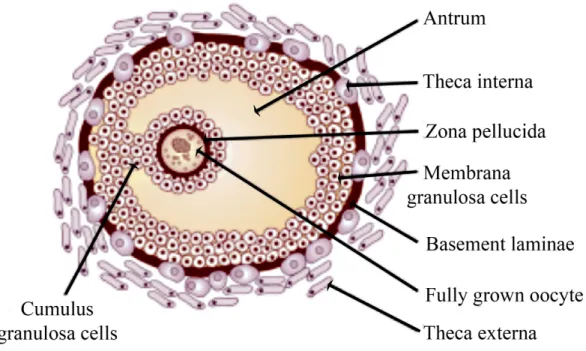
Each follicle consists of an oocyte surrounded by the zona pellucida and one or more layers of somatic cells referred to as cumulus granulosa cells (CGC), the antrum and the basal lamina which separates mural granulosa cells from
the theca cells, which are of stromal origin and are considered to be the interstitial tissue of the follicle (Figure 1) (Gospodarowicz et al., 1974).
Figure 1: Schematic representation of a pre-ovulatory mammalian follicle. The cell types comprising the follicle are shown; the fully grown oocyte and cumulus granulosa cells. Also pointed out are theca cells, granulosa cells, extracellular matrix produced by the oocyte (zona pellucida)
2.1.1 Oocyte
The oocyte is the female germ cell prior to fertilization; the number of oocytes in the mammalian ovary is fixed early in life (Conner et al., 2005). The development of the oocyte starts with the primordial germ cells (PGC) which undergo meiosis to form an oogonium. PGC have the ability to perform extensive migration from the place of their formation to the developing gonad. This process is regulated by somatic germ cell interactions and some additional factors including FGF; in mice FGF2 has been reported to be a mitogenic factor that affects motility of PGC by mediating activation of the MAP-kinase pathway, and FGF7 has also been found to have a role in regulating PGC numbers by activation of FGFRIIIb (Takeuchi et al., 2005). Once the gonad is assembled from PGC, the cells start differentiating and proliferating resulting in the formation of the oogonia. In many organisms the oogonia divide several times forming clusters of interconnected cells; after each division cytoplasmic bridges remain allowing continuous communication between cells and coordinated development. The oogonia differentiate by meiosis into primary oocytes that arrest in prophase and form the major reserve of oocytes in primordial follicles (Voronina and Wessel, 2003).
The best-documented stimulators of oocyte maturation are hormones and growth factors. Some mechanisms of maturation have been proposed and they include: 1) the production of a maturation-inducing substance by follicular cells that drives oocytes to mature, possibly involving activation of membrane receptors by steroid hormones; 2) inactivation of follicle-derived maturation inhibitor; and 3) inhibition of gap junction-mediated transport to prevent transfer of a follicle-derived inhibitor (Conner et al., 2005).
2.1.2 Granulosa cells
GC are important for oocyte maturation as they provide nutrients that support further development. As follicles grow and the antrum cavity is formed, the GC separates into two anatomically and phenotypically different subtypes: the cumulus granulosa cells (CGC), which are in direct contact with the oocyte, have a high rate of proliferation, low steroidogenic capacity, low LH receptor (LHCGR) expression and high levels of insulin growth factor I (IGF-1); and the mural granulosa cells (MGC) which have a primarily endocrine function and support follicle growth, and which undergo terminal differentiation to luteal cells after ovulation. The interaction between oocytes and CGC is complex; CGC express characteristics distinct from the MGC that are acquired under the influence of the oocyte and which promote cell differentiation and development of the GC (Albertini et al., 2001). The oocyte achieves this by secreting labile paracrine signaling factors, and perturbation of this signaling results in the production of an oocyte unable to undergo normal maturation (Yeo et al., 2009). It is possible that MGC are antagonist or insufficient for supporting the last stages of oocyte maturation (Eppig et al., 1997).
GC lack a vascular supply, therefore they require contact with their neighboring cells via gap junctions; these gap junctions contain different connexins such as connexin 32, 43 and 45. Connexin 43 has been studied widely in the mouse where it is has been detected from the onset of folliculogenesis just after birth and persists through ovulation. In later stages it has been found that coupling between GC is mediated specifically by connexin 43 and is essential for continued follicular growth, expansion of the GC population during early stages of follicular development, and that
mutations in this gap junction lead to a retarded oocyte growth, poor development of the zona pellucida of both granulosa cells and oocytes (Ackert et al., 2001; Gospodarowicz et al., 1974).
2.1.3 Theca cells
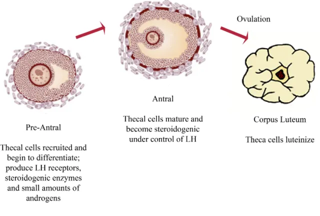
Theca cells (TC) are endocrine cells that play essential roles within the ovary by producing androgen substrates under LH control that are required for ovarian estrogen biosynthesis, and they provide structural support of the growing follicle as it progresses through various developmental stages (Figure 2). They are highly vascularized and through this vascularization they provide the rest of the follicle with essential nutrients and endocrine hormones from the pituitary axis (Magoffin, 2005; Young and McNeilly, 2010).
TC are believed to be recruited from surrounding stromal tissue; the hypothesis of the origin of TC is that growing follicles secrete a series of signals that stimulates TC differentiation and some evidence suggests that these signals involve unknown small molecular-weight proteins secreted by GC (Magoffin, 2005). During development, the majority of follicles undergo atresia, and the TC are often the final follicular cell type to die. For those follicles that ovulate, the TC then undergo hormone-dependent differentiation into luteinized TC of the corpus luteum (Young and McNeilly, 2010).
Figure 2: Thecal cell development and function. Thecal cells are essential for folliculogenesis. TC are required for the production of androgens and they form the vascular compartment of the follicle. After ovulation, thecal cells luteinize and form cells of the corpus luteum (Young
and McNeilly, 2010).
2.2 Follicular growth and development
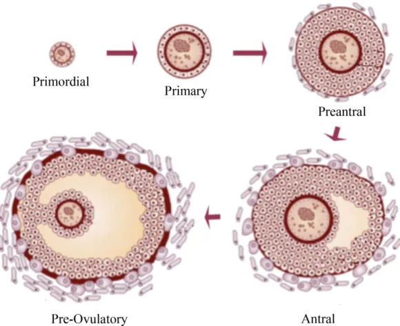
Folliculogenesis describes the formation of the primordial follicle and its progression through the successive stages of preantral, antral and finally preovulatory growth (Figure 3). The development from primordial follicle to preovulatory follicle is a time-consuming event, estimated in cows to take 180 days. It appears that FSH (follicle stimulating hormone) plays a
predominant role in follicle selection and final preovulatory growth. After the LH surge, there is a series of events that lead to ovulation; it is known that LH activates progesterone (P4) receptor in GC, the expression of prostaglandin synthase 2 and the epidermal growth factor-like ligands such as amphiregulin and epiregulin that induce changes in CGC (Gospodarowicz et al., 1974).
Figure 3: Folliculogenesis. Formation of the primordial follicle and its progression through different stages (Erickson et al., 1985; Young and
In cattle a rise in blood FSH concentrations recruits a cohort of small antral follicles into a phase of growth. The largest of these follicles becomes the 'dominant follicle', and secretes high levels of estrogen and inhibins, which then suppress pituitary FSH secretion, which in turn induces atresia in the remaining follicles in the cohort (McGee and Hsueh, 2000; Sisco et al., 2003). The dominant follicle also produces higher levels of autocrine and paracrine factors that stimulate an increase in vasculature and FSH responsiveness. One of these factors is IGF1, which serves to enhance GC responsiveness to FSH by increasing expression of the FSH receptor. A new factor that has been studied is cell-cell adhesion and cell-extracellular matrix (ECM) interactions as they are related to changes in the follicular basal lamina and may have an effect on differentiation of GC and TC (Albertini et al., 2001; McGee and Hsueh, 2000).
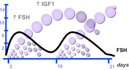
Studies using ultrasonic imaging have documented that follicular growth in cattle occurs in a wave-like pattern and that the majority of estrous cycles in cattle consist of two or three waves. The first wave starts on the day after ovulation (Day 0), the second wave occurs around day 9 and, in the case of a third, it emerges around day 15 (Adams et al., 2008). Each follicular wave is preceded by an increase in FSH that begins about day 2.5 before the wave and starts to decrease about the time of the appearance of the cohort of follicles in the wave (Figure 4) (Bao and Garverick, 1998). During follicular growth, three major events take place: recruitment, selection and dominance. The recruitment begins with the growth of 8-41 small follicles between 3-4 mm, which continue to grow at a similar rate for two days. After this period, one of the follicles is selected and continues growing until it becomes dominant, the rest of the follicles become atretic and regress (Adams et al., 2008).
Figure 4: FSH regulation. Representation of a two follicular wave bovine cycle, each follicular wave is preceded by an increase in FSH blood levels
(Adams et al., 2008).
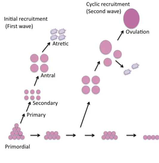
2.2.1 Recruitment
Follicle activation or recruitment takes place in two phases: 1) a continuous recruitment of the dormant primordial follicles into the growing follicle pool; and 2) a cyclical recruitment in response to FSH (Figure 5). It has been demonstrated that FSH can bind to GC of preantral follicles making them responsive to FSH and permitting them to follow a wave-like pattern in response to periodical endogenous surges of FSH (Adams et al., 2008; McGee and Hsueh, 2000). Follicle recruitment is associated with initiation of simultaneous expression of P450ssc and CYP19a1 mRNA
in GC of the recruited cohort of follicles, which are likely to be increased by circulating FSH (Bao and Garverick, 1998).
During gowth of the cohort, follicles grow from 5 mm to 8-9 mm diameter, and the GC express CYP19a1 and P450scc mRNA, but not 3β-HSD mRNA, and the TC express LHCGR, P450scc, P450c17, 3β-HSD, and StAR mRNA. This suggests that GC start to metabolize androgens coming from TC to estradiol (E2) and cholesterol to pregnenolone, but not pregnenolone to P4 because of the lack of 3β-HSD (Bao and Garverick, 1998). Follicles at this stage of development are all antral, and most will undergo atretic degeneration, leaving just the dominant follicle to the reach preovulatory stage (Kolpakova et al., 1998; McGee and Hsueh, 2000). This stage of follicular development is considered to be gonadotropin dependent (Bao and Garverick, 1998).
Figure 5: Recruitment of bovine ovarian follicles. Bovine follicle recruitment in two-wave pattern, showing initial and cyclic recruitment (McGee and Hsueh, 2000).
2.2.2 Selection and dominance
In monovulatory species, selection is the process where a single follicle is chosen from the cohort of medium size growing follicles for further development while the rest become atretic (Fortune et al., 2004). In polyovulatory species, multiple follicles are selected and grow synchronously
until ovulation. The exact process of how a follicle is selected remains unknown, although it has been suggested that the selected follicle shows increased expression of FSHR, LHCGR and 3β-HSD in GC, permitting them to be responsive to LH and continue developing in the face of lowered FSH concentrations (Aerts and Bols, 2010). It has also been proposed that the increased follicular growth rate is due to an increase in IGF1 bioavailability in the dominant follicle (Lucy, 2007). It has been established that the development of one antral follicle until it becomes dominant requires 42 days in the cow, or the equivalent of two estrous cycles (Aerts and Bols, 2010). A dominant follicle has higher concentrations of E2 in follicular fluid, higher LHCGR mRNA levels in TC and GC, higher levels of 17α-hydroxylase and aromatase in GC compared with non-dominant growing follicles (Fortune et al., 2004). Another characteristic of dominant follicles is the high expression of StAR mRNA in TC, which may assure enough cholesterol transport to the mitochondria for androgen production (Bao and Garverick, 1998).
If the dominant follicle becomes the preovulatory follicle, a cascade of events started by the preovulatory LH surge results in ovulation. LH increases the synthesis of progesterone receptors, prostaglandins and epidermal growth factor (EGF)-like factors in GC, and induces the primary oocyte to complete meiosis I. There is also an up-regulation of the expression of proteases thought to play critical roles in follicular rupture (Russell and Robker, 2007).
2.2.3 Atresia
Follicular atresia occurs by apoptosis or programmed cell death. Atresia occurs in the dominant follicle if it does not become the preovulatory follicle, for example as first-wave dominant follicle, and also in the subordinate follicles recruited in a cohort, where atresia has been associated with a suppression of E2 secretion and CYP19a1 expression (Bao and Garverick, 1998). Major intracellular effectors of atresia include the B-cell lymphoma 2 family and the caspase family (Hengartner, 2000).
3. Steroidogenesis
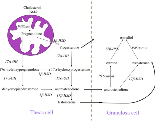
Steroid hormones are derivates of cholesterol; they can be classified into five categories: glucocorticoids (cortisol), mineralocorticoids (aldosterone), androgens (testosterone), estrogens (estradiol and estrone) and progestins (progesterone). In the bovine follicle five enzymes are required for the production of estradiol. Steroidogenesis starts with the internalization of blood-borne low-density lipoproteins, and once inside the cell cholesterol is maintained as liquid droplets (cholesterol esters), which are converted to free cholesterol by the enzyme cholesterol ester hydrolase. Free cholesterol is then mobilized to the mitochondria by the steroidogenic acute regulatory protein (StAR) where it is converted to pregnenolone by the enzyme cytochrome P450 cholesterol side-chain cleavage. Pregnenolone can follow two different routes, 1) conversion to progesterone by the enzyme 3ß-hydroxysteroid dehydrogenase (3ß-HSD) or 2) to 17α-hydroxypregnenolone by the enzyme cytochrome P450 hydroxylase (CYP11a1). 17α-hydroxypregnenolone can be converted to androstenedione by CYP11a1 and 3ß-HSD. Androstenedione is converted into testosterone by 17ß-hydroxyosteroidehydrogenase (17ß-HSD); TC secrete androstenedione and
testosterone and GC can convert androstenedione to estradiol by 17ß-HSD and testosterone to estrone by CYP19a1. Progesterone can be mobilized directly from TC to GC (Figure 6) (Miller and Auchus, 2011).
Figure 6: Diagram of the major steroidogenic pathways in ruminants (Miller and Auchus, 2011).
3.1 Estradiol
E2 regulates the structure and function of the female reproductive system. One characteristic of the growing follicle is its considerable capacity for E2 production. Once the increase of E2 synthesis within the follicle has begun, it has the capacity of self-augmenting by up-regulating androgen synthesis in TC and pregnenolone in GC (Beg and Ginther, 2006). In cattle, E2 promotes development of preantral follicles and stimulates steroidogenesis. Shortly before the beginning of deviation between the largest follicle and the second largest follicle, there is a marked difference in concentrations of E2 in the follicular fluid of the two follicles, and enhance a rapid increase in E2 content is a key characteristic of a dominant follicle. In addition, E2 concentrations decrease in subordinate follicles while the dominant follicle continues growing. As the rate of growth of the follicle slows, estradiol concentrations do not decrease until the follicle starts to regress. All these make E2 a marker for health or atresia of follicles (Beg and Ginther, 2006; Fortune et al., 2004; Price et al., 1995).
3.2 Progesterone
P4 is a steroid hormone involved in pregnancy and embryogenesis. P4 is produced in TC and GC. During the beginning of follicular growth there are no differences in P4 levels between the two largest follicles, however some studies have found that after the second largest follicle starts regressing, there is an increase in P4, making unclear the role of progesterone in the process of growth and differentiation (Beg and Ginther, 2006). The role of P4 is essential not only for the establishment but also for the maintenance of pregnancy, as it supports ovulation and uterine and mammary gland
development (Kim et al., 2010). The major source of P4 during pregnancy is the corpus luteum and in some species the placenta. The genomic actions of P4 are mediated by the intracellular progesterone receptors, and blocking P4 binding sites results in abortion (Arck et al., 2007).
4. Growth factors
Ovarian folliculogenesis is modulated by diverse growth factors, including insulin-like growth factors (IGF), epidermal growth factors (EGF), transforming growth factor β (TGF-β) and FGF (Ben-Ami et al., 2006).
4.1 Insulin-like growth factors
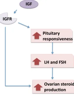
The IGF family includes two ligands, six binding proteins and two receptors. They are produced in ovarian follicles. In the ovary, their main role is during follicular development where they can stimulate the growth of antral follicles and proliferation of GC, and they synergize with gonadotropins to promote differentiation of follicle cells and to inhibit apoptosis (Beg and Ginther, 2006). IGF also increases the expression of FSHR and LHCGR and stimulates the synthesis and secretion of E2, P4, testosterone, oxytocin, inhibin A, activin-A and prostaglandins (Figure 7) (Quirk et al., 2004). The bioavailability of IGF is regulated within the follicle by a family of six binding proteins, which are non-glycosylated peptides that act as carriers for IGF in the serum and regulate the half life of IGF (Beg and Ginther, 2006).
Figure 7: IGF effect on steroid production. Relationship between IGF receptor (IGFR) and the increase in the production of ovarian steroids
(Poretsky et al., 1999).
4.2 Epidermal growth factor
EGF is a protein of 53 amino acids which plays a crucial role in reproduction. Other members of the EGF family include TGF-α, amphiregulin (Areg), epiregulin (Ereg), betacellulin (BTC), epigen, neuregulins and heparin-binding EGF-like growth factor. These proteins can work through four types of transmembrane receptors (Ben-Ami et al., 2006). The main functions of EGFR within the follicle are stimulating proliferation of GC, increasing P4 secretion, and controlling of the release of E2. In oocytes, EGF affects maturation and cumulus expansion, and inhibits apoptosis.
The role of EGF has also been investigated as a paracrine mediator of LH induced ovulation (Quirk et al., 2004; Sirotkin, 2010). The EGF receptors also play important roles in cell proliferation, survival, adhesion, motility, invasion, and angiogenesis in normal and in malignant cells, including ovarian tumors (Jiang et al., 2011).
4.3 Transforming growth factor beta
The TGF-β superfamily of extracellular signaling molecules includes over 35 structurally related but functionally diverse proteins. These proteins function as extracellular ligands involved in numerous physiological processes. This superfamily has been classified into several subfamilies: the TGF-β subfamily (TGF-β1, TGF-β2, TGF-β3), the bone morphogenetic protein (BMP) subfamily, the growth and differentiation factor (GDF) subfamily, the activin/inhibin subfamily, the glial cell-derived neurotrophic factor (GDNF) subfamily and other members such as anti-Mullerian hormone (AMH) (Knight and Glister, 2006). Within the ovary, GDF9, BMP15, inhibins, activins, and AMH are all expressed (Knight and Glister, 2003).
Functions of TGF-β subfamily members vary widely from regulating folliculogenesis to regulating proliferation. GDF9 and BMP15 are expressed in the oocyte from the preantral stage of development and play key roles in promoting preantral follicle growth. Studies on later stages of follicle development indicate an important positive role for granulosa cell-derived activin, BMP2, BMP5 and BMP6, theca cell-derived BMP2, BMP4 and BMP7 and oocyte-derived BMP6 in promoting granulosa cell proliferation, follicle survival and prevention of premature luteinization and/or atresia.
Secretion of TGF-β from theca cells increases LH receptor production by granulosa cells in response to FSH stimulation, whereas it inhibits androgen production by theca cells (Knight and Glister, 2003, 2006).
5. Fibroblast growth factors
The fibroblast growth factors (FGFs) are a large family of growth factors that consists of at least 22 small related proteins between 17 and 34 kDa. They have a high affinity for heparan sulfate proteoglycans and require it to activate the seven cell surface FGF receptors (Oulion et al., 2012). They are found in organisms ranging from nematodes to humans and most FGFs are highly conserved across species. They are characterized by a central domain of 120 to 130 amino acids and an internal core region with 28 highly conserved and 6 identical amino acid residues which interact with the FGF receptor (Ornitz and Itoh, 2001). FGFs interact with heparin which stabilizes FGFs and prevents thermal denaturation, proteolysis and is required for FGF receptor activation (Itoh and Ornitz, 2004).
FGFs have an effect on a variety of cells in different biological processes in both developing and adult tissues, which include stimulating mitogenesis, angiogenesis, morphogenesis, differentiation and tissue injury repair (Kolpakova et al., 1998). They were first isolated from bovine pituitary glands and were reported to control the division of an ovarian cell line maintained in tissue culture (Itoh and Ornitz, 2004; Oktem and Oktay, 2008). Most FGF (3-8, 10, 17-19, 21 and 23) have an N-terminal signal peptide and are readily secreted from cells. On the other hand FGF9, 16 and 20 lack the signal peptide but are still secreted. Instead these FGF have a N-terminal hydrophobic sequence that is required for secretion.
FGF1 and 2 are not secreted through classical pathways but can be released from damaged cells or by an exocytotic mechanism. FGF22 has a putative N-terminal signal peptide and remains attached to the cell surface rather that being secreted. FGF11-14 lack signal peptides and have an intracellular function in a receptor independent manner (Itoh and Ornitz, 2004).
5.1 FGF families
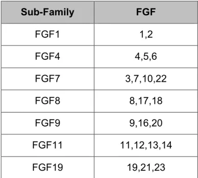
In vertebrates, FGFs can be classified into different subfamilies that share sequence similarity, and biochemical and developmental properties (Ornitz and Itoh, 2001). Phylogenic analysis divides the human FGF genes into seven subfamilies (Table 1). The chromosomal locations of all human FGF genes (except FGF16) are known and most human FGF genes are scattered through the genome indicating that they were generated by gene duplications and translocations during evolution (Itoh and Ornitz, 2004). The human FGF11 and FGF7 subfamilies each consist of four closely related members. They may have arisen from an ancestral gene by two successive genome duplications (Itoh and Ornitz, 2004).
Table 1: Evolutionary relationship within the human FGF gene family. Twenty-two genes have been identified, and phylogenic analysis suggests that these genes can be arranged into seven subfamilies, each containing two or four members (Ornitz and Itoh, 2001).
Sub-Family FGF FGF1 1,2 FGF4 4,5,6 FGF7 3,7,10,22 FGF8 8,17,18 FGF9 9,16,20 FGF11 11,12,13,14 FGF19 19,21,23 5.2 FGF receptors
FGFs act by binding to transmembrane tyrosine kinase receptors (RTKs) on the cell surface (Fantl et al., 1993). RTKs are plasma membrane receptors that control multiple fundamental cellular processes during development and adult life, including cell cycle, migration, metabolism, survival, proliferation, and differentiation (Lemmon and Schlessinger, 2010). RTKs are single-pass membrane proteins with an extracellular ligand-binding domain and an intracellular kinase domain that distinguishes RTK
from all other receptors. Humans have 58 known RTKs, which fall into 20 subfamilies based on their amino acid sequence similarities, their structural architectures and biological functions (Bae and Schlessinger, 2010). These families include the epidermal growth factor receptors (EGFRs), the fibroblast growth factor receptors (FGFRs), the insulin and the insulin-like growth factor receptors (IR and IGFR), the platelet derived growth factor receptors (PDGFRs), the vascular endothelial growth factor receptors (VEGFRs), the hepatocyte growth factor receptors (HGFRs), and the nerve growth factor receptors (NGFRs) (van der Geer et al., 1994).
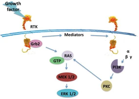
Despite the diversity of RTKs, there is a great degree of commonality in the types of intracellular signaling pathways initiated by these proteins. In mammalian systems, biochemical and molecular genetic analyses have shown that for all RTKs, the binding of ligand to the extracellular domain activates the tyrosine kinase in the cytoplasmic domain. This leads to downstream activation of a number of common signaling molecules. Frequently activated proteins include phospholipase C-γ, phosphatidylinositol 3-kinase (PI3K), GTPase-activating protein, pp60c-src, p21ras, Raf-l kinase, ERK 1 and ERK 2 kinases (also referred to as MAP kinases), and S6 ribosomal kinases (Figure 8). Ultimately, the activation of signaling pathways involving these molecules leads to changes in gene expression and a change in the phenotypic state of the cell (Fantl et al., 1993).
Figure 8: RTK signaling. Transactivation of multiple pathways of ERK/MAPK signaling by RTK activation (Wetzker and Bohmer, 2003).
The family of FGFRs includes four major receptors (FGFR1-4) that like other RTKs are activated by ligand induced receptor dimerization followed by tyrosine kinase activation and autophosphorylation of specific tyrosine residues in the cytoplasmic region, a process shown to be mediated by a precise and sequentially ordered reaction (Furdui et al., 2006).
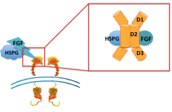
One characteristic of the FGF activation of the FGFRs is that it requires the cooperation of the accessory molecule heparin sulfate proteoglycan (HSPG) to stabilize FGFR dimers at the cell surface under normal physiological conditions (van der Geer et al., 1994). Another crucial factor for
binding with the FGF ligands is the extracellular regions of FGFR, which contain three Ig-like domains (D1-D3) of which D2 and D3 are essential for FGF binding (Figure 9). Receptor affinity is determined by the receptor genes and also by alternative splicing of the FGFR mRNA. One RNA splicing event in the third Ig-like domain results in three different versions of D3, usually called IIIa, IIIb and IIIc variants for FGFR 1-3 (FGFR4 is not alternatively spliced). The IIIb and IIIc variants of each receptor are expressed on the cell surface and are fully active receptors. However, the IIIa splice variant is usually inactive, but a further splicing event that removes the entire third Ig domain results in a variant that is activated by low concentrations of FGF1 (Tomlinson and Knowles, 2010).
Figure 9: FGFR domains. Extracellular regions containing three Ig-like domains (Lemmon and Schlessinger, 2010)
FGF1 activates all receptor proteins, whereas FGF2 activates only the “c” splice variants of FGFR1, FGFR2 and FGFR3 as well as FGFR4. Receptor activation pattern tends to be similar within FGF subfamilies, for example, both FGF7 and FGF10 activate almost exclusively only FGFR2b, and members of the FGF8 subfamily preferentially bind to FGFR3c and FGFR4 (Zhang et al., 2006). In the bovine ovary, all FGFR are expressed. FGFR3 and 4 are both activated by FGF8 and FGF18. FGFR3c is expressed in granulosa and theca cells while FGFR4 is only expressed in theca cells. Their expression appears to be involved with follicular growth; FGFR4 mRNA levels decrease as follicular size increases, and FGFR3c mRNA levels increase in healthy follicles and is positively correlated with follicular estradiol levels (Buratini et al., 2005b). It is now well established that several FGFR mutations are linked to developmental disorders, including a variety of human cancers that are caused by gain or loss of function. It has been suggested that changes in the alternative-splicing pattern of the receptors are correlated with the progression of several tumors towards malignancy (Ezzat and Asa, 2005; Tomlinson and Knowles, 2010).
5.3 FGF signaling pathways
FGF signaling is generally mediated by a dual-receptor system consisting of high affinity FGFR and low affinity heparan sulfate proteoglycan receptors that are most often lacking of signaling capabilities but enhance ligand presentation to the receptors. FGF binding results in receptor oligomerization, activation of the cytoplasmic tyrosine kinase domains and receptor autophosphorylation. Intracellular signaling is mediated by tyrosine phosphorylation of key substrates and activation of downstream pathways. The main activated pathways are the mitogen activated protein kinases (MAPK), protein kinase C (PKC) and PI3K (Kornmann et al., 1998) .
Figure 10: FGF signaling pathways. Main pathways activated by FGFs include MAPK, PKC and PI3K (Kornmann et al., 1998).
MAPKs are widely believed to be responsible for the mitogenic responses of cells to FGF actions. Activation of MAPK is always observed in response to FGF (Portela et al., 2010). Signaling pathways include ERK 1/2, p38 and JNK kinases. The activation of ERK 1/2 and p38 has been observed in all cell types examined, and involves receptor mediated recruitment to the receptor of the docking protein FRS2a along with tyrosine phosphatase Shp2, the adaptor Grb2 and the docking protein GAB1. It seems that FGF signals induce a MAPK mediated negative feedback loop that leads to a reduction in the recruitment of the adaptor Grb2. This negative loop also includes signals via activation of the Sprouty proteins that also inhibit the
recruitment of Grb2. Meanwhile XFLRT3, a member of a leucine-rich-repeat transmembrane protein family, is also activated by FGFs and acts as a positive modulator (Dailey et al., 2005). It has also been suggested that MAPK phosphorylation, Sprouty protein and XFLRT3 expression are modulated in strength and in duration by FGFs and that different levels of activation of these molecules may lead to differential responses to each FGF (Dailey et al., 2005).
The activation of the PKC pathway involves the release of intracellular calcium, and requires the recruitment of PLC-y to the FGFR domain. Inhibition of this pathway seems to have no effect on the proliferative response of cells (Dailey et al., 2005). PI3K is known as the survival pathway activated by FGFs by leading to activation of the AKT pathway, which affects the expression of pro-apoptotic factors. Akt also blocks the activity of GSK-3β, which enhances antiapoptotic signal (Dailey et al., 2005; Hacohen et al., 1998).
5.4 FGF early-response genes
Sprouty proteins (SPRY) are a family of four cysteine-rich proteins that were first described as antagonists of FGF-stimulated apical branching of the airway in Drosophila. FGF signaling induces Sprouty expression, and Sprouty acts in a competitive fashion to inhibit intracellular FGF signaling (Hacohen et al., 1998). In cattle, FGF2 (Figure 11) stimulates the expression of SPRY1, 2 and 4 in bovine granulosa cells through ERK 1/2 and Akt signaling, as well as the PKC pathway (Jiang et al., 2011). In the case of SPRY2, intracellular calcium flux is critical and sufficient for its up-regulation in granulosa cells independently of the PKC pathway (Jiang et al., 2011). Studies in mouse oocytes have showed that FGF8 cooperates with BMP15
to suppress cumulus cell expression of Spry2 mRNA in growing follicles but to promote Spry2 mRNA levels stimulated by EGF in periovulatory follicles (Sugiura et al., 2009).
Figure 11: FGF8 signaling pathway; FGF8 activation of MAP-kinase cascade (Niehrs and Meinhardt, 2002).
FGF has also been linked to expression of members of the NR4A family of orphan nuclear receptors, which are involved in cell cycle mediation, inflammation and apoptosis. In bovine granulosa cells, NR4A1, NR4A2 and NR4A3 are rapidly induced by FGF, and in the case of NR4A1, the activation by FGF2 seems to be dependent on intracellular calcium signaling. Overexpression of NR4A1 resulted in a repression of aromatase transcription that may explain the inhibitory effects of FGFs on E2 production (Jiang et al., 2011; Ohno et al., 2009).
As FGFs have a major role during organ development in the embryo, they have been associated with the ETS family of transcription factors. In limb buds, FGF signaling through ETV4 and ETV5 control proximal-distal limb outgrowth and promotes sonic hedgehog expression in the posterior limb. In the mouse, ETV4 and ETV5 have been found in GC and CC during folliculogenesis while in the bovine ovary they are present only in GC and their mRNA levels can be stimulated by FGF2. The function of ETVs has not been described in the follicle (Zhang et al., 2009) (Jiang et al., 2011).
5.5 Role of FGF
The expression patterns of FGFs suggest that they have an important role in development, and in the development and progression of various malignant diseases. FGF2 exerts mitogenic effects and is over-expressed in human tumor cell lines, however FGF2 expression may also be associated with favorable prognosis in ovarian and breast cancer. FGF1 can display biological activities similar to those of FGF2. FGF3 was initially identified as an oncogene implicated in mouse mammary tumors. FGF4 is also called “human cancer transforming factor-1”, and its over-expression also increased the metastic potential of breast cancer cells in association with
altered expression of matrix metallo-proteinases. In addition, FGF5 is also expressed in breast cancer and in some gastrointestinal and urinary tract cancers along with FGF6. In the case of FGF8, it has been proposed to enhance murine mammary tumorogenesis in cooperation with Wnt-1 proto-oncogene (Kornmann et al., 1998).
FGFs are known for their non-pathological roles in the developing embryo. FGF10 in vertebrates is critical for limb development and FGF2, FGF9 and FGF18 play roles in gonadal development and sex differentiation (Ornitz and Itoh, 2001; Portela et al., 2010).
5.6 Role in the ovary
FGFs control cell proliferation and in early studies they were found to be one of the most potent mitogens, specially in GC were they produced a 300-3000 fold change in GC mitogenesis (Gospodarowicz et al., 1977). In rat granulosa cells, FGF inhibited the ability of FSH to stimulate E2 production and to induce LH receptors, but with suboptimal concentrations of FSH, FGFs enhanced the synthesis of P4 (Baird and Hsueh, 1986). This demonstrated the ability of FGFs to differentially regulate steroidogenesis in GC.
FGF2 is well known as an inhibitor of steroidogenesis, and one of its roles is the regulation of angiogenesis. Early studies performed with rat GC demonstrated the effects of FGF2 on E2 and P4 production, where treatment with FGF2 inhibited FSH-induced E2 and P4 production in mature GC but increased E2 levels in FSH-primed GC; these opposite effects indicated a role for FGF2 as a mediator of follicular development, ovulation and luteinization. Also, FGF2 inhibited SERPINE2 expression in bovine GC,
and SERPINE2 is correlated with E2 secretion (Cao et al., 2006). FGF2 has a mitogenic effect on monolayer cultures of GC (Lavranos et al., 1994). In bovine GC, FGF2 leads to a rapid up-regulation of the orphan nuclear receptor NR4A1, which is correlated with an inhibition of steroidogenesis (Jiang et al., 2011). FGF2 increased the levels of GADD45B mRNA in bovine GC, which has been associated with cell proliferation and survival in non-ovarian cell types (Jiang et al., 2011).
FGF7 is also known as keratocyte growth factor. In rats FGF7 is localized in the follicle from early preantral stages, specifically in mesenchymal cells, and in primordial follicles FGF7 interacts with the epithelial growth factor kit ligand (KITL) to promote transition to the primary stage. This interaction creates a feedback loop where primordial follicles will produce KITL and thus promote TC formation, which in turn will produce FGF7 that promotes the production of KITL from GC (Kezele et al., 2005). In cattle FGF7 is present in TC and GC along with its receptors FGFR2IIIb and FGFR3IIIc, and mRNA levels increase with follicular growth supporting a role for this FGF in folliculogenesis and angiogenesis (Berisha et al., 2004).
FGF9 mRNA and protein, and its receptor FGFR3, are present in rat ovaries, mainly in GC where they have been linked to P4 production. Studies in vitro have shown that FGF9 combined with FSH stimulated P4 production by GC, and this was associated with increased P450 side-chain cleavage mRNA levels (Drummond et al., 2007). In cattle, FGF9 may act as an autocrine differentiation factor regulating ovarian function as it is present in higher amounts in small follicles (1-5mm) compared to large follicles. FGF9 stimulated GC proliferation and inhibited FSHR and CYP11A1 mRNA abundance; FGF9 increased proliferation and also inhibited LHCGR, CYP11A1 and CYP17A1 mRNA levels in TC. In summary FGF9 regulates
ovarian function by inhibiting gonadotropin receptors and the cAMP signaling cascade while stimulating proliferation (Hacohen et al., 1998; Schreiber and Spicer, 2012; Schreiber et al., 2012).
FGF10 mRNA has been detected in oocytes and TC of preantral and antral bovine follicles while its protein in oocytes, GC and TC from antral follicles. FGF10 expression varies during follicular growth, decreasing as follicle E2 content increases. This, coupled with the inhibition of E2 secretion caused by the addition of FGF10 to granulosa cells in vitro, led to the hypothesis that FGF10 acts to restrain GC differentiation in small growing follicles, and as FGF10 levels decrease, the GC differentiate and secrete greater amounts of E2 (Buratini et al., 2007).
6. FGF8 and FGF18
The FGF8 subfamily consists of three members, FGF8, FGF17 and FGF18. They have 70-80% amino acid sequence identities, similar receptor binding properties and some overlapping sites of expression (Ornitz and Itoh, 2001). The three members are closely linked to the nucleophosmin genes indicating that these FGF might have arisen from a common ancestral gene (Itoh and Ornitz, 2004). Another important characteristic is that the ligands from this family have similar receptor activation patterns and it could be proposed that they would have the same actions on bovine granulosa cells.
FGF8 is a mitogenic growth factor, and in adult mice it is only detected in the oocyte. Studies in knockout mice demonstrated that the lack of FGF8 produced abnormalities of the estrous cycle and a reduction in GC proliferation (Lan et al., 2008). Again in mice, FGF8 acts as a paracrine
factor to promote glycolysis in cumulus cells, and does so in cooperation with BMP15 (Sugiura et al., 2009). Studies in rats demonstrated that FGF8 suppressed FSH-induced E2 production in GC while not affecting P4 and cAMP levels, but in the presence of BMPs there was a suppression of P4 secretion and cAMP levels, making this interaction critical in the regulation of steroidogenesis (Miyoshi et al., 2010). In cattle FGF8 is detected in the oocyte, TC and GC, and both FGF8 receptors, FGFR3c and FGFR4, are expressed within the follicle (Buratini et al., 2005a). In antral follicles, FGFR3c is expressed in GC and TC, and FGFR4 exclusively in TC. FGFR3c expression is up-regulated by FSH, and increased along with E2 levels, a known marker for follicular health (Buratini et al., 2005b).
FGF18 has been detected in oocytes in mice (Zhong et al., 2006), whereas in cattle it was detected in GC and TC but not in oocytes (Portela et al., 2010). FGF18 mRNA abundance is lower in healthy dominant follicles compared to the regressing follicles suggesting a down-regulation during follicular growth and up-regulation during follicular atresia. In addition, FGF18 inhibits E2 and SerpinE2 secretion, which are considered markers of non-atretic follicles, and it reduces the secretion of P4 (Portela et al., 2010). Interestingly, FGF18 reduced the expression of GADD45B, a cell cycle regulator known for its role in the protection of GC from apoptosis; FGF18 also increased the proportion of atretic cells as determined by DNA fragmentation and cell cycle analysis (Portela et al., 2010). This was the first reported incidence of an apoptotic action for an FGF in the follicle.
Hypothesis and Objectives
As mentioned in the preceding paragraph, FGF8 and FGF18 are members of the same FGF subfamily and possess similar receptor binding. It could be proposed that they would have the same effects on bovine granulosa cells, but FGF8 is a mitogenic growth factor that increases follicular health while the FGF18 appears to be a pro-apoptotic factor.
Our hypothesis is that upon FGFR activation, FGF8 and FGF18 activate different signaling pathways in bovine granulosa cells.
The objective of the present study was to determine the early response genes in FGF8 and FGF18 pathways, and gain insight into how FGF18 is pro-apoptotic whereas the typical FGF signaling is pro-survival.
Materials and methods Primary cell culture
All materials were obtained from Life Technologies Inc (Burlington, ON, Canada). Bovine granulosa cells were cultured in serum-free conditions that maintain estradiol secretion and responsiveness to FSH (Gutierrez et al., 1997; Sahmi et al., 2004; Silva and Price, 2000). Under these conditions, cells respond to FGF2 with phosphorylation of ERK1/2 and Akt, and acute increases in abundance of mRNA encoding SPRY2, SPRY4 and NR4A1 (Jiang et al., 2011). Bovine ovaries were obtained from adult cows, irrespective of stage of the estrous cycle, at an abattoir and transported to the laboratory at 30°C in phosphate-buffered saline (PBS) containing penicillin (100 IU/ml), streptomycin (100 mg/ml) and fungizone (1 mg/ml). Granulosa cells were harvested from follicles 2 – 5 mm diameter, and the cell suspension was filtered through a 150 mesh steel sieve (Sigma-Aldrich Canada, Oakville ON). Cell viability was assessed by Trypan blue dye exclusion. Cells were seeded into 24-well tissue culture plates (Sarstedt Inc., Newton, NC) at a density of 1 million viable cells in 1 ml DMEM/F12 containing sodium bicarbonate (10 mmol/l), sodium selenite (4 ng/ml), bovine serum albumin (BSA) (0.1%; Sigma-Aldrich), penicillin (100 U/ml), streptomycin (100 mg/ml), transferrin (2.5 mg/ml), nonessential amino acid mix (1.1 mmol/l), bovine insulin (10 ng/ml), androstenedione (10-7 M at start of culture and 10-6 M at each medium change) and bovine FSH (10 ng/ml starting on day 2; AFP5346D; National Hormone and Peptide Program, Torrance, CA). Cultures were maintained at 37°C in 5% CO2, 95% air for 5 days, with 70% medium being replaced on days 2 and 4.
Cell lines
Different cell lines were used including three human cell lines: KGN (Nishi et al., 2001), HEK293t (Graham et al., 1977) and SVG (Major, 1987), and a bovine granulosa cell line, A1 (donated by Khampoune Sayasith, CRRA, Université de Montréal). KGN, HEK293t, and A1 cells were cultured in DMEM/F12 medium containing fetal bovine serum (10%) and gentamycin (50 µg/ml). SVG cells were cultured in Opti-MEM medium containing fetal bovine serum (5%) and gentamycin (50 µg/ml). Cells were cultured in 20 ml flasks (Sarstedt Inc., Newton, NC) until they reached confluence, after which they were removed from the flask using phosphate-buffered saline (PBS) and trypsin (0.05% for SVG cells and 0.25% for the other lines). Cells were then seeded in 24-well tissue culture plates (Sarstedt Inc., Newton, NC) at a density of 3 X 10⁻⁵/well and maintained at 37°C in 5% CO2, 95% air until cells reached confluence and then they were treated with FGFs.
Experimental treatments
The effect of FGF8 and FGF18 on granulosa cells was assessed in separate cultures with time and dose response experiments. Recombinant human FGF8 and FGF18 (PeproTech) were added on day 5 for 0, 1, 2, 4 and 8 h at a dose of 10 ng/ml in PBS. For the microarray experiment, the 2 h and 0 h time points were compared for each FGF. For every experiment, a pool of cells collected on a specific day constituted one replicate, and all experiments were performed with three independent replicates.
Total RNA extraction and RT-PCR
After treatments, the culture medium was removed and total RNA was extracted using Trizol according to the manufacturer’s instructions. Total RNA was quantified by absorbance at 260 nm. Reverse transcription was performed on 1 µg DNase-treated total RNA in the presence of 1 mmol/l oligo (dT) primer and 4 U Omniscript RTase (Qiagen, Mississauga, ON, Canada), 0.25 mmol/l dideoxynucleotide triphosphate (dNTP) mix and 19.33 U RNase Inhibitor (GE Healthcare, Baie D’Urfé, QC, Canada) in a volume of 20 µl at 37°C for 1 h. The reaction was terminated by incubation at 93°C for 5 min.
Microarray analysis
RNA from the FGF8 time-course and the FGF18 time-course experiments were used for microarray analysis to detect early-response genes activated by each FGF. The 2 h and 0 h time points were compared for each FGF. RNA samples were amplified with the RiboAmp HSPlus RNA Amplification kit (Life Technologies Inc) and labeled with Cy3 and Cy5 with the ULS Fluorescent Labeling kit (Kreatech Inc, Durham NC). The EmbryoGENE bovine microarray contains 42,242 probes and has been described in detail elsewhere (Robert et al., 2011). Samples were hybridized with the array in a dye-swap design for 17 h at 65°C, followed by washes in Expression Wash Buffer 1 for 1 min at room temperature, in Expression Wash Buffer 2 at 65°C for 3 min, for 10 sec in 100% acetonitrile, and for 30 sec in Stabilization and Drying Solution (Agilent Technologies Canada, Mississauga ON). The array was scanned with a PowerScanner (Tecan US Inc, Durham, NC) and fluorescence intensities analyzed on the ELMA platform (elma.embryogene.ca). After background subtraction and
normalization (Loess), genes that were significantly (P<0.05) altered at least 2-fold at 2 h of treatment compared to time 0 control were identified with the Limma algorithm. Allocation of genes to common pathways was investigated with Ingenuity Pathway Analysis (IPA; Ingenuity Systems, Redwood City, CA). Raw and normalized microarray data for FGF8 (GEO: GSE41489) and FGF18 (GEO: GSE41480) have been deposited in the NCBI GEO database. To verify microarray results, further cultures were performed with addition of FGF8 or FGF18 (10 ng/ml for each) for 0, 1, 2, 4 and 8 h, and abundance of mRNA measured by real-time PCR as described above. Primers for the genes identified by microarray are given in Table 2, and were designed so that the amplicon spans an exon/exon junction. Amplicon authenticity was verified by sequencing all products.
Table 2: Primer sequences for microarray validation.
Gene symbol
Forward primer Reverse primer
EGR1 AAGCGAGCAGCCCTACGA GCAGCCGGGTGGTTTG
FOS ATGGGTTCTCCCGTCAATGC GGTCGAGATGGCAGTCACTGT
BAMBI TCGCCACTCCAGCTACATCTT TGGGCTGCATCACAGTAGCA
FOSL1 AGTGCAGGAACCGGAGGAAA TCTCTCGCTGCAGTCCAGATT
XIRP1 CAAACAAGAGGAACCGACAGA GGCATTGGCCATCCTTCT
PLK2 GAACCCTTGGAACACAGGAGAA TTCACAGCCGTGTCCTTGTTT
Real-time PCR
Real-time PCR was performed on a 7300 Real-Time PCR system (Applied Biosystems, Streetsville ON, Canada) with Power SYBR Green PCR Master Mix. The bovine-specific primers for target genes other than those listed in Table 1 have previously been published (Jiang et al., 2011). Common thermal cycling parameters (3 min at 95°C, 40 cycles of 15 sec at 95°C, 30 sec at 59°C, and 30 sec at 72°C) were used to amplify each transcript. Melting-curve analyses were performed to verify product identity. Samples were run in duplicate and were expressed relative to histone H2AFZ as housekeeping gene. This gene is routinely used in our laboratory, and shows similar stability to cyclophilin A, both of which were more stable in granulosa cells than glyceraldehyde-3-phosphate dehydrogenase as determined by geNorm software (Ramakers et al., 2003). Data were normalized to a calibrator sample using the ΔΔCt method with correction for amplification efficiency (Pfaffl, 2001).
Statistical analyses
All statistical analyses were performed with JMP software (SAS institute, Cary NC). Data were transformed to logarithms if they were not normally distributed (Shapiro-Wilk test). The data are presented as least square means ± SEM.
Results
Microarray analysis of FGF8 and FGF18 activated early response genes In order to explore early-response genes activated by FGF8 and FGF18, a microarray analysis was performed on samples collected 0 h and 2 h after challenge with FGF. The microarray software identified 12 genes up-regulated by FGF8 (P<0.05) (Table 1) including genes already known to be FGF target genes, SPRY2 and NR4A1. Also up-regulated were genes that had never been studied in relation with FGFs, such as EGR1, XIRP1, HAS2, FOS, BAMBI, PKL2 and CTGF. These genes had a significant role in reproduction or were related to a growth factor network by Ingenuity Pathway Analysis (IPA) (Fig 12). In contrast, no gene was significantly regulated by FGF18.
Table 3: Genes significantly regulated by FGF8 in bovine granulosa cells.
Gene symbol
Gene name Fold
change
EGR1 Early growth response 1 3.90
XIRP1 Xin actin-binding repeat containing 1
3.68
HAS2 Hyaluronan synthase 2 3.61
FOS FBJ murine osteosarcoma viral oncogene homolog
2.49
BAMBI BMP and activin membrane-bound inhibitors homolog
2.93
SPRY2 Sprouty homolog 2 3.14
NR4A1 Nuclear receptor subfamily 4, group A, member 1
2.62
PLK2 Polo-like kinase 2 2.13