HAL Id: inserm-00667523
https://www.hal.inserm.fr/inserm-00667523
Submitted on 7 Feb 2012HAL is a multi-disciplinary open access archive for the deposit and dissemination of sci-entific research documents, whether they are pub-lished or not. The documents may come from teaching and research institutions in France or abroad, or from public or private research centers.
L’archive ouverte pluridisciplinaire HAL, est destinée au dépôt et à la diffusion de documents scientifiques de niveau recherche, publiés ou non, émanant des établissements d’enseignement et de recherche français ou étrangers, des laboratoires publics ou privés.
Farnesyl diphosphate synthase is involved in the
resistance to zoledronic acid of osteosarcoma cells.
Benjamin Ory, Gatien Moriceau, Valérie Trichet, Frédéric Blanchard, Martine
Berreur, Françoise Rédini, Michael Rogers, Dominique Heymann
To cite this version:
Benjamin Ory, Gatien Moriceau, Valérie Trichet, Frédéric Blanchard, Martine Berreur, et al.. Farnesyl diphosphate synthase is involved in the resistance to zoledronic acid of osteosarcoma cells.: resistance of osteosarcoma to nitrogen bisphosphonates. Journal of Cellular and Molecular Medicine, Wiley Open Access, 2008, 12 (3), pp.928-41. �10.1111/j.1582-4934.2008.00141.x�. �inserm-00667523�
Farnesyl diphosphate synthase is involved in the resistance to zoledronic
acid of osteosarcoma cells
Ory B.1, 2, Moriceau G. 1, 2, Trichet V. 1, 2, Blanchard F. 1, 2,
Berreur M. 1, 2, Rédini F. 1, 2, Rogers M3, Heymann D.1, 2, 4 *
1. INSERM, ERI 7, Nantes, F-44035 France
2. Université de Nantes, Nantes Atlantique Universités, Laboratoire de Physiopathologie
de la Résorption Osseuse et Thérapie des Tumeurs Osseuses Primitives, EA3822, Nantes, F-44035 France
3. Bone & Musculoskeletal Research Programme, Institute of Medical Sciences, University of Aberdeen, UK.
4. Hospital, Nantes, F-44035 France
*Corresponding author : Laboratoire de Physiopathologie de la Résorption Osseuse et
Thérapie des Tumeurs Osseuses Primitives, EA3822 - INSERM ERI 7, Faculté de Médecine, 1 rue Gaston Veil. 44035 Nantes cedex 1 France.
Tel : 33 240 412 845 ; Fax : 33 240 412 860 ; Email :
dominique.heymann@univ-nantes.fr
Key words: osteosarcoma; bisphosphonate; zoledronic acid; metabolic resistance;
farnesyl diphosphate synthase
Abstract
We recently demonstrated original anti-tumor effects of zoledronic acid (Zol) on osteosarcoma cell lines independently of their p53 and Rb status. The present study investigated the potential Zol-resistance acquired by osteosarcoma cells after prolonged treatment. After 12 weeks of culture in the presence of 1µM Zol, the effects of high doses of Zol (10 to 100µM) were compared between the untreated rat (OSRGA, ROS) and human (MG63, SAOS2) osteosarcoma cells and Zol-pretreated cells in terms of cell proliferation, cell cycle analysis, migration assay and cytoskeleton organization. Long-term treatment with 1µ M Zol reduced the sensitivity of osteosarcoma cells to high concentrations of Zol. Furthermore, the Zol-resistant cells were sensitive to conventional anti-cancer agents demonstrating that this resistance process is independent of the multidrug resistance phenotype. However, as similar experiments performed in the presence of clodronate and pamidronate evidenced that this drug resistance was restricted to the nitrogen containing bisphosphonates, we then hypothesized that this resistance could be associated with a differential expression of farnesyl diphosphate synthase (FPPS) also observed in human osteosarcoma samples. The transfection of Zol-resistant cells with FPPS siRNA strongly increased their sensitivity to Zol. This study demonstrates for the first time the induction of metabolic resistance after prolonged Zol treatment of osteosarcoma cells confirming the therapeutic potential of Zol for the treatment of bone malignant pathologies, but points out the importance of the treatment regimen may be important in terms of duration and dose to avoid the development of drug metabolic resistance.
Introduction
Osteosarcoma is the most frequent malignant primary bone tumor that develops mainly in the young, the median age of diagnosis being 18 years [1]. Despite recent improvements in chemotherapy and surgery, the problem of non-response to chemotherapy remains. Thus, current strategies for the treatment of high-grade osteosarcoma fail to improve its prognosis [2, 3], mainly because of chemotherapy resistance. This poor prognosis of osteosarcoma warrants new therapeutic strategies to improve the overall rate of survival.
Bisphosphonates (BPs) are stable synthetic analogues deriving from endogenous pyrophosphate (PPi) [4]. Various side chains can be added to the central carbon atom, thus producing a range of BPs with differential clinical activity and potency [5]. The most common therapeutic application of BPs is osteoporosis, and their use has been extended to the treatment of malignant osteolysis and hypercalcemia. Two groups of BPs can be identified including non-nitrogen-containing and nitrogen-containing BPs. The BPs that lack a nitrogen atom, closely related to PPi (such as clodronate, etidronate and tiludronate) are metabolized intracellularly to cytotoxic analogues of ATP and decrease osteoclast survival [5]. In contrast, nitrogen-containing bisphosphonates (such as pamidronate, alendronate, risedronate, ibandronate and zoledronate) induce apoptosis of osteoclasts by inhibiting enzymes of the mevalonate pathway, especially farnesyl diphosphate synthase (FPPS) [6, 7] FPPS prevents the biosynthesis of cholesterol and isoprenoid lipids (FPP and geranylgeraniol diphosphate) which are required for the prenylation of small GTPases (i.e. Ras, Rho, and Rac), a biochemical reaction essential for the anchorage of small GTPases to cell membranes and to protein-protein interactions
[8]. In addition to their powerful anti-bone resorption effects, recent in vitro studies
cells (myeloma, carcinoma and sarcoma) [9, 10]. Preclinical data confirmed the Zol anti-tumor activity in experimental models of bone anti-tumors. Among these studies, we reported recently the enhancement of tumor regression and tissue repair when Zol is combined with ifosfamide in rat osteosarcoma [11] and that Zol suppresses lung metastases and prolongs overall survival of osteosarcoma-bearing mice [12]. Furthermore, recent clinical trials in patients suffering from malignant bone diseases demonstrated that Zol was safe and well tolerated at the approved dose of 4 mg i.v. every 3-4 weeks [4]. Because the main difficulty encountered in treating cancer relates to mutations carried by many tumor cells in key genes such as p53, Rb or proteins affecting caspase signalling, we demonstrated selective and original anti-tumor effects of Zol on several osteosarcoma cell lines independently of their p53 and Rb status [13]. Indeed, Zol inhibited osteosarcoma cell proliferation through a cell cycle arrest in S and G2/M phases and induced atypical apoptosis independent of caspase activation, characterized by the translocation of Apoptosis Inducing Factor and Endonuclease-G [13]. These data now allow to consider these molecules as potential therapeutic agents in clinical trials of tumor bone pathologies independently of the p53 and Rb status of the tumor.
The optimization and increase in specificity of cancer treatments has improved their efficacy and reduced the associated adverse effects, but unfortunately has not yet resulted in a cure for the majority of patients. Studies of the mechanisms by which tumor cells escape treatment is essential to circumvent drug resistance in cancer cells and to design new therapeutic protocols that are not subject to these drug-resistances [14]. Two types of resistance mechanism have been identified [15]. The first one results in resistance restricted to a specific drug or limited to a small number of related drugs which can be bypassed by modification of the chemotherapeutic agent. The second mechanism confering multi-resistance to many unrelated drugs, is called multidrug resistance (MDR)
and is responsible for many failures of cancer treatment [16]. The most common mechanisms responsible for the various forms of resistance are the overexpression of efflux pumps, inhibition of apoptosis, increased repair of DNA damage, mutations in key cell cycle checkpoint genes and increased or altered drug targets [14]. Similar to non-osseous malignancies, osteosarcomas frequently exhibit a MDR phenotype explaining why patient survival has not improved since the mid-1980s despite advances in anticancer therapies. Because Zol represents a potential novel anti-neoplastic agent for the therapy of osteosarcoma, the present study investigated the potential development of innate and/or acquired resistance to Zol and the molecular mechanisms involved in this phenomenon.
Material and Methods
Patients
This study included 7 patients (3 females aged 41-93 years, 4 males aged 16-79 years) that were referred to our institution for the treatment of osteosarcoma. All cases were diagnosed as osteogenic osteosarcoma based on histological samples obtained by open biopsies. The experimental procedures followed in the present study were in accordance with the ethical standards of the responsible institutional committee on human experimentation and with Helsinki Declaration of 1975, revised in 1983. The study was approved by the institutional ethic committee.
Cells, culture conditions and establishment of Zol-resistant cell lines
The rat osteosarcoma OSRGA cell line was initially established from a radio-induced osteosarcoma [17, 18], the rat ROS17/2.8 cell line was kindly provided by Prof H.J. Donahue (Penn State University, USA) and the human MG63 and SAOS2 cell lines were purchased from ATCC (USA). These cell lines were cultured in DMEM (BioWhittaker, Belgium) supplemented with 5% Fetal Calf Serum (Hyclone, France) and
2 mM L-glutamine (BioWhittaker). Rat and human osteosarcoma cell lines resistant to Zol (MG53res, SAOS2res, ROSres, OSRGAres) were established by 3 months of continuous treatment with 1µM Zol.
Cell growth and viability
Cell growth and viability were determined by a cell proliferation reagent assay kit
using sodium
3’[1-(phenylaminocarbonyl)-3,4-tetrazolium]-bis(4-methoxy-6-nitro)benzene sulfonic acid hydrate (XTT) (Roche Molecular Biomedicals, Germany). Two thousand cells/well were plated into 96-well plates and cultured for 72h in culture
medium in the presence or the absence of 10-12 to 10-4 M Zol. Zol was provided by Novartis Pharma AG (Switzerland) as the disodium hydrate form. In another set of
experiments, cells were treated for 72 h in the presence or the absence of 10-9 to 10-6 M
methotrexate (Sigma, France), doxorubicine (Sigma) and 1 to 50 µg/ml mafosfamide (Baxter, France), 10 to 1000 µM clodronate (Sigma), 1 to 500 µM pamidronate (Sigma) and 5µM verapamil (Sigma). After the culture period, XTT reagent was added to each well and incubated for 5h at 37°C, the corresponding absorbance was then determined at 490 nm. Cell viability was also assessed by trypan blue exclusion and live and dead cells were scored manually. Cell death was also monitored microscopically after Hoechst
n°33258 staining (Sigma). In this experiment, cells were seeded at 104 cells/well in a
24-well plate and treated or not with 10 µM Zol for 48 hours or 100 nM staurosporine (Sigma) for 16 hours, stained by 10 µg/ml Hoechst reagent for 30 min at 37°C and then observed under UV microscopy (DMRXA, Leica, Germany).
Western blot analysis
Zol-treated cells were lysed in RIPA buffer (150 mM NaCl, 5% Tris pH 7.4, 1% NP-40, 0.25% Na deoxycholate, 1 mM Na3VO4, 0.5 mM PMSF, 10 µ g/ml leupeptin, 10
µg/ml aprotinin). Protein concentration was determined by the BCA kit(Pierce Chemical,
USA). Fiftyµ gof total cell lysate protein were run on SDS-PAGE, electrophoretically
transferred to Immobilon-P membrane (Millipore, MA, USA). The membrane was blotted with antibodies anti-p-Rb (Ser 807/811), -p-cdc2 (tyr15), -actin (Cell Signaling
Technologies, USA), -p21WAF1(BD Biosciences USA) and the unprenylated form of
Rap1A (Santa Cruz, USA) to indirectly quantified FPPS enzymatic activity, in PBS,
0.05%Tween 20, and 3%bovine serum albumin (BSA). The membrane was washed and
was then visualized with the enhanced chemoluminescence system (ECL Kit; Roche
Molecular Biomedicals). The band densities were measured using the GeneTools
computer software program (SynGene). Caspase -1, -3 and -8 activities
Caspase -1, -3 and -8 activities were assessed on 10 µl of total Zol-treated or not
cell lysates using the kit CaspACETM Assay System, “Fluorometric” (Promega, USA)
following the manufacturer’s recommendations. Cells treated with UV light for 30 seconds 24 h before harvesting were used as a positive control. Results were expressed in arbitrary units referred to the total protein content.
Cell cycle analysis
OSRGA, MG63 and SAOS2 cells were incubated in the absence or the presence of 10 µM Zol for 48 hours, trypsinized, washed twice and incubated in PBS containing 0.12% Triton X-100, 0.12 mM EDTA and 100 µg/ml ribonuclease A. Then 50 µg/ml propidium iodide were added to each sample for 20 min at 4°C. Cell cycle distribution was analyzed by flow cytometry (FAC Scan), based on 2N and 4N DNA content.
Time-lapse microscopy and confocal microscopic analysis
For time-lapse experiments, cells were seeded at 5 x 10⁴ cells/well and cultured in
6-multiwell plates in the absence or the presence of 10 µM Zol. Phase-contrast photographs (Leica) were taken every 10 min during 60 h and edited using the
MetamorphTM software. Cell divisions and apoptotic cells were then manually scored. To
study cell migration, cells plated in 6-well plates and cultured until confluence were treated or not with 10 µ M Zol for 24 h before a slit was made in the cell monolayer. Actin filament detection was performed after cell treatment with or without 10 µM Zol fixed in
4% paraformaldehyde and stained with FITC-conjugated phalloidin (0.25 µg/ml; Sigma). Cover glasses were fitted with the Long Pro Kit (Molecular probes). Images were collected on a Leica TCS-SP1 confocal microscope with 63/1.4x oil immersion lens. The digital images were visualized with a 24-bit imaging system including Leica’s TCS-NT software and projections were generated from z-stacks.
siRNA gene silencer
The FPPS gene expression was knocked down using specific human and rat FPPS
siRNA (Ambion, France) and the INTERFERinTM transfection reagent (Polyplus
transfection, France). Cells were seeded at 40% confluency in a 24-well plate one day before transfection. In each well 10 nM siRNA duplexes diluted in serum-free medium
were incubated with 2 µl of INTERFERinTM for 30 min at room temperature. Then, 100
µl mixture per well were added onto the cells and incubated at 37°C. The 72h-Zol treatment started 24 h after siRNA transfection. For each condition tested, a negative siRNA control was used (Santa Cruz biotechnology, Germany). Additional experiments were performed in the presence of geranylgeraniol (GGO) (Sigma, France).
RT-PCR analysis
Total RNA was isolated from cultured OSRGA, MG63 and SAOS2 cells using the TRIzol reagent (Invitrogen, France). First, RNA was reversed-transcribed (RT), using 400 U MMLV-RT from Invitrogen, then 2 µl of the RT reaction mixture were subjected to PCR using upstream and downstream primers to determine the expression of rat and human FPPS [Human FPPS sense: AGATCTGTGGGGGTCTTCCT, anti sense: TCCCGGAATGCTACTACCAC; Rat FPPS sense: AGTACAATCGGGGTCTGACG, anti sense: CGCGATAGGCAGGTAGAAAG] and 0.25 µl of 5 U/µl Taq polymerase
(Eurobio, France). After the number of PCR cycles was increased, a plot was done for each sample, the cycle values corresponding to the linear part of the amplification curve were then determined (28 cycles, Tm=58°C) and used to quantify the message versus the 18S signal determined in the same way. The PCR products were electrophoresed in 1% agarose gel containing ethidium bromide. The band densities were measured using the GeneTools computer software program. Three independent experiments were performed for each gene and a representative experiment is shown in the Results section.
Results
Osteosarcoma cell lines develop Zol-resistance after long-term continuous treatment
with low dose Zol
Consistent with previous results [11, 12, 19], Zol treatment of Zol-sensitive rat ROS, OSRGA(Figure 1A) and human MG63, SAOS2 (Figure 1B) osteosarcoma cells strongly reduced their proliferation. Thus, 0.1 to 100 µM Zol decreased the viable cell number in a dose-dependent manner (IC50: 1 to 8 µM) as revealed by the XTT assay.
After 3 month continuous treatment with 1 µM Zol, rat and human osteosarcoma cells
became less sensitive to Zol and resistant cell lines (OSRGAres, ROSres, MG63res, SAOS2res) were then progressively established (Figure 1A). Indeed, the potency of Zol to
affect cell proliferation was strongly reduced on human resistant cell lines and Zol was
ineffective on rat resistant cell lines (Figures 1A).
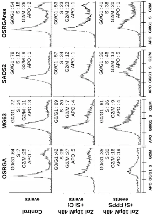
The influence of this resistance process was also assessed on the other known activities of Zol on tumor cells [cell cycle (Figure 1B), DNA checkpoints (Figure 1B), cytoskeleton (Figure 1C), cell migration (Figure 1C) [13]]. Cell cycle analysis was performed after 48h of 10 µM Zol-treatment. The results obtained confirmed that 48h of Zol-treatment induced a strong cell cycle arrest in S and G2/M phases in Zol-sensitive OSRGA cells (Figure 1B, [13]) and showed that Zol-treatment did not modulate the cell cycle in OSRGAres cells (Figure 1B). Indeed, the number of cells in S, G2/M phases strongly increased from 35% to 53% for OSRGA cells in the presence of 10 µM Zol
concomitantly with a decrease of cells in G0/G1 phase: 42% vs. 64% (Figure 1B). A
similar phenomenon was observed in human osteosarcoma cell lines (data not shown). We therefore investigated by western blot whether the DNA checkpoint proteins were involved in the cell cycle blockade observed in the presence of Zol. Thus, the treatment of sensitive OSRGA cells by 10 µM Zol increased the inactive form of cdc2 (p-cdc2, Tyr15)
after 72 hours of treatment. Simultaneously, Zol strongly reduced p21 expression and transiently upregulated Rb phosphorylation (Ser 807 and 811) after 24h of Zol treatment (Figure 1B). No modulation of p-cdc2, Rb and p21 was observed in OSRGAres cells regardless of the duration of Zol treatment (Figure 1B).
As Zol has been shown to disturb cytoskeletal organization and to inhibit cell migration [13], we wondered whether Zol could alter such parameters in OSRGAres cells. Confocal microscopy revealed a major disorganization of the actin stress fibres associated with membrane ruffling in sensitive OSRGA cells treated with 10 µM Zol for 72 h, this was never observed in OSRGAres cells (Figure 1C). Moreover, as shown by the time-lapse assay, 10 µM Zol totally blocked the migration of sensitive OSRGA cells but was not able to abolish migration of OSRGAres cells (Figure 1C).
.
The molecular mechanism involved in the reduced-Zol sensitivity is not associated
with a multidrug resistance (MDR) phenotype and is restricted to the
nitrogen-containing bisphosphonates
The potential role of the MDR phenotype in the Zol resistance phenomenon was assessed by XTT assays. The MDR phenotype is conventionally defined as the resistance of cells to conventional chemotherapeutic agents such as mafosfamide, methotrexate and doxorubicin [20, 21]. The XTT assays revealed that OSRGAres cells were still always sensitive to increasing doses of mafosfamide, methotrexate, and doxorubicin (Figure 2). Furthermore, 5µM verapamil, a P-gp pump inhibitor [22] was not able to abolish the Zol resistance (Figure 2). Overall, these data demonstrate that the Zol resistance was not associated with MDR phenotype. In addition, similar experiments performed in the presence of clodronate, a non-nitrogen containing-BP [4], revealed that OSRGAres are as
sensitive to clodronate as they are to lower concentrations of Zol (Figure 2). When, osteosarcoma cells were treated with another nitrogen-containing BP, pamidronate which also targets FPPS, it significantly reduced Zol-sensitive OSRGA proliferation in contrast to OSRGAres cells which are also resistant to pamidronate (Figure 2). Similar results have been obtained with the osteosarcoma cell lines MG63 and SAOS2 (data not shown). These experiments demonstrated that the Zol-resistance phenomenon in osteosarcoma cells appears to be MDR-independent and is apparently restricted to nitrogen-containing BPs.
Farnesyl diphosphate synthase (FPPS) is implicated in the Zol-resistance mechanism
of osteosarcoma cell lines
FPPS being the main molecular target of nitrogen containing BPs [23], the FPPS transcript expression was analyzed by RT-PCR and compared in sensitive OSRGA and OSRGAres cells (Figure 1D). Thus, the Zol resistant cells expressed a higher level of FPPS mRNA than the sensitive cells. To further determine the involvement of FPPS in the Zol-resistance mechanism of human and rat osteosarcoma cells, the effect of Zol on OSRGA, OSRGAres, MG63 and SAOS2 was analyzed after transfection with FPPS siRNA. Semi-quantitative RT-PCR analysis was used to evaluate the efficacy of FPPS siRNA on FPPS mRNA expression. In all experiments, FPPS mRNA levels were significantly decreased in FPPS siRNA transfected cell lines compared to the siRNA control (Figure 3A). Inhibition of FPPS activity was then assessed indirectly by the expression of the unprenylated form of the small GTPase Rap1A (unRAP1A) that is expressed after inhibition of FPPS [24, 25] (Figure 3B). The transfection of Zol-sensitive cells with FPPS siRNA strongly increased their sensitivity to Zol in all osteosarcoma cell lines studied. Indeed, FPPS siRNA transfection modified the unRAP1A expression kinetic in OSRGA, MG63 and SAOS2 cells. In the
presence of FPPS siRNA, unRAP1A expression was strongly induced by 1µM Zol treatment for 24h whereas its expression was only observed with 10 µM Zol treatment for 48h in control siRNA transfected cells (Figure 3B). In OSRGAres cells, a very weak expression of unRAP1A was observed after Zol treatment. Interestingly, FPPS siRNA re-induced the sentivity to Zol treatment in these resistant cells to a level comparable to parental OSRGA cells transfected with FPPS siRNA. Thus, the unRAP1A expression was observed after 24h treatment with 1µM Zol in FPPS siRNA-OSRGAres transfected (Figure 3B). Similarly, micoscopic observations confirmed the FPPS siRNA effects on the sensitization of osteosarcoma cells to Zol treatment (Figure 3C). Thus, an increase of floating cell number associated with an inhibition of cell proliferation was observed after transfection of all osteosarcoma cell lines with FPPS siRNA (Figure 3C).
XTT analyses were performed to determine the impact of FPPS siRNA on Zol activity (Figure 3D). Transfection with FPPS siRNA significantly increased the sensitivity to Zol treatment of all osteosarcoma cell lines analyzed (Figure 3D). The sensitivity to 10µM Zol was up-modulated by 22%, 31%, 53% and 42% in OSRGA, OSRGAres MG63 and SAOS2 respectively in the presence of FPPS siRNA compared to the control siRNA (Figure 3D). Furthermore, the efficacy of FPPS siRNA occured for lower doses of Zol in OSRGAres compared to OSRGA cells (respectively 22% and 1% increase of sensitivity in the presence of 0.1 µ M Zol) (Figure 3D).
siRNA FPPS increases the Zol-induced blockade of the cell cycle in S, G2/M phases
in osteosarcoma cell lines
We previously demonstrated that Zol induces osteosarcoma cell cycle arrest in S, G2/M phases in OSRGA sensitive cells [13]. To determine whether FPPS siRNA could modulate this sensitivity, the cell cycle of FPPS siRNA transfected osteosarcoma cells
was analyzed by flow cytometry. Figure 4 reveals that FPPS siRNA accentuates the Zol-induced effects observed on cell cycle distribution, leading to a significant increase of cells blocked in S phase compared to the control siRNA. Indeed, the number of cells in S phase increased from 26% to 30% for OSRGA, from 20% to 26% for MG63, from 34% to 46% for SAOS2 and from 23% to 38% for OSRGAres cells in the presence of FPPS siRNA compared to the control siRNA after 48h of treatment with 10 µM Zol (Figure 4). Furthermore, these observations were concomitant with a significant reduction of the cell number in G0/G1 phase: 35% vs. 42% for OSRGA, 61% vs. 69% for MG63, 36% vs. 57% for SAOS2 and 41% vs 53% for OSRGAres.
Geranyl geraniol (GGO) reversed the FPPS siRNA effects in osteosarcoma cell lines
To determine whether the effects previously demonstrated for the FPPS siRNA in osteosarcoma cells are reversible, FPPS siRNA transfected cells treated with increasing doses of Zol were cultured in the presence of 25µM geranylgeraniol, the FPPS metabolic product (Figure 5). GGO protected rat and human osteosarcoma cell lines from the effects of Zol in the FFPS siRNA transfected cells and totally reversed FPPS siRNA effects (Figure 5A). We therefore investigated by western blot the expression kinetic of unRAP1A in the presence of 25µM GGO in FPPS siRNA transfected cells (Figure 5B). GGO totally abolished unRAP1A expression similar to what had been observed in Zol-resistant cell lines (Figure 3B). Overall, these data then strengthen our conclusion that FPPS is involved in the Zol-resistance mechanism.
Dual origin of Zol resistance: innate and/or acquired
To explain the origin of the Zol-resistance observed in osteosarcoma cell lines, two hypotheses can be proposed: (i) an innate resistance mechanism linked to differential
levels of FPPS expression and associated with selection of a sub-population of cells expressing a higher FPPS activity, (ii) an acquired resistance mechanism linked to an increased FPPS transcription level as a feedback response to long-term, low dose Zol treatment. To distinguish between these two hypotheses, OSRGA osteosarcoma cell lines
were treated with low Zol concentrations (1 pM to 104 pM) for 72 h (Figure 6A). Low
concentrations of Zol induced a 60% increase of viable cells and up-modulated the expression of FPPS mRNA in a dose dependent manner (Figure 6A), these results support acquired resistance to Zol. Since a potential mechanism of innate resistance could be also envisaged, OSRGA cell line was cloned by limiting dilution and the expression of FPPS was analyzed by semi-quantitative RT-PCR (Figure 6B). Several clones were isolated with heterogenous sensitivity to Zol treatment (Figure 6B). Furthermore, the isolated clones expressed differential levels of FPPS related to their sensitivity to Zol treatment, these results support innate resistance to Zol (Figure 6B). Similarly, we analyzed the transcriptional expression of FFPS in 7 human osteosarcoma samples analyzed by semi-quantitative RT-PCR before any chemotherapy (Figure 6C). The results revealed that a very high heterogeneity of FPPS expression in these patients strengthening the hypothesis of innate resistance to Zol.
Discussion
The first effects of bisphosphonates on calcium metabolism were discovered over 30 years ago, and these drugs have become the most widely used agents in the treatment of bone diseases associated with excessive resorption (osteoporosis, malignant osteolysis, etc). The recent evidences of an anti-tumor effect of nitrogen-containing BPs has led to investigation of the potential acquired resistance mechanism. Indeed, failure of anti-cancer therapies often occur from innate or acquired drug resistance of the tumor cells to the chemotherapeutic agents [26]. In this context, the elucidation of potential resistance mechanisms to the zoledronic acid (Zol) will allow adaptation of the treatment regimen in terms of duration and dose to avoid the development of drug resistance. The present study
demonstrated that after 3 months of continuous treatment with 1µM Zol, osteosarcoma
cell lines became less sensitive to Zol inhibition and resistant cell lines were then
progressively established. Furthermore, this resistance appeared to be independent of the
MDR phenotype and was clearly related to a differential expression of farnesyl diphosphate synthase (FPPS).
To exert its activities, Zol must be internalized by cells.Although the mode of Zol
internalization is still controversial, two mechanisms have been proposed: first, cellular uptake of Zol may require fluid-phase endocytosis in osteoclasts [27]; in the second case, integrins located at the cell membrane could represent a binding site for Zol which could explain why Zol is able to inhibit cell adhesion and that RGD peptide prevents the Zol effects on osteosarcoma cell lines [13]. However, it remains unclear whether cell types
other than osteoclasts can internalize BPs [27]. Recently, Notarnicola et al demonstrated
that high FPPS activity level correlates to a stronger inhibition of cellular apoptosis in colorectal cancer cells [28]. Similarly, Ortiz-Gomez et al demonstrated that overexpression of FPPS confers resistance to risedronate in Leishmania major and that
the degree of resistance was correlated with an increase in this enzymatic activity [29]. These data strongly support our present results and strengthen the pivotal function of
FPPS in the Zol-resistance mechanism. Athough FPPS is considered as the main target of
nitrogen containing-BPs, the inhibition of prenylation being the most likely explanation for their biological effects, van Beek et al evidenced that undetermined additional mechanisms could be involved which may be also proposed for specific resistance mechanisms in certain specific cell types [30].
In the present study, we wondered what could be the origin of the Zol-induced resistance mechanism: an innate or an acquired resistance mechanism? In fact, the results did not allow us to distinguish between these two hypotheses. The main argument in favor of an innate resistance mechanism is the differential FPPS expression of OSRGA osteosarcoma sub-clones composing the heterogenous “parental” OSRGA cell line. Indeed, Zol treatment exerts a selective inhibitory effect on cancer cells expressing less FPPS and after several weeks of culture, FPPS overexpressing cells become predominant and emerge from the parental population (Figure 6B)[28]. On the other hand, the effect of Zol treatment on FPPS expression is in favor of an acquired resistance mechanism.
Indeed, 72h treatment with low doses of Zol (1 to 104pM) increased FPPS expression in
OSRGA cells (Figure 6A) inducing the development of FPPS overexpressing tumor cells (Figure 1D). Similar involvement has been envisaged in myloma cells [31]. This hypothesis was also strengthened by Ortiz-Gomez et al who obtained resistant cell lines by stepwise selection in the presence of risedronate, resulting in the development of resistant promastigotes exhibiting increased levels of FPPS at the transcriptional and the translational levels [29]. These authors considered that as a result of drug pressure, cells overcame the effects of risedronate by overexpressing the target protein. Such modification has been already observed in osteosarcoma patients treated with
chemotherapy. Indeed, after comparison of primary biopsy tissue with that removed after metastasectomy, genetic changes acquired by the tumors have been demonstrated [32, 33]. An acquired resistance to bisphosphonates was also reported by Papapoulos et al in Paget’s disease [34]. These authors argued that resistance to the action of bisphosphonates in Paget’s disease is caused by disease-related factors rather than decreased responsiveness of the molecular target in contrast to the present data. They supported this hypothesis with studies using statins that target the same intracellular biochemical pathway upstream of FPPS, these studies showed no evidence of development of resistance to their action [35, 36]. They also presented data suggesting that acquired resistance is specific for pamidronate and does not extend to other nitrogen-containing BPs. In summary, various and concomitant resistance mechanisms can not be excluded: direct or indirect effects on FPPS, innate and/or acquired mechanisms.
Chemotherapy resistance in osteosarcoma is well documented [37]. Osteosarcoma cells are subjected to genetic disturbances such as alterations in the tumor suppressor pathways centered on p53 and Rb [38, 39], changes in oncogenes / anti-oncogenes such as
deletions in p16INK4A (cyclin-dependent kinase inhibitor 2A), c-fos overexpression and
amplification of cyclin-dependent kinase 4 [40-42]. These genetics instabilities lead to heterogenic cell populations within the same tumor and to the emergence of resistant tumor cells. The most described resistance phenomena concern widely used chemotherapeutic agents such as cisplatin, doxorubicin or methotrexate. In these cases, the resistance mechanisms involved are: mutation of the drug target, up- or down-regulation of the drug target, decreased drug uptake, drug inactivation, increased drug elimination and increased DNA repair [43-45]. Multidrug resistance phenotype (MDR), due to P-gp or related protein overexpression is the most reported resistance mechanism. In osteosarcoma, MDR1 [46] or P-gp [47] expression could be used as a prognostic
marker for sensitivity to chemotherapy, allowing the selection of patients for whom alternative treatments may be considered. Recently, other prognostic factors have been described, such as the expression level of clusterin/apolipoprotein J [48] or expression of a pregnane xenobiotic receptor (PXR) a major inducer of cytochrome P450 3A4 [49]. Therefore, these factors may also represent predictive markers correlating with the response of cancer cells to chemotherapy.
We described in ostesarcoma a Zol-resistance mechanism specific to nitrogen-containing BPs which did not confer simultaneous resistance to other unrelated drugs. In this context, drug resistance could be circumvented using multiple drugs with different cellular targets and different mechanisms of action. For instance, when Zol is associated with ifosfamide in rat osteosarcoma, enhanced tumor regression and tissue repair have been observed [11]. In the future, Zol could be combined with other chemotherapeutic agent to increase therapeutic efficacy and avoid the emergence of resistance mechanism [50].
Acknowledgement
Zoledronic acid was kindly provided by Novartis Pharma AG (Basel, Switzerland) and masfosamide by Baxter Oncology (Dr Martinez, France). We thank Dr Jonathan Green for helpful discussions, Caroline Colombeix from the confocal microscopy platform (Institut Fédératif de Recherche 26, Nantes) and Dr Philippe Juin (INSERM U601, Nantes) for help in time lapse microscopy. This work was supported by INSERM and the Région des Pays de la Loire. Benjamin ORY received a fellowship from INSERM and the Région des Pays de la Loire.
Figure legends
Figure 1: Osteosarcoma cell lines develop Zol-resistance after long-term of
continuous treatment with low doses of Zol
A: rat (OSRGA, ROS) and human (MG63, SAOS2) sensitive and resistant (corresponding cell Name-res) osteosarcoma cell lines were treated with increasing concentrations of Zol (0.1 µM to 100 µM) for 72 h. The number of viable cells was then measured using the XTT assay. Graphs represent the mean values of three independent experiments performed in triplicate.
B: Cell cycle distribution of OSRGA and OSRGAres, treated or not with 10 µM Zol for
48 h were analyzed by propidium iodide staining and FACS analysis. G1/S and G2/M DNA checkpoints were analyzed by western blot and compared between sensitive and resistant OSRGA osteosarcoma cell lines in the presence or absence of 10 µM Zol for 24, 48 and 72 h. All experiments were repeated 3 times and a representative blot is shown. C: Zol effects on organization of actin stress fibres were observed by confocal
microscopy after phalloïdine staining. The actin network reorganization was associated with membrane ruffling (white arrow) in Zol-sensitive OSRGA cell line (Original magnification: x1000). Zol effects on cell migration were also analyzed by time lapse microscopy. The horizontal bars represent the limit of the slit cut performed on the cell monolayer at the start of the experiment (Original magnification: x100).
D: Farnesyl diphosphate synthase (FPPS) transcription level was determined by semi
quantitative RT-PCR in OSRGA sensitive and resistant cell lines. 18S was used as a control.
Figure 2: The molecular mechanism involved in the reduced-Zol sensitivity is not
associated with a multidrug resistance (MDR) phenotype and is restricted to the
nitrogen-containing bisphosphonates
OSRGA and OSRGAres sensitivity to conventional anti-cancer agents mafosfamide, methotrexate, doxorubicin and sensitivity to Zol in the presence or absence of a P-gp pump inhibitor (5µM verapamil) was analyzed by the XTT assay. Similar experiments were performed in the presence of clodronate and pamidronate. Graphs represent the mean values of three independent experiments performed in triplicate. Error bars represent the standard deviation.
Figure 3: Involvement of farnesyl diphosphate synthase (FPPS) in the Zol-induced
resistance mechanism in osteosarcoma
A: Farnesyl diphosphate synthase (FPPS) transcription level was determined by semi
quantitative RT-PCR in FPPS siRNA transfected cell lines compared to the siRNA control cells. 18S was used as a control.
B: Western blot analysis of unprenylated RAP1A (unRAP1A) from OSRGA cell lines
transfected with FPPS siRNA and control siRNA, treated 24 and 48h with 1 and 10 µM Zol All experiments were repeated 3 times, and a representative blot is shown.
C: Photomicrographs of FPPS siRNA transfected cells after 48 h with 10 µM Zol
compared to control siRNA. Original magnification: x100.
D: Rat (OSRGA, OSRGAres) and human (MG63, SAOS2) osteosarcoma cell lines were
transfected with FPPS siRNA and treated after 24h of culture by increasing concentrations of Zol (0.1 µM to 10 µM) for 72 h. The number of viable cells was then
determined using the XTT assay. Histograms represent the percentage of the increased sensitivity to Zol in the presence of FPPS siRNA compared to control siRNA. Values are mean of three independent experiments performed in triplicate. Error bars represent the standard deviation.
Figure 4: FPPS siRNA increases the Zol-induced blockade of the cell cycle in S
phases in osteosarcoma cell lines
Cell cycle distribution of osteosarcoma cell lines (FPPS siRNA vs control siRNA) treated or not with 10 µM Zol for 48 h were analyzed by propidium iodide staining and FACS analysis.
Figure 5: Geranylgeraniol (GGO) reverses FPPS siRNA effects in osteosarcoma cell
lines
A: Rat (OSRGA) and human (MG63, SAOS2) osteosarcoma cell lines were transfected
with FPPS siRNA and treated 24h after with increasing concentrations of Zol (0.1 µM to 100 µM) for 72 h in the presence or not of 25 µM GGO. The number of viable cells was then determined using the XTT assay. Graphs represent the mean values of three independent experiments performed in triplicate. Error bars represent the standard deviation.
B: Western blot analysis of unprenylated RAP1A (unRAP1A) form. Cells transfected
with control siRNA or with FPPS siRNA combined with 25 µM GGO were treated with 1 and 10µM Zol for 24 and 48 h. All experiments were repeated 3 times, and a representative blot is shown.
A: OSRGA osteosarcoma cell lines were treated with increasing low concentration of Zol
(1 pM to 104 pM) for 72 h. The number of viable cells was then determined using a XTT
assay. Graphs represent the average values of three independent experiments performed in triplicate. Error bars represent the standard deviation. Farnesyl diphosphate synthase (FPPS) transcription level was determined by semi-quantitative RT PCR under the same conditions of Zol treatment. 18S was used as a control.
B: Similar experiments were performed with higher concentrations of Zol (0.1 µM to 100
µM) in two OSRGA clones obtained by limiting dilution.
C: Transcriptional analysis of FFPS in 7 human osteosarcoma samples analyzed by
References
1. Link MP, Goorin AM, Horowitz M, Meyer WH, Belasco J, Baker A, Ayala AShuster J. Adjuvant chemotherapy of high-grade osteosarcoma of the extremity. Updated results of the Multi-Institutional Osteosarcoma Study. Clin Orthop Relat Res. 1991(270): p. 8-14.
2. Rosen G, Murphy ML, Huvos AG, Gutierrez MMarcove RC. Chemotherapy, en bloc resection, and prosthetic bone replacement in the treatment of osteogenic sarcoma. Cancer. 1976; 37(1): p. 1-11.
3. Provisor AJ, Ettinger LJ, Nachman JB, Krailo MD, Makley JT, Yunis EJ, Huvos AG, Betcher DL, Baum ES, Kisker CTMiser JS. Treatment of nonmetastatic osteosarcoma of the extremity with preoperative and postoperative chemotherapy: a report from the Children's Cancer Group. J Clin Oncol. 1997; 15(1): p. 76-84.
4. Heymann D, Ory B, Gouin F, Green JRRedini F. Bisphosphonates: new therapeutic agents for the treatment of bone tumors. Trends Mol Med. 2004; 10(7): p. 337-43.
5. Rogers MJ, Gordon S, Benford HL, Coxon FP, Luckman SP, Monkkonen JFrith JC. Cellular and molecular mechanisms of action of bisphosphonates. Cancer. 2000; 88(12 Suppl): p. 2961-78.
6. Rogers MJ. New insights into the molecular mechanisms of action of bisphosphonates. Curr Pharm Des. 2003; 9(32): p. 2643-58.
7. Russell RG. Bisphosphonates: mode of action and pharmacology. Pediatrics. 2007; 119 Suppl 2: p. S150-62.
8. Coxon FP, Helfrich MH, Van't Hof R, Sebti S, Ralston SH, Hamilton ARogers MJ. Protein geranylgeranylation is required for osteoclast formation, function, and survival: inhibition by bisphosphonates and GGTI-298. J Bone Miner Res. 2000; 15(8): p. 1467-76.
9. Mackie PS, Fisher JL, Zhou HChoong PF. Bisphosphonates regulate cell growth and gene expression in the UMR 106-01 clonal rat osteosarcoma cell line. Br J Cancer. 2001; 84(7): p. 951-8.
10. Sonnemann J, Eckervogt V, Truckenbrod B, Boos J, Winkelmann Wvan Valen F. The bisphosphonate pamidronate is a potent inhibitor of human osteosarcoma cell growth in vitro. Anticancer Drugs. 2001; 12(5): p. 459-65.
11. Heymann D, Ory B, Blanchard F, Heymann MF, Coipeau P, Charrier C, Couillaud S, Thiery JP, Gouin FRedini F. Enhanced tumor regression and tissue repair when zoledronic acid is combined with ifosfamide in rat osteosarcoma. Bone. 2005; 37(1): p. 74-86.
12. Ory B, Heymann MF, Kamijo A, Gouin F, Heymann DRedini F. Zoledronic acid suppresses lung metastases and prolongs overall survival of osteosarcoma-bearing mice. Cancer. 2005; 104(11): p. 2522-9.
13. Ory B, Blanchard F, Battaglia S, Gouin F, Redini FHeymann D. Zoledronic acid activates the DNA S-phase checkpoint and induces osteosarcoma cell death characterized by apoptosis-inducing factor and endonuclease-G translocation independently of p53 and retinoblastoma status. Mol Pharmacol. 2007; 71(1): p. 333-43.
14. Gottesman MM. Mechanisms of cancer drug resistance. Annu Rev Med. 2002; 53: p. 615-27.
15. Gottesman MM, Ambudkar SV, Ni B, Aran JM, Sugimoto Y, Cardarelli COPastan I. Exploiting multidrug resistance to treat cancer. Cold Spring Harb Symp Quant Biol. 1994; 59: p. 677-83.
16. Ozben T. Mechanisms and strategies to overcome multiple drug resistance in cancer. FEBS Lett. 2006; 580(12): p. 2903-9.
17. Jasmin C, Allouche M, Jude JG, Klein B, Thiery JP, Perdereau B, Gongora R, Gongora GMazabraud A. [An experimental model of osteosarcomas in rats ]. Sem Hop. 1982; 58(28-29): p. 1684-9.
18. Klein B, Pals S, Masse R, Lafuma J, Morin M, Binart N, Jasmin JRJasmin C. Studies of bone and soft-tissue tumours induced in rats with radioactive cerium chloride. Int J Cancer. 1977; 20(1): p. 112-9.
19. Evdokiou A, Labrinidis A, Bouralexis S, Hay SFindlay DM. Induction of cell death of human osteogenic sarcoma cells by zoledronic acid resembles anoikis. Bone. 2003; 33(2): p. 216-28.
20. Biedler JLRiehm H. Cellular resistance to actinomycin D in Chinese hamster cells in vitro: cross-resistance, radioautographic, and cytogenetic studies. Cancer Res. 1970; 30(4): p. 1174-84.
21. Gottesman MMPastan I. Biochemistry of multidrug resistance mediated by the multidrug transporter. Annu Rev Biochem. 1993; 62: p. 385-427.
22. Tsuruo T, Iida H, Tsukagoshi SSakurai Y. Overcoming of vincristine resistance in P388 leukemia in vivo and in vitro through enhanced cytotoxicity of vincristine and vinblastine by verapamil. Cancer Res. 1981; 41(5): p. 1967-72.
23. Gibbs JBOliff A. The potential of farnesyltransferase inhibitors as cancer chemotherapeutics. Annu Rev Pharmacol Toxicol. 1997; 37: p. 143-66.
24. Suri S, Monkkonen J, Taskinen M, Pesonen J, Blank MA, Phipps RJRogers MJ. Nitrogen-containing bisphosphonates induce apoptosis of Caco-2 cells in vitro by inhibiting the mevalonate pathway: a model of bisphosphonate-induced gastrointestinal toxicity. Bone. 2001; 29(4): p. 336-43.
25. Reszka AA, Halasy-Nagy JRodan GA. Nitrogen-bisphosphonates block retinoblastoma phosphorylation and cell growth by inhibiting the cholesterol biosynthetic pathway in a keratinocyte model for esophageal irritation. Mol Pharmacol. 2001; 59(2): p. 193-202.
26. Kruh GD. Introduction to resistance to anticancer agents. Oncogene. 2003; 22(47): p. 7262-4.
27. Coxon FP, Thompson KRogers MJ. Recent advances in understanding the mechanism of action of bisphosphonates. Curr Opin Pharmacol. 2006; 6(3): p. 307-12.
28. Notarnicola M, Messa C, Cavallini A, Bifulco M, Tecce MF, Eletto D, Di Leo A, Montemurro S, Laezza CCaruso MG. Higher farnesyl diphosphate synthase activity in human colorectal cancer inhibition of cellular apoptosis. Oncology. 2004; 67(5-6): p. 351-8.
29. Ortiz-Gomez A, Jimenez C, Estevez AM, Carrero-Lerida J, Ruiz-Perez LMGonzalez-Pacanowska D. Farnesyl diphosphate synthase is a cytosolic enzyme in Leishmania major promastigotes and its overexpression confers resistance to risedronate. Eukaryot Cell. 2006; 5(7): p. 1057-64.
30. van Beek ER, Cohen LH, Leroy IM, Ebetino FH, Lowik CWPapapoulos SE. Differentiating the mechanisms of antiresorptive action of nitrogen containing bisphosphonates. Bone. 2003; 33(5): p. 805-11.
31. Salomo M, Jurlander J, Nielsen LBGimsing P. How myeloma cells escape bisphosphonate-mediated killing: development of specific resistance with
preserved sensitivity to conventional chemotherapeutics. Br J Haematol. 2003; 122(2): p. 202-10.
32. Ifergan I, Meller I, Issakov JAssaraf YG. Reduced folate carrier protein expression in osteosarcoma: implications for the prediction of tumor chemosensitivity. Cancer. 2003; 98(9): p. 1958-66.
33. Zhou H, Randall RL, Brothman AR, Maxwell T, Coffin CMGoldsby RE. Her-2/neu expression in osteosarcoma increases risk of lung metastasis and can be associated with gene amplification. J Pediatr Hematol Oncol. 2003; 25(1): p. 27-32.
34. Papapoulos SE, Eekhoff EMZwinderman AH. Acquired Resistance to Bisphosphonates in Paget's Disease of Bone. J Bone Miner Res. 2006; 21 Suppl 2: p. P88-91.
35. Endo A. The discovery and development of HMG-CoA reductase inhibitors. J Lipid Res. 1992; 33(11): p. 1569-82.
36. Doggrell SA. Statins in the 21st century: end of the simple story? Expert Opin Investig Drugs. 2001; 10(9): p. 1755-66.
37. Chou AJGorlick R. Chemotherapy resistance in osteosarcoma: current challenges and future directions. Expert Rev Anticancer Ther. 2006; 6(7): p. 1075-85.
38. Arndt CACrist WM. Common musculoskeletal tumors of childhood and adolescence. N Engl J Med. 1999; 341(5): p. 342-52.
39. Sandberg AABridge JA. Updates on the cytogenetics and molecular genetics of bone and soft tissue tumors: osteosarcoma and related tumors. Cancer Genet Cytogenet. 2003; 145(1): p. 1-30.
40. Benassi MS, Molendini L, Gamberi G, Ragazzini P, Sollazzo MR, Merli M, Asp J, Magagnoli G, Balladelli A, Bertoni FPicci P. Alteration of pRb/p16/cdk4 regulation in human osteosarcoma. Int J Cancer. 1999; 84(5): p. 489-93.
41. David JP, Mehic D, Bakiri L, Schilling AF, Mandic V, Priemel M, Idarraga MH, Reschke MO, Hoffmann O, Amling MWagner EF. Essential role of RSK2 in c-Fos-dependent osteosarcoma development. J Clin Invest. 2005; 115(3): p. 664-72. 42. Wei G, Lonardo F, Ueda T, Kim T, Huvos AG, Healey JHLadanyi M. CDK4
gene amplification in osteosarcoma: reciprocal relationship with INK4A gene alterations and mapping of 12q13 amplicons. Int J Cancer. 1999; 80(2): p. 199-204.
43. Grem JL, King SA, Wittes RELeyland-Jones B. The role of methotrexate in osteosarcoma. J Natl Cancer Inst. 1988; 80(9): p. 626-55.
44. Siddik ZH. Cisplatin: mode of cytotoxic action and molecular basis of resistance. Oncogene. 2003; 22(47): p. 7265-79.
45. Beretta GL, Gatti L, Tinelli S, Corna E, Colangelo D, Zunino FPerego P. Cellular pharmacology of cisplatin in relation to the expression of human copper transporter CTR1 in different pairs of cisplatin-sensitive and -resistant cells. Biochem Pharmacol. 2004; 68(2): p. 283-91.
46. Gomes CM, van Paassen H, Romeo S, Welling MM, Feitsma RI, Abrunhosa AJ, Botelho MF, Hogendoorn PC, Pauwels ECleton-Jansen AM. Multidrug resistance mediated by ABC transporters in osteosarcoma cell lines: mRNA analysis and functional radiotracer studies. Nucl Med Biol. 2006; 33(7): p. 831-40. 47. Serra M, Pasello M, Manara MC, Scotlandi K, Ferrari S, Bertoni F, Mercuri M, Alvegard TA, Picci P, Bacci GSmeland S. May P-glycoprotein status be used to stratify high-grade osteosarcoma patients? Results from the Italian/Scandinavian Sarcoma Group 1 treatment protocol. Int J Oncol. 2006; 29(6): p. 1459-68.
48. Lourda M, Trougakos IPGonos ES. Development of resistance to chemotherapeutic drugs in human osteosarcoma cell lines largely depends on up-regulation of Clusterin/Apolipoprotein J. Int J Cancer. 2007; 120(3): p. 611-22. 49. Mensah-Osman EJ, Thomas DG, Tabb MM, Larios JM, Hughes DP, Giordano
TJ, Lizyness ML, Rae JM, Blumberg B, Hollenberg PFBaker LH. Expression levels and activation of a PXR variant are directly related to drug resistance in osteosarcoma cell lines. Cancer. 2007; 109(5): p. 957-65.
50. Ory B, Moriceau, G, Redini, F, Heymann, D. mTOR inhibitors (rapamycin and their derivatives) and nitrogen bisphosphonates: bi-functional conpounds for the treatment of bone tumors. Current Medicinal Chemistry. 2007; 14: p. 1381-87.
Figure 1 Control Zol 72h 10µM O S R G A 0h 48h 0h 48h Control Zol 10µM O S R G A O S R G A re s O S R G A re s D S APO G0/G1 G2/M C o n tr o l Z o l 1 0 µ M 4 8 h e v e n ts e v e n ts G0/G1S G2/M APO OSRGA OSRGAres B G0/G1 :64 S :7 G2/M :28 APO :1 G0/G1 :42 S :26 G2/M :27 APO :5 G0/G1 : 53 S : 23 G2/M : 23 APO : 1 G0/G1 : 54 S : 18 G2/M : 26 APO : 2 C FPPs 18S OSRGA OSRGAres 0 20 40 60 80 100 120 0 0.1 1 10 100 O D (% c o m p a re d to t h e c o n tr o l) Zol (µM) SAOS2 SAOS2 res MG63 res MG63 0 20 40 60 80 100 120 0 0.1 1 10 100 O D (% c o m p a re d to t h e c o n tr o l) Zol (µM) OSRGA OSRGA res ROS ROS res A 0 24 48 72 OSRGA p-Rb (ser 811/807) p-cdc2 (tyr 15) Zol 10µM, Time (h) actin p21 OSRGAres 0 24 48 72 p-cdc2 (tyr 15) Zol 10µM, Time (h) actin p21 p-Rb (ser 811/807)
Ory et al
0 2 0 4 0 6 0 8 0 1 0 0 1 2 0 0 1 5 1 0 5 0 F ig u re 2 OD (% c om pa re dt o t he c on tro l) O S R G A O S R G A r e s 0 2 0 4 0 6 0 8 0 1 0 0 1 2 0 0 0 .1 1 1 0 1 0 0 OD (% c om pa re dt o t he c on tro l) O S R G A O S R G A r e s M a fo s fa m id e (µ g /m l) M e th o tr e x a te (n a n o M ) 0 2 0 4 0 6 0 8 0 1 0 0 1 2 0 0 0 .1 1 1 0 1 0 0 OD (% c om pa re dt o t he c on tro l) O S R G A O S R G A r e s Zo l (µ M ) O S R G A r e s + v e ra p a m il 5 µ M 0 2 0 4 0 6 0 8 0 1 0 0 1 2 0 0 1 0 1 0 0 5 0 0 1 0 0 0 OD (% c om pa re dt o t he c on tro l) C lo d ro n a te (µ M ) O S R G A O S R G A r e s 0 2 0 4 0 6 0 8 0 1 0 0 1 2 0 0 1 1 0 1 0 0 5 0 0 OD (% c om pa re dt o t he c on tro l) P a m id ro n a te (µ M ) O S R G A O S R G A r e s 0 2 0 4 0 6 0 8 0 1 0 0 1 2 0 0 1 1 0 1 0 0 1 0 0 0 OD (% c om pa re dt o t he c on tro l) O S R G A O S R G A r e s D o x o ru b ic in (n a n o M )
F
ig
u
re
F ig u re 4 ol ntr Co ev en ts ev en ts G 0 /G 1 G 2 /M S A P O G 0 /G 1 G 2 /M S A P O G 0 /G 1 : 6 4 S : 7 G 2 /M : 2 8 A P O : 1 G 0 /G 1 : 4 2 S : 2 6 G 2 /M : 2 7 A P O : 5 G 0 /G 1 : 6 9 S : 2 0 G 2 /M : 7 A P O : 4 G 0 /G 1 : 7 2 S : 1 4 G 2 /M : 1 1 A P O : 3 M G 6 3 O S R G A re s G 0 /G 1 : 3 5 S : 3 0 G 2 /M : 1 6 A P O : 1 9 G 0 /G 1 : 6 1 S : 2 6 G 2 /M : 9 A P O : 4 Zo l1 0µ M 4 8h +S i F PP S ts en ev O S R G A S A O S 2 G 0 /G 1 G 2 /M S A P O G 0 /G 1 : 5 7 S : 3 4 G 2 /M : 1 2 A P O : 1 G 0 /G 1 : 7 8 S : 1 2 G 2 /M : 9 A P O : 1 G 0 /G 1 : 3 6 S : 4 6 G 2 /M : 1 3 A P O : 5 G 0 /G 1 G 2 /M S A P O G 0 /G 1 : 5 3 S : 2 3 G 2 /M : 2 3 A P O : 1 G 0 /G 1 : 5 4 S : 1 8 G 2 /M : 2 6 A P O : 2 G 0 /G 1 : 4 1 S : 3 8 G 2 /M : 2 0 A P O : 1 Zo l1 0µ M 4 8h +S i C t
0 0 .1 1 1 0 1 0 0 Z o l (µ M ) 0 0 .1 1 1 0 1 0 0 Z o l (µ M ) 0 0 .1 1 1 0 1 0 0 Z o l (µ M ) O S R G A M G 6 3 S A O S 2 OD (% c om pa re dt o t he c on tro l) OD (% c om pa re dt o t he c on tro l) OD (% c om pa re dt o t he c on tro l) 0 2 0 4 0 6 0 8 0 1 0 0 1 2 0 0 2 0 4 0 6 0 8 0 1 0 0 1 2 0 2 0 4 0 6 0 8 0 1 0 0 1 2 0 1 4 0 S i F P P S + G G O 2 5 µ M S i F P P S + G G O 2 5 µ M S i F P P S + G G O 2 5 µ M S i F P P S S i F P P S S i F P P S C t 1 1 1 0 1 0 µ M 2 4 h 4 8 h u n R A P 1 A Si FP PS a c ti n C t 1 1 1 0 1 0 µ M 2 4 h 4 8 h C t 1 1 1 0 1 0 µ M 2 4 h 4 8 h GG O 25 µM +s iF PP S A B u n R A P 1 A a c ti n GG O 25 µM +s iF PP S GG O 25 µM +s iF PP S Si FP PS F ig u re 5 Si FP PS
Figure 6 O D (% c o m p a re d to t h e c o n tr o l) 80 100 140 160 0 1 10 102 103 104 Zol (pM), 72h 0 1 10 102 103 104 Zol (pM) FPPs 18S A clone B clone A 0 20 40 60 80 100 120 0 0.1 1 10 100 O D (% c o m p a re d to t h e c o n tr o l) Zol (µM), 72h clone B clone A B C FPPs 18S FPPs 18S Patients 1 2 3 4 5 6 7