1
REPUBLIQUE ALGERIENNE DEMOCRATIQUE ET POPULAIRE
MINISTERE DE L’ENSEIGNEMENT SUPERIEUR ET DE LA
RECHERCHE SCIENTIFIQUE
UNIVERSITE FERHAT ABBES SETIF
FACULTE DES SCIENCES DE LA NATURE ET DE LA VIE
Département de Biochimie
Mémoire
Présenté par
AOUACHRIA Sana
Pour l’obtention du titre de
MAGISTER en BIOCHIMIE
Option:
Biochimie et Physiologie Expérimentale
THEME
The in vitro evaluation of antioxidant properties of
Cachrys libanotis L. roots extracts
Soutenu le ………..devant le jury :
Président: Pr. ARRAR Lekhmici Professeur U.F.A Sétif Rapporteur: Pr. BAGHIANI Abderrahmane Professeur U.F.A Sétif Examinateurs: Pr. KHENNOUF Seddik Professeur U.F.A Sétif Pr. SENATOR Abderrahmane Professeur U.F.A Sétif
2
Dedication
To my dear parents
To my uncle and my aunt, my “second parents”
To my sisters and my brother
To my family
To my promotion and all my friends
3
Acknowledgement
First of all, I am grateful to Al-mighty ALLAH who give me strength and perseverance, whatever I am today is just because Him (ALLAH subhana-Wa-Taala).
I would like to express my appreciation and very grateful thanks to my supervisor Pr. BAGHIANI Abderrahmane for accepting me into his group and for his guidance, advices and availability. It was his supervision that allowed this research to occur.
I want to express my thanks to Pr. ARRAR Lekhmici, Pr. KHENNOUF Seddik and Pr. SENATOR Abderrahmane who accepted to be in the committee of my thesis. I am grateful also to Pr. ARRAR Lakhmissi for help, encouragement and valuable advices. My special thanks go to Dr. BOUMERFEG Sabah for her availability, advices and consultation. I would like to thank Mr. ABDEDDAIM for allowing me performs the RP-HPLC in his laboratory in agronomy department at university El-Hadj Lakhdar-Batna. I want to thankful BAASSISS Salima who helped me in lyophilization step in the same department.
I want to acknowledge all my professors during magister study, especially Pr. GHARZOULI Kamel and Pr. SENATOR Abderrahmane who helped me to grow in both my competence and into the scientific way of thinking.
I am thankful to AMENI Djamila, DJERMOUNI Meriem and ADJEDJ Moufida for introducing me to basics of assays. I am especially thankful to BENSLAMA Abderrahim, thanks for ideas, conversations and help in every aspect. I express my thanks to BOUSSOUALIM Naouel for her help, constant support and encouragement.
I am very very grateful to my dear parents, my “second parents”, my grandmother, brother and sisters for their help in plant collections and plant powder preparation, thanks for your prayers, constant support and continuous encouragement.
4
Summary
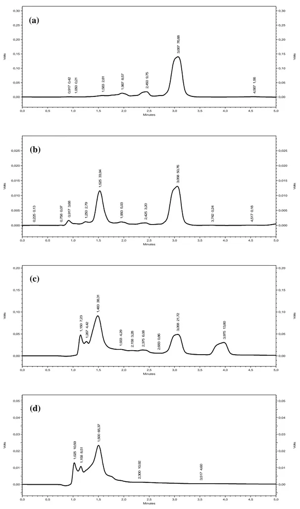
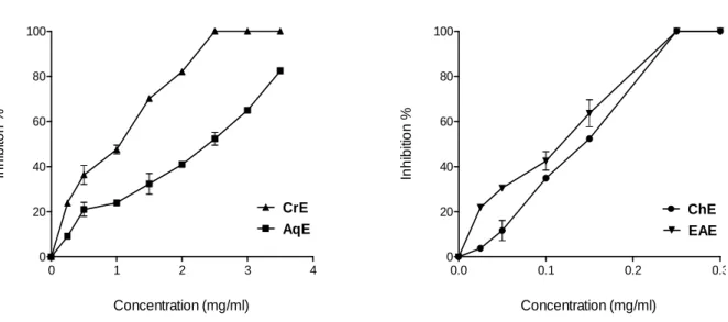
The aim of this study consisted to evaluate the antioxidant activity of Cachrys libanotis roots extracts, used in folk medicine. The roots were submitted to extraction and fractionation to give crude extract (CrE), chloroformic extract (ChE), ethyl acetate extract (EAE) and aqueous extract (AqE). RP-HPLC analysis revealed the presence of catechin and epicatechin in ChE, and myrectin and gossypin in EAE. The highest total polyphenols and flavonoids amounts were recorded in EAE (83.23 µg gallic acid equivalent/mg of extract and 0.63 µ g quercetin equivalent/mg of extract, respectively). Xanthine oxidoreductase (XOR) was purified from bovine milk with yield of 17.97 mg/l, PFR of 7.2, and XOR specific activity of 2261 nmole/min/mg. Both enzymatic and non-enzymatic methods were used to evaluate the antioxidant activity of extracts. Results demonstrated that EAE had the highest XOR inhibitory effect (IC50= 0.11 mg/ml) followed by ChE, CrE and AqE. ChE presented the
highest effect both in cytochrome c test and in NBT assay with an IC50 of 0.22 mg/ml and
0.21 mg/ml, respectively. All extracts inhibited linoleic acid oxidation, with remarkable percentage. In fact, the highest effect was showed by EAE (76.21%) followed by CrE and AqE with an approximately similar effect (65.17% and 64.57%, respectively), and ChE (56.95%). DPPH scavenging assay showed that CrE and EAE exhibited the highest effect with an IC50 of 0.41 and 0.59 mg/ml, respectively. Both CrE and AqE had a considerable
chelating activity on ferrous iron (Fe+2), 9-folds lower than that of EDTA (IC50= 6.1 µg/ml).
Reducing power of extracts was evaluated using FRAP. Extracts exhibited a moderate reducing power, and the highest power was recorded for EAE (TAP= 6.12 mM Fe+2/g of extract). These results can be useful as a starting point of view for further applications of
Cachrys libanotis roots in healthcare. In addition, the ability of roots extracts to inhibit XOR
can be of great use in some diseases where the inhibition of XOR is warranted.
5
ﺺﺨﻠﻣ
ﻟﺍ ﻢﻴﻴﻘﺗ ﱃﺇ ﺔﺳﺍﺭﺪﻟﺍ ﻩﺬﻫ ﻑﺪ
ﺩﺎﻀﳌﺍ ﻁﺎﺸﻨ
ﺓﺪﺴﻛﻸﻟ
ﳉ
ﺭﻭﺬ
Cachrys libanotisﻟﺍ ﺐﻄﻟﺍ ﰲ ﺔﻠﻤﻌﺘﺴﳌﺍ
ﱯﻌﺸ
.
ﰎ
ﺹﻼﺨﺘﺳﺇ
ﺕﺍﺪﻳﻮﻧﻮﻓﻼﻔﻟﺍ
ﻦﻣ
ﺭﻭﺬﺟ
Cachrys libanotisﻰﻄﻋﺃ ﻱﺬﻟﺍﻭ ،
ﺕﺎﺼﻠﺨﺘﺴﻣ ﺔﻌﺑﺭﺃ
:
ﺺﻠﺨﺘﺴﳌﺍ
ﻡﺎﳋﺍ
(CrE)ﻡﺭﻮﻓﻭﺭﻮﻠﻜﻟﺍ ﺺﻠﺨﺘﺴﻣﻭ
(ChE)ﺕﺎﺘﻴﺳﻷﺍ ﻞﻴﺛﺇ ﺺﻠﺨﺘﺴﻣﻭ
(EAE)ﻲﺋﺎﳌﺍ ﺺﻠﺨﺘﺴﳌﺍ ﻭ
(AqE).
ﺪﻗ ﻭ
ﻞﻴﻠﲢ ﻒﺸﻛ
RP-HPLCﻋ
ﻦ
ﺩﻮﺟﻭ
catechinﻭ
/
ﻭﺃ
epicatechinﰲ
ChEﺩﻮﺟﻭ ﻦﻋﻭ ،
myrecetinﻭ
gossypinﰲ
EAE.
ﹼﻥﺃ ﺕﺍﺪﻳﻮﻧﻮﻓﻼﻔﻟﺍ ﻭ ﺕﻻﻮﻨﻴﻔﻟﺍ ﺔﻴﻤﻛ ﺮﻳﺪﻘﺗ ﺮﻬﻇﺃ ﺎﻤﻛ
EAE اﺣ
ﺘﻮ
ﻯ
ﺔﻴﻤﻛ ﱪﻛﺃ
) 83.23ﻍﻭﺮﻜﻴﻣ
ﻟﺎﻐﻟﺍ ﺾﲪ ﺊﻓﺎﻜﻣ
ﻴ
ﻚ
/
ﻭ ﺺﻠﺨﺘﺴﻣ ﻎﻣ
0.63ﻍﻭﺮﻜﻴﻣ
ﺊﻓﺎﻜﻣ
quercetin/
ﻎﻣ
ﺐﻴﺗﺮﺘﻟﺍ ﻰﻠﻋ ،ﺺﻠﺨﺘﺴﻣ
(.
ﺔﻴﻘﻨﺗ ﺖﲤ
xanthine oxidoreductase (XOR)
ـﺑ ﺭﺪﹸﻗ ﺩﻭﺩﺮﲟ ﺮﻘﺒﻟﺍ ﺐﻴﻠﺣ ﻦﻣ
.97 17ﻎﻣ
/
ﻝ
،
ﻭ
7.2=
PFR،
ـﺑ ﺕﺭﺪﻗ ﺔﻴﻋﻮﻧ ﺔﻴﻃﺎﺸﻧﻭ
2261ﻝﻮﻣﻮﻧﺎﻧ
/
ﺔﻘﻴﻗﺩ
/
ﻎﻣ
.
ﻭ ﺔﻴﳝﺰﻧﺇ ﻕﺮﻃ ﺖﻠﻤﻌﺘﺳﺍ
ﺕﺎﺼﻠﺨﺘﺴﻤﻠﻟ ﺓﺪﺴﻛﻸﻟ ﺩﺎﻀﳌﺍ ﻁﺎﺸﻨﻟﺍ ﻢﻴﻴﻘﺘﻟ ﺔﻴﳝﺰﻧﺇ ﲑﻏ
.
ﻗﻭ
ﹼﻥﺃ ﺞﺋﺎﺘﻨﻟﺍ ﺖﻔﺸﻛ ﺪ
EAEﲑﺛﺄﺗ ﻰﻠﻋﺃ ﺮﻬﻇﺃ
ﻰﻠﻋ ﻲﻄﻴﺒﺜﺗ
XOR ) IC50 = 0.11ﻎﻣ
/
ﻞﻣ
(ـﺑ ﺎﻋﻮﺒﺘﻣ
ChEﻭ
CrEﻭ
AqE .ﻥﺃ ﺎﻀﻳﺃ ﺖﻨﻴﺑ ﺎﻤﻛ
ChEﻚﻠﳝ
ﺭﺬﳉ ﺔﻴﺣﺍﺯﺇ ﺓﺭﺪﻗ ﻰﻠﻋﺃ
O2•¯ﺈـﺑ ﺍﺀﺍﻮﺳ
ﺭﺎﺒﺘﺧ
cytochrome cﺈـﺑ ﻭﺃ
ﺭﺎﺒﺘﺧ
NBT ) IC50 = 0.21 أﻭ
0.22ﻎﻣ
/
ﺐﻴﺗﺮﺘﻟﺍ ﻰﻠﻋ ،ﻞﻣ
.(
ﺎﻣﺃ ﻭ
ﺞﺋﺎﺘﻧ
ﺭﺎﺒﺘﺧﺇ
ﺾﻴﻴﺒﺗ
ﲔﺗﻭﺭﺎﻛﺎﺘﻴﺒﻟﺍ
ﺘﺒﺛﺃ ﺪﻘﻓ
ﺖ
ﻥﺃ
ﺩﺎﻀﻣ ﻁﺎﺸﻧ ﻚﻠﲤ ﺕﺎﺼﻠﺨﺘﺴﳌﺍ
ﺓﺪﺴﻛﻸﻟ
ﱪﺘﻌﻣ
،
ﺚﻴﺣ
ﺮﻬﻇﺃ
EAEﻟﺍ
ﻁﺎﺸﻨ
ﻷﺍ
ﱪﻛ
ﺔﺒﺴﻨﺑ
76.21 %ﻦﻣ ﻞﻛ ﻪﻴﻠﻳ
CrEﻭ
AqEﺪﺟ ﺐﺴﻨﺑ
ﺔﺑﺭﺎﻘﺘﻣ
:
%65.17ﻭ
%64.57،
ﺐﻴﺗﺮﺘﻟﺍ ﻰﻠﻋ
ﻁﺎﺸﻧ ﻥﺎﻛ ﲔﺣ ﰲ ؛
ChEﺔﺒﺴﻨﺑ ﱏﺩﻷﺍ
56.95 %.
ﲔﺑ ﻚﻟﺬﻛﻭ
ﺭﺬﺟ ﺔﺣﺍﺯﺇ ﺭﺎﺒﺘﺧﺇ
DPPHﻥﺃ
CrEﻭ
EAEﺔﻴﺣﺍﺯﺇ ﺓﺭﺪﻗ ﻰﻠﻋﺃ ﻥﺎﻜﻠﳝ
) IC50 = 0.41ﻭ
0.59ﻎﻣ
/
ﻞﻣ
ﻰﻠﻋ ،
ﺐﻴﺗﺮﺘﻟﺍ
(.
ﻦﻣ ﻞﻛ ﺮﻬﻇﺃ
CrEﻭ
AqEﻲﺋﺎﻨﺜﻟﺍ ﺪﻳﺪﳊﺍ ﺕﺎﻧﻮﻳﺃ ﺏﻼﺨﺘﺳﺍ ﰲ ﺓﱪﺘﻌﻣ ﺪﺟ ﺓﺭﺪﻗ
)
Fe+2(
ﻞﻗﺃ ﻲﻫﻭ ،
ﺓﺭﺪﻗ ﻦﻣ
EDTAـﺑ
9ﻂﻘﻓ ﻑﺎﻌﺿﺃ
.
ﺎﻤﻛ
ﺔﻴﻨﻘﺘﺑ ﺕﺎﺼﻠﺨﺘﺴﳌﺍ ﻒﻠﺘﺨﳌ ﺔﻴﻋﺎﺟﺭﻹﺍ ﺓﺭﺪﻘﻟﺍ ﺮﻳﺪﻘﺗ ﰎ
FRAP،
ﺬﻟﺍﻭ
ﲔﺑ ﻱ
ﺔﻄﺳﻮﺘﻣ ﺔﻴﻋﺎﺟﺭﺇ ﺓﺭﺪﻗ ﺎﳍ ﺕﺎﺼﻠﺨﺘﺴﳌﺍ ﹼﻥﺃ
،ﹼﻥﺃﻭ
ﺓﺭﺪﻘﻟﺍ
ﺔﻴﻋﺎﺟﺭﻹﺍ
ﻞﻀﻓﻷﺍ
ــﻟ ﺩﻮﻌﺗ
EAE)
TAP = 6.12ﺮﻟﻮﻣ ﻲﻠﻴﻣ
/
ﺺﻠﺨﺘﺴﳌﺍ ﻦﻣ ﻍ
(.
ﻩﺬﻫ ﻝﻼﻐﺘﺳﺍ ﻦﻜﳝ
ﻝﺎﻤﻌﺘﺳﻹ ﺔﻳﺍﺪﺑ ﺔﻄﻘﻨﻛ ﺞﺋﺎﺘﻨﻟﺍ
ﺭﻭﺬﺟ
Cachrys libanotisﻱﻭﺍﺪﺘﻟﺍ ﻝﺎﳎ ﰲ
ﺔﻓﺎﺿﺇ ،
ﱘﺰﻧﻹ ﺔﻴﻄﻴﺒﺜﺘﻟﺍ ﺎﺭﺪﻗ ﹼﻥﺃ ﱃﺇ
XORﺪﻗ
ﰲ ﺎﳍﺎﻤﻌﺘﺳﺍ ﻦﻣ ﻦﹼﻜﻤﻳ
ﻂﻴﺒﺜﺗ ﺐﻠﻄﺘﺗ ﱵﻟﺍ ﺽﺍﺮﻣﻷﺍ ﺾﻌﺑ ﺝﻼﻋ
ﱘﺰﻧﺇ
.
XORﺢﻴﺗﺎﻔﳌﺍ ﺕﺎﻤﻠﻜﻟﺍ
:
،ﻱﺪﺴﻛﺄﺘﻟﺍ ﺩﺎﻬﺟﻹﺍ
،ﺔﻴﺒﻄﻟﺍ ﺕﺎﺗﺎﺒﻨﻟﺍ
ﺍ ﱘﺰﻧﻹﺍ
ﺆﻤﻠ
ﲔﺜﻧﺍﺰﻜﻠﻟ ﻊﺟﺮﳌﺍ ﺪﺴﻛ
) XOR (ﺕﺍﺩﺎﻀﻣ ،
،ﺓﺪﺴﻛﻷﺍ
ﺓﺩﺪﻌﺘﻣ
ﺕﻻﻮﻨﻴﻔﻟﺍ
.
6
Résumé
Le but de cette étude consiste à évaluer l’activité biologique des extraits des racines de
Cachrys libanotis, utilisées dans la médecine traditionnelle. Les racines ont été soumises à
une extraction et fractionnement pour avoir l’extrait brut (CrE), chloroformique (ChE), d’acétate d’éthyle (EAE) et aqueux (AqE). L’analyse par RP-HPLC a révélé la présence du catéchine et épicatéchine dans ChE, et du myréctine et gossypine dans EAE. La quantité en polyphénols et en flavonoids la plus élevée est présenté par EAE (83.23 µg équivalent d’acide gallique/mg d’extrait et 0.63 µg équivalent de quercétine/mg d’extrait, respectivement). La xanthine oxydoréductase (XOR) a été purifiée du lait bovin avec un rendement de 17.97 mg/l, PFR de 7.2, et une activité spécifique de 2261 nmole/min/mg. Des méthodes enzymatiques et non-enzymatiques ont été utilisées pour évaluer l’activité antioxydante des extraits. Les résultats montrent que EAE présent l’effet inhibiteur le plus puissant sur XOR (IC50= 0.11
mg/ml) suivi par ChE, CrE et AqE. ChE montre l’effet le plus élevé dans le test du cytochrome c et le test du NBT avec un IC50 de 0.22 mg/ml et 0.21 mg/ml, respectivement.
Tous les extraits inhibent remarquablement l’oxydation de l’acide linoleique. En effet, l’effet le plus élevé est observé pour EAE (76.21%) suivi par CrE et AqE ayant un effet approximativement similaire (65.17% et 64.57%, respectivement), et ChE (56.95%). Le test du scavenger du DPPH a montré que CrE et EAE présentent l’effet le plus puissant avec un IC50 de 0.41 et 0.59 mg/ml, respectivement. CrE et AqE montrent une activité chélatrice
remarquable, 9-fois moins celle d’EDTA (IC50= 6.1 µg/ml). Le pouvoir réducteur des extraits
est évalué utilisant la technique de FRAP. Les extraits exercent un pouvoir réducteur modéré, et le pouvoir le plus élevée est celui d’EAE (TAP= 6.12 mM Fe+2/g d’extrait). Ces résultats peuvent être investiguer comme un point de départ pour des applications de ces racines dans la santé. En plus, le pouvoir de ces racines d’inhiber la XOR peut avoir une utilisation importante pour certaines maladies dont l’inhibition de la XOR est nécessaire.
7
Abbreviations
AqE: aqueous extract
BHT: butylated hydroxyl-toluene
BMXOR: bovine milk xanthine oxidoreductase ChE: chloroform extract
CrE: crude extract
DPPH: 1,2-diphenyl-2-picrylhydrazyl hydrate DTT: dithiothreitol
DW: dry weight
EAE: ethyle acetate extract
EDTA: etylenediamine-tetraacetic acid FCR: Folin-Ciocalteu reagent
FlOH: flavonoid antioxidant
FRAP: ferric acid reducing antioxidant power GAE: gallic acid equivalent
GPx: glutathione peroxidase GSH: glutathione
IR: ischemia-reperfusion mt-DNA: mitochondrial DNA NBT: nitro-blue tetrazolium NOS: nitric oxide synthase PFR: protein/flavin ratio
PMS: 5-methylphenazinium methyl sulfate QE: quercetin equivalent
RE: rutin equivalent
RNS: reactive nitrogen species
RONS: reactive oxygen and nitrogen species ROS: reactive oxygen species
RP-HPLC: reverse polarity-high performance
liquid chromatography
SOD: superoxide dismutase TPTZ: 2,4,6- tripyridyl-s-triazine XOR: xanthine oxidoreductase XDH: xanthine deshydrogenase XO: xanthine oxidase
8
List of figures
Fig. 1. Molecular structure of the XDH………... Fig. 2. The enzymatic process catalyzed by XO..………... Fig. 3. Role of XOR in ischemia reperfusion injury……… Fig. 4. Basic structure of flavan nucleus……… Fig. 5. Scavenging of ROS (R.) by flavonoids………..
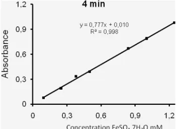
Fig. 6. Binding sites for trace metals………. Fig. 7. Cachrys libanotis L. plant……… Fig. 8. Extraction procedure of flavonoids from C. libanotis roots……….. Fig. 9. Calibration curve of gallic acid……….. Fig. 10. Calibration curve of quercetin and rutin……….. Fig. 11. Calibration curve of FeSO4.7H2O………
Fig. 12. Separated extracts peaks in RP-HPLC (a): ChE, (b): EAE, (c): CrE and (d): AqE……… Fig. 13. BMXOR inhibition by C. libanotis roots extracts………. Fig. 14. IC50, inhibitory concentration of C. libanotis extracts for 50% of BMXOR activity…….
Fig. 15. Superoxide- scavenging effect of C. libanotis extracts using cytochrome c assay………. Fig. 16. Comparison between IC50 of C. libanotis extracts resulting from XOR inhibition and
O2 .-
scavenging assays ………...
Fig. 17. Superoxide- scavenging effect of C. libanotis extracts using NBT assay…………... Fig. 18. IC50, inhibitory concentration of C. libanotis extracts for 50% of superoxide generation using NBT assay………..………
Fig. 19. Kinetics of β-carotene-bleaching in the presence of C. libanotis extracts, water,
methanol and BHT during 48h………
Fig. 20. Antioxidant activity of C. libanotis extracts compared with BHT, methanol and water… Fig. 21. DPPH scavenging activity of C. libanotis extracts and standards……….. Fig. 22. Ferrous iron-chelating ability of extracts from the roots of C. libanotis……... Fig. 23. EC50, effective concentration of C. libanotis extracts for 50% of chelating activity...
15 16 18 20 22 23 25 28 29 30 36 41 43 44 46 46 47 48 49 50 52 54 54
9
Fig. 24. FRAP reaction kinetics of reagent blank and C. libanotis extracts (73.5 µg/ml)……….. Fig. 25. Comparison between TAP of C. libanotis extracts at 4 min and 30 min….………... Fig. 26. TAP, total antioxidant power of C. libanotis extracts and gallic acid at 30 min…………
56 57 58
List of tables
Table I. Nomenclature of RONS ………...……….... Table II. Physiological roles that involve free radicals or their derivatives……… Table III. Some pathologies result from oxidative stress…..……… Table IV. Yields of extraction of C. libanotis roots……….. Table V. Total polyphenols and flavonoids content of C. libanotis extracts……… Table VI. Retention time of standards……….. Table VII. Characteristics of purified BMXOR ……… Table VIII. DPPH scavenging activity of C. libanotis extracts and standards……… Table IX. EC1 and TAP values at 4 min and 30 min of reaction using FRAP Assay………
4 8 10 37 38 40 42 51 58
10
Contents
Title page ... i Dedication ... ii Acknowledgements ... iii Summary ... ivﺺﺨﻠﻣ
... v Sommaire ... vi Abbreviations ... viiList of figures ... viii
List of tables ... ix List of schemes ... ix Contents ... x Introduction ... 1 1. Oxidative stress ... 3 1.1. Oxidants ... 3
1.1.1. Important reactive oxygen and nitrogen species (RONS) ... 3
1.1.2. Sources of RONS ... 6
1.1. 3. Physiological roles of RONS ... 8
1.1.4. Biological consequences of oxidative stress ... 8
1.1.5. Pathological implications of oxidative stress ... 10
1.2. Antioxidant defense ... 10
1.2.1. Enzymatic antioxidants... 11
1.2.2. Non-enzymatic antioxidants ... 12
2. Xanthine oxidoreductase (XOR) ... 14
2.1. Structure ... 14
2.3. Distribution ... 15
2.4. Catalysis mechanism of XOR... 16
2.5. Physilogical functions ... 17
2.6. Pathological implications ... 17
2.6.1. Hyperuricemia ... 17
2.6.2. Ischemia-reperfusion injury (IR injury) ... 17
11
3. Polyphenols ... 19
3.1. Phenolic acids ... 19
3.1. Tannins ... 20
3.1. Flavonoids ... 20
3.3.1. Antioxidant activity of flavonoids ... 21
4. Plant (Cachrys libanotis L.) ... 24
4.1. Taxonomy ... 24
4.2. Botanical description ... 25
4.3. Traditional uses ... 25
Materials and methods ... 26
1. Materials ... 26
1.1. Biological materials ... 26
1.2. Chemicals and materials ... 26
2. Methods ... 27
2.1. Flavonoids extraction ... 27
2.2. Analyse of C. libanotis extracts ... 29
2.2.1. Determination of total polyphenols content ... 29
2.2.2. Determination of flavonoids content ... 29
2.2.3. Reverse polarity -high performance liquid chromatography (RP-HPLC)... 30
2.3. Purification of bovine milk xanthine oxidoreductase (BMXOR) ... 31
2.4. Evaluation of antioxidant activity of C. libanotis by enzymatic methods... 31
2.4.1. XOR inhibition ... 31
2.4.2. Cytochrome c test ... 32
2.5. Evaluation of antioxidant activity of C. libanotis by non-enzymatic methods ... 32
2.5.1. NBT assay ... 32
2.5.2. β-carotene/linoleic acid bleaching method ... 33
2.5.3. DPPH scavenging assay ... 34
2.5.4. Ion chelating assay... 34
2.5.5. FRAP assay ... 35
Statistical analysis ... 36
Results and discussion ... 37
1. Flavonoids extraction ... 37
12
2.1. Determination of total polyphenols and flavonoids content ... 38
2.2. RP-HPLC ... 39
3. Purification of bovine milk xanthine oxidoreductase (BMXOR) ... 42
4. Evaluation of antioxidant activity of C. libanotis by enzymatic methods ... 43
4.1. XOR inhibition ... 43
4.2. Cytochrome c test ... 45
5. Evaluation of antioxidant activity of C. libanotis by non-enzymatic methods…….47
5.1. NBT assay ... 47
5.2. β-carotene/linoleic acid bleaching method ... 48
5.3. DPPH-scavenging assay ... 51
5.4. Ion chelating assay... 53
5.5. FRAP assay ... 56
General discussion ... 60
14
Introduction
Reactive oxygen and nitrogen species are well recognized for playing a dual role as both deleterious and beneficial species. Beneficial effects of reactive oxygen and nitrogen species occur at low or moderate concentrations and involve physiological roles in cellular signaling pathways. In contrast, the generation of reactive oxygen and nitrogen species, where xanthine oxidoreductase is considered as one of their major sources, beyond the antioxidant capacity of a biological system gives rise to oxidative stress, a deleterious process that can be an important mediator of damage to cell structures, including lipids and membranes, proteins, and DNA. Oxidative stress facilitates the development of a variety of human diseases such as inflammations, cardiovascular diseases, neurodegenerative diseases, diabetes mellitus and cancer. One solution to this problem is compensating this disequilibrium with antioxidant compounds that are contained in natural plant sources.
Medicinal plants have been used as sources of medicine in virtually all cultures. During the last decade, the use of traditional medicine has expanded globally and is gaining popularity. The interest in many traditional herbs and plants is due to our increasing knowledge of the role of free radicals and antioxidants in human health and disease. However, still there is not enough knowledge and data about most of them.
Plant materials contain secondary metabolites that polyphenols constitute one of the principal classes. These compounds arouse a big interest by their numerous beneficial effects toward human health. Polyphenols are subdivided to simple phenols, flavonoids and tannins. In particular, flavonoids are recognized for their antioxidant properties, antimicrobial, anti-inflammatory, etc. These activities have the most often a link with their antioxidant activity and particularly their capacity to inhibit enzymes responsible for radical generation, scavenge free radicals and chelating metal ions.
15
The research on plants and herbs with alleged folkloric used as pain relievers, anti-inflammatory agents, should therefore be viewed as beneficial and logical strategy in the search of antioxidants drug. From that came our choice of Cachrys libanotis L., having traditional claims for the treatment of rheumatism, as subject of our study.
Up to date, there are no scientific reports neither on the extraction of flavonoids from the
Cachrys libanotis L. roots nor on its antioxidant properties. In this context, the aim of our
16
1. Oxidative stress
Oxidative stress denotes an imbalance between oxidants (reactive species) and antioxidants in favor of the oxidants at the cellular or individual level, leading to a disruption of redox signaling and control and /or molecular damage (Lykkesfeldt and Svendsen, 2007). Oxidative stress might occur when the antioxidant defense system is overwhelmed by an increased oxidant burden or a decrease in antioxidant supply (Kirschvink et al., 2008).
1.1. Oxidants
Oxidants are compounds capable of oxidizing target molecules. This can take place by one of three actions: abstraction of a hydrogen atom, abstraction of an electron or the addition of oxygen (Lykkesfeldt and Svendsen, 2007). Reactive oxygen and nitrogen species (RONS) are the major types of reactive species exist. Some of them are free radicals and some are not (Table I). A "free radical" is any atom or group of atoms that contains one or more unpaired electrons in an outer orbital, which is capable of independent existence (Gilbert, 2002). The presence of unpaired electrons can result in a species that is highly reactive. However, the reactivity of RONS, whether free radicals or not, can give an idea about their activity and specificity of reaction with other molecules. The reactivity of RONS will determine their half-life in biological systems and how far they can travel by diffusion from the site of their generation (Aust, 2004).
1.1.1. Important reactive oxygen and nitrogen species (RONS)
Under normal conditions, molecular oxygen (O2) is present in a triplet, diradical form, having
two unpaired electrons of parallel spin. This diradical form is not very reactive to organic molecules because most organic molecules are in a singlet state and due to the quantum mechanical restriction; a spin restriction creates a barrier to the insertion of a pair of electrons simultaneously, preventing its reaction with biomolecules (Laranjinha, 2009). However, O2
17
can be reduced to H2O by a series of reduction reactions, either enzymatically or
non-enzymatically, requiring four electrons in total (Auts, 2004; Powers et al., 2008). The incomplete reduction of oxygen leads to formation of reactive oxygen species (ROS) that the most important are superoxide radical (O.-2, one electron), hydrogen peroxide (H2O2, 2
electrons) and hydroxyl anion, OH. (Berg, 2007; Laranjinha, 2009).
Table I. Nomenclature of RONS (Halliwell and Whiteman, 2004).
Reactive oxygen and nitrogen species
Free radicals Nonradicals
Superoxide (O.-2) Hydroxyl (OH.) Hydroperoxyl (HO.2) Peroxyl (RO.2) Alkoxyl (RO.) Carbonate (CO.-3)
Carbon dioxide (CO.-2)
Hydrogen peroxide (H2O2)
Hypobromous acid (HOBr) Hypochlorous acid (HOCl) Ozone O3
Singlet oxygen (O12)
Organic peroxides (ROOH) Peroxynitrite (ONOO-) Peroxynitrous acid (ONOOH) Nitric oxide (NO.)
Nitrogen dioxide (NO.2)
Nitrous acid (HNO2) Nitrosyl cation (NO+) Nitroxyl anion (NO-) Dinitrogen tetroxide (N2O4)
Dinitrogen trioxide (N2O3)
Peroxynitrite (ONOO-) Peroxynitrous acid (ONOOH) Nitronium (nitryl) cation (NO2+)
Alkyl peroxynitrites (ROONO) Nitryl chloride (NO2Cl)
O.-2 is considered as primary ROS, and can further interact with other molecules to generate
secondary ROS, either directly or prevalently through enzyme- or metal-catalyzed processes
18
H2O2 is formed by dismutation of O.-2 in the presence of superoxide dismutase (SOD) or
spontaneously (Valko et al., 2005):
H2O2 does not contain unpaired electrons in the valence orbitals and, therefore, is not a free
radical molecule but, upon lysis of the O-O bond by ferrous iron, known as Fenton reaction, yields the most powerful oxidant known in a biological setting, HO. (Valko et al., 2005):
Endogenous iron is usually present in chelated or bound forms as part of hemoglobin, myoglobin, several enzymes and the transport protein, tranferrin, and therefore not readily available for reaction (de Beer et al., 2002).
O.-2 and H2O2 can react with transition metals such as iron or copper to form the strong
oxidant, OH. , following Haber-Weiss reaction (Sorg, 2004):
Conversely to O.-2 and H2O2, which are less reactive and therefore, more selective in its
targets, the OH., upon formation, oxidizes indiscriminately and site-specifically any biomolecule (Sorg, 2004).
As reported by Nivière and Fontecave (1995), NO., an intercellular messenger, is produced from oxygen by various nitric oxide synthase (NOS):
O
O
O
O
2222+ arginine + NADPH NO
+ arginine + NADPH NO
+ arginine + NADPH NO
+ arginine + NADPH NO
....+ citrulline + H
+ citrulline + H
+ citrulline + H
+ citrulline + H
2222O + NADP
O + NADP
O + NADP
O + NADP
The simultaneous generation of NO. and O.-2 leads to the formation of ONOO-. ONOOH,
formed by its protonation, is a powerful oxidant itself, but can also decompose to yield further
SOD
NOS
NOS
NOS
NOS
19
oxidants with the chemical reactivity of NO2., OH. and NO+2. NO. is poorly reactive with most
molecules in the human body (non-radicals), but as a free radical it can react extremely rapidly with other free radicals such as O.-2 , amino acid radicals, and certain transition metal
ions (Gutteridge and Halliwell, 2002).
1.1.2. Sources of RONS
The principal sources of RONS are devised in two types according to their origin: a. Endogenous sources of RONS
• Mitochondria
ROS are commonly generated as byproducts of the mitochondrial electron transfer reaction for the production of ATP. Approximately 2-4% of the total oxygen consumed during electron transport is reduced not to water by cytochrome c oxidase but rather to O2.-, due to
the "leakage" of unpaired electrons to O2 during the proton-motive quinone cycle. Under the
physiological condition, the O2.- generated by the respiratory chain in mitochondria is
scavenged by SOD in the mitochondrial matrix to form H2O2 (Chen and Castranova, 2004).
• NADPH oxidase
The key component of the respiratory burst system is the membrane-bound multisubunit enzyme complex termed the NADPH oxidase in both phagocytic cells and nonphagocytes, which act as a pivotal defense system against a range of infectious agents (Babior, 1999). NADPH oxidase is inactive in resting cells and upon stimulation by a variety of soluble mediators and by particulate stimuli that interact with cell surface receptors, this oxidase is rapidly activated to produce O2.- and other ROS, such as H2O2, OH., and, through the
one-electron reduction mechanism (Babior, 1999; Robinson, 2009):
NADPH oxidase
NADPH oxidase
NADPH oxidase
NADPH oxidase
20
• Xanthine oxidoreductase (XOR)
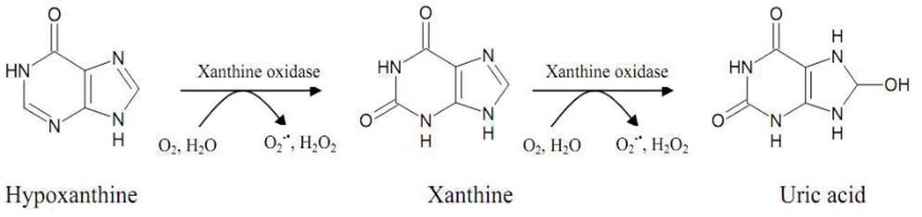
Xanthine oxidoreductase (XOR) catalyzes the reduction of O2 to yield O.-2 and H2O2 during
uric acid formation from hypoxanthine. Kelley and their collaborators (2010) reported that H2O2 is the major oxidant product of XOR. Moreover, XOR has recently been shown to
catalyze the anaerobic reduction of inorganic nitrite to NO.. In the presence of oxygen this also is reduced, to O.-2, which reacts rapidly with NO. to give ONOO- (Harrison, 2006).
• Nitric oxide synthase (NOS)
Three NOS isoforms have been identified in mammalian tissues: neuronal NOS (nNOS or type I), inducible NOS (iNOS or type II), and endothelial NOS (eNOS or type III). NOS (EC 1.14.13.39) catalyzes the oxygen- and NADPH-dependent oxidation of L-arginine to NO. and citrulline (del Rio et al., 2002).
In addition, ROS generation can make by enzymes such as membrane oxidases. In fact, cytochrome P-450, P-450 reductase and cytochrome b-5 reductase in the endoplasmic reticulum under certain conditions generate O.-2 and H2O2 during their catalytic cycle
(Bandyopadhyay et al., 1999). Peroxisomal oxidases and flavoproteins, as well as D-amino acid oxidase, L-hydroxy acid oxidase, and fatty acyl oxidase participate in ROS generation (Bandyopadhyay et al., 1999; Valko et al., 2007). Furthermore, Reactions catalyzed by lipoxygenases and cyclooxygenase also are important cellular sources of free radicals. Cyclooxygenase react with H2O2 producing ferryl heme and tyroxyl radical. Such ‘‘activated
enzyme’’ can induce one-electron oxidation of arachidonic acid and also other substrates producing free radicals (Bartosz, 2003).
21
A number of environmental factors, such as ultraviolet light, ionizing radiation and ultrasound can stimulate the RONS generation endogenously after interaction with tissues or cells (Mark
et al., 2000; Bartosz, 2003; Chen and Castranova, 2004).
Various other exogenous sources also contribute directly or indirectly to the total oxidant load. These include effects of air pollution and natural toxic gases, such as ozone, as well as chemicals and toxins, including oxidizing disinfectants. Moreover, foreign microorganisms induce secondary oxidant formation and release in the host via the immune system, in addition to their sometimes directly oxidizing capabilities. Diets containing inadequate amounts of nutrients may also indirectly result in oxidative stress by impairing cellular defense mechanisms (Lykkesfeldt and Svendsen, 2007).
1.1.3. Physiological roles of RONS
Sorg (2004) reported that an adequate level of certain RONS can have a physiological role, as for instance the catalysis of many biochemical reactions, the defense against invading pathogens or the capacitation of spermatozoa. Moreover, in the nervous system, it acts as a neuromodulator and plays a role in synaptic plasticity and long-term memory, whereas in the vascular system, it controls blood pressure, inhibits platelet aggregation (Table II).
Table II. Physiological roles that involve free radicals or their derivatives (Dröge, 2002).
Species
Source
Physiological effects
Nitric oxid (NO.) Different NOS •Smooth muscle relaxation and others various cGMP-dependent functions
Superoxide (O2.-)
and related ROS
NAD(P)H oxidase
•Control of ventilation
•Control of erythropoietin production and other hypoxia - inducible functions
•Smooth muscle relaxation
•Signal transduction from various membrane receptors / enhancement of immunological
22 functions Superoxide (O2.-)
and related ROS Any sources
•Oxidative stress responses and the maintenance of redox homeostasis
1.1.4. Biological consequences of oxidative stress
RONS may cause oxidative damage to proteins, lipids, and DNA, which inhibits the normal functions of proteins and lipids, and facilitates DNA mutagenesis; thus, these species play a pivotal role in various clinical conditions (Rice-Evans, 1994).
DNA and RNA damage are major consequences of oxidative stress. Exposure of nucleic acids to reactive species may result in base modification, cross-linking of DNA and DNA-proteins, sister chromatid exchange and single- or double-strand breaking leading to disruption of transcription, translation and DNA replication (Goetz and Luch, 2008). Mitochondrial DNA (mtDNA), which is transiently attached to the inner mitochondrial membrane where a large amount of ROS is produced, is particularly vulnerable to oxidative damage. Moreover, DNA repair mechanisms in the mitochondria are less efficient than in the nucleus. Therefore, ROS-mediated mtDNA damages may contribute to mitochondrial dysfunction generated by endogenous reactive intermediates which act directly on mitochondrial proteins (Valko et al., 2006).
Oxidation of proteins may occur directly as protein side chains are oxidized leading to a loss of function of proteins and a deactivation of enzymes. Often, thiols of proteins involved in the regulation of enzyme activity are directly oxidized. Increase of malondialdehyde has been suggested to lead to intra- and inter-molecular cross-links of proteins. Conformational changes leading to an increase in hydrophobicity may result in aggregation or precipitation of proteins, which can no longer be subjected to the normal protein degradation pathway (Stadtman and Levine, 2006; Valko et al., 2006). Additionally, oxidative damage of proteins
23
may occur by the adduction of secondary products like oxidation of sugar (glycoxidation), or of polyunsaturated lipids (lipoxidation) (Stadtman and Levine, 2006).
Lipid peroxidation of membranes occurs as a consequence of direct reaction of fatty acids of polar lipids with oxygen or a reaction catalyzed either by metals like iron or by NADPH cytochrome P-450 reductase (Devasagayam et al., 2003). Lipid peroxidation is initiated by the
formation of a Fenton-derived oxidant that results in the production of a lipid radical. In the presence of oxygen, this radical is converted to a hydroperoxide radical, which then abstracts hydrogen from other lipids. These steps result in chain propagation and, consequently, enhanced lipid peroxidation (Wink et al., 2002).
1.1.5. Pathological implications of oxidative stress
An oxidative stress is often associated to all kinds of diseases, although it is not always easy to determine whether it is a cause or a consequence of the observed condition. A selection of pathologies, for which the mechanism of oxidative stress is well documented, is described in Table III.
Table III. Some pathologies result from oxidative stress.
Pathology
References
Cancer (Goetz and Luch, 2008)
Cardiovascular diseases (Vijaya Lakshmi et al., 2009) Metabolic syndrome
Diabetes mellitus
(Calhau and santos, 2009) (Maritim et al., 2003) Inflammation
Rheumatoid arthritis
Ischemic reperfusion injury
(Libetta et al., 2011)
(Hitchon and El-Gabalawy, 2004) (Brennan and Eaton, 2006)
Neurodegenerative diseases Alzheimer’ disease
Parkinson disease
(Anderson, 2004) (Cai and Yan, 2007) (Cohen, 2002)
24
1.2. Antioxidant defense
Antioxidants was defined as substances that are able, at relatively low concentrations, to compete with other oxidizable substrates and, thus, to significantly delay or inhibit the oxidation of these substrates (Dröge, 2002). According to Shi and their collaborators (2001), there are several lines of defense as follows:
(i) inhibiting the formation of active oxygen species and free radicals, (ii) preventing chain initiation by scavenging initiating radicals,
(iii) repairing, de novo and clearance of oxidatively damaged lipids, proteins and DNA.
1.2.1. Enzymatic antioxidants
They include the glutathione redox system [consisting of glutathione reductase, glutathione-S-transferase, glutathione peroxidase (GPx), and glucose-6-phosphate dehydrogenase], SOD and catalase.
• Superoxide dismutase (SOD)
SOD (EC1.15.1.1.) are a family of metalloenzymes that catalyze the disproportionation of O2.- radicals to H2O2 and O2, and play an important role in protecting cells against the toxic
effect of superoxide radicals produced in different cellular compartments (del Rio et al., 2002).
• Glutathione peroxidase (GPx)
GPx is a selenoenzyme whose catalytic function depends on the presence of the mineral in the enzyme. It catalyzes the reduction of both H2O2 and lipid hydroperoxides. In general
terms the enzyme functions in a cycle with glutathione reductase which uses reducing equivalents derived from glucose via the pentose phosphate pathway and NADPH (Diplock, 1994).
25
• Catalase
In human tissues, there is a very high concentration of the enzyme in liver peroxisome and erythrocytes, and there is relatively much less in brain, heart and skeletal muscle (Diplock, 1994). The general reaction catalyzed is the reduction of H2O2 to water and O2. However,
H2O2 can cross membranes and it may be that the protective effect of GPx is overwhelmed,
when intracellular concentrations of H2O2 are high. Under these circumstances the protective
function of catalase may be of great importance (Diplock, 1994; Forman, 2008).
1.2.2. Non-enzymatic antioxidants
They includes different chemical compounds such as tocopherol (vitamin E), ascorbic acid (vitamin C), Caretinoids, glutathione (GSH), phenolic compounds, ubiquinol (Coenzyme Q10), phospholipids (proteoglycans and hyaluronic acid), lipoic acid, proteins binding free iron and copper such as ceruloplasmin, , transferrin, taurine, albumin), protein hydrolysates, bilirubin, melatonin, uric acid, mucin, surfactant, amino acids, peptides, and phytates (Vallyathan, 2004; Kirschvink et al., 2008).
• Vitamine E
The vitamine E is a free radical scavenger which yields a long-lived radical upon hydrogen abstraction, thereby interrupting the chain reaction. This property is optimized in α-tocopherol which is a remarkable scavenger of peroxyl radicals in phospholipid membrane bilayers. Vitamin E may reduce the possibility of forming reactive OH. and ONOO-, and thus protect against oxidative damage. Also, it may modulate the activation and/or expression of redox-sensitive biological response modifiers, and thereby attenuates the cellular events leading to the onset of cardiovascular, cancer, aging and other degenerative diseases (Chow, 2008).
26
The role of vitamin C as an antioxidant is indicated by its known free radical-scavenging action. As a reducing and antioxidant agent, it directly reacts with O2.-, OH. and various
lipid hydroperoxides. In addition, it can restore the antioxidant properties of oxidized vitamin E, suggesting that a major function of vitamin C is to recycle the vitamin E radical. However, vitamin C shown a prooxidant action which resides in its ability to reduce Fe+3 to Fe+2 state; Fe+2 is known as a potent free radical inducer (Yu, 1994).
• Glutathione (GSH)
GSH, the tripeptide glutathione (γ-L-glutamyl-L-cysteinylglycine), has a very important role in protection against free radical damage by providing reducing equivalents for the several key enzymes referred to above. GSH is also a scavenger of OH. and O12; in the case of OH.,
the thiyl radical is produced, which is also a product of the oxidation of GSH by peroxidases or by O2 in the presence of transition-metal ions. Thiyl radicals may initiate radical reactions
although they are much less reactive than OH.. A slow reaction of GSH with O2.-, may also
lead to the formation of O12. Thus, although the overwhelming function of GSH in living cells
is protective, the possibility of undesirable secondary reactions must be borne in mind (Diplock, 1994). Vitamin E, vitamin C, and GSH may act in combination through NADPH-glutathione reductase system (Bandyopadhyay et al., 1999)
• Carotinoids
Carotinoids have long been considered antioxidants because of their capacity to scavenge free radicals. Carotinoids protect lipids against peroxidation by quenching free radicals and other reactive oxygen species, notably singlet oxygen. β-carotene displays an efficient biological radical-trapping antioxidant activity through its inhibition of lipid peroxidation induced by the XO system (Yu, 1994).
27
• Polyphenols
Polyphenols are a big group of compounds found in plants and uniquely characterize by the presence of more than one phenol group in the molecule. They are considered the most abundant antioxidant in the diet although the diversity of their structures makes them different from other antioxidants. Moreover, the amount of different polyphenols found to date in plants and plant foods, several thousands, make them a complex family of compounds with very interesting therapeutic properties against cancer, cardiovascular diseases, inflammation and other diseases (Chow, 2008).
2. Xanthine oxidoreductase (XOR)
XOR known initially as Schardinger’s enzyme, it was discovered in 1902 by Austrian biochemist Franz Schardinger. It was later isolated, purified, and studied by Malcolm Dixon and Sylva Thurlow in the 1920’s, and in 1938 it was suggested by Booth that Schardinger enzyme be there after referred to as xanthine oxidase (XO).
2.1. Structure
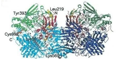
XOR belongs to the family of molybdenum-containing hydroxylases comprising a group of closely related metalloflavoproteins (Parks et al., 2002). The enzymes from various sources are proteins of 290 kDa, and are composed of two identical and catalytically independent subunits (Enroth et al., 2000). The 3D structure (Fig. 1) of each subunit is organized into three major domains: Modomain, FeS domain, and flavin adenine dinucleotide (FAD) -domain. The Mo-domain is the largest and contains the Mo active site. The Fe-S domain contains two [2Fe–2S] centers whereas the FAD-domain contains the flavin cofactor (Brondino et al., 2006).
The enzyme occurs in two forms, xanthine dehydrogenase (XDH, EC 1.1.1.204), which predominates in vivo, and xanthine oxidase (XO, EC 1.1.3.22). Conversion of XDH to XO
28
can occur reversibly by oxidation of essential sulfhydryl groups or through limited proteolytic cleavage of the amino terminus (Harisson, 2006; Nishino et al., 2008). XDH shows a preference for nicotinamide adenine dinuclotide (NAD+) reduction at FAD-domain, whereas XO fails to react with NAD+ and exclusively uses O2 as its substrate, leading to the formation
of O2-. and H2O2 (Enroth et al., 2000).
Fig. 1. Molecular structure of the XDH. XDH dimer divided into the three major domains and
two connecting loops. The two monomers have symmetry related domains in the same colors, in lighter shades for the monomer on the left and in darker shades for the monomer on the right. From N to C terminus, the domains are: ironysulfur-center domain (residues 3–165; red), FAD domain (residues 226–531; green), and Mo-pt domain (residues 590–1,331; blue). The loop connecting the ironysulfur domain with the FAD domain (residues 192–225) is shown in yellow, the one connecting the FAD domain with the Mo-pt domain (residues 537–589) is in brown, and the N and C termini are labeled. The FAD cofactor, the two ironysulfur centers, the molybdopterin cofactor, and the salicylate also are included (Enroth et al., 2000).
XDH and XO are products of the same gene; however, XOR is transcribed as a single gene product, xanthine dehydrogenase (XDH). The gene encoding human XOR is approximately 60 kb, comprises 36 exons and 35 introns (Xu et al., 1996), and its location was confirmed on chromosome band 2p23 (International Human Genome Sequencing Consortium, 2001). The nucleotide sequence (1,332 residues) is highly homologous with that of the bovine milk enzyme (1332 residues) showing 91% homology (Xu et al., 1994).
2.3. Distribution
XOR was found in liver parenchymal cells and in vesicles in Kupffer cells and sinusoidal endothelial cells, using histochemical techniques which rely on detection of enzyme activity
29
in tissue homogenates (Frederiks and Vreeling-Sindelarova, 2002). Further, immuno-localization techniques with XOR antibodies recognize active and inactive forms of the enzyme in intact tissues such as human liver, brain and heart (Meneshian and Bulkley, 2002; Martin et al., 2004). Wright and their collaborators (1993) found XOR in human liver, lung, skeleton muscle and pancreas, using tissue hybridation techniques.
Subcellular localization methods have demonstrated the presence of XOR in both the cytoplasm (Frederiks and Vreeling-Sindelarova, 2002) and the cell membranes (Rouquette et
al., 1998) with cell surface binding likely mediated by glycosaminoglycans (Radi et al.,
1997). In addition, the enzyme is a major protein component of the milk fat globule membrane, which envelops fat droplets in freshly expressed milk (Mather, 2000). XOR is generally present in mammalian blood and, because of endogenous proteases, occurs largely in the XO form (Kooij et al., 1992).
2.4. Catalysis mechanism of XOR
XOR reacts with many substrates including purines, purine ribonucleosides, and 2-deoxyribonucleosides. It plays a pivotal role in the catabolism of purines (Fig. 2) when both forms catalyze the terminal two steps of purine degradation in humans, hypoxanthine to xanthine and xanthine to uric acid (Hille and Nishino, 1995).
30
Reaction mechanism occurs in two phases, the reductive half- reaction and the oxidative half oxidative. The reductive half-reaction involves binding and reaction at molybdenum center of the enzyme of up successive xanthine molecules (Enroth, 2000). In contrast, the oxidative half-reaction takes place at FAD-domain. Electrons are subsequently transferred from FAD to NAD+ or O2 (Hille and Nishino, 1995).
2.5. Physiological functions
The primary functions of XOR can be including purine metabolism, oxidant and, antioxidant production, nitric oxide regulation, and drug metabolism (Parks et al., 2002). In addition, XOR implicate in innate immunity (Meneshian and Bulkley, 2002; Harrison, 2006), and it is essential to the process by which the milk fat globule becomes enveloped by the apical cell membrane during secretion (Harrison, 2006).
2.6. Pathological implications
As a result of its ability to generate RONS, implicated in both tissue structural damage and cell signaling interference, XO form has received considerable attention as a patho-physiological cause of some pathological states.
2.6.1. Hyperuricemia
Hyperuricemia is the most cited pathology involving XOR. It is a pathological state that arises from overproduction (by XOR) or under excretion (renal tubule disorders) of uric acid. As a result of hyperuricemia, insoluble uric acid forms microscopic crystals in the capillary vessels of joints. These crystals cause inflammation and sharp pain, which is termed acute gouty arthritis or acute gout (Sachs et al., 2009).
31 2.6.2. Ischemia-reperfusion injury (IR injury)
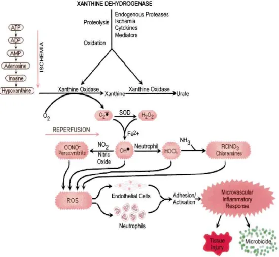
Much of the injury sustained by an organ is a consequence of ischemia actually occurred at the time of reperfusion, triggered by ROS generated by XO upon the reintroduction of oxygen (Parks et al., 2002). XO may serve as the initial source of reactive oxygen species generation in post-IR injury (Fig. 3). During ischemia, mitochondrial oxidative phosphorylation ceases and ATP levels decrease. The levels of AMP increase, and AMP is metabolized to adenosine, inosine, and hypoxanthine. Normally, hypoxanthine is converted by XDH to xanthine and uric acid. Concurrently, transmembrane ion gradients are dissipated, allowing elevated cytosolic concentrations of calcium that activates various proteolytic and phospholipase enzymes. A rapid Ca+2-dependent proteolytic conversion of XDH to XO takes place, which in the phase of reperfusion uses O2 as an electron acceptor instead of nucleotide radical, thus
catalyzing O.-2 production. These reactive oxygen species can interact to yield O2.-, and
subsequently H2O2, OH·, OONO., HOCl, or the chloramines leading to the cell and tissue
32
Fig. 3. Role of XOR in ischemia reperfusion- injury (Meneshian and Bulkley, 2002).
In side of these pathologies, XO has been implicated to play a significant role in other pathological states such as cardiovascular diseases (Berry and Hare, 2004), neurological dysfunction (Han et al., 2007), and rheumatoid arthritis and in other joint inflammation (Al-Mouhtaseb et al., 2011).
2.7. XOR inhibition
Allopurinol, a potent XO inhibitor with a purine backbone, has been used clinically for more than 40 years. Unfortunately, this drug has infrequent but severe side effects (Pacher et al., 2006). Febuxostat is a potent non-purine, selective inhibitor of both the oxidized and reduced forms of XOR, and could be useful for the treatment of hyperuricemia and gout, as report Yasuhiro and their collaborators (2005). Currently, no clinically effective xanthine oxidase inhibitor for the treatment of hyperuricemia has been developed since allopurinol. Therefore,
33
new inhibitor devoid of undesired side effects has been investigated. Many studies of natural polyphenols, especially flavonoids, in the form of plants or purified extracts show that they could be used as XOR inhibitors (Niu et al., 2010).
3. Polyphenols
With nearly 10,000 individual compounds, phenolics compounds are the most abundant secondary metabolites in plants. They are defined, chemically, as compounds that have one or more hydroxyl groups attached directly to a benzene ring and that have more than one phenolic hydroxyl group attached to one or more benzene rings. Plant phenolics are biosynthesized by several different routes which two basic pathways are involved: the shikimic acid pathway and the malonic acid pathway (Taiz and Zeiger, 2003). They can be classified into phenolic acids, tannins and flavonoids.
3.1. Phenolic acids
Phenolic acids are by their structure simple phenols and include two major subgroups: hydroxybenzoic acids and hydroxycinnamic acids. Phenolic acids are commonly present under two principal forms in all plant-derived foods: a free and a bound form. The latter is found more frequently and occurs in the form of esters, glycosides and bound complexes (Andjelkovic et al., 2006).
3.2. Tannins
The term tannin was first used to describe compounds that could convert raw animal hides into leather in the process known as tanning. There are two categories of tannins: condensed and hydrolysable. Condensed tannins are compounds formed by the polymerization of flavonoid units. Hydrolysable tannins are heterogeneous polymers containing phenolic acids, especially gallic acid, and simple sugars. They are smaller than condensed tannins and may be hydrolyzed more easily; only dilute acid is needed (Taiz and Zeiger, 2003).
34 3.3. Flavonoids
To date, more than 8000 structures have been classified as members this class of natural products (Quideau et al., 2011).
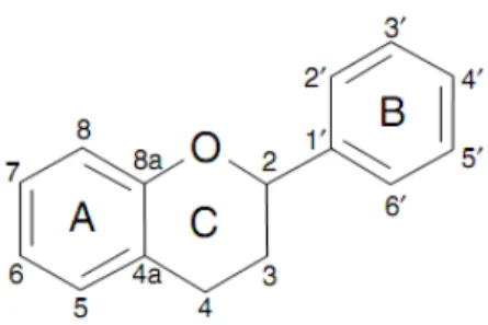
Flavonoids are a group of polyphenolic compounds diverse in chemical structure and characteristics. They occur naturally in fruit, vegetables, nuts, seeds, flowers, and bark and are an integral part of the human diet (Cook and Samman, 1996), although they are considered as non-nutrients (Negrào and Faria, 2009). All flavonoids share a basic C6-C3-C6
structural skeleton, consisting of two aromatic C6 rings (A and B) and a heterocyclic ring (C)
that contains one oxygen atom (Fig. 4).
Fig. 4. Basic structure of flavan nucleus (Negrào and Faria, 2009).
On the basis of the position of and the modifications to the A, B and C rings, flavonoids are grouped into the following structural classes: flavanones, dihydroflavonols, leuco-anthocyanidins (flavan-3,4-diols), flavan-3-ols, leuco-anthocyanidins, anthocyanins, flavones, flavonols and isoflavones (Negrào and Faria, 2009). Most flavonoid compounds are glycosides, which are often accumulated in the vacuoles of plant cells. Glycosides can be either O- or C-linked (Rice-Evans, 2001).
Many of these flavonoids have potential beneficial effects on human health. They have been found to have effects on: cardiovascular diseases prevention by virtue of their capacity to inhibit lipoproteins oxidation (Mink et al., 2007), neuro-degenerative diseases prevention (Singh et al., 2008) and reduction in cancer incidence (Singh and Khar, 2006). In addition, flavonoids exhibit a wide range of biological activities, including antioxidant,
anti-35
inflammatory (Negrào and Faria, 2009), antimicrobial, anti-ulcer, hepatoprotective actions and as enzymes inhibitors (Harborne and Williams, 2000). Most of the beneficial health effects of flavonoids are attributed to their antioxidant abilities (Negrào and Faria, 2009).
3.3.1. Antioxidant activity of flavonoids
a. Scavenging activity
Flavonoids express their radical-scavenging action through two mechanisms. The first is based on the capacity of the flavonoids functional group to donate a hydrogen atom to a free radical R. (R represents superoxide anion, peroxyl, alkoxyl, and hydroxyl radicals).
Through this so-called hydrogen-atom transfer mechanism, the flavonoid antioxidant (FlOH) itself becomes a free radical (Fl-O.) which may react with a second radical, acquiring a stable quinone structure (Fig. 5). The second mechanism is the single-electron transfer from FlOH to a free radical R. with formation of a stable radical cation FlOH.+ (Wright et al., 2001).Due to their lower redox potentials (0.54 ˂ E7 ˂ 0.7V), flavonoids are thermo-dynamically able to
reduce highly oxidizing free radicals with redox potentials in the range (1.0-2.13V), such as superoxide, peroxyl, alkoxyl, and hydroxyl radicals (Jovanovic et al., 1994).
36
Fig. 5. Scavenging of ROS (R.) by flavonoids (Pietta, 2000).
All these reactions provoke inhibition or reduction in the formation of free radicals, they interrupt the propagation of free radical chain reactions, or they delay the start or reduce the reaction rate (Wright et al., 2001). Besides scavenging, flavonoids may stabilize free radicals involved in oxidative processes by complexing with them (Pietta, 2000).
There are three functional groups that have been attributed to an increase in the ROSscavenging potential among the flavonoids: the odihydroxy structure of the B ring; the C2 -C3 double bond in concert with a 4-oxo functionality of the C ring; and the additional presence of both a 3- and a 5-hydroxyl moiety of the C and A rings, respectively (Heim et al., 2002).
b. Chelating activity
Flavonoids can also act as antioxidants by chelating metal ions (Fig. 6) such as Fe+2/ Cu+ and Fe+3/Cu+2 ions that are involved in the conversion of O2 and H2O2 into highly aggressive OH.
through Haber-Weiss/Fenton-type reactions (Engelmann et al., 2005). Thus, chelating agents may inactivate metal ions and potentially inhibit the metal-dependent processes (Andjelkovic
et al., 2006).
37
The flavonoids with better Fe+3 reducing activity are those with a 2, 3-double bond and possessing both the catechol group in the B-ring and the 3-hydroxyl group. The copper reducing activity seems to depend largely on the number of hydroxyl groups. However, it must also be recalled that plant polyphenols bearing catechol and/or pyrogallol moieties can, under certain circumstances, exert prooxidant properties, notably by reducing Fe+3 or Cu+2 ions that they chelate (Mira et al., 2002).
c. Implication in lipidic peroxidation process
The previous two action modes may explain the effect of flavonoids on lipidic peroxidation, since lipidic peroxidation implies the presence of free radicals and metallic ions. They are able to directly scavenge free radicals and then interrupt the propagation step. Moreover, as potent chelators, they chelate the free iron (Engelmann et al., 2005). Finaly, flavonoids present on membrane surface are able to regenerate the vitamin E, one of antioxidant essential to cell membrane protection (Negre-Salvayre et al., 1995).
d. Enzymes inhibition
Flavonoids are also known to be potent inhibitors for several enzymes, such as XO, NOS cyclooxygenase, lipooxigenase, and phosphoinositide 3-kinase. The presence of numerous aromatic hydroxyl groups on flavonoids enables them to easily attach to enzyme surfaces averting potent inhibition(Middleton et al., 2000).
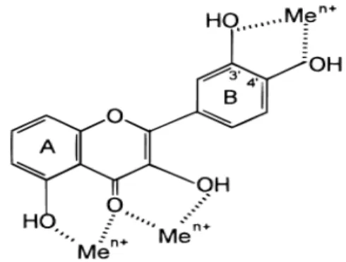
The hydroxyl groups at C-5, C-7, C3’ and C4’ and the carbonyl group at C4, which contribute favorable hydrogen bonds and electrostatic interactions between inhibitors and the active site, and the double bond between C-2 and C-3 in C-ring, which maintain planar structure, were essential for a high inhibitory activity on xanthine oxidase (Cos et al., 1998; Van Hoorn et al., 2002; Lin et al., 2002; Heim et al., 2002).
38
4. Plant (Cachrys libanotis L.)
The Apiaceae (Umbellifereae) is a cosmopolitan family. It is a large family with about 300 genera and more than 3000 species (Heywood et al., 1996). In Algeria, it has about 55 genera and 130 species. Cachrys is a genus belonging to the family of Apiaceae. This genus consists of more than 100 species and is widely distributed in Mediterranean basin (Quezel and Santa, 1963).
4.1. Taxonomy
Kingdom: Plantae, subkingdom: Tracheobionta, super division: Spermatophyta, division: Magnoliophyta, class: Magnoliopsida, subclass: Rosidae, order: Apiales, family: Apiaceae, genus: Cachrys, specie: Cachrys libanotis L. (C. libanotis).
Cachrys libanotis has many synonyme such as Cachrys echinophora var. leiocarpa Guss.,
Hippomarathrum libanotis var. typicum Fiori, Hippomarathrum libanotis Koch, and Hippomarathrum bocconei Boiss.
Cachrys libanotis is called: ighni (local name), amarint(h)e or armarint(h)e à fruit lisse
(French), and with the common Persian name of horse fennel. However, only the name of genus (Cachrys) was found in English, basil. Cachrys was derived from the romans word
kaio, to burn, on account of the carminative qualities of the plants (Don, 1834).
4.2. Botanical description
Cachrys libanotis is a perennial plant distributed in Italy, Sardinia, North Africa and South
West of Europe. It’s of 40-80 cm height; its leaves are divided into linear strips. Flowers are yellow and organized in umbel. Its fruits are smooth and ovoid (10-15 mm) and its seeds are extremely acrid. Its roots are gross, white, branching, and aromatic (Fig. 7). The plant grows in arid places and in hedges (Quezel and Santa, 1963).
39
4.3. Traditional uses
Cachrys libanotis is heating, astringent and anti-jaundice (de Lamarck and Poiret, 1783).
Local healers have traditional knowledge about their uses. In fact, they use this plant for treatment of rheumatism. Noting that, in our knowledge, there is no scientific studies on its biologic proprieties, excepting its dermatitis effect (Ena et al., 1989; Ena et al., 1991).
Arial part Leaves
Flowers Roots
Fig. 7. Cachrys libanotis L. plant. Arial part, leaves and flowers available from: http://www.actaplantarum.org/acta/albums1.php?id=3824
Materials and methods
1. Materials
1.1. Biological materials
The medicinal plant C. libanotis L. was gathered in November 2010 from Ain-Touta, Batna. The sample was authenticated by Pr. Oudjhih Bachir, university Elhadj Lakhdar, Batna.
40
dark, and powdered using traditional mill. The powder was conserved in glass bottle at ambient temperature until its use. Bovine milk was obtained in February 2011 from local farm (Guedjel) and stored at 4°C prior to its use.
1.2. Chemicals and materials
Many of chemical materials were used : butanol, methanol, hexane, chloroform, acetate ethyle, acetic acid, Folin-Ciocalteu, aluminium chloride (AlCl3), Caffeic acid, catehin, ellagic
acid, epicatechin, feslin, flavon, propyl gallat, gallic acid, gossypin, kaemferol, morin, myricetin, naringin, naringenin, quercetin, rutin, etylenediamine-tetraacetic acid (EDTA), dithiothreitol (DTT), ammonium sulfate, sodium phosphate (Na2HPO4 and NaH2PO4), bicine,
acrylamide, bis-acrylamide, brillant bleu R250, glycine, sodium dodecyl sulphate (SDS), coumassie blue, xanthine, NAD+, allopurinol, cytochrome c, 1,2-diphenyl-2-picrylhydrazyl hydrate (DPPH), β-carotene, linoleic acid, tween 40, butylated hydroxytoluene (BHT), nitro bleu tetrazolium (NBT), 5-methylphenazinium methyl sulfate (PMS), ferrozine, iron II chloride (FeCl2), 2,4,6- tripyridyl-s-triazine (TPTZ), iron III chloride (FeCl3). All these
reagents are from Sigma, Fluka and Prolab. Among used apparatus: rotavapor (Rotavapor Germany, bÜchi461), centrifuge (Sigma) 3K30, spectrophotometer (TechomP, UV/VIS-8500), electro-migration apparatus (pharmacia-LKB), HPLC apparatus (Shimadzu 10 vp).
2. Methods
2.1. Flavonoids extraction
Cachrys libanotis powder was soaked, according to Markham method (1982), in 85%
aqueous-methanol with a ratio of plant material and extracting solvent of 1:10 w/v, under agitation overnight at 4°C. The extract was filtered on filter paper then on sintered glass to obtain the first filtrate. This procedure was repeated on the residue using 50% aqueous – methanol under agitation for 4h to obtain the last filtrate. The first and the last filtrates were
41
combined then the methanol was removed under reduced pressure on a rotavapor below 45°C. The milk white crude extract was coded as CrE. A defined portion of total CrE volume was lyophilized and stored at -20°C until it use.
CrE was subjected to fractionation using liquid-liquid extraction. CrE was successively extracted with different solvents of increasing polarity: hexane for defatting, chloroform for aglycone flavonoids extraction and ethyl acetate for glycoside flavonoids extraction (Fig. 8). The obtained organic layer of each partition was evaporated under reduced pressure on a rotavapor below 45°C to dryness and to afford hexane, chloroform, ethyl acetate and aqueous fractions coded as HxE, ChE, EAE and AqE, respectively. All of these fractions were stored at -20°C prior to use.
Crude extract
- Methanol 50% extraction - Agitation for 4h
- Filtration on sintered glass
Combined filtrates Filtrate Residue
Filtrate Residue
- Methanol 85% extraction - Agitation for 24h
- Filtration on sintered glass
Roots powder
42
Fig. 8. Extraction procedure of flavonoids from C. libanotis roots (Markham, 1982).
2.2. Analyse of C. libanotis extracts
2.2.1. Determination of total polyphenols content
Total polyphenols content was estimated by the Folin–Ciocalteu method (Li et al., 2007). This method consists of the phosphotungstic (WO4-2)-phosphomolybdic (MoO4-2) acid (Folin-Ciocalteu’s reagent, FCR) reduction by the phenolic hydroxyl groups, resulting in the formation of a blue product in alkaline solution. Briefly, 200 µl of appropriate dilution of each
43
extract were added to 1 ml of 1:10 diluted FCR. After 4 min, the reaction mixture was neutralized with 800 µl of saturated sodium carbonate (75 g/l). Subsequently, the shaken mixture was allowed to stand for 2h at room temperature, and then measured at 765 nm using VIS spectrophotometer. Gallic acid (20-140 mg/l) was used for the standard calibration curve (Fig. 9). The results were expressed as µg gallic acid equivalent (GAE)/mg of each extract.
Fig. 9. Calibration curve of gallic acid.
2.2.2. Determination of flavonoids content
The AlCl3 method (Bahorun et al., 1996) was used for determination of the flavonoids content
of the C. libanotis extracts, employing the reaction of complex formation between flavonoids and aluminum chloride. Aliquots of 1 ml of each extract were added to equal volumes of a solution of 2% AlCl3.6H2O (2 g in 100 ml methanol). The mixture was vigorously shaken,
and absorbance was read at 430 nm after incubation in dark at room temperature of 10 min. Quercetin and rutin (1-40 mg/l) were used as standards for calibration curve (Fig. 10). Flavonoids contents were expressed as µg quercetin and rutin equivalent (QE and RE, respectively)/mg of each extract.