Effects of kinesiotaping on symptoms, functional
limitations, and underlying deficits on individuals with
rotator cuff tendinopathy
Thèse
Fabio Carlos Lucas de Oliveira
Doctorat en sciences cliniques et biomédicales
Philosophiæ doctor (Ph. D.)
Effects of kinesiotaping on symptoms, functional
limitations, and underlying deficits on individuals with
rotator cuff tendinopathy
Thesis
Fábio Carlos Lucas de Oliveira
Under the supervision of:
Dr. Jean-Sébastien Roy, Supervisor
Dr. Laurent Julien Bouyer, Co-supervisor
RÉSUMÉ
Le kinesiotape est une ressource complémentaire largement utilisée dans les cliniques pour le traitement de nombreuses pathologies musculosquelettiques, qui a été suggéré comme un traitement efficace pour diminuer la douleur et les limitations fonctionnelles chez les individus avec une tendinopathie de la coiffe des rotateurs (TCR) par l’augmentation du retour proprioceptif, qui contribuerait à l’amélioration du contrôle neuromusculaire de l’épaule. L’objectif de cette thèse était de déterminer si le kinesiotape engendre des gains supplémentaires à la réadaptation des individus avec TCR à moyen et long terme.
L’efficacité du kinesiotape à moyen et long terme a été étudiée lorsqu’utilisé en association avec un programme de réadaptation de six semaines, basé sur l’entraînement sensorimoteur pour restaurer le contrôle neuromusculaire de l’épaule. Pour atteindre nos objectifs, 52 individus diagnostiqués avec une TCR unilatérale ont participé à un traitement composé de 10 séances de physiothérapie et d’exercices à la maison. Les participants ont été assignés, aléatoirement, à l’un des deux groupes (KT [expérimental] ou No-KT [contrôle]), dans lesquels le groupe KT a reçu une application thérapeutique de kinesiotape, spécifique pour la TCR, en plus du programme de réadaptation, alors que le groupe No-KT a reçu seulement le programme de réadaptation. Le programme de réadaptation était le même pour les deux groupes, incluant un entraînement sensorimoteur, la rééducation du patient, des exercices résistés pour le renforcement musculaire, de la thérapie manuelle, et des étirements. Un plan de traitement individuel a été personnalisé et mis en place pour chaque participant. Les techniques utilisées variaient en fonction des besoins spécifiques de chacun.
Le niveau de symptômes et les limitations fonctionnelles ont été évalués avec le questionnaire Disabilities of the Arm, Shoulder, and Hand (DASH), le Brief Pain Inventory (BPI), et le Western Ontario Rotator Cuff (WORC) à cinq moments (évaluation initiale, 3, 6 et 12 semaines, et 6 mois), alors que les amplitudes de mouvement (ADM) de l’épaule, sans douleur et complète, et la distance acromio-humérale (DAH) au repos et à 60° d’abduction active de l’épaule, ont été évaluées avant (évaluation initiale) et après le traitement (semaine 6). De plus, l’effet immédiat du kinesiotape sur l’augmentation de la DAH et sur l’amélioration de la capacité de repositionnement articulaire actif des individus avec TCR a également été évaluées avant la première séance de physiothérapie chez les participants du groupe KT (devis transversal).
Globalement, 78.8% des participants ont rapporté un changement positif significatif de leur condition à la fin du traitement. Les résultats de l’essai randomisé contrôlé (ECR) montrent que les deux groupes ont présenté une amélioration similaire et significative de leurs symptômes et limitations fonctionnelles au fil du temps. Par conséquent, le kinesiotape n’a apporté aucun bénéfice
supplémentaire au processus de réadaptation pour réduire les symptômes et les limitations fonctionnelles chez les individus avec TCR à moyen et long terme. De plus, les résultats de l’étude transversale ont montré que le kinesiotape seul a entraîné une augmentation immédiate de la DAH chez les individus avec TCR alors qu’aucun changement immédiat de la capacité proprioceptive chez ces mêmes individus n’a été observé.
ABSTRACT
Kinesiotaping, an adjunct resource widely used in clinics for treating several musculoskeletal disorders, has been suggested to be effective in immediately reducing pain and functional limitations in individuals with rotator cuff tendinopathy (RCTe) through improvement of the proprioceptive feedback, which may contribute to improving shoulder control. The objective of this thesis was to determine whether kinesiotaping provides additional benefits for the rehabilitation of individuals with RCTe in the mid and long-term.
The effectiveness of kinesiotaping in the mid and long-term was investigated when used in conjunction with a 6-week rehabilitation programme based on sensorimotor training for the restoration of shoulder neuromuscular control. To reach our objectives, 52 individuals diagnosed with unilateral RCTe took part in a treatment composed of 10 physiotherapy sessions and home exercises. Participants were randomly assigned to one of two groups (KT [experimental] or No-KT [control]), in which KT group received a therapeutic kinesiotaping application, specific for RCTe, in addition to the rehabilitation programme, whereas No-KT group received only the rehabilitation programme. The physiotherapy rehabilitation programme was the same for both groups, including sensorimotor training, patient re-education, resisted exercises for muscular strengthening, manual therapy, and stretching exercises. An individual rehabilitation plan was customized for each participant. The techniques used varied according to the specific needs of each participant.
The level of symptoms and functional limitations were assessed using the Disabilities of the Arm, Shoulder, and Hand (DASH) questionnaire, Brief Pain Inventory (BPI) scores, and the Western Ontario Rotator Cuff (WORC) index, in five-time points (at baseline, week-3, week-6, week-12 and 6-months follow-up), whereas pain-free and full shoulder active range of motion (ROM), and acromiohumeral distance (AHD) at rest and at 60º of active shoulder abduction were evaluated before (baseline) and after the treatment (week-6). In addition, the immediate effect of kinesiotaping in increasing AHD and improving the active joint repositioning ability of individuals with RCTe was also assessed before the first physiotherapy session in the participants of the KT-group (cross-sectional design).
In general, 78.8% of the participants reported a significant positive change in their shoulder condition at the end of the treatment. The results of the randomized controlled trial (RCT) show that both groups presented a similar and significant improvement of their symptoms and functional limitations over time. Therefore, kinesiotaping did not provide additional benefits to the rehabilitation process for reducing symptoms and functional limitations of individuals with RCTe in the mid- and long-term. In addition, the results of the cross-sectional study showed that kinesiotaping alone provided an
immediate increase of AHD in individuals with RCTe, whereas no immediate changes in the proprioceptive ability of these individuals were observed.
TABLE OF CONTENT
RÉSUMÉ ... III ABSTRACT ... V TABLE OF CONTENT ... VII LIST OF TABLES ... XIII LIST OF FIGURES ... XIV LIST OF ABBREVIATIONS ...XV LIST OF ANNEXES ... XVI LIST OF APPENDICES ... XVII DEDICATION ... XVIII ACKNOWLEDGMENTS ... XIX PREFACE ...XX CHAPTER 1. INTRODUCTION ... 1 1.1. The shoulder ... 1 1.1.1. Shoulder stability ... 2
1.1.2. Shoulder neuromuscular control ... 6
1.1.2.1. Internal model ... 6
1.1.2.2. Proprioception ... 6
1.2. The Rotator Cuff Tendinopathy (RCTe) ... 7
1.2.1. Epidemiology of rotator cuff tendinopathy ... 7
1.2.2. Main causes of the rotator cuff tendinopathy ... 8
1.2.2.1. Intrinsic and extrinsic factors ... 8
1.2.2.2. Central changes related to a rotator cuff tendinopathy ... 12
1.2.3. Impairments observed in individuals with rotator cuff tendinopathy ... 13
1.2.3.1. Muscle activation during dynamic movements ... 13
1.2.3.2. Mechanical alterations during arm elevation ... 14
1.2.3.3. Acromiohumeral distance (AHD) ... 15
1.2.3.4. Range of motion (ROM)... 16
1.2.4. Symptoms caused by rotator cuff tendinopathy ... 17
1.3. Treatments for rotator cuff tendinopathy ... 17
1.4. The Kinesiotaping ... 22
1.4.1. Characteristics of the kinesiotaping ... 22
1.4.2. The rationale behind the kinesiotaping functioning ... 23
1.4.3. Current knowledge about kinesiotaping for rotator cuff tendinopathy ... 24 1.4.3.1. What is currently known about the effects of kinesiotaping on pain and
1.4.3.2. What is currently known regarding the effects of kinesiotaping on
proprioception? ... 26
1.4.3.3. What is currently known regarding the effects of kinesiotaping on acromiohumeral distance? ... 27
1.4.3.4. What is currently known regarding the effects of kinesiotaping on the range of motion? ... 28
1.5. Objectives and hypotheses ... 29
1.5.1. Pattern of EMG activity of RC muscles in individuals with RCTe (manuscript 1) . 30 1.5.2. Research protocol for determining the effects of kinesiotaping used in conjunction with a conventional physiotherapy programme for individuals with RCTe (manuscript 2) ... 30
1.5.3. Immediate effects of kinesiotaping on subacromial space and shoulder proprioception (manuscript 3) ... 30
1.5.4. Benefits of kinesiotaping for the rehabilitation of individuals with RCTe in the short, mid- and long-term (manuscript 4) ... 30
CHAPTER 2. ELECTROMYOGRAPHIC ANALYSIS OF ROTATOR CUFF MUSCLES IN PATIENTS WITH ROTATOR CUFF TENDINOPATHY: A SYSTEMATIC REVIEW ... 32
2.1. Résumé ... 33
2.2. Abstract ... 34
2.3. Introduction ... 35
2.4. Methods ... 36
2.4.1. Identification and selection of studies... 36
2.4.2. Assessment of characteristics of studies ... 36
2.4.2.1. Qualitative analysis (critical appraisal) ... 36
2.4.2.2. EMG scale of assessment ... 37
2.4.2.3. Data extraction ... 39
2.4.2.4. Data analysis ... 39
2.5. Results ... 40
2.5.1. Characteristics of the studies ... 41
2.5.1.1. Diagnostic criteria and labeling ... 41
2.5.1.2. Methodological quality ... 41
2.5.2. EMG activity of RC muscles in patients with RCTe ... 42
2.5.2.1. Impact of RCTe on EMG activity during isometric contractions ... 42
2.5.2.2. Impact of RCTe on EMG activity during isokinetic contractions ... 42
2.6.1. EMG activity of RC muscles in patients with RCTe ... 45
2.6.1.1. Normalization of EMG values in symptomatic patients ... 45
2.6.1.2. Impact of RCTe on EMG activity during isometric contractions ... 45
2.6.1.3. Impact of RCTe on EMG activity during isokinetic contractions ... 46
2.6.1.4. Impact of RCTe on EMG activity during isotonic contractions ... 46
2.6.1.5. Impact of RCTe on EMG activity during unrestrained active movements 46 2.6.1.6. Impact of RCTe on EMG activity in sports movements ... 47
2.6.2. Strengths and limitations of the study ... 48
2.6.3. Future research directions ... 48
2.7. Conclusions ... 49
2.8. Footnotes ... 49
2.8.1. Competing interests ... 49
2.8.2. Source(s) of support ... 49
CHAPTER 3. METHODOLOGY ... 60
3.1. Manuscript 2 (Effects of kinesiotaping added to a rehabilitation programme for patients with rotator cuff tendinopathy: protocol for a single-blind randomised controlled trial addressing symptoms, functional limitations, and underlying deficits) ... 61
3.1.1. Résumé ... 62
3.1.2. Abstract ... 63
3.1.3. Introduction ... 64
3.1.3.1. Objectives and hypotheses... 65
3.1.4. Methods and Analysis ... 66
3.1.4.1. Study design ... 66
3.1.4.2. Participants ... 68
3.1.4.3. Randomization, blinding and allocation concealment ... 68
3.1.4.4. Rehabilitation programme (independent variable) ... 68
3.1.4.5. KT techniques ... 70
3.1.4.6. Data collection ... 72
3.1.4.6.1. Outcome measures (dependent variables) ... 72
3.1.4.6.2. Primary outcome ... 72
3.1.4.6.2.1. Symptoms and functional limitations ... 72
3.1.4.6.3. Secondary outcomes ... 73
3.1.4.6.3.1. BPI and WORC index ... 73
3.1.4.6.3.2. Range of motion (ROM) ... 73
3.1.4.6.3.3. Acromiohumeral distance (AHD) and muscle activity 73 3.1.4.6.3.4. Global Rating of Change (GRC) ... 75
3.1.4.7. Sample size ... 75
3.1.4.8. Recruitment of patients ... 75
3.1.4.9. Withdrawal of individual participants ... 76
3.1.4.10. Data integrity and analysis ... 76
3.1.4.11. Statistical analysis ... 76
3.1.5. Discussion ... 77
3.1.5.1. Strength and limitations of this study ... 77
3.2. Manuscript 3 (Immediate effects of kinesiotaping on acromiohumeral distance and shoulder proprioception in individuals with symptomatic rotator cuff tendinopathy) ... 78
3.2.1. Participants ... 78
3.2.2. Study design ... 78
3.2.3. Outcome measure ... 79
3.2.3.1. Active joint repositioning ... 79
3.2.3.2. Ultrasound imaging ... 81
3.2.4. Kinesiotaping techniques ... 82
3.2.5. Sample size ... 84
3.2.6. Statistical and data analysis ... 84
CHAPTER 4. IMMEDIATE EFFECTS OF KINESIOTAPING ON ACROMIOHUMERAL DISTANCE AND SHOULDER PROPRIOCEPTION IN INDIVIDUALS WITH SYMPTOMATIC ROTATOR CUFF TENDINOPATHY ... 85
4.1. Résumé ... 86 4.2. Abstract ... 87 4.3. Introduction ... 88 4.4. Methods ... 89 4.5. Results ... 89 4.6. Discussion ... 91 4.6.1. Limitations ... 93 4.7. Conclusions ... 93 4.8. Footnotes ... 93 4.8.1. Funding ... 93 4.8.2. Competing interest ... 94 4.8.3. Ethics approval ... 94
5.1. Résumé ... 96 5.2. Abstract ... 97 5.3. Introduction ... 99 5.4. Methods ... 100 5.4.1. Design ... 100 5.4.2. Participants ... 100 5.4.3. Sample size ... 100
5.4.4. Randomisation, blinding and allocation concealment ... 101
5.4.5. Intervention ... 101
5.4.5.1. Rehabilitation programme ... 101
5.4.5.2. Kinesiotaping application ... 101
5.4.6. Outcome measures ... 103
5.4.6.1. Symptoms and functional limitations (primary outcome) ... 103
5.4.6.2. Pain intensity and RC-specific symptoms ... 105
5.4.6.3. Range of motion ... 105
5.4.6.4. Acromiohumeral distance ... 105
5.4.7. Data handling and statistical analyses... 106
5.5. Results ... 106
5.5.1. Flow of participants through the study ... 106
5.5.2. Effects of kinesiotaping ... 108
5.5.3. Description of the time effect for all outcomes ... 108
5.6. Discussion ... 114
5.6.1. Strength and limitations ... 115
5.6.2. Clinical implications ... 116 5.7. Conclusion ... 116 5.8. Footnotes ... 116 5.8.1. Contributors ... 116 5.8.2. Funding ... 116 5.8.3. Competing interests ... 117 5.8.4. Ethics approval ... 117
5.8.5. Data sharing statement ... 117
CHAPTER 6. DISCUSSION ... 118
6.1. Altered muscle activity in individuals with RCTe ... 118
6.2. Acromiohumeral distance and shoulder proprioception immediately after kinesiotaping application ... 119
CHAPTER 7. CONCLUSIONS ... 124
7.1. Research avenues arising from this doctoral thesis and clinical recommendations ... 124
ANNEXES ... 126
APPENDICES ... 155
LIST OF TABLES
CHAPTER 1
Table 1.1. Summary of evidence on the effectiveness of interventions (passive modalities)
used for improving pain and functional limitations ... 20
Table 1.2. Classification of kinesiotaping according to the tension applied (by percentage) ... 23
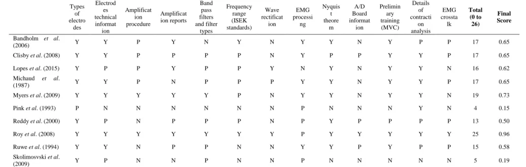
CHAPTER 2 Table 2.1. Assessment of methodological quality (critical appraisal) after a consensus between the researchers ... 37
Table 2.2. Scores of EMG scale of assessment after a consensus between the researchers ... 38
Table 2.3. Evidence table of included studies ... 50
Table 2.4. Overview of the level of evidence for the outcomes ... 57
Table 2.5. Muscle investigated and general findings of the included studies ... 58
Table 2.6. Overview of the level of evidence for the functional grouping ... 59
CHAPTER 4 Table 4.1. Demographic characteristics (n=22) ... 89
Table 4.2. Descriptive statistics of the acromiohumeral distance (AHD; in mm) in two conditions (with and without kinesiotaping) (n=22) ... 90
Table 4.3. Mean absolute error scores during the joint repositioning task for testing proprioception in two conditions (without [No-KT] and with kinesiotaping [KT]) (n=22) ... 90
CHAPTER 5 Table 5.1. Baseline characteristics of participants (n=52) ... 110
Table 5.2. Group mean scores for all outcomes. Data expressed as mean ± standard deviation ... 111
Table 5.3. Results (p-values) of ANOVAs statistical tests for the intention-to-treat analysis 112 Table 5.4. Outcomes changes over time (mean improvements) compared to baseline values throughout treatment (overall sample, n=52; intention-to-treat analysis) ... 113
LIST OF FIGURES
CHAPTER 1
Figure 1.1. Contribution of scapulothoracic muscles for shoulder functioning through scapular
force couple ... 5
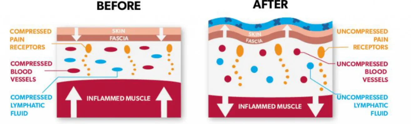
Figure 1.2. Mechanisms of rotator cuff tendinopathy ... 11 Figure 1.3. Hypothetical effects of kinesiotaping underneath the skin (lifting effects) ... 24
CHAPTER 2
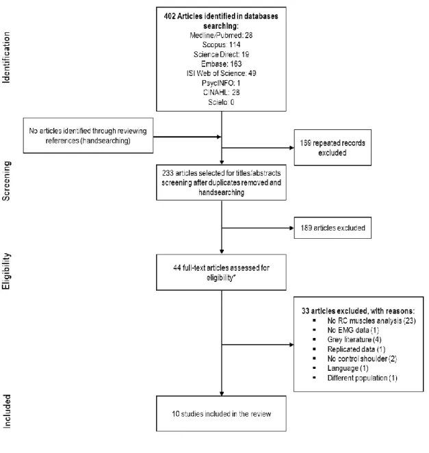
Figure 2.1. Flowchart describing the article selection process ... 40
CHAPTER 3
Figure 3.1. Schematic diagram of the study design ... 67 Figure 3.2. Kinesiotaping application. First strip (1: Y-shape surrounding deltoid muscles),
second strip (2: I-shape in functional correction for multiaxial shoulder instability over the glenohumeral joint, supraspinatus, trapezius, and middle deltoid muscles) and third strip (3: I-shape in mechanical correction for glenohumeral joint) ... 71
Figure 3.3. Kinesiotaping application ... 79 Figure 3.4. Placement of the IMU wireless sensor used in active joint repositioning task ... 81 Figure 3.5. Kinesiotaping technique for RCTe and ultrasonography illustrating the AHD
measurements at rest (0º). ... 83
CHAPTER 5
Figure 5.1. Kinesiotaping technique for RCTe and underlying deficits ... 103 Figure 5.2. Flowchart for the kinesiotaping application ... 104 Figure 5.3. Flow diagram of participants through the study ... 107 Figure 5.4. Mean group scores to symptoms and functional limitations (DASH, BPI, and
LIST OF ABBREVIATIONS
ADM Amplitude de mouvement
AHD Acromiohumeral distance
ANOVA Analysis of variance
BPI Brief Pain Inventory
CI Confidence interval
CID Clinically important difference
CIRRIS Centre for interdisciplinary research in rehabilitation and social integration
CIUSSS-CN Center Integrated University Health and Social Services (Centre intégré
universitaire de santé et des services sociaux de la Capitale Nationale, in French)
CNS Central nervous system
DAH Distance acromiohumérale
DASH Disabilities of the Arm, Shoulder, and Hand questionnaire
ECR Essai randomisé contrôlé
EMG Electromyography
ER External rotation
ES Effect size
FL Functional limitations
GRC Global Rating of Change
ICC Intraclass correlation coefficient
IC Intervale de confiance
IRDPQ Institut de réadaptation en déficience physique de Québec
IR Internal rotation
ISEK International Society of Electrophysiology and Kinesiology
KT Kinesiotaping
MDC Minimal detectable change
RC Rotator cuff
RCTe Rotator cuff tendinopathy
RCT Randomised controlled trial or randomized controlled trial
RMS Root mean-square
ROM Range of motion
SEM Standard error of measurement
SENIAM Surface EMG for Noninvasive Assessment of Muscles
SRM Standardized response mean
TCR Tendinopathie de la coiffe de rotateurs
LIST OF ANNEXES
Annex A
Information sheets and consent form... 126
Annex B
Edinburgh Handedness Inventory ... 133
Annex C
Disabilities of the Arm, Shoulder and Hand (DASH) – French-Canadian version ... 135
Annex D
Brief Pain Inventory (BPI) ... 140
Annex E
Western Ontario Rotator Cuff (WORC) index ... 142
Annex F
LIST OF APPENDICES
Appendix A
Search strategy (manuscript 1) ... 155
Appendix B
Checklist for assessing EMG (manuscript 1) ... 157
Appendix C
Recruiting email ... 163
Appendix D
Recruiting poster ... 165
Appendix E
Telephone interview – Primary screening ... 167
Appendix F
Eligibility criteria form and clinical tests ... 169
Appendix G
Sociodemographic questionnaire ... 171
Appendix H
Anamnese ... 174
Appendix I
Outcome assessments form – acromiohumeral distance (AHD), range of motion (ROM), and shoulder proprioception ... 176
Appendix J
Assessor and participants blinding evaluation (manuscript 4) ... 181
Appendix K
Home exercises adherence form ... 184
Appendix L
DEDICATION
This thesis is dedicated to my mother (in memoriam) that, despite we have passed not much more than eight years living together, taught me how to persevere even with adversities and that the real objectives are achievable, but only with hard work they could be achieved.
I also dedicate this doctoral thesis to my grandmother, Hercília de Oliveira Machado (in memoriam), who was the main responsible for my growth. Thank you for your hard words and actions that, at the right time, shaped me with important teachings such as character and solidarity. No doubt, I owe you everything! Both of you are unforgettable and will always be in my heart and in my prayers!
Lastly, but not least, a special dedication to my lovely daughter (Gabriela). Although she did not agree in most of the time, even so, she realized that daddy had to work a little more instead of going to play. Because of you, daddy had to take some forced breaks, which, after all, were always very helpful for renewing my energies. Thank you for brightening up my hazy days with your smile, and your sincere, naïve and funny words. My love, I dedicate to you not only this thesis but every day of my life. I love you more than everything in this life!
The faith in the victory has to be unshakeable! Anjos (Pra quem tem fé). O Rappa
Dedico esta tese à minha mãe (in memoriam) que, apesar dos poucos mais de oito anos vividos juntos, me ensinou como é possível perseverar mesmo com as dificuldades e que os reais objetivos são alcançáveis, mas somente com muita luta eles poderiam ser alcançados.
Também dedico esta tese de doutorado à minha avó, Hercília de Oliveira Machado (in memoriam), que foi a principal responsável pela minha criação. Obrigado pelas duras palavras e ações que, no momento certo, me moldaram com importantes ensinamentos, tais como caráter e solidariedade. Sem dúvidas, eu devo tudo a você! Vocês são inesquecíveis e estarão sempre no meu coração e nas minhas orações!
Finalmente, porém não menos importante, uma dedicatória especial à minha adorável filha (Gabriela). Embora ela não tenha concordado na maioria das vezes, ela compreendeu que o papai precisava trabalhar mais um pouco ao invés de ir brincar. Graças a você, o papai teve que fazer algumas pausas forçadas, que, no final das contas, sempre foram muito úteis para renovar as energias. Obrigado por alegrar os meus dias mais nebulosos, com o seu sorriso e suas sinceras, ingênuas e engraçadas palavras. Meu amor, dedico a você não apenas esta tese, mas todos os dias da minha vida. Eu te amo mais do que tudo nesta vida!
ACKNOWLEDGMENTS
First of all, I thank God, our father, and Lord of all the universe, for enlightening my paths and giving me the opportunity to be enrolled in a Ph.D. programme. Thank you, my Lord, for the faith that increases every day, the patience, health, and all friendship provided to me throughout this journey. I thank greatly, Dr. Jean-Sébastien Roy to accept me in his team and for teaching me all aspects of scientific rigor. I also thank him for all encouragements to go further in search of answers. Undoubtedly, his guidance was crucial for me to overcome the hard path along these last three years. I could write a book of acknowledgments to him.
Thanks to Dr. Laurent Bouyer for his expertise, critical and assertive comments, always aiming at improving our work.
Thanks to Dr. Benoît Pairot de Fontenay, for all exchange, friendship, and advice. His engagement in this project was elementary.
I express my gratitude to all CIRRIS members for their backstage support. Special thanks to Mr. Steve Forest and Dr. Jean Leblond, whose availability contributed immensely to this project. Finally, my sincere thanks also go to every funding source for this project. All my gratitude to the CAPES Foundation that provided me a generous grant for pursuing my doctoral studies.
PREFACE
This thesis is composed of four scientific manuscripts, divided into seven chapters, based on aspects related to the rehabilitation process of individuals with a rotator cuff tendinopathy (RCTe). In here, you will find an introduction to the subject (chapter I), where the rationales behind the articles included in this thesis are presented, followed by four publications, which were extracted from the data collected over the last three years on the journey in search of my doctorate in clinical and biomedical sciences. They are presented in chapters II to V.
The first manuscript (chapter II) is a systematic review synthesizing the evidence on muscle activation of the rotator cuff (RC) muscles in symptomatic individuals with RCTe. Published in the
Journal of Electromyography and Kinesiology on June 07th, 2017, this study allowed us to identify important aspects of the RC muscle activity as a possible trigger source of painful symptoms of RCTe and to understand its association with impairments observed in this population. Findings from this review gave us a notion about how and which muscles could be targeted in a clinical research protocol (second manuscript) to determine the effects of kinesiotaping added to a rehabilitation programme for symptoms and functional limitations associated with RCTe. This research protocol was published in the British Medical Journal Open on September 1st, 2017 and it is presented in chapter III with detailed information on the methodology applied in our research protocol.
To address some underlying aspects related to RCTe, a third manuscript, presented in chapter IV, investigated the immediate effects of kinesiotaping on the acromiohumeral distance (AHD) and active shoulder joint repositioning ability, as a part of proprioception, in symptomatic individuals with RCTe. This study was published in the Clinical Biomechanics on November 10th, 2018. Finally, chapter V brings the central study of this doctoral project (fourth manuscript). It is a randomized
controlled clinical trial aiming to identify whether the kinesiotaping add some advantages when included in a rehabilitation programme for individuals with RCTe. This manuscript was submitted to the Journal of Physiotherapy in October 2018.
As the primary author of all these studies, I participated directly in the conception of the study, designing, and preparation of the procedures. Additionally, I was responsible for the data collection, recruitment, interpretation, data analyses, implementation of the rehabilitation programme, and writing the manuscripts. All works were greatly supervised by Dr. Jean-Sébastien Roy, co-supervised
analyses, and supervision of the manuscripts writing. I would like to take advantage of this opportunity to thank you all co-authors involved in these research: Amanda Ager, who was a partner in evaluating the articles for the systematic review (manuscript 1); Dr. François Desmeules, who collaborated in the conception and revision of the main study of this doctoral research (manuscript 2 and 4); and Dr. Benoît Pairot de Fontenay, who participated directly in the preparation of the procedures, data collection, outcomes assessments, interpretation and data analysis, and he has commented several versions of the manuscripts extracted from this research project (manuscript 2 to 4).
Following the presentation of these manuscripts, a discussion is provided in chapter VI, and a general conclusion (chapter VII), based on the studies composing this thesis.
CHAPTER 1. INTRODUCTION
The shoulder is one of the most affected joints in humans. It is estimated that one out of three people in the general population will have, or already had, an episode of shoulder pain. One of the most common causes of shoulder pain is the rotator cuff tendinopathy (RCTe) that can generate long-lasting symptoms and limitations affecting the capacity to work. A high absenteeism or presenteeism rate and an increased loss of productivity, caused by the RCTe, provide an important socioeconomic impact for societies and governments.1,2 According to the Quebec workers’ compensatory board (CNESST), the estimated annual expenses for work-related shoulder injuries in the province of Quebec (Canada) exceeded $ 128 000 000 between 2013 and 2015. Therefore, effective treatments for RCTe is a priority.
Among several methods of treatment described in the literature, rehabilitation is the main choice for managing shoulder disorders3-5 and shoulder control exercises are the basis of a rehabilitation programme for an RCTe due to its effectiveness in reducing shoulder motor deficits.3,4 Previous studies have demonstrated that a rehabilitation based on sensorimotor training is very effective for restoration of the shoulder neuromuscular control.6,7 Notwithstanding, because not all individuals with RCTe present motor abnormalities,8 and due to long-lasting motor deficits and the limitations generated by RCTe, new approaches for the treatment of this injury are encouraged to optimize the effects of a rehabilitation programme based on a sensorimotor training. Understanding the shoulder functioning in normal and pathological conditions as well as the abnormalities that affect the shoulder girdle joints during arm elevation are important for building an efficient rehabilitation strategy for individuals with RCTe.
This introduction section provides a review of the shoulder functioning, followed by a brief approach on RCTe and the conservative methods of treatment commonly used for this injury, prior to dive into a specific topic on the kinesiotaping as a promising therapeutic resource for providing a new approach for individual with RCTe, besides its effects on the population with RCTe.
1.1. The shoulder
The clavicle, through the acromioclavicular and sternoclavicular joints, concatenates the upper limbs and the trunk10. Both the acromioclavicular and the sternoclavicular joints contribute to scapulothoracic upward rotation,12 and possibly to the overall scapulothoracic motion. The coordination between these joints results in an adequate scapulohumeral rhythm.13 Although the scapulothoracic joint is not a true joint, due to the absence of joint capsule and ligamentous stability, it articulates the ribs and scapula,9,10 having, therefore, an important role in the scapular movements during upper limbs motion. Serving as a site for attachment of several muscles, an adequate scapular movement is crucial for the proper functioning of the shoulder.14 The scapula glides over the ribs 2 through 7 during the arm elevation movements, whereas the scapulothoracic joint contributes to maintaining the glenohumeral joint properly aligned throughout arm elevation.9 The synchronous rhythm between the scapula and the glenohumeral joint is essential for a normal shoulder kinematics. For instance, during a 30º arm elevation, the scapula is required to move slightly, whereas, in higher movements of the upper limbs in abduction, the scapula is required to rotate counterclockwise in 2:1 ratio from glenohumeral to scapulothoracic joint motion,15,16 throughout the entire movement. This means that for each 2º of glenohumeral motion, there is 1º of scapulothoracic motion contributing for arm elevation in the frontal plane.17 Thus, in the horizontal abduction at 90º, 30º came from scapulothoracic joint and 60º from glenohumeral joint.
Known as the shoulder joint, the glenohumeral joint is formed by the humeral head and the glenoid fossa of the scapula.9 The glenohumeral joint has a special role in the shoulder kinematics and functionality of the upper limbs.9 Its dynamic stability is provided by glenohumeral muscles, which control the dynamic centralization of the humeral head in the glenoid fossa during arm movements.9,12,18-20 As the glenoid fossa is shallowly concave, with a joint surface smaller than the humeral head, which has a hemispheric and convex format, this articulation has great mobility and poor congruency.9,10 Factors such as the loose connection capsule, the limited bony contour and the lack of ligamentous support makes the glenohumeral joint more susceptible to instability,21 resulting in loss of shoulder stability.
1.1.1. Shoulder stability
Great mobility accompanied by potential instability is the main characteristic of the shoulder.22,23 The shoulder stability is related to a proper glenohumeral joint functioning through an adequate intersegmental control of the humerus, scapula, and clavicle, and this stability is assured by the cooperation among bony architecture and soft tissues such as muscles, ligaments, tendons and joint
capsule.22 The elements necessary to ensure glenohumeral joint stability and, hence, a normal functioning of shoulder, include the adequate size of the glenoid fossa and proper mobility of the scapula, retroversion of the humeral head, an intact joint capsule and glenoid labrum, and, finally, a proper functioning of RC muscles in terms of force, endurance, flexibility and integrity.22-25
According to the principles of the stability as described by Panjabi,26 there are three subsystems working together to create a joint stability: passive, active, and neural control.26 In the shoulder, the stability is provided by static and dynamic contenders17,22,27 corresponding to passive and active subsystems, respectively. Passive or static joint stability is provided by bony structures, ligaments, and glenoid labrum, whereas active or dynamic stability is provided by the RC (supraspinatus superiorly, subscapularis anteriorly, infraspinatus and teres minor inferiorly), deltoid and biceps brachii muscles. Additionally, both long heads of the biceps and triceps brachii also participated in the stability mechanism of the shoulder, reinforcing the shoulder joint capsule. The long head of the biceps brachii, for instance, acts in the stabilization against a superior translation of the humerus, contributes for anterior glenohumeral joint stability, and resists to humeral torsion force when the shoulder is abducted and externally rotated.17,27
The scapulohumeral and scapulothoracic muscles are the main responsible for the dynamic stability being categorized into three groups: glenohumeral muscles, including the deltoid, pectoralis major, teres major, and the four RC muscles; scapulothoracic muscles, including trapezius (upper, middle, lower fibers), serratus anterior (superior, middle, inferior), rhomboids (major and minor), pectoralis minor, levator scapulae, and subclavius muscles; and multi-joint muscles, including the pectoralis major, latissimus dorsi, biceps and triceps brachii.28 Multi-joint muscles contribute to the motion and forces generated on the glenohumeral joint during movement, whereas the scapulothoracic muscles play an important role in the shoulder control due to the close relationship between scapulothoracic and glenohumeral joints,29 allowing smooth gliding motion of the scapula over the thoracic cage during arm movements. Notwithstanding, the glenohumeral muscles are the primary source of stability for the shoulder since only the RC muscles and the three portions of the deltoid play a significant role to maintain congruency between the scapula and humeral head17 during static and dynamic activities.
that raises the arm up to 30º. Thereafter, the arm is raised by the middle deltoid, which also participates in the shoulder stabilization and provides the strength necessary to lift the arm higher than 30º. When the arm approaches 90º of abduction, the external rotators muscles (infraspinatus, teres minor) rotate the humerus externally, in an attempt to avoid impingement of the structures placed in the subacromial space.30,31 In this context, a dynamic interplay among RC muscles is essential for the stability of the glenohumeral joint since it maintains the humeral head tightly in the glenoid fossa, avoiding impingement of the subacromial structures throughout the arm elevation movement.32 Simultaneously, the acromion raises and moves in posterior tilting, whereas the superior angle of scapula moves downward, and the inferior angle of the scapula moves laterally, in upward rotation.12,14 The scapula can also move into protraction, abduction, and elevation when the shoulder is in normal condition, depending on the direction of the movement.14 These scapular movements require an effective activation of the scapulothoracic muscles and result in clavicular movements, which contributes to important rotational and tipping adjustments of the scapula during movements.33 The scapula is stabilized through a synchronized combination of forces in two planes: the balance between upper trapezius, levator scapulae muscle, and the arm weight, in the frontal plane; and pectoralis minor, rhomboids, and serratus anterior muscles, in the sagittal and transverse planes. A dysfunction in any of these muscles added to a laxity of the joint capsule may affect the shoulder muscle synchronism, which compromises the shoulder stability and the scapular kinematics, resulting in an important alteration in the shoulder neuromuscular control.34 It is well accepted that all these above-mentioned mechanisms are regulated by the central nervous system (CNS) to ensure an adequate shoulder neuromuscular control through activation or co-activation of muscular system to produce a coordinated movement, without compensation or excessive mobility, and without painful symptoms.
Figure 1.1. Contribution of scapulothoracic muscles for shoulder functioning through scapular force couple.
1.1.2. Shoulder neuromuscular control
1.1.2.1. Internal model
Proper neuromuscular control is based on the interaction between the central and peripheral nervous system, besides motor and sensory systems.35-38 The internal model principle of control theory39 is often used to describe the neuromuscular functioning. This theory suggests that all movements once learned, have a model built in the CNS. This pre-structured representation is exploited every time that a known motor task is required. In this context, the intern model builds the movement and triggers the peripheral structures necessary to perform the task properly. Therefore, motor learning plays an important role in the creation and maintenance of built models.35,39 The internal model’s theory includes two components: an inverse and a forward model. The inverse model is based on the determination of the motor command required to achieve the desired movement trajectory. The forward model predicts the sensory feedback that may result from a movement and the consequences of a specific motor command.35 When a command does not result in a proper movement, the command is updated. However, neuroanatomical abnormalities, such as a proprioceptive deficit, may result in malfunctioning of the internal model.40
As the shoulder neuromuscular control relies, in part, on the efferent responses related to the proprioceptive afferent impulses, proprioception has been proposed to assist building the internal representation of the limbs, acting in the conversion of kinematic information to motor commands, resulting in suitable forces to perform a movement.41
Impairments in a mechanical or sensorial structure involved in the shoulder functioning may lead to inadequate movement patterns and functional instability, indicating an improper shoulder neuromuscular control. Previous studies have demonstrated that impairments, such as altered muscle activation of RC30,42,43 and scapulothoracic muscles,25,44-47 associated with a reduced proprioceptive input48 and contributing for decreasing shoulder neuromuscular control, are often observed in individuals with RCTe.49,50
1.1.2.2. Proprioception
Proprioception is an essential part of shoulder stability and neuromuscular control.38,51 For a movement production, a very fast, complex and important exchange of sensory information is accomplished between the peripheral structures and the CNS.36-38 Proprioceptive information originates at the level of mechanoreceptors48 that are present in muscles, tendon, fascia, ligament,
skin and joint capsule,48,52 such as Meissner’s corpuscles (touch-sensitive nerve receptor), Ruffini corpuscle (pressure-sensitive nerve endings), Pacinian corpuscles (vibratory pressure, touch and stretch sensitive receptor), Merkel’s disks (tactile sensory nerve-endings), and free nerve endings (afferent and efferent endings) and are responsible for providing proprioceptive feedback to the CNS. Muscle spindles located in the skeletal muscles,36,53 also plays an important role in the transmission of proprioceptive feedback to the CNS. They respond to changes in the skeletal muscle length.36 Additionally, Golgi tendon organs, located in the musculotendinous junction of skeletal muscles, also participate in the proprioceptive feedback mechanism acting in conjunction with muscles to ensure a proper interconnection between the CNS and the peripheral structures (afferent38 and efferent endings36). This mechanism ensures a proper agonist, antagonist and synergistic muscle activation and, thereafter, provides a synchronous shoulder control.54 Injuries in the joint capsules, ligaments or muscles may also affect the proprioceptive input, resulting in muscular imbalance and impaired shoulder neuromuscular control, triggering a series of problems that can generate an RCTe.
1.2. The Rotator Cuff Tendinopathy (RCTe)
The RCTe is a broad term encompassing several diagnoses related to painful signs and symptoms at the subacromial structures (RC tendon, long head of the biceps, bursa)3,4,55-57 combining pain and impaired performance.58,59 Subacromial structures are involved with inflammation and degeneration in the context of an RCTe. RCTe is frequently termed subacromial pain (impingement) syndrome, based on the underlying mechanism that includes encroachment of the subacromial space soft tissues underneath the coracoacromial arch as the arm is elevated secondary to a dynamic narrowing of the subacromial space.60,61
1.2.1. Epidemiology of rotator cuff tendinopathy
The RCTe is the commonest pathology of the shoulder62-66 and the second most common cause of musculoskeletal complaints in humans.64 According to the systematic review of Luime et al.,67 shoulder pain has an incidence of 0.9% to 2.5% in the general population, and a prevalence rate as follows: wide-ranging point (7% to 26%), monthly (19% to 31%), annual (5% to 47%), and estimated
from this study shown an incidence of 0.3% to 5.5%, and point prevalence from 2.4% to 21%.69 In the working-age population, the prevalence was 2.4 to 14%, with a monthly prevalence of 2.8% and annual ranging from 0.5% to 7.4%.69 Although more frequent in men,70 the symptoms of an RCTe seem to be more incapacitating in women.71 Men are likely more affected than women possibly since most of the awkward72 and physically strained works,73 requiring high physical workload,73 are occupied by men. This may contribute to increase the occurrence of RCTe in this population. Previous literature67,71,74 on the prevalence of RCTe have reported that women present more severe pain and worse shoulder function than men. Hill et al.71 compared the SPADI scores between men and women and found a difference of 9.6 points for pain and stiffness suggesting that women are 40% more likely to have more severe shoulder pain and impaired function than men (41.6 SPADI points vs.32.0 SPADI points, respectively). According to Ge et al.,75 gender differences in pain modulation is pointed out as a possible explanation for the higher severity of pain observed in women.75 For the authors, women feel more pain than men due to greater susceptibility in developing temporal summation mechanisms of muscle pain, besides having different motor control strategies to adapt to an injured condition,76 generating a different circuit of pain interpretation in the women compared to the men.
The RCTe has a very important socio-economic impact on the societies, contributing to increasing the rate of absenteeism and sick leave as the limitations caused by an RCTe may remain for 12 months or more64 in up to 40% of the cases77 involving manual workers. Considering these long-lasting remaining limitations, efficient methods of treatment are strongly needed. Howbeit, an effective treatment is dependent on the accuracy of the diagnosis and the identification of the causes of RCTe, which is a troublesome challenge.
1.2.2. Main causes of the rotator cuff tendinopathy
1.2.2.1. Intrinsic and extrinsic factors
The RCTe may originate from multiple factors and many theories have emerged as a possible explanation of RCTe, including tension overload, degeneration of the RC tendons, abnormal anatomy of the coracoacromial arch or humeral head, and shoulder control impairments.78 Extrinsic, intrinsic, or a combination of both mechanisms are classically described as factors playing an important role in generating an RCTe (Figure 1.2).79
Intrinsic factors are directly related to degeneration of the substance of the tendon, which is commonly caused by genetic,80 aging,81 insufficient vascularity,82 alterations in the biological production or concentration of collagen,83 and mechanical overload in the RC tendon.84 Defined as structural alterations in the tendon, a degeneration, particularly of RC tendons, is inevitable and inexorably progressive.17 Even though the human body has a high capacity to withstand mechanical stress, an excessive level of stress such as overuse and overload, may provide a tissue breakdown, generating structural degeneration.79 Considering the concept stated by Meyer85 that establishes that shoulder tendon is doomed to degeneration85 based on the wear-and-tear effects, aging could contribute to the appearance of an RCTe since the degeneration process occurs slowly. However, limited evidence exists to support this theory that aging is related to the onset of an RCTe.4
The supraspinatus tendon is the most affected tendon in an RCTe. Because it crosses the subacromial space close to the undersurface of the coracoacromial arch, the supraspinatus tendon can possibly be pinched when the arm is elevated over the shoulder level. Due to very poor vascularization of the area where the supraspinatus tendon is constantly pinched, this area is considered critical.13,86 Therefore, it is likely that repeated bouts of hypoxia in the critical zone may induce degeneration of the RC tendon.87 The prompt response for all the repetitive encroachment is a chronic irritation, followed by inflammation, which generates swelling and thickening of the tendon. Combination of these inflammatory events reduces the subacromial space and hampers the passage of the supraspinatus tendon.88,89
The extrinsic factors are those in which structures outside the tendon contribute to repetitive subacromial compressions,61 such as subacromial and acromioclavicular joint osteophytes, pectoralis minor shortening, anatomical variations in the shape of the acromion, inadequate posture and reduced thoracic spine mobility, glenohumeral joint capsule stiffness, shoulder kinematic alterations, RC and scapular muscle impairments, and pathological conditions, such as arthritis in the acromioclavicular joint, that trigger degenerative changes17,79 of the tendon fibers. Abnormalities in the acromion, which is classified according to its shape (type I, flat; type II, curved; and type III, hooked undersurface),90 have been suggested as contributors to the extrinsic mechanism of RCTe. Despite prior studies have documented a relationship between the acromion shape and severity of RCTe,91-93 abnormalities in the acromion shape as a cause of RCTe is very controversial. Balke et al.94 investigated the correlation of the acromial morphology with RCTe and no association between the type of acromion to RC injury
kinematics, contributing to mechanical impingement,61,95 than the shape of the acromion. Therefore, the acromial morphology alone, without combination with any other factor, may not be necessarily a definitive factor causing RCTe.79
It is suggested that an interaction between intrinsic and extrinsic factors exist, as both factors produce alterations resulting in compression of subacromial structures. Although there is no consensus on etiological mechanisms of RCTe,96,97 both intrinsic and extrinsic factors result in impairments in the proprioceptive feedback, compromising the connection between CNS and peripheral structures49,98 and then, contributing to the progression of RCTe.
Figure 1.2. Mechanisms of rotator cuff tendinopathy.
1.2.2.2. Central changes related to a rotator cuff tendinopathy
Individuals with RCTe often report lasting pain. Studies have demonstrated that this long-lasting pain may be associated with alterations in the central motor representations affecting the movements,99,100 such as changes in the corticospinal excitability of shoulder muscles. The presence of persistent pain can lead to changes in the daily habits of individuals with RCTe, such as alterations of the pattern of movements,101 in an attempt to protect the injured tissue from further damage. These changes seem to be positive in the short-term, but in the mid- and long-term, they generate important abnormalities in the motor pattern of shoulder movements.102 Among these possible abnormalities, it can be highlighted the alterations in the level of activation and coordination of RC and scapulothoracic muscles and glenohumeral joint kinematics, which affect the shoulder stability, resulting in progression of motor deficits, and contribute to the recurrence of an RCTe. Recently, Ngomo et al.100 investigated the motor representations of the infraspinatus muscle in 39 individuals with RCTe, using transcranial magnetic stimulation. The authors found that the affected shoulder presented an active motor threshold larger than the unaffected shoulder, suggesting a lower cortical excitability of the infraspinatus in the affected shoulder compared to unaffected shoulder, in the targeted population.100 Interestingly, a moderate correlation (r=.45, p=.005) between the asymmetry in the cortical excitability and the duration of pain was observed. Thus, these alterations in the central motor representations may, partially, explain the chronicity of symptoms100 and the motor deficits100 caused by an RCTe. They may also be responsible for the lack of effectiveness of treatments of individuals with chronic RCTe.22,103
Another interesting point that may play an important role in pain complaints reported by individuals with RCTe, is the central sensitization. Described by Woolf et al.104 as “an amplification of neural
signaling within the CNS that elicits pain hypersensitivity”, central sensitization has been reported as
a hypersensitivity generator (i.e., reduction of the pressure-pain threshold)105 in individuals with RCTe.106 Thus, nociceptors become hypersensitized,104 reducing the pain threshold of individuals with RCTe.106,107 The consequence is an amplified and misrepresented response from the CNS to little (nociceptive) or normal (non-nociceptive) somatosensory input.108
Even supposing that a minority of individuals with RCTe presents predominant central sensitization, evidence has supported the clinical importance of the hypersensitivity in chronic musculoskeletal pain since these individuals often present a poor prognosis and outcomes, not responding adequately to peripheral treatment.108
Although most of the current treatments for RCTe has focused in interventions for the peripheral structures,16,109-116 specific interventions for reversing this maladaptive reorganization, such as pain education and sensorimotor training, have been demonstrated to be effective for reducing painful symptoms and motor deficits associated with RCTe.7,103,117
1.2.3. Impairments observed in individuals with rotator cuff tendinopathy
Based on the all above-mentioned information, one can suppose that the shoulder motor deficits may explain part of the progression or even the severity level of the RCTe. Among these impairments, alterations in the muscle activation of the glenohumeral and scapulothoracic muscles during arm elevation as well as the kinematic alterations of glenohumeral, scapulothoracic, and sternoclavicular joints, are pointed out as strong indications of motor deficits.19,20,45,118 In the next subsections, the main motor deficits observed in individuals with RCTe will be approached, followed by the description of two clinical outcomes that can be used for evaluating the shoulder motor deficits in this population.
1.2.3.1. Muscle activation during dynamic movements
Alterations in muscle activation patterns of the RC muscles could explain, in part, the dynamic narrowing of the subacromial space and the alterations in upper limb kinematics that have been observed in individuals with RCTe during arm elevation.7,8,19 These alterations, along with shoulder mechanical alterations, are consistent indicative of motor deficits.19,20,45,118,119
As the shoulder is an inherently unstable joint, activation of RC muscles plays an important role in the shoulder stability since it increases glenohumeral joint stiffness, thereby maintaining a stabilizing congruency between the humeral head and the glenoid fossa. Therefore, the harmonic functioning of the shoulder is dependent on synchronous activation among RC muscles.30,42,120 The RC muscles are activated alongside other muscles, such as scapulothoracic muscles to properly align the humeral head with respect to the glenoid fossa,30 thereby preventing the impingement of the subacromial structures during arm elevation.32 Thus, scapulothoracic muscles also play an exceptional role in the
of coordination of trapezius and serratus anterior, may partially explain the scapular kinematics impairments observed in individuals with RCTe.121,122 Because alterations in the muscle activity of scapulothoracic muscles can provide alterations in scapular position, an optimal dynamic interplay among deltoid, scapulothoracic and RC muscles is crucial for maintaining or restoring shoulder function.7,8 The neuromuscular impairments, such as muscle activation of RC muscles, have been targeted by several investigations.7,123-126 Systematic reviews of EMG activity of the shoulder complex25,43,44 have concluded that individuals with RCTe may present an altered muscle activity such as increased muscle activity in upper trapezius,25,44 and reduced muscle activity in the lower trapezius and serratus anterior.25 A delayed onset of activation in lower trapezius43,44 and serratus anterior43 has been also reported. As most of the systematic reviews have addressed the EMG activity of scapulothoracic muscles, examination of RC muscle activation is constitutive for a thorough evaluation of shoulder neuromuscular control. In chapter 2, we present a study,42 published in the
Journal of Electromyography and Kinesiology in June 2017, that synthesized the evidence on the
EMG activity of the RC muscles in healthy compared to individuals with RCTe. This study reports that RC muscle activity of individuals with RCTe may be altered; however, analyses involving isometric, isotonic and isokinetic contractions do not expose these alterations. In contrast, dynamic motion analysis favors the identification of the EMG deficits.42
1.2.3.2. Mechanical alterations during arm elevation
As stated in previous studies, mechanical alterations of the shoulder, especially in the glenohumeral and scapulothoracic joints,19,31,127-129 are one of the main causes of symptoms of individuals with RCTe. A superior translation of the humeral head occurs during arm elevation, even in healthy people. In a normal shoulder, the humeral head remains centered in the glenoid fossa, throughout the movement. On the other hand, several mechanical alterations have been observed at the glenohumeral joint and in the scapular motion in a presence of an RCTe. In this condition, the superior translation of the humeral head is increased, resulting in loss of congruency between the humerus and glenoid fossa and, hence, in impingement by compression of the subacromial structures.20 This alteration could be, therefore, the main cause of impairments observed in individuals with RCTe.
Among the kinematic alterations observed in individuals with RCTe, it is highlighted the decreased scapular posterior tilting,19 associated with decreased activation of the serratus anterior;19,118 decreased upward rotation,19,129 which may keep the acromion in a lower anterolateral position20 as a result of reduction in the lower trapezius muscle activation;19,45 increased internal rotation20,119
associated with a reduced infraspinatus activation30,130,131 and increased subscapularis activation;30,132,133 and excessive elevation of the shoulder girdle134 associated with an increased upper trapezius muscle activation.19,135 Restrained scapular motion results in impossibility of the acromion moving away from the humeral head during arm elevation, leading to a dynamic narrowing of the subacromial space7 and compression of subacromial structures.20
In addition to scapular neuromuscular impairments, shortening of the pectoralis minor,19,119,136,137 posterior shoulder tightness,138 and an increase in thoracic spine flexion19,116 are possible factors that could be responsible for the scapular alterations. Thoracic kyphosis has been suggested to contribute to RCTe since it diminishes the scapular posterior tilt, impacting directly the width of subacromial space 139 and reduction of the AHD.
1.2.3.3. Acromiohumeral distance (AHD)
Faulty shoulder neuromuscular control in individuals with RCTe often includes hampered contribution of RC and scapulothoracic muscles30 in keeping the subacromial space intact. The width of subacromial space is estimated through the AHD, which is defined as the tangential distance between humeral head bony landmarks and acromion inferior edge,140 usually determined using magnetic resonance imaging (MRI),141-143 ultrasonography,140,144-146 and radiographs.143,147-149
Previous studies have stated that the AHD varies between 9 and 12 mm in asymptomatic subjects,150,151 however, it is reduced in individuals with RCTe7,141,142,152 during arm elevation or compared to healthy.7,140,152-154 Recently, Navarro-Ledesma et al.152 compared the AHD of 76 individuals with RCTe to 40 healthy subjects. Their results indicated that, at 60º of shoulder abduction, the AHD reduced by 0.33 mm in the symptomatic shoulder in comparison to asymptomatic shoulder, whereas the reduction in comparison to the healthy subjects corresponded to 0.51mm.152 As a reduced subacromial space may compromise the shoulder functioning, methods contributing to increasing the AHD are always welcome. In this context, we tested the effectiveness of the kinesiotaping, as a promising adjunct therapeutic resource, in increasing the AHD. The results can be consulted in chapter 4.
1.2.3.4. Range of motion (ROM)
The ROM is the maximal amount of movement produced within a joint,157 without compensation. In this context, the shoulder has the largest ROM of the body with a maximal elevation amplitude of approximately 180º.157 However, muscular or kinematic abnormalities of the shoulder functioning may compromise this large amplitude. For this reason, evaluation of the ROM is important for determining the strategy for an effective rehabilitation of an RCTe.
In individuals with RCTe, the ROM is often reported to be reduced due to several factors such as joint stiffness and posterior capsule tightness.157,158 Thus, limited ROM is one of the most common impairments observed in this population. A significant moderate relationship (r=-.50, p=.006) between loss of internal rotation ROM and the increased posterior capsule tightness has been previously reported.158 Therefore, the presence of posterior shoulder tightness in the glenohumeral joint along with the reduction of glenohumeral internal rotation may explain the impaired ROM observed in individuals with RCTe.157
Tyler et al.158 quantified alterations in the shoulder ROM of dominant and non-dominant upper-limbs of 31 individuals with RCTe compared to 33 healthy controls. Their findings indicated that individuals with RCTe presented an increased posterior capsule tightness and decreased internal rotation ROM compared to healthy controls. The more expressive decrease of ROM was observed in individuals with RCTe in the nondominant upper-limbs in comparison to those who had the dominant upper-limbs affected.158
The presence of a posterior shoulder tightness is indicative of asymmetric tension within the capsule158,159 that, in turn, leads to arthrokinematics alterations157 such as anterior and superior migration of the humeral head during forward flexion of the shoulder158 and limited posterior translation of the humeral head when the arm is raised in abduction. Both limiting the normal shoulder ROM. In addition, a study using cadaveric shoulder models160 demonstrated that a posterior capsular contracture associated with a glenohumeral internal rotation deficit may restrain the humerus to externally rotate when the arm is elevated in abduction resulting in impingement and, consequently, restricted ROM due to mechanical alterations in the glenohumeral joint and pain, which is the main symptom of this population.
1.2.4. Symptoms caused by rotator cuff tendinopathy
In general, individuals with an RCTe may present a series of symptoms such as joint stiffness, weakness, and functional limitations observed as difficult to reach objects requiring arm elevation. However, pain is the main symptom associated with RCTe.3,161 In this section, the most common symptoms observed in individuals with RCTe are presented in detail.
As the main symptom reported by individuals suffering from an RCTe, pain usually occurs when the arm is elevated in forward flexion and vertical abduction, especially at a higher level than 60º, but it can also occur in lower amplitude. In general, pain is manifested during dynamic activities requiring overhead movements; however, studies have demonstrated that pain at rest and at night162 are also important complaints of people with RCTe.
Individuals suffering from an RCTe have also difficulties in performing repetitive activities requiring arm elevation, not necessarily at shoulder levels or overhead, throwing, pushing, pulling, or swinging the arm. Littlewood et al.69 reported strong evidence that these functional limitations are often related to the pain caused by an RCTe.69 These findings were related to studies determining the prognostic factors relating to shoulder pain,163-165 and the impairments caused by pathological shoulder conditions such as RCTe. The consequences of these impairments are important functional limitations that incapacitate to work by restraining overhead movements and to perform the daily activities as dressing or combing hair by limiting reaching behind the back, lifting loads, and sleeping on the injured shoulder.
Several methods are used in clinics to control pain caused by RCTe. In the next section, we present full details on the methods and therapeutic resources used to relieve pain and improve function as well as the level of evidence on their effectiveness in the treatment of individuals with RCTe.
1.3. Treatments for rotator cuff tendinopathy
Several recommendations for treating RCTe have been provided in systematic reviews and meta-analysis; however, there is no specific rehabilitation protocol for guiding clinicians or physiotherapists about how to treat this injury. Although surgery is widely performed in individuals
conservative rehabilitation should be a priority for the treatment of RCTe, whereas surgical treatments should be used only when conservative interventions have failed.161
Several types of conservative interventions have been tested to obtain better results for the impairments associated with RCTe. All these treatments have similar goals, which are the reduction of common pain-related impairments,5 and improvements of upper limbs and shoulder function.161 However, different level of evidence exists for each modality. The modalities are classified as active (exercise programs) and passive modalities (medication such as anti-inflammatory non-steroids and corticosteroids injections, electrotherapy, manual therapy, and kinesiotaping). Reviewing all these modalities exceed the objective of this thesis, therefore evidence on passive modalities are summarized in Table 1.1, whereas the current section will focus on interventions demonstrating clinical effectiveness in treating an RCTe such as therapeutic exercises.
Evidence has shown that rehabilitation programme based on exercises is as effective as surgery in the long-term (1-, 2-, 4-, 5-, up to 10-year follow-up)169,171,172 for reducing pain and improving functional limitations, with the advantages to be cheaper and providing very little or no risk of post-intervention complications. Hanratty et al.3 reviewed systematically 16 studies that investigated the effects of therapeutic exercises for treating symptoms and functional limitations of 1162 individuals with RCTe. The authors reported strong evidence that exercises reduce pain and improve function in the short-term for this population, with specific exercise providing better results than non-specific exercises.5 Dong et al.161 concluded that exercise-based programs are one of the most important types of treatment for individuals RCTe, whereas a systematic review conducted by Steuri et al.,5 reported that exercises are more effective than the conventional physiotherapy that not include exercises. Many other interventions, such as mobilization with movements173,174 and with exercises,175,176 movement training,7 and strengthening exercises,6 have targeted the RCTe impairments in rehabilitation programme19,121,122,135,177 to decrease symptoms, disability, and functional loss.126,178 However, as impairments observed in individuals with RCTe, are associated with inadequate motor control, restoration of shoulder neuromuscular control is the key to the success of the rehabilitation process.22,179 Therefore, treatments that might correct the shoulder neuromuscular control using movement training to reduce motor deficits are considered a good option for treating this population. In this context, the sensorimotor training emerges as a solution since it can optimize the scapular motion and re-educate muscular recruitment, resulting in improvements of the activation of RC muscles and maximization of the synchronism among RC muscles.6,7 Studies using programs of exercises based on motor training for correcting scapular kinematic and glenohumeral joint control