Université de Montréal
Analyse fonctionnelle des fimbriae de type chaperon-placier chez Salmonella enterica sérovar Typhi
Par Karine Dufresne
Département de microbiologie, infectiologie et immunologie, Faculté de médecine
Thèse présentée en vue de l’obtention du grade de Philosophiae Doctor en microbiologie et immunologie
Décembre 2019
Université de Montréal
Département de microbiologie, infectiologie et immunologie, Faculté de médecine
Cette thèse intitulée
Analyse fonctionnelle des fimbriae de type chaperon-placier de Salmonella enterica sérovar Typhi
Présenté par Karine Dufresne
A été évaluée par un jury composé des personnes suivantes Marc Drolet Président-rapporteur France Daigle Directeur de recherche Mario Jacques Membre du jury Salim Timo Islam Examinateur externe
Christian Baron Représentant de la doyenne
Résumé
Salmonella enterica sérovar Typhi est une bactérie pathogène humain-spécifique et l’agent étiologique de la fièvre typhoïde. Parmi ses facteurs de virulence, il y a 14 systèmes d’adhésion putatifs nommés fimbriae qui ont été identifiés dans le génome de S. Typhi. Les fimbriae sont regroupés en opérons qui codent pour des structures protéiques extracellulaires, pour une machinerie de sécrétion et d’assemblage et parfois pour des régulateurs. Ceux-ci sont peu exprimés en conditions de laboratoire et peu étudiés chez S. Typhi. Parmi les 14 fimbriae de S. Typhi, 12 appartiennent à la classe des chaperon-placier, c’est-à-dire qu’ils possèdent un chaperon et un placier qui leur sont dédiés pour la formation de la structure fimbriaire. Je crois que ces fimbriae sont importants pour la pathogenèse de S. Typhi. Le but de ce projet est l’analyse fonctionnelle des fimbriae de type chaperon-placier chez S. Typhi. Pour ce faire, j’ai voulu établir une caractérisation générale des 12 fimbriae de type chaperon-placier, puis j’ai concentré l’étude sur la régulation de 2 de ces fimbriae, c’est-à-dire Fim et Std.
La caractérisation générale des fimbriae de type chaperon-placier consistait à déterminer l’expression des promoteurs fimbriaires lors de la croissance en différentes conditions de culture mimant l’infection, à déterminer la présence et la morphologie des fimbriae à la surface de la bactérie et à évaluer l’effet des fimbriae sur la pathogenèse de S. Typhi (formation de biofilm, interactions avec les cellules de l’hôte et motilité bactérienne). L’expression maximale des fimbriae a été obtenue principalement en milieu minimal. J’ai observé pour la première fois 6 des 12 fimbriae par microscopie électronique à transmission. Chaque fimbria présentait des effets sur au moins une étape testée sur la pathogénèse. La régulation de std et fim a été étudiée en déterminant le rôle de régulateurs globaux et par criblage d’une banque de mutants par insertion de transposon. Principalement, j’ai découvert que le promoteur std était activé par Crp, responsable de la répression catabolique, tandis que fim voit son expression modulée par la chaîne de transport d’électrons (Ndh) et des perturbations de l’enveloppe (OmpR). Finalement, nos résultats démontrent que les fimbriae de type chaperon-placier sont importants pour la
pathogenèse de S. Typhi et que deux de ceux-ci sont régulés par des signaux environnementaux importants rencontrés par la bactérie lors de l’infection.
Abstract
Salmonella enterica serovar Typhi is a human-specific pathogenic bacteria and the etiologic agent of typhoid fever. Among its virulence factors, there are 14 putative adhesion systems named fimbriae identified in the S. Typhi genome. Each fimbria is clustered in an operon that encodes for extracellular proteinaceous structures, for the secretion and assembly machinery and sometime for regulators. Fimbrial genes are poorly expressed under laboratory conditions, with few studied in S. Typhi. Among the 14 fimbriae, 12 belong to the chaperone-usher class, where each one encodes a dedicated chaperone and usher that form the fimbrial structure. I propose that fimbriae are important for S. Typhi pathogenesis. The aim of this project is the functional analysis of all the chaperone-usher fimbriae of S. Typhi. My goals were to establish a general characterization of the 12 chaperone-usher fimbriae, and to study specifically the regulation of 2 fimbriae, Fim and Std.
The general characterization of chaperone-usher fimbriae includes the determination of the expression of fimbrial promoters in different growth conditions mimicking infection, the observation of the presence and morphology of fimbriae at the bacterial surface, and the evaluation of the role of fimbriae on S. Typhi pathogenesis (biofilm formation, host-cells interactions and motility). Fimbrial expression was generally higher when cells were grown in minimal medium. I was able to observe for the first time the presence of 6 out of 12 fimbriae by transmission electron microscopy. Regarding the role of fimbriae in pathogenesis, each fimbria was involved in at least one step. Regulation of std and fim was studied by evaluating the implication of several general regulators and by screening a transposon-based library. Overall, I discovered that the std promoter was activated by Crp, responsible of catabolic repression, and that fim was modulated by the activity of the electron transport chain and by envelope perturbations. Finally, my results demonstrated that the chaperone-usher fimbriae are important for S. Typhi pathogenesis and two of them are regulated by important environmental signals encountered during bacterial infection.
Table des matières
Résumé ... 5
Abstract ... 7
Table des matières ... 9
Liste des tableaux ... 15
Liste des figures ... 17
Liste des sigles et abréviations ... 19
Remerciements ... 23
Chapitre 1 – Revue de littérature ... 25
1.1 Historique de Salmonella ... 25
1.2 Particularités génétiques de Salmonella ... 26
1.3 Salmonella enterica sérovar Typhi ... 28
1.3.1 Prévalence et cycle d’infection ... 28
1.3.2 Facteurs de virulence de S. Typhi ... 29
1.4 Fimbriae de Salmonella enterica ... 33
1.4.1 Article 1: Salmonella Fimbriae: What is the Clue to Their Hairdo? ... 35
1.4.1.1 Abstract ... 35
1.4.1.2 Introduction ... 36
1.4.1.3 Fimbrial biogenesis pathways ... 37
1.4.1.3.1 Chaperone/usher pathway ... 38
1.4.1.3.2 Nucleation/precipitation pathway ... 41
1.4.1.3.3 Type IV fimbriae ... 44
1.4.1.4 Salmonella fimbriome ... 47
1.4.1.6 Fimbriae as a tool ... 59
1.4.1.6.1 Salmonella detection using fimbrial genes ... 59
1.4.1.6.2 Vaccine development ... 60
1.4.1.7 Conclusion ... 61
1.5 Régulation génique ... 63
1.5.1 Facteurs sigma ... 63
1.5.2 Régulateurs liant l’ADN ... 64
1.5.2.1 Régulateurs transcriptionnels de type LysR (LTTR) ... 64
1.5.2.2 Facteurs de transcription de la famille CAP ... 65
1.5.2.3 Autres types de régulateurs liant l’ADN ... 65
1.5.3 Systèmes à deux composantes ... 65
1.6 Réponse à l’environnement ... 66
1.6.1 Déstabilisation des membranes bactériennes ... 66
1.6.2 Limitation en nutriments essentiels ... 67
1.6.3 Respiration aérobie et espèces réactives à l’oxygène ... 69
1.7 Hypothèse, but et objectifs ... 70
Chapitre 2 – Caractérisation des fimbriae de type chaperon-placier chez Salmonella enterica sérovar Typhi ... 73
2.1 Préface au chapitre ... 73
2.2 Article 2: Functional Analysis of the Chaperone-Usher Fimbrial Gene Clusters of Salmonella enterica serovar Typhi ... 74
2.2.1 Abstract ... 74
2.2.3.6 Interactions with human epithelial intestinal cells ... 81
2.2.3.7 Interactions with macrophages ... 81
2.2.3.8 Biofilm assays... 81
2.2.3.9 Motility assays ... 82
2.2.3.10 Statistical Analysis ... 82
2.2.4 Results ... 82
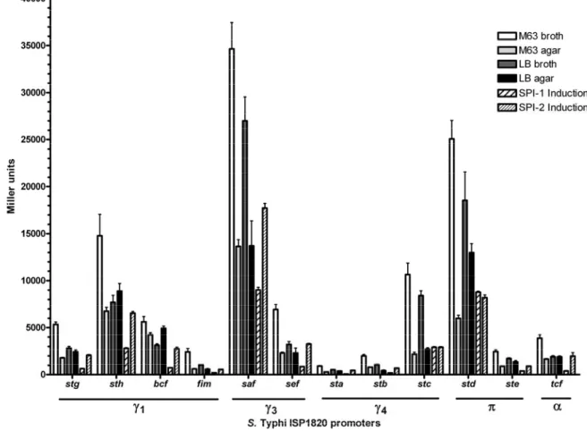
2.2.4.1 S. Typhi CU fimbriae are better expressed in minimal media ... 82
2.2.4.2 Production and visualization of CU fimbriae in a S. Typhi afimbrial mutant ... 84
2.2.4.3 Adhesion and invasion of epithelial cells ... 87
2.2.4.4 Uptake and survival within macrophages ... 89
2.2.4.5 Fimbriae and motility ... 91
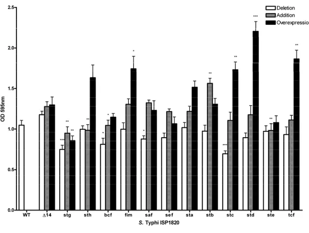
2.2.4.6 Role of fimbriae on biofilm production ... 91
2.2.5 Discussion ... 93
2.2.6 Authors contributions ... 101
2.2.7 Conflict of interest statement ... 101
2.2.8 Acknowledgments ... 101
2.2.9 References ... 101
Chapitre 3 – Régulation du fimbria Std chez Salmonella enterica sérovar Typhi ... 123
3.1 Préface au chapitre ... 123
3.2 Article 3 – Identification of Crp as a novel regulator of the Std fimbrial expression in Salmonella ... 124
3.2.1 Abstract ... 125
3.2.2 Importance ... 125
3.2.3 Introduction ... 125
3.2.4 Materials and methods ... 127
3.2.4.1 Bacteria, plasmids and growth conditions ... 127
3.2.4.2 Cloning of stdA promoters ... 127
3.2.4.5 Western Blot ... 128
3.2.4.6 qPCR assay ... 129
3.2.4.7 Statistical Analysis ... 129
3.2.5 Results ... 129
3.2.5.1 Identification of novel regulators for S. Typhi std operon ... 129
3.2.5.2 Crp activates std expression in S. Typhi ... 134
3.2.5.3 Production of Std fimbriae and mRNA expression ... 136
3.2.5.4 Crp binds to a distal region of the std promoter ... 136
3.2.6 Discussion ... 138
3.2.7 References ... 140
3.2.8 Supplementary data ... 142
3.3 Expression de std entre S. Typhi et S. Typhimurium ... 162
3.4 Criblage de régulateurs de l’expression de std ... 166
Chapitre 4 – Régulation du fimbria Fim chez Salmonella enterica sérovar Typhi ... 169
4.1 Préface au chapitre ... 169
4.2 Article 4: Identification of novel regulators of Type 1 fimbriae (fim) in Salmonella enterica serovar Typhi ... 170
4.2.1 Abstract ... 171
4.2.2 Introduction ... 172
4.2.3 Materials and methods ... 173
4.2.3.1 Bacteria, plasmids and growth conditions ... 173
4.2.3.2 Cloning of fimA promoter and chromosomal deletion of putative regulator genes ... 173
4.2.4.1 Putative candidate regulators of S. Typhi fim ... 175
4.2.4.2 Identification of novel fim regulators by transposon-based library screening ... 177
4.2.4.3 Ndh activates Fim specific phenotypes ... 179
4.2.5 Discussion ... 181
4.2.6 References ... 183
Chapitre 5 – Discussion ... 193
5.1 Caractérisation des fimbriae de type chaperon-placier chez S. Typhi ... 193
5.2 Régulation du fimbria Std ... 195
5.3 Régulation du fimbria Fim ... 199
Chapitre 6 – Conclusion ... 201
Références bibliographiques ... 203
Liste des tableaux
Salmonella fimbriome ... 48
Salmonella fimbrial type ... 50
Fimbrial distribution ... 53
Summary of fimbrial expression and pathogenesis phenotypes. ... 96
Bacterial strains and plasmids used for this study ... 109
Primers used in this study ... 116
List of all putative regulators tested for std promoter expression ... 130
Bacterial strains and plasmids used for this study ... 144
Primers used in this study ... 151
Identification of transposon insertions for significant modulators of fim ... 178
Bacterial strains and plasmids used in this study ... 185
Primers used in this study ... 190 Détails des acronymes pour chacun des fimbriae de type chaperon-placier chez S. Typhi 222
Liste des figures
Figure 1. – Chaperone/usher pathway. ... 40
Figure 2. – Nucleation/precipitation pathway. ... 43
Figure 3. – Type IV pathway. ... 46
Figure 4. – fim regulation. ... 58
Figure 5. – S. Typhi CU fimbrial operons organization. ... 77
Figure 6. – Expression of S. Typhi ISP1820 CU fimbrial promoters. ... 83
Figure 7. – Fimbrial major subunits in extracellular structures extract... 84
Figure 8. – Visible fimbriae in transmission electron microscopy. ... 86
Figure 9. – Fimbrial interactions with INT-407 intestinal epithelial cells. ... 88
Figure 10. – Fimbrial interactions with THP-1 macrophages. ... 90
Figure 11. – Impact of fimbriae on biofilm formation. ... 92
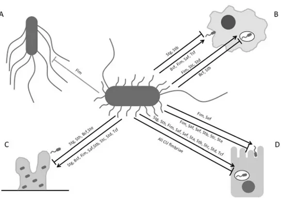
Figure 12. – Impact of S. Typhi fimbriae on motility, host cell interactions and biofilm formation. 121 Figure 13. – Significant modulators of std promoter expression in S. Typhi ISP1820. ... 132
Figure 14. – Crp is an activator of std expression. ... 135
Figure 15. – Truncation of std promoter region affects modulation by Crp. ... 137
Figure 16. – Crp activates std promoter in S. Typhimurium. ... 142
Figure 17. – Effect of glucose concentration on std expression in WT and crp strain. ... 143
Figure 18. – Expression du promoteur std de S. Typhi chez différentes souches bactériennes. 163 Figure 19. – Expression du promoteur std de S. Typhi chez différents sérovars de S. enterica. 165 Figure 20. – Expression relative de std chez les mutants par insertion de transposon Tn10. 167 Figure 21. – Expression of fim promoter in putative regulators. ... 176
Figure 24. – Courbe de croissance de la souche afimbriaire avec vecteur inductible portant l’opéron fim 217
Figure 25. – Motilité des souches mutantes pour chacun des systèmes fimbriaires de S. Typhi 218
Figure 26. – Motilité des souches portant pWSK29 avec chacun des opérons fimbriaires natifs de S. Typhi 219
Figure 27. – Motilité des souches portant pMMB207c avec chacun des opérons fimbriaires de S. Typhi 220
Liste des sigles et abréviations
ADN : acide déoxyribonucléique Ap : ampicilline
ARN : acide ribonucléique ARNm : ARN messager ATP : adénosine triphosphate
cAMP: adénosine monophosphate cyclique CFU : colony-forming unit
Cm : chloramphénicol DAP: diaminopimelic acid EBP: enhanced binding protein EIIAGlc: enzyme IIAGlc
IPTG: Isopropyl-β-thiogalactopyranoside Kan: kanamycine
Kb : kilobase
LB : Luria-Bertani ou lysogeny broth LPS : lipopolysaccharides
LTTR : Régulateurs transcriptionnels de type LysR MAM : molécules d’adhésion multivalentes pb : paire de bases
PMA : M phorbol 12-myristate 13-acétate SCV : vacuole contenant Salmonella
SEM : erreur standard de la moyenne (standard error of the mean) S. Enteritidis : Salmonella enterica sérovar Enteritidis
S. Paratyphi : Salmonella enterica sérovar Paratyphi S. Typhi: Salmonella enterica sérovar Typhi
S. Typhimurium: Salmonella enterica sérovar Typhimurium SPI : Îlot de pathogénicité de Salmonella
T1SS : système de sécrétion de type I T3SS : système de sécrétion de type III T5SS : système de sécrétion de type V T6SS : système de sécrétion de type VI TCS: système à deux composantes TLR : récepteurs de type ‘’Toll’’
‘’It is not the strongest of the species that survive, nor the most intelligent, but the one most responsive to change.’’
Charles Darwin
Remerciements
Je remercie tout d’abord les organismes subventionnaires qui ont supporté le projet et ma recherche. Par la suite, j’aimerais remercier mon comité de thèse pour leur temps et leurs précieux conseils.
J’aimerais remercier les professeurs, personnel administratif et collègues qui m’ont aidé et soutenu durant mes études au département de microbiologie, infectiologie et immunologie. Je remercie particulièrement Dr Drolet et Dr Harel qui m’ont encouragé à chacune des étapes de mon doctorat, m’ont apporté de très bonnes idées durant mon parcours et ont grandement contribué à l’évolution de ma pensée scientifique. Un merci spécial à Imène Kouidmi qui a été présente à la majorité des moments importants de mes études en microbiologie et m’a soutenu dans mes décisions. Merci à Sean Giacomucci, Clément Baret-Micaelli, Julien Brochu, Émilie Vlakos, Maxime Parent-Michaud, Maxime Raymond et Renaud Balthazar pour les rencontres de corridors et les discussions scientifiques ou moins scientifiques.
Je remercie ma famille pour le soutien constant. Ils m’ont appris le travail acharné et me motive à avancer toujours plus loin dans tout ce que j’entreprends. Ils sont une source d’inspiration et me guident. Merci de croire en moi et de me soutenir en tout temps. Merci particulièrement à ma mère Diane et mon frère Karl de m’encourager, de me rassurer et de me motiver depuis très longtemps!
Merci à Francis-Simon qui m’accompagne à chaque moment et a une écoute inépuisable. Merci d’être dans ma vie, d’être si doux, attentionné et blagueur. Tu me fais sourire, rire et tu rends la vie beaucoup plus intéressante! Je suis chanceuse de t’avoir près de moi.
Je remercie mes amis pour les rires et les joies, mais aussi pour tout le support dans les moments difficiles et les pleurs. Grâce à vous, je deviens une meilleure personne à chaque jour, j’évolue et je suis plus heureuse. Vous êtes la famille que je me suis constituée et vous êtes irremplaçables. Merci à Philippe Mandeville-Gauthier, Marie-Claude Farmer, Andréanne Cousineau, François M. Bernard, William Forget-Parent, Benoit Chabot, Philippe Beaudet, Christopher Normandeau, Patrice Chrétien-Raymer, Sandra Landry, Catherine Desjardins, Simon Fortin-Fournier, Benjamin Lara, Catherine Leclerc, Phebe Courville-Gagnon et Georges Nehmé pour quasiment 14 ans d’amitié. Une mention d’honneur pour Will et Phil qui ont été là dans les moments les plus stressants et qui ont aussi partagé les plus grandes victoires : merci d’être mes amis fidèles et mes guerriers de la nuit! Un autre merci très spécial à Georges qui, malgré la distance Alberta-Québec, prend le temps de nous appeler ou nous rejoindre dans les moments importants. Finalement, merci à Andréanne d’embarquer dans mes plans les plus fous : entre des aller-retours éclairs à Gatineau et des courses, on ne s’ennuie jamais!
Je remercie ensuite tous les membres du laboratoire de France que j’ai rencontré durant mon doctorat. Je pense premièrement aux anciens qui ont tracé la voie pour ce projet (Chantal Forest, Jean-Mathieu Leclerc, Élise David et Yoan Houde). Par la suite, je remercie Eve-Lyne Quevillon, Julie Saulnier-Bellemare, Claudie Murret-Labarthe, Maud Kerhoas, Valérie Labelle et Clémence Abriat pour les réflexions, les discussions, les rires, les niaiseries et les critiques musicales! Un autre merci à Maud pour tout le temps passé ensemble, à discuter en travaillant! Merci aussi à Clémence qui a commencé son doctorat très peu de temps après moi : les discussions ensemble ont été si enrichissantes, j’ai été très contente d’avoir eu quelqu’un avec qui parler de ce que je vivais. Merci à chacun des stagiaires que j’ai côtoyés : nos échanges m’ont
Chapitre 1 – Revue de littérature
1.1 Historique de Salmonella
Les bactéries du genre Salmonella ont été identifiées pour la première fois à la fin du 19e siècle par
l’épidémiologiste Theobald Smith sous la direction de Dr Daniel Elmer Salmon au « Bureau of Animal Industry » (BAI), aux États-Unis. La bactérie fût alors nommée Salmonella cholerasuis, puis renommée Salmonella enterica en 1986 (1). Cette espèce fût longtemps considérée comme la seule du genre Salmonella, mais une deuxième espèce, Salmonella bongori, fût identifiée par comparaison génétique de génomes dans les années 1980. L’espèce Salmonella enterica est elle-même classée en 6 sous-espèces : enterica, salamae, arizonae, diarizonae, houtenae et indica. Ces sous-sous-espèces sont ensuite subdivisées en plus de 2600 sérovars (ou sérotypes) observés à ce jour. Les sérovars sont classés par la classification de Kauffman-White selon la présentation de trois antigènes de surface, c’est-à-dire les antigènes O (lipopolysaccharide), F (flagelle) et Vi (capsule). L’antigène O détermine la sous-espèce à laquelle appartient une souche, puis l’antigène F en détermine le sérovar. La capsule n’est retrouvée que chez quelques rares sérovars dont S. Typhi (2).
Des sérovars de Salmonella, nous dénombrons aujourd’hui plusieurs bactéries pathogènes d’importance clinique présentant deux pathologies bien distinctes. Les bactéries de certains sérovars tels que Typhimurium et Enteritidis causent une infection dite localisée : cette infection est caractérisée par une inflammation au niveau de l’intestin causant une gastro-entérite. Ce type d’infection à Salmonella est habituellement pris en charge rapidement par les défenses de l’hôte et la bactérie est rapidement éliminée au niveau de l’intestin. La deuxième présentation clinique est due à une infection systémique où les défenses de l’hôte sont contrecarrées par plusieurs facteurs de la bactérie menant lentement celle-ci à se disséminer vers les autres organes de l’hôte. Cette deuxième manifestation est retrouvée dans le cas d’une infection par les sérovars Typhi, Paratyphi et Gallinarum (3).
Une deuxième différence majeure entre sérovars est le spectre d’hôte de ces bactéries pathogènes : certains sérovars peuvent infecter une large variété d’hôtes, tandis que certains sérovars sont hôte-spécifiques. Majoritairement, les sérovars hôte-spécifiques causent une infection systémique, tandis que les sérovars à large spectre d’hôte cause une gastro-entérite (3).
Salmonella enterica sérovar Typhi (S. Typhi) est l’agent étiologique de la fièvre typhoïde. Cette pathologie est retrouvée dans les pays en voie de développement, particulièrement dans les pays ayant une problématique au niveau de l’eau potable et des installations sanitaires (4). Cette bactérie humain-spécifique est transférée d’un hôte à l’autre par contact avec de l’eau ou de la nourriture contaminées par les fèces ou l’urine d’un porteur. De ce fait, la fièvre typhoïde est retrouvée chez près de 17 millions de personnes par année, engendrant environ 200 000 morts par année. Les signes et symptômes de la fièvre typhoïde comprennent une fièvre prolongée, des malaises, une perte d’appétit, des céphalées, une constipation ou une diarrhée, des taches rosées sur la poitrine, ainsi qu’une augmentation de la taille de la rate et du foie (5).
1.2 Particularités génétiques de Salmonella
Plusieurs particularités génétiques discriminent chaque sérovar un de l’autre permettant les différences de présentation clinique et de spécificité d’hôte. Parmi ces particularités, nous comptons la présence d’îlots de pathogénicité, de pseudogènes et de prophages dans le génome de ces bactéries, ainsi que la présence de plasmides variés.
génétique distincte chez S. Typhimurium (6). Parmi ces SPI, certains portent des facteurs de virulence majeurs pour chacune de ces bactéries, tel que SPI-1 et SPI-2 qui codent pour des systèmes de sécrétion de type III (T3SS) respectivement liés à l’invasion et à la survie face aux cellules de l’hôte. SPI-6 porte les opérons fimbriaires de saf et tcf, ainsi qu’un système de sécrétion de type VI (T6SS), tandis que le SPI-7 porte les gènes nécessaires à la formation de la capsule Vi, SPI-10 porte le fimbria Sef, SPI-11 code pour la toxine typhoïde chez S. Typhi et SPI-18 code pour plusieurs gènes importants à l’invasion chez S. Typhi (6).
La présence de pseudogènes est une caractéristique majeure de différenciation entre sérovars hôte-spécifiques ou généralistes. Un pseudogène est un gène muté créant la présence d’un codon d’arrêt (STOP) prématuré, la présence d’un changement de cadre de lecture ou la formation d’une protéine tronquée, qui normalement causerait l’inactivation du gène en question. En fonction des sérovars, nous retrouvons plus de 5% du génome qui possède des pseudogènes. Par exemple, 210 pseudogènes sont retrouvés chez S. Typhi (CT18 et Ty2), tandis que S. Typhimurium (LT2) possède seulement 39 pseudogènes (7). Il est proposé que cette inactivation de gènes crée la spécification vers un hôte de choix, laissant ainsi de côté les fonctions qui n’ont plus besoin d’être remplies chez cet hôte. Parmi les pseudogènes communs entre S. Typhi et S. Paratyphi A, plusieurs opérons fimbriaires possèdent des gènes tronqués, proposant un rôle important de ces systèmes dans la spécificité d’hôte (8).
Le génome de Salmonella possède plusieurs prophages et vestiges de ceux-ci. Ces phages sont porteurs de plusieurs gènes menant à la virulence bactérienne en finalité. Chez S. Typhimurium SL1344, il y a 4 prophages : Gifsy-1, -2 et -3 et SopE. S. Typhi possède plusieurs éléments venant de prophages. Les différentes souches de S. Typhi sont très clonales entre elles, à l’exception de ces insertions phagiques qui créent des différences d’une souche à l’autre (6, 9).
gènes de virulence tels que l’opéron spv, impliqué dans la survie à l’intérieur des macrophages de l’hôte, et le fimbriae Pef. Chez S. Typhi CT18, pHCM1 est retrouvé et code pour des gènes de résistance à divers antibiotiques et métaux lourds (6).
Une particularité du génome de S. Typhi est qu’il a subi un réarrangement par recombinaison entre différents opérons d’ARN ribosomaux (ARNr) ou différents éléments IS200. Ce réarrangement fait diverger le squelette génomique de S. Typhi comparativement à S. Typhimurium et est possiblement responsable de différences entre les deux sérovars vu que certains gènes sont régulés différemment selon un contexte spatial et génétique différent (10).
1.3 Salmonella enterica sérovar Typhi
1.3.1 Prévalence et cycle d’infection
S. Typhi est une bactérie pathogène humain spécifique et l’agent étiologique de la fièvre typhoïde. Sa voie d’entrée est par le système digestif, un environnement hostile semé d’obstacles. Premièrement, différentes molécules antibactériennes sont présentes directement au niveau de la bouche, puis la bactérie doit vivre la présence d’une forte acidité au niveau de l’estomac. Suite au passage à l’estomac, la bactérie parcours l’intestin grâce au péristaltisme qui s’y produit, mais aussi grâce à ses flagelles jusqu’aux cellules M présentes à la plaque de Peyer au niveau du colon. Ces cellules spécialisées seraient le point d’entrée de S. Typhi vers les macrophages présents qui servent de réservoirs pour la multiplication et le déplacement de la bactérie (4). Salmonella forme alors un compartiment, nommé vacuole contenant Salmonella (SCV ou ‘’Salmonella-containing vacuole’’), à l’intérieur des
S. Typhi, suite à une infection aiguë, persiste par formation de biofilm à l’intérieur de la vésicule biliaire. Ainsi, l’hôte infecté devient porteur sain, c’est-à-dire qu’il ne présente plus de signes ou symptômes de la fièvre typhoïde. Le porteur aura alors périodiquement des relâches de S. Typhi dans ses fèces (12).
1.3.2 Facteurs de virulence de S. Typhi
Plusieurs facteurs de virulence ont été identifiés pour S. Typhi, communs avec S. Typhimurium ou uniques à S. Typhi. Certains de ces facteurs expliquent en partie la spécificité d’hôte et la présentation clinique de S. Typhi.
S. Typhi produit une toxine de type A2B5 nommée toxine typhoïde. Celle-ci est codée par le gène cdtB
et nécessite l’action d’un complexe de PltA et 5 PltB (ou PltC), des homologues des composantes de la toxine de Bordetella pertussis. Les complexes PltAB ou PltAC permettent de livrer CdtB de l’intérieur de la bactérie vers une cellule cible (13). Lorsque CdtB est en complexe avec PltA et PltB, son effet est plus cytotoxique, tandis que le complexe CdtB, PltA et PltC augmente la leucopénie (14). CdtB reconnaît spécifiquement des glycanes sialylés terminaux des glycoprotéines présentes à la surface des cellules humaines, mais absentes des cellules d’autres animaux. Comparativement à la majorité des toxines AB, la toxine typhoïde ne peut être produite que lorsque la bactérie a formé sa vacuole dans la cellule hôte. Alors, S. Typhi forme la toxine et la délivre de la SCV vers l’espace extracellulaire avec l’aide de Rab29, une GTPase de l’hôte, qui semble diriger la vacuole de façon à permettre la sortie de la toxine (13). Le mécanisme de sécrétion de la toxine typhoïde serait similaire au système holine/endolysine des bactériophages. TtsA jouerait un rôle d’endolysine qui permettrait le passage de la toxine à travers la paroi cellulaire, tandis qu’une holine inconnue formerait un pore pour la sortie de TtsA vers le périplasme. Peu est connu par rapport au mécanisme d’exportation de la toxine encore aujourd’hui. Suite à la libération de la toxine typhoïde, les cellules hôtes sont distendues et leur noyau s’élargie jusqu’à deux fois sa taille normale, phénotype typique d’un arrêt du cycle cellulaire entre les phases G2 et M (15). Cet effet cellulaire est dû à l’activité DNase de CdtB qui endommage l’ADN hôte. La régulation
peuvent être formés. Pour le complexe comprenant PltB, PhoPQ semble avoir le plus grand effet. Pour le complexe comprenant PltC, SsrAB et OmpR/EnvZ jouent un rôle plus important et l’effet de PhoPQ est quasiment nul (14, 16, 17).
Plusieurs structures de surface au niveau de la membrane externe de l’enveloppe jouent un rôle important pour la virulence chez S. Typhi. Parmi celles-ci, il y a les lipopolysaccharides (LPS), la capsule Vi, les systèmes de sécrétion de type III (T3SS), le système de sécrétion de type VI (T6SS), les flagelles, les adhésines non fimbriaires et les fimbriae.
Le LPS de S. Typhi est intriqué dans la membrane externe de la bactérie et en sont une des composantes majeures (18). Le LPS est formé de trois parties : le lipide A, les cœurs interne et externe d’oligosaccharides et une chaîne variable de polysaccharides nommée l’antigène O (19). La chaîne de polysaccharides ou antigène O varie en composition et en longueur chez Salmonella et permet de déterminer les sous-espèces. Chez S. Typhi, les antigènes O une grande hétérogénéité par rapport à leur longueur selon l’environnement. Entre autres, la longueur de l’antigène O augmente en phase stationnaire lorsque les nutriments se font plus rares et qu’il y a activation de RpoS et RpoN. Les chaînes de polysaccharides des LPS de S. Typhi sont relativement courtes, car un régulateur majeur, Wzz, est non-fonctionnel, ce qui diminue la reconnaissance par le TLR4 (20). Les LPS sont essentiels à la virulence pour S. Typhi en infection de souris humanisées (hu-SRC-SCID) (21). Les LPS sont masqués en présence de la capsule Vi (22). Chez S. Enteritidis, le facteur Dam régule wzz par l’intermédiaire de RcsB et PmrA (23).
gènes tviB, tviC et tviE importants pour la synthèse du polysaccharide et aussi les gènes vexA-E impliqués dans le transport du polysaccharide. Le complexe VexA-D est un transporteur de type ABC (ATP-binding cassette transporter) servant à l’export du polysaccharide Vi. Finalement, TviD semble important pour la formation de la capsule, mais son rôle est indéterminé (26). La présence de la capsule semble limiter l’accès aux structures de surface de Salmonella comme les T3SS et les fimbriae. La capsule de S. Typhi est finement régulée par le régulateur majeur TviA, mais est aussi activée par des systèmes à deux composantes (TCS) tels que OmpR/EnvZ et le système Rcs. La capsule est principalement réprimée par RpoS (22).
Il y a deux T3SS chez Salmonella, ceux-ci codés par SPI-1 et -2. Le T3SS-1 est impliqué dans l’invasion des cellules hôtes, tandis que le T3SS-2 est responsable de la survie à l’intérieur des macrophages. Ces machineries complexes de plus de 20 protéines forment un pore à la membrane externe et une aiguille moléculaire qui injecte une série d’effecteurs à la cellule hôte permettant de la remodeler et de la manipuler (27-29). Le T3SS de SPI-1 est régulé principalement par une série de gènes retrouvésdans l’îlot de pathogénicité. HilA est le principal régulateur du T3SS-1 et est activé par HilD et HilC. HilD est réprimé dans certaines conditions par HilE (30, 31). Ces gènes régulateurs spécifiques au T3SS-1 sont affectés par plusieurs régulateurs (Lon, Fur, FimYZ, PhoPQ, FliZ, OmpR/EnvZ, CsrA, SirA/BarA, Lrp, Dam, Mlc et PhoBR) selon les conditions environnementales rencontrées par la bactérie (30, 32, 33). HilA active la synthèse du système de sécrétion, mais aussi invF qui permet la synthèse des effecteurs du T3SS-1. Chez S. Typhi, l’alarmone (p)ppGpp régulerait conjointement le T3SS-1, la capsule et les flagelles (34). Le T3SS de SPI-2 est régulé principalement par un TCS nommé SsrAB. Le T3SS-2 serait aussi régulé par OmpR et PhoP (35-38). Chez S. Typhi, SPI-2 serait membre du régulon LeuO (39, 40).
Le T6SS pourrait être comparé à une seringue moléculaire pour la bactérie. C’est une machinerie de sécrétion très similaire à la queue et la gaine de certains bactériophages, ce qui confère à ce système une force contractile pour injecter ses effecteurs vers la cellule hôte ou autres cellules bactériennes cibles. Chez S. Typhi, les gènes codant pour ce système sont retrouvés sur le SPI-6. Parmi ces gènes SciI
cholerae (41). Le T6SS semble toutefois fonctionnel et augmente la cytotoxicité vis-à-vis les cellules hôtes (42). Ce système serait régulé principalement par PmrA, le système Rcs et Hfq (42, 43).
Salmonella porte généralement plusieurs flagelles péritriches. Un flagelle est un T3SS modifié avec une extrusion de flagellines formant une longue structure protéique extracellulaire qui active le TLR5 présent sur les monocytes, les cellules dendritiques et les cellules épithéliales. Le rôle des flagelles est principalement lié à la motilité bactérienne et la chimiotaxie, mais ils ont aussi été associés à l’invasion et la survie dans les macrophages (44, 45). Un vaste réseau de régulation chapeauté par FlhCD est établi pour les flagelles. Les flagelles et le T3SS-1 sont corégulés. Chez S. Typhi, TviA régule directement les gènes flagellaires pour diminuer leur transcription et éviter la reconnaissance de la bactérie par TLR5 (46). Les flagelles seraient aussi atténués lorsqu’il y a augmentation de la température d’incubation (47). Comparativement à S. Typhimurium qui code deux structures flagellaires (FliC et FljB), S. Typhi code seulement pour fliC. Cependant, des variants antigéniques (Hj et Hz66) sont retrouvés chez certaines souches de S. Typhi, entre autres originaires d’Indonésie. Ceux-ci produisent le même type de réaction au TLR5 que le variant majeur Hd, mais varient quant à leur structure flagellaire, leur motilité et leur immunogénicité (45).
Les adhésines dites non-fimbriaires incluent les adhésines sécrétées par un T1SS, T5SS, PagC, STY0351 et STY1980. Deux adhésines sont sécrétées par un T1SS, c’est-à-dire SiiE et BapA. Cependant, SiiE est un pseudogène suggérant une perte de fonction chez S. Typhi. BapA serait important pour l’autoaggrégation et la formation de biofilm chez S. Enteritidis (48). Il serait régulé par CsgD principalement (49). Les adhésines non-fimbriaires sécrétées par un T5SS sont dites auto-transportées.
1.4 Fimbriae de Salmonella enterica
Chaque sérovar de Salmonella enterica possède un répertoire particulier de fimbriae lui conférant une spécificité d’adhésion aux surfaces biotiques et abiotiques. S. Typhi a une combinaison unique de 14 fimbriae qui pourrait expliquer sa spécialisation pour l’hôte humain, ainsi que sa pathogenèse. Trois types de fimbriae sont présents chez Salmonella et nommés selon leur mode d’assemblage, c’est-à-dire les pili de type IVb, les fimbriae curli et les fimbriae de type chaperon-placier. Dans la section 1.4.1 (article 1), ces trois modes d’assemblage sont détaillés en plus de fournir une distribution et une occurrence des fimbriae à travers les différents sérovars de Salmonella. Les aspects généraux de la régulation des fimbriae chez Salmonella sont ensuite abordés dans une revue sur la régulation du fimbria de type 1 de S. Typhimurium, c’est-à-dire le fimbria Fim. Finalement, l’utilisation des fimbriae comme moyens de détection pour Salmonella ou comme possibles cibles thérapeutiques contre cette bactérie est discutée à la fin de la section 1.4.1. L’article 1 est un chapitre de livre proposé à Intech et soumis à un processus de révision par les pairs.
Article 1: Dufresne, K. D., F. (2017). “Salmonella Fimbriae: what is the clue to their hairdo?” in Current Topics in Salmonella and Salmonellosis, ed M. Mares (Rijeka: InTech.), 59–79.
Contribution des auteurs:
J’ai réalisé les figures et ai rédigé l’article. France Daigle a réalisé les tableaux et aidé à la rédaction. Tous les auteurs ont révisé l’article. France Daigle a fourni le support financier.
1.4.1 Article 1: Salmonella Fimbriae: What is the Clue to Their Hairdo?
Karine Dufresne and France Daigle
Department of microbiology, infectiology and immunology, Université de Montréal, Montréal, Canada, france.daigle@umontreal.ca
1.4.1.1 Abstract
Fimbriae are important virulence factors for Salmonella pathogenesis. They mediate adhesion to host cells (including plants), food, stainless steel and much more. The fimbrial systems are organized in gene clusters of four to fifteen genes that code for structural, assembly and regulatory proteins. There are three kinds of fimbriae depending on their mode of assembly. The chaperone/usher (CU) fimbriae use a dedicated chaperone and usher protein to coordinate the subunits biogenesis on the cell surface. The curli fimbriae are assembled by nucleation/precipitation pathway. The type IV fimbria assembly requires a transmembrane apparatus and ATP to energize the reaction. Several fimbriae are conserved among Salmonella serovars, while some are present in a limited set or only specific serovars. Expression and regulation of fimbrial genes are not well understood and most Salmonella fimbriae are poorly expressed during in vitro culture, which further complicates research concerning their regulation and role during infection. However, Salmonella fim gene cluster, coding for type-1 fimbriae, was widely studied and presents its own set of regulators. Investigating fimbrial distribution, expression and regulation will further elucidate their roles in bacterial pathogenesis and host specificity. Furthermore, fimbriae are important for developing efficient diagnostic tests and antimicrobial strategies against Salmonella.
1.4.1.2 Introduction
Multiple virulence factors are implicated in Salmonella pathogenesis. These factors include type 3 secretion systems (T3SS) encoded in Salmonella Pathogenicity Islands (SPI) -1 and -2, other SPIs, flagella, capsule, plasmids and adhesion systems (6, 53). Among those factors, fimbriae represent a major player in pathogenesis and a source of diversity for Salmonella serovars. Fimbriae are the most common adhesion systems and are differentially expressed and found in a specific pattern among each serovars (54, 55).
Historically, the first observation of fimbriae was described in 1901 in Bacillus anthracis by Hinterberger and Reitman which hypothesized that the filaments were implicated in nutrients acquisition (56). Then, in 1949, Anderson suggested that the filaments were artefacts due to sample preparation for electron microscopy (57). However, many other studies contradicted Anderson and confirmed the presence of non-flagellar appendages on the bacterial surface. In 1950, Houwink and Van Iterson observed the appendages and described them as shorter and more rigid filaments than the flagella from Escherichia coli and suggested that the fibres were implicated in attachment to surface (58). The name fimbria (Latin word for fibres) was suggested in 1955 by Duguid et al. to describe the filamentous structures (58, 59). The term fimbria is preferable to use to describe non-flagellar filaments than pili, which is used to designate structures implicated in conjugation (60, 61). In 1966, Duguid et al. classified fimbriae in seven types (types 1 to 6 and F) according to the morphology and haemagglutination patterns. However, another classification, based on serology, better predicted genetic relatedness of fimbrial antigens. Nowadays, fimbriae are designated by the mode of assembly of the fibril (59).
the functional redundancy complicates their studies (61). However, fimbriae are implicated during infection and in a variety of other roles, like biofilm formation, seroconversion, haemagglutination, cellular invasion and macrophage interactions (6, 58, 63-67). In mice model, S. Typhimurium fimbriae demonstrate a role in intestinal cells attachment, caecum colonisation and persistence in gut (68-70). Moreover, fimbriae are important determinants of host adaptation by Salmonella (71).
In this chapter, an overview of Salmonella fimbriae is presented. First, the three pathways for fimbrial biogenesis (CU, precipitation/nucleation, type IV fimbriae) are described. Second, the distribution of fimbrial genes among Salmonella subspecies and serovars is presented. Third, the regulation of fimbrial genes is described and fim FGC regulation is detailed. Finally, the use of fimbriae as diagnostic and therapeutic tools is discussed.
1.4.1.3 Fimbrial biogenesis pathways
Three pathways for fimbrial assembly exist in Salmonella, the chaperone/usher (CU), the nucleation/precipitation and the type IV pathway (72). Fimbriae of the CU pathway employ dedicated chaperones and ushers for the fimbrial assembly. The nucleation/precipitation pathway forms an aggregative fibre by precipitation of the subunits in presence of the nucleator in the extracellular environment. Finally, the type IV fimbrial pathway uses complex machinery for the fimbriae formation and needs ATP to drive the assembly reaction. Furthermore, the type IV fimbriae can be retracted and diassembled (72).
The three pathways produce quite different fimbriae. CU fimbriae have the classic fimbrial shape with the repetition of major subunits emerging from the usher inserted in the outer membrane. The major subunits can be accompanied by minor subunits and/or adhesins (59). The fimbriae produced by the nucleation/precipitation pathway have an aggregated shape, due to the precipitation of major subunits together. This kind of fimbriae is highly stable and hardly depolymerised (73). The type IV
through the periplasm and the outer membrane reaching the extracellular medium (74). Here, the three fimbrial assembly mechanisms will be detailed.
1.4.1.3.1 Chaperone/usher pathway
The CU fimbriae represent the largest and most diversified class of adhesion systems (75, 76). Multiple CU fimbriae are present in Salmonella suggesting a functional redundancy (9, 74). The assembly is characterised by an interaction between the subunits, a periplasmic chaperone, and an outer membrane usher in order to form a mature fibre (Figure 1) (77). Each fimbriae produced by this pathway have their own unique and specific chaperone and usher (62). Usher sequence is a good discrimination tool and is used to subdivide the CU fimbriae into six phylogenetic clades (α, κ, π, σ, γ, β) (9, 61).
The biogenesis of the CU fimbriae begins with the production of the subunits in the cytoplasm and their export through the inner membrane by the general secretory pathway (GSP) (74, 77, 78). It consists in a post-translational translocation implying the SecYEG complex and SecDF/YajC proteins. When the pre-protein is produced, it can be targeted directly to the accessory factor SecA or transported to SecA by the general chaperone SecB. Then, SecA catalyzes the hydrolysis of ATP to energize the translocation through SecYEG. Use of ATP, in combination with proton-motive force, triggers the transport of the pre-protein to the periplasm. During the translocation across the inner membrane, the N-terminal signal peptide is cleaved by periplasmic peptidases (77, 79). To prevent early folding of the subunits, the fimbrial chaperone instantly forms a complex with the translocated subunit in the periplasm (80).
primary β-strands (80-82). Hydrophobic residues are alternated in the seven strands, facing the internal part of the barrel. These residues form the hydrophobic core of the domain that is implicated in the binding of the subunit. The fimbrial chaperones have an extended loop that lies at the extremity of one arm of the L-shaped molecule. This loop contains a conserved motif that is involved in the complex formation between the chaperone and subunits (80). The subunit and the chaperone have a similar structure, but the subunit is missing the seventh β-strand of the C-terminal extremity (78). The chaperone transfers the missing β-strand to the subunit to complete its structure: this mechanism is called the donor strand complementation (76). The chaperone preserves the folding energy of the subunit to drive the last steps of the assembly due to lack of energy source (ATP) in the periplasmic space (83). The chaperone also prevents premature fimbrial formation in the periplasm and primes the assembly through the usher (80, 84).
Then, the uncapping of the chaperone by the usher expose the interactive surface of the subunit to the outer membrane usher and assembly of subunits at the surface can occur (83). The transfer of the subunit from the chaperone to the usher happens very rapidly in vivo. In absence of the usher in vitro, only a slow and inefficient assembly was observed. This suggests that the uncapping of the chaperone is important for the efficiency of mature fimbriae assembly (78, 80). An interaction between the usher and the subunit, and also between the usher and the chaperone is required (81). This triangular interaction is important for the usher to discriminate subunit-loaded from unloaded chaperone (83). Fimbrial usher forms a ring in the outer membrane with a transient twin-pore of 2-3 nm diameter to allow passage of subunits to the extracellular environment (85). The usher catalyses fimbrial polymerisation by involving donor strand exchange where the N-terminal sequence of the subunit is replaced by a short sequence of the last subunit in the polymerized fibril with a zip-in-zip-out mechanism (83). This step is triggered in part by the chaperone required for the strand exchange between the new subunit and the forming fimbria. The quaternary structure of the subunit is achieved when the protein passes through the pore. The final morphology and structure (rigid or flexible), the length (1 to 3 μm) and width (2 to 10 nm) of the fibre of the CU fimbriae depend on the subunits
Figure 1. – Chaperone/usher pathway.
The subunit proteins are synthesised in the cytoplasm and translocated through the GSP. When the signal peptide is cleaved from the subunit, the chaperone complements the missing strand of the subunit in a process called donor strand complementation. The energy from the folding of the subunit is preserved by the chaperone. The chaperone drives the subunit to the usher and exchanges the donor strand. The subunit is then translocated by the usher to the extracellular medium and added to other subunits to form the fibril. IM=inner membrane; OM=outer membrane.
1.4.1.3.2 Nucleation/precipitation pathway
Curli fimbriae were initially discovered in Escherichia coli and are very conserved among the Enterobacteriaceae family, compared to any other types of FGC. The amyloids fibrils are particularly known for their role in biofilm formation and its recognition by the immune system (86). The FGC for curli is named csg (curli subunit gene) for E. coli and agf (thin aggregative fimbriae) for Salmonella, but the term csg is now commonly used for Salmonella. Curli formation depends on two divergent operons, csgBAC and csgDEFG. The csgBAC genes encode for CsgA, the major subunit, CsgB, the nucleator, and CsgC, an oxidoreductase of unknown function. The csgDEFG genes encode for the transcription regulator of the operon (CsgD) and for the assembly proteins located in the periplasm (CsgE) or in the outer membrane (CsgG and CsgF) (87).
The curli assembly mechanism is characterised by the exportation of the subunits and their precipitation to each other in the presence of a nucleator that fixes the fibril on the bacterial surface. Exportation of curli proteins also uses the GSP to pass through the inner membrane to the periplasm. Then, the CsgA and CsgB proteins are secreted by the lipoprotein CsgG. CsgG is composed of nine anticodon-binding-domain-like units that form a 36-stranded β-barrel complex that is inserted in the outer membrane. CsgG forms a pore in the outer membrane that permits the passage of the subunits and the nucleator. CsgG is accompanied by the accessory proteins CsgE and CsgF. CsgE is a specificity factor that forms a nonameric adaptor that binds to CsgG and closes the periplasmic space. The presence of CsgE optimizes the uptake of CsgA by CsgG and translocation of CsgA (88). CsgF helps the nucleation activity of CsgB. It was suggested that CsgF has a role in specific localisation and/or chaperoning of the nucleator, so CsgB will reach its full activity. Moreover, CsgF depends on CsgG and CsgE for its stability (89).
Once at the bacterial surface, the nucleator polymerises the subunits together into thin aggregative fimbriae (fibrils). This process happens only in the extracellular environment and requires
insoluble cross β-sheet molecules (9). CsgB anchors the curli fimbriae on the surface of the bacterial cell (Figure 2). In E. coli, it was observed that CsgB, in addition to its role of nucleator, is also part of the fimbriae with the CsgA subunits. A structurally different fibril made of CsgB subunits can be formed in the absence of CsgA (90). CsgA and CsgB share 30% of sequence identity and have the same predicted length (87). In E. coli, interbacterial complementation between a nucleator mutant and a subunit mutant is possible. However, in Salmonella, this complementation cannot happen, suggesting that the curli fimbriae are different in their nucleation process. However, the interbacterial complementation was observed in Salmonella when a lipopolysaccharide O-antigen mutant was used (91). The nucleation/precipitation pathway is still poorly understood and research is actually performed on the different aspects of the curli fimbrial formation.
Figure 2. – Nucleation/precipitation pathway.
The subunit CsgA is synthesized in the cytoplasm and translocated by the GSP. CsgA passes through the periplasm and is translocated in the extracellular medium by CsgG, helped by CsgE. The nucleator CsgB is also translocated by CsgG and supported by CsgF for its stability on the bacterial surface. When CsgA is in presence of the nucleator in the extracellular environment, the subunits precipitate in an aggregated fibril. CsgC is an oxidoreductase, but its specific role is still unknown. IM= inner membrane; OM=outer membrane.
1.4.1.3.3 Type IV fimbriae
Type IV fimbriae are usually from 1 to 5 μm long and are composed of repeated subunits of a single pilin. Type IV fimbriae are subdivided in two groups based on homology of the major subunits: type IVa and type IVb fimbriae (9). The difference between the two types is in the length of the peptide sequence and the mature major pilin sequence. Specific assembly mechanisms for type IVb fimbriae from Salmonella have yet to be characterised (92).
Type IV fimbriae pathway have the most complex machinery. They form an apparatus, composed of various proteins, that goes through the inner and outer membranes allowing the anchor of the fibre and energy accessibility for fimbrial assembly. The gene cluster also encodes numerous proteins with diverse functions, as the fibril is not only assembled but also disassembled. Type IV fimbriae are frequently compared to the type II secretion system (T2SS) which possesses similar structure and mechanism of assembly. Type IV fimbriae are implicated in adherence and twitching motility (62).
Type IV fimbriae are present in a variety of organisms including human pathogens such as Neisseria gonorrhoeae, Neisseria meningitidis, Pseudomonas aeruginosa and Vibrio cholerae. For Salmonella, they are found in S. bongori, S. enterica serovars Heidelberg, Parathyphi B and Typhi (92). S. bongori type IV fimbria is encoded by the sbe operon that remains uncharacterised and is located on a plasmid, as well as in S. Paratyphi B. While the type IVb gene cluster is located on the chromosome of S. Heidelberg and S. Typhi (9).
added one by one, but at three sites simultaneously, each corresponding to a strand to form a three-helix bundle (94). An ATPase inserted in the inner membrane supplies the energy required for the assembly of the type IV fimbriae. The secretin proteins are inserted in the outer membrane and form a channel that permits the passage of the intact pilus through the bacterial surface (9). These proteins form complexes that are then assembled in a cage-like final structure (94). Other proteins are also involved in the assembly/disassembly mechanisms, such as another ATPase dedicated for the disassembly of the fimbriae, lipoproteins of the secretin complex (pilotins), inner membrane proteins or gene products involved in peptidoglycan remodelling to permit the passage of the fibril through the periplasm (73, 94, 95). This assembly pathway is less understood and requires further investigations (94).
Figure 3. – Type IV pathway.
The prepilins are transported and translocated through the inner membrane (IM) to the periplasm by the GSP. A peptidase cleaves the signal peptide of the pre-pilin and the pilin can be assembled on the platform protein. An ATPase triggers the reaction. The pilins form a three-helix structure that passes through the outer membrane (OM) by a secretin supported by pilotins. The type IV fimbriae can also retract depending of the environmental conditions.
1.4.1.4 Salmonella fimbriome
Each fimbrial pathway described above is present in Salmonella creating a great element of genetic diversity. CU fimbriae are the most common fimbriae detected in the Salmonella genome. Curli (csg) is found in all Salmonella genome, whereas only a few serovars have the type IV fimbriae. There are 38 unique FGCs identified so far in 111 sequenced genomes from 34 different serovars (Tableau 1) (96, 97). Each serovar have their own repertoire of FGCs but there are seven FGCs that are highly conserved in most Salmonella strains forming the core of Salmonella FGCs. Most of the FGCs are sporadic or found only in a few strains constituting the signature of each serovar.
Salmonella fimbriome
Fimbriae CU
clade Prevalence Distribution Fimbriae
CU
clade Prevalence Distribution
Bcf γ1 core absent in IV sdj γ4 sporadic IIIb diarizonae
Csg curli core all Salmonella sdk/sfi π sporadic IIIb, VI
Fim γ1 core absent in
bongori sdl π sporadic IIIb diarizonae
Lpf γ1 conserved absent in ID sef γ3 sporadic IB, D (pseudo)
Mrk γ4 sporadic only in
Montevideo sib β sporadic VI indica
Pef κ sporadic only in IA, IC
and bongori fae/skf κ sporadic IB, IE
Peg γ4 conserved IB, IC, IIIa, VI,
bongori ssf γ4 sporadic II salamae
Peh γ4 sporadic only in
Montevideo sta γ4 sporadic ID
Pil sporadic Type IV; ID, IE,
bongori stb γ4 core I, II, IIIb;
Saf γ3 conserved ssp. I stc γ4 conserved IA, IB, ID
Sba γ4 sporadic Bongori std π core II, IIIa, missing in
Gallinarum
sbb/sbf π sporadic Bongori ste π conserved missing in IA, IE
sbc/spf κ sporadic IV, VI, bongori stf π conserved missing in ID, IE
Sbs β sporadic II salamae stg γ1 sporadic ID, bongori
sdc/sas σ sporadic IIIa arizonae sth γ1 core missing IIIa and
IIIb sdd /smf γ1 sporadic IE, II, IIIa, IV sti γ1 conserved missing in ID
Sde γ3 sporadic Tennessee (IE) stj β sporadic IA, IE
Sdh γ4 sporadic IE stk γ4 sporadic IE
Each Salmonella strain contains 5 to 14 different CU fimbriae with an average of 12 fimbriae in S. enterica. Representatives from all the six phylogenetic clades are present in Salmonella (Tableau 2) (9). The γ-fimbriae constitute the largest clade with 22 FGCs and include the highly conserved FGCs (bcf, fim and sth) that belong to the clade γ-1. The most diverse clade is γ-4, with the conserved stb and stc or peg (stc-peg) and many of the new sporadic FGCs. While the α clade (for alternate CU), also known as class 5 fimbriae, has one FGC, tcf which is found in several serovars. The σ clade also had only one FGC representative, sdc, that was only found in S. enterica subspecies IIIa (arizonae).
Salmonella fimbrial type
CU clade Fimbriae
Α Tcf
Β sbs, sib, stj
γ1 bcf, fim, lpf, sdd /smf, stg, sth, sti γ3 saf, sde, sef
γ4 mrk, peg, peh, sba, sdh, sdi, sdj, ssf, sta, stb, stc, stk Κ fae/skf, pef, sbc/spf
Π sbb/sbf, sdk, sdl, std, ste, stf
The distribution of the 38 FGCs gave a signature for each species, subspecies and serovars (Tableau 3). Seven FGCs, curli and the CU fim, bcf, sth, stb, stc-peg and std, represent the conserved (core) fimbriae of Salmonella (positive in more than 90% of strains). The fim fimbriae were found in all S. enterica strains, only missing in S. bongori. The bcf cluster was only missing in S. enterica ssp. IV (houtenae) and the sth cluster was only missing in S. enterica ssp. IIIa and IIIb. The stb cluster was present in S. enterica ssp. I, II, IIIb and the std cluster was not detected in S. enterica serovar Gallinarum, ssp. II, IIIA and S. bongori. The FGC stc and peg had probably emerged from a common ancestor: they belong to the same clade (γ-4), are inserted at the same position in the genome (between thiM and mrp), their distribution is mutually exclusive, and either one is present in the majority of Salmonella strains.
Most cases of salmonellosis in humans are caused by S. enterica ssp. I and many of the sequenced serovars were from ssp. I. Thus, 27 out of the 38 FGCs are found in ssp. I. The ssp. I was divided into 5 classes using previous phylogenetic analysis (96, 97) (Tableau 3). The class IA contains broad host range serovars involved in gastroenteritis, mainly serovar Typhimurium. The class IB is formed by serovar Dublin, Enteritidis, Pullorum and Gallinarum, all sharing similar O-antigens and FGCs. The class IC contains serovar Choleraesuis and Paratyphi C and class ID contains the human specific serovars Typhi and Paratyphi A. A separate branch of class IA, including serovar Heidelberg, Virchow, and Hadar, that had the highest number of FGCs, as well as serovars Montevideo, Schwarzengrund, Welterveden, Javiana, Kentucky and Tennessee, were commonly isolated in association with edible plants, constitute the class IE.
In addition of the 7 core FGCs, 5 highly conserved FGCs (saf, ste, stf, sti and lpf) were associated with S. enterica ssp. I (Tableau 3). The sti, lpf and stf clusters are missing in human specific serovars (class ID). The ste cluster is missing in class IA serovars and in some of the class IE serovars. Thus, S. enterica ssp. I harbours the core FGCs (fim, bcf, sth, stb, stc-peg and std), the conserved FGCs (saf, ste, stf, sti and lpf) and some sporadic FGCs unique to each serovar. Many FGCs of Salmonella are sporadic and form the unique repertoire in each serovars.
Despite the presence of many FGCs, extensive gene degradation was observed in most of the host-restricted and warm-blooded host adapted serovars, mainly Gallinarum, Choleraesuis, Paratyphi A and Typhi. Genome degradation of FGCs may correspond to the loss of genes rendered unnecessary by niche specialization or by selective pressure in order to diminish antigen presentation at the bacterial surface during systemic disease. Intriguingly, most of FGCs were intact in Paratyphi B.
There are 11 FGCs that are not in ssp. I, with only sbc and sdk that are shared by more than one serovars. The low numbers of FGCs might be specific for cold-blooded animals colonization. A conserved signature specific for each subspecies was observed. As more diverse strains will be sequenced, new FGCs will likely be discovered.
Fimbrial distribution
Subspecies Core Conserved Accessory Absent
Salmonella enterica I. enterica bcf, csg, fim, sth, stb, std stc-peg
sti, , saf, ste, stf, lpf
A pef, stj Ste
B fae, sef C pef, tcf,
D sef, sta, stg, tcf, pil sti, lpf, stf E fae, mrk, peh, sdd, sde, sdh, stj, stk, pil lpf, ste, stf
VI. indica sbc, sdk, sib stb
II. salamae sdd, ssf, sbs std IV. houtenae sbc, sdd bcf, stb IIIb. diarizonae sdi, sdj, sdk, sdl sth IIIa. Arizonae sdc, sdd sth, stb, std Salmonella bongori lpf sba, sbb, sbc, sbe, stg(sbd) fim, stb, std
1.4.1.5 Fimbrial regulation
Salmonella fimbriae are usually not expressed constitutively, and rarely expressed under laboratory condition, except for Fim fimbriae, a type-1 fimbria (54). Fimbriae are important during infection (70, 98, 99), suggesting that their expression is tightly regulated. Little is known about the regulation mechanisms that promote fimbrial expression. In general, fimbrial expression is positively or negatively regulated at the genetic level. Some regulators are unique to a specific fimbria, like the regulation of curli by CsgD, while others are global, like Dam, H-NS and Lrp (Leucine-responsive regulatory protein) (100). These mechanisms include regulatory proteins, DNA methylation, cyclic di-GMP, and small RNAs (100). In Salmonella, a regulation network exists between the virulence factors. Here, we present the regulation of fimbrial genes including the interaction with motility and invasion. Then, we propose an example of regulation of the fim FGC expression in S. Typhimurium, the most characterised fimbriae of Salmonella.
1.4.1.5.1 General regulation of fimbrial genes
Genes implicated in different aspects of virulence including motility, adhesion, invasion of host cells and intestinal persistence, are all regulated during infection. It was proposed that there is a temporal hierarchy between the T3SS of SPI-1 (invasion), flagellar and fimbrial genes, where SPI-1 is first activated, followed by flagellar genes and then type-1 fimbrial genes (fim). The crosstalk between these systems seems to be critical for bacterial pathogenesis (101). Each element of virulence is linked via a large regulatory network that is not completely understood. DNA adenine methylation (Dam) regulates many virulence genes in Salmonella (102): it is required for SPI-1 and pef expression but it also represses many genes, including the std, csg and flagellar genes (102-104). It was also shown that
Crosstalk regulation also occurs between the capsule and the type IVb fimbriae in S. Typhi. Both virulence factors are encoded on SPI-7 and facilitate invasion of monocytes, suggesting a regulation overlapped. However, the exact regulation elements that act on those two systems are unknown (107).
One of the posttranscriptional regulation mechanisms uses the binding of small RNAs and the Hfq chaperone. In an hfq mutant strain, the expression of fimbrial gene sefA was activated when most of the other fimbrial subunit genes were repressed in S. Enteritidis. Overall, the hfq deletion decreased adherence compared to wild-type strain. Thus, Hfq seems to regulate fimbrial expression of most fimbrial genes from S. Enteritidis (108). There is probably more sRNAs regulation of fimbrial gene expression awaiting to be discovered.
Phase variation is a transcriptional mechanism that controls the switch between fimbriated (ON) and afimbriated (OFF) cells within a bacterial population. In Salmonella, expression of lpf and pef was shown to be controlled by phase variation. The regulators of this mechanism are various and depend of the FGCs concerned (104, 109).
The secondary messenger cyclic-di-GMP controls virulence and biofilm formation in Salmonella (110). In Salmonella, curli expression was activated by AdrA, a GGDEF- domain protein that increases intracellular level of cyclic-di-GMP (111). Fimbrial production regulated by the cyclic-di-GMP level was also observed in other species such as Klebsiella pneumonia, E. coli and P. aeruginosa (112).
In spite of all those known elements of regulation, how Salmonella passes from being afimbriated in vitro to a fimbriated form in vivo is still unknown.
1.4.1.5.2 Regulation of fim in S. Typhimurium
The fim FGC codes for six genes (fimAICDHF). This cluster is the most studied and one of the most conserved fimbriae of Salmonella enterica and was mainly characterised in S. Typhimurium. These fimbriae have a binding specificity for mannose residues (112). The Fim fimbria of Salmonella is not homologous with its homonym from E. coli, except for sharing some morphological and mechanistic features (113, 114). Regulation and amino acid sequences of fimbrial proteins are divergent between the two species. The transition from afimbriated to fimbriated stage occurs for fim, but there is no inversion of the promoter region as observed for E. coli phase variation (115). For S. Typhimurium, the major subunit FimA is accompanied by two other subunits, FimI and FimF, and by the adhesin, FimH. The fimC and fimD genes encode respectively the chaperone and the usher (116). Ancillary genes, fimZYW, and a rare arginine transfer RNA (tRNA) fimU and STM0551, inserted between fimY and fimW, directly regulate fim expression (117).
Ancillary genes fimZYW regulate the expression of fimA (100, 118-120). FimZ, a sensor DNA-binding protein, is the principal positive regulator of fimA (121). FimY upregulates fimZ expression by binding to the fimZ promoter. FimY and FimZ then form a complex that activates the fimA promoter (112, 119). fimY is itself regulated by the arginine tRNA fimU (122). Lrp is another regulator that binds and activates the fimZ promoter, probably by antagonizing the binding of the global repressor protein H-NS to this promoter region (116, 120, 123). H-NS has a high affinity for AT-rich DNA region and fimZ gene has an unusual AT-rich sequence (123).
PhoPQ activates hilE, one of the negative regulators of hilA, by a FimYZ-dependant manner. FimZ also down-regulates flhDC, genes implicated in flagella expression (125). Flagellar gene fliZ also represses fimZ on a posttranscriptional manner, reinforcing the fact that there is an alternated expression of flagella (motility) and fimbriae (adhesion) (101) and confirming a regulation network between SPI-1 (invasion) and fimbrial expression (126). Thus, a combination of factors directly implicated in fimbrial genes regulation can also impact on other virulence systems of Salmonella.
Figure 4. – fim regulation.
The fim FGC is activated and inhibited by diverse regulators. FimZ, accompanied by FimY, is the principal activator of fimA. Lrp and c-di-GMP also activate fimA at the promoter level. H-NS and FimW inhibit fimA expression by linking its promoter. FimW also reduces fimA expression by linking FimZ and decreases the availability of this activator. The fimZ gene is activated by FimZ itself and by FimY, but is repressed by FliZ in a post-transcriptional manner. The tRNA fimU regulates fimY. FimZ down-regulates flhCD, genes implicated in flagellar expression.