2014 ESC/EACTS Guidelines on myocardial revascularization
The Task Force on Myocardial Revascularization of the European Society of Cardiology (ESC)
and the European Association for Cardio-Thoracic Surgery (EACTS)
Developed with the special contribution of the European Association of Percutaneous
Cardiovascular Interventions (EAPCI)
Authors/Task Force members, Philippe Kolh* (EACTS Chairperson) (Belgium), Stephan Windecker*
(ESC Chairperson) (Switzerland), Fernando Alfonso (Spain), Jean-Philippe Collet (France), Jochen Cremer
(Germany), Volkmar Falk (Switzerland), Gerasimos Filippatos (Greece), Christian Hamm (Germany), Stuart
J. Head (Netherlands), Peter Jüni (Switzerland), A. Pieter Kappetein (Netherlands), Adnan Kastrati
(Germany), Juhani Knuuti (Finland), Ulf Landmesser (Switzerland), Günther Laufer (Austria),
Franz-Josef Neumann (Germany), Dimitrios J. Richter (Greece), Patrick Schauerte (Germany), Miguel Sousa
Uva (Portugal), Giulio G. Stefanini (Switzerland), David Paul Taggart (UK), Lucia Torracca (Italy),
Marco Valgimigli (Italy), William Wijns (Belgium) and Adam Witkowski (Poland)
ESC Committee for Practice Guidelines, Jose Luis Zamorano (Chairperson) (Spain), Stephan Achenbach (Germany), Helmut Baumgartner (Germany), Jeroen J. Bax (Netherlands), Héctor Bueno (Spain), Veronica Dean (France), Christi Deaton (UK),Çetin Erol (Turkey), Robert Fagard (Belgium), Roberto Ferrari (Italy), David Hasdai (Israel), Arno W. Hoes (Netherlands), Paulus Kirchhof (Germany/UK), Juhani Knuuti (Finland), Philippe Kolh (Belgium), Patrizio Lancellotti (Belgium), Ales Linhart (Czech Republic), Petros Nihoyannopoulos (UK), Massimo F. Piepoli (Italy), Piotr Ponikowski (Poland), Per Anton Sirnes (Norway),
Juan Luis Tamargo (Spain), Michal Tendera (Poland), Adam Torbicki (Poland), William Wijns (Belgium) and Stephan Windecker (Switzerland)
EACTS Clinical Guidelines Committee and Miguel Sousa Uva (Chairperson) (Portugal)
Document reviewers, Stephan Achenbach (ESC Review Coordinator) (Germany), John Pepper (EACTS Review Coordinator) (UK), Anelechi Anyanwu (USA), Lina Badimon (Spain), Johann Bauersachs (Germany), Andreas Baumbach (UK), Farzin Beygui (France),
National Cardiac Societies document reviewers:listed in Addenda
The content of these European Society of Cardiology (ESC) and European Association for Cardio-Thoracic Surgery (EACTS) Guidelines has been published for personal and educational use only. No commercial use is authorized. No part of the ESC Guidelines may be translated or reproduced in any form without written permission from the ESC. Permission can be obtained upon submission of a written request to Oxford University Press, the publisher of the European Heart Journal and the party authorized to handle such permissions on behalf of the ESC.
Other ESC entities having participated in the development of this document:
Associations:Acute Cardiovascular Care Association (ACCA), European Association for Cardiovascular Prevention & Rehabilitation (EACPR), European Association of Cardiovascular Imaging (EACVI), European Heart Rhythm Association (EHRA), Heart Failure Association of the ESC (HFA).
Working groups:Working Group on Cardiac Cellular Electrophysiology, Working Group on Cardiovascular Magnetic Resonance, Working Group on Cardiovascular Pharmacology and Drug Therapy, Working Group on Cardiovascular Surgery, Working Group on Coronary Pathophysiology and Microcirculation, Working Group on Nuclear Cardiology and Cardiac Computed Tomography, Working Group on Peripheral Circulation, Working Group on Thrombosis, Working Group on Valvular Heart Disease.
Councils:Council for Cardiology Practice, Council on Cardiovascular Primary Care, Council on Cardiovascular Nursing and Allied Professions.
Disclaimer 2014:The ESC/EACTS Guidelines represent the views of the ESC and of the EACTS and were produced after careful consideration of the scientific and medical knowledge and the evidence available at the time of their dating.
The ESC and the EACTS are not responsible in the event of any contradiction, discrepancy and/or ambiguity between the ESC/EACTS Guidelines and any other official recommendations or guidelines issued by the relevant public health authorities, in particular in relation to good use of healthcare or therapeutic strategies. Health professionals are encouraged to take the ESC/EACTS Guidelines fully into account when exercising their clinical judgment as well as in the determination and the im-plementation of preventive, diagnostic or therapeutic medical strategies; however, the ESC/EACTS Guidelines do not in any way whatsoever override the individual re-sponsibility of health professionals to make appropriate and accurate decisions in consideration of each patient’s health condition and, where appropriate and/or necessary, in consultation with that patient and the patient’s care provider. Nor do the ESC/EACTS Guidelines exempt health professionals from giving full and careful consideration to the relevant official, updated recommendations or guidelines issued by the competent public health authorities, in order to manage each patient’s case in light of the scientifically accepted data pursuant to their respective ethical and professional obligations. It is also the health professional’s responsibility to verify the applicable rules and regulations relating to drugs and medical devices at the time of prescription.
© 2014 European Society of Cardiology. Reproduced with kind permission. For permissions please email: journals.permissions@oup.com.
ESC/ EA CT S GU IDELINES
European Journal of Cardio-Thoracic Surgery 46 (2014) 517–592 doi:10.1093/ejcts/ezu366 Advance Access publication 29 August 2014
Nikolaos Bonaros (Austria), Marco De Carlo (Italy), Christi Deaton (UK), Dobromir Dobrev (Germany), Joel Dunning (UK), Eric Eeckhout (Switzerland), Stephan Gielen (Germany), David Hasdai (Israel), Paulus Kirchhof (UK/Germany), Heyman Luckraz (UK),
Heiko Mahrholdt (Germany), Gilles Montalescot (France), Domenico Paparella (Italy), Ardawan J. Rastan (Germany), Marcelo Sanmartin (Spain), Paul Sergeant (Belgium), Sigmund Silber (Germany), Juan Tamargo (Spain), Jurrien ten Berg (Netherlands), Holger Thiele (Germany), Robert-Jan van Geuns (Netherlands), Hans-Otto Wagner (Germany), Sven Wassmann
(Germany), Olaf Wendler (UK) and Jose Luis Zamorano (Spain)
The disclosure forms of the authors and reviewers are available on the ESC websitewww.escardio.org/guidelines
* First and corresponding authors: Philippe Kolh, Cardiovascular Surgery Department, University Hospital (CHU, ULg) of Liege, Sart Tilman B 35, 4000 Liege, Belgium. Tel: +32 4 366 7163; Fax: +32 4 366 7164; Email: philippe.kolh@chu.ulg.ac.be. Stephan Windecker, Cardiology, Bern University Hospital, Freiburgstrasse 4, CH-3010 Bern, Switzerland. Tel: +41 31 632 47 70; Fax: +41 31 632 42 99; Email: stephan.windecker@insel.ch
Keywords:Acute coronary syndromes• Bare-metal stents • Coronary artery bypass grafting • Coronary artery disease • Drug-eluting stents • EuroSCORE • Guidelines • Heart Team • Myocardial infarction • Myocardial ischaemia • Myocardial revascularization • Medical therapy • Percutaneous coronary intervention• Recommendation • Revascularization • Risk stratification • Stents • Stable angina • Stable coronary artery disease• ST-segment elevation myocardial infarction • SYNTAX score
TABLE OF CONTENTS
Abbreviations and acronyms . . . 519
1. PREAMBLE . . . 522
2. INTRODUCTION . . . 523
3. SCORES AND RISK STRATIFICATION . . . 524
4. PROCESS FOR DECISION-MAKING AND PATIENT INFORMATION . . . 527
4.1 Patient information and informed consent . . . 527
4.2 Multidisciplinary decision-making (Heart Team) . . . 527
4.3 Timing of revascularization andad hoc percutaneous coronary intervention . . . 528
5. STRATEGIES FOR DIAGNOSIS: FUNCTIONAL TESTING AND IMAGING . . . 529
5.1 Non-invasive tests . . . 529
5.2 Invasive tests . . . 529
5.3 Detection of myocardial viability . . . 529
6. REVASCULARIZATION FOR STABLE CORONARY ARTERY DISEASE . . . 530
6.1 Rationale for revascularization . . . 530
6.2 Evidence basis for revascularization . . . 530
6.2.1 Revascularization with the use of percutaneous coronary intervention . . . 532
6.2.2 Percutaneous coronary intervention with drug-eluting stents vs. bare-metal stents . . . 532
6.2.3 Revascularization with the use of coronary artery bypass grafting . . . 533
6.3 Percutaneous coronary intervention vs. coronary artery bypass grafting . . . 533
6.3.1 Proximal left anterior descending coronary artery disease . . . 534
6.3.2 Left main coronary artery disease . . . 534
6.3.3 Three-vessel coronary artery disease . . . 534
7. REVASCULARIZATION IN NON-ST-SEGMENT ELEVATION ACUTE CORONARY SYNDROMES . . . 536
7.1 Early invasive vs. conservative strategy . . . 536
7.2 Timing of angiography and intervention . . . 537
7.3 Type of revascularization . . . 537
7.3.1 Coronary artery bypass surgery . . . 538
7.3.2 Percutaneous coronary intervention . . . 538
8. REVASCULARIZATION IN ST-SEGMENT ELEVATION MYOCARDIAL INFARCTION . . . 539
8.1 Time delays . . . 539
8.2 Selection of reperfusion strategy . . . 539
8.3 Primary percutaneous coronary intervention . . . 540
8.4 Fibrinolysis . . . 541
8.5 Secondary percutaneous coronary intervention . . . 542
8.6 Coronary artery bypass surgery . . . 543
9. REVASCULARIZATION IN PATIENTS WITH HEART FAILURE AND CARDIOGENIC SHOCK . . . 543
9.1 Chronic heart failure . . . 543
9.1.1 Revascularization . . . 543
9.1.2 Myocardial viability and revascularization . . . 543
9.1.3 Ventricular reconstruction . . . 544
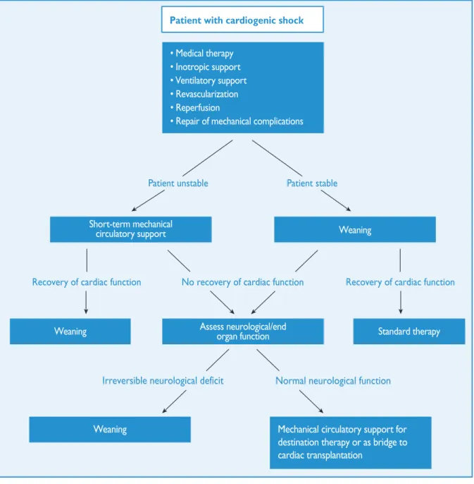
9.2 Cardiogenic shock . . . 544
9.2.1 Revascularization . . . 545
9.2.2 Mechanical circulatory support . . . 545
9.2.3 Right ventricular failure . . . 546
9.2.4 Mechanical complications . . . 546
10. REVASCULARIZATION IN PATIENTS WITH DIABETES . . . 547
10.1 Evidence for myocardial revascularization . . . 547
10.1.1 Stable coronary artery disease . . . 547
10.1.2 Acute coronary syndromes . . . 548
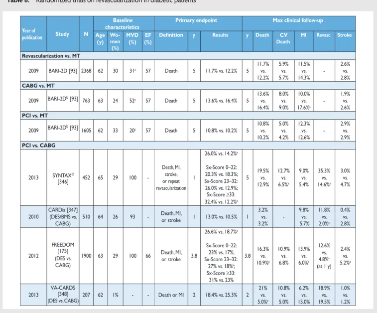
10.2 Type of myocardial revascularization . . . 548
10.2.1. Randomized clinical trials . . . 548
10.2.2 Meta-analyses . . . 549
10.3 Revascularization with the use of percutaneous coronary intervention . . . 549
10.4 Revascularization with the use of coronary artery bypass grafting . . . 549
10.5 Antithrombotic pharmacotherapy . . . 549
10.6 Anti-diabetic medications . . . 550
11. REVASCULARIZATION IN PATIENTS WITH CHRONIC KIDNEY DISEASE . . . 550
11.1 Evidence-base for revascularization . . . 550
11.1.1 Patients with moderate chronic kidney disease . . . 551
11.1.2 Patients with severe chronic kidney disease and end-stage renal disease or in haemodialysis . . . 551
11.2 Prevention of contrast-induced nephropathy . . . 551
12. REVASCULARIZATION IN PATIENTS REQUIRING VALVE INTERVENTIONS . . . 552
12.1 Primary indication for valve interventions . . . 552
12.2 Primary indication for coronary revascularization . . . . 553
13. ASSOCIATED CAROTID/PERIPHERAL ARTERY DISEASE . . . . 554
13.1.1 Risk factors for stroke associated with myocardial
revascularization . . . 554
13.1.2 Preventive measures to reduce the risk of stroke after coronary artery bypass grafting . . . 554
13.1.3 Carotid revascularization in patients scheduled for myocardial revascularization . . . 554
13.1.4 Type of revascularization in patients with associated carotid and coronary artery disease . . . 555
13.2 Associated coronary and peripheral arterial disease . . . 556
14. REPEAT REVASCULARIZATION AND HYBRID PROCEDURES . 556 14.1 Early graft failure . . . 556
14.2 Disease progression and late graft failure . . . 557
14.3 Acute percutaneous coronary intervention failure . . . . 557
14.4 Repeat percutaneous coronary intervention . . . 558
14.5 Hybrid procedures . . . 558
15. ARRHYTHMIAS . . . 560
15.1 Ventricular arrhythmias . . . 560
15.1.1 Revascularization for prevention of sudden cardiac death in patients with stable coronary artery disease and reduced left ventricular function . . . 560
15.1.2 Revascularization for treatment of electrical storm . 560 15.1.3 Revascularization after out-of-hospital cardiac arrest . . . 560
15.2 Atrial arrhythmias . . . 560
15.2.1 Atrialfibrillation complicating percutaneous coronary intervention . . . 560
15.2.2 Atrialfibrillation complicating coronary artery bypass grafting . . . 560
15.2.3 Post-operative atrialfibrillation and stroke risk . . . . 561
15.3 Concomitant surgical procedures for atrialfibrillation or stroke treatment . . . 561
16. PROCEDURAL ASPECTS OF CORONARY ARTERY BYPASS GRAFTING . . . 562
16.1 Pre-operative management . . . 562
16.2 Blood management . . . 562
16.2.1 Blood salvage interventions . . . 562
16.2.2 Pharmacological strategies . . . 562 16.2.3 Blood transfusion . . . 562 16.3 Surgical procedures . . . 562 16.3.1 Conduit harvest . . . 562 16.3.2 Coronary vessel . . . 562 16.3.3 Completeness of revascularization . . . 562
16.3.4 Construction of central anastomosis . . . 562
16.3.5 Bypass grafts . . . 562
16.3.6 On-pump and off-pump procedures . . . 563
16.3.7 Minimally invasive procedures . . . 564
16.4 Reporting perioperative outcome . . . 564
17. PROCEDURAL ASPECTS OF PERCUTANEOUS CORONARY INTERVENTION . . . 564
17.1 Percutaneous coronary intervention devices . . . 564
17.1.1 Balloon angioplasty . . . 564
17.1.2 Coronary stents . . . 564
17.1.3 Bioresorbable stents . . . 564
17.1.4 Drug-coated balloons . . . 566
17.1.5 Other devices . . . 566
17.2 Adjunctive invasive diagnostic tools . . . 566
17.2.1 Intravascular ultrasound . . . 566
17.2.2 Optical coherence tomography . . . 567
17.2.3 Pressure-derived fractionalflow reserve . . . 567
17.3 Specific lesion subsets . . . 568
17.3.1 Bifurcation stenosis . . . 568
17.3.2 Chronic total coronary occlusion . . . 568
17.3.3 Ostial lesions . . . 568
18. ANTITHROMBOTIC TREATMENTS . . . 569
18.1 Percutaneous coronary intervention in stable coronary artery disease . . . 569
18.1.1 Oral antiplatelet therapy . . . 569
18.1.2 Intravenous antiplatelet therapy . . . 569
18.1.3 Anticoagulation . . . 569
18.2 Non-ST-segment elevation acute coronary syndrome . . 570
18.2.1 Oral antiplatelet therapy . . . 570
18.2.2 Intravenous antiplatelet therapy . . . 571
18.2.3 Anticoagulation . . . 572
18.3 ST-segment elevation myocardial infarction . . . 573
18.3.1 Oral antiplatelet therapy . . . 573
18.3.2 Intravenous antiplatelet therapy . . . 573
18.3.3 Anticoagulation . . . 574
18.4 Points of interest and special conditions . . . 575
18.4.1 Pre-treatment with P2Y12inhibitors . . . 575
18.4.2 Intravenous P2Y12inhibitors . . . 576
18.4.3 Anticoagulation after percutaneous coronary intervention in acute coronary syndrome patients . . . 576
18.4.4 Anticoagulation during percutaneous coronary intervention in patients on oral anticoagulation . . . 577
18.4.5 Antithrombotic therapy after percutaneous coronary intervention in patients requiring oral anticoagulation . . . 577
18.4.6 Duration of dual antiplatelet therapy after percutaneous coronary intervention . . . 578
18.4.7 Drug interactions: a clopidogrel-related topic . . . . 579
18.4.8 Renal dysfunction . . . 579
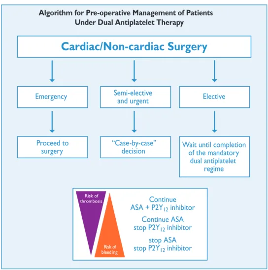
18.4.9 Surgery in patients on dual antiplatelet therapy . . . 580
18.4.10 Antiplatelet therapy monitoring and genetic testing . . . 582
18.4.11 Patients with hypersensitivity to acetylsalicylic acid . . . 582
18.4.12 Heparin-induced thrombocytopaenia . . . 582
19. VOLUME–OUTCOME RELATIONSHIP FOR REVASCULARIZATION PROCEDURES . . . 583
19.1 Coronary artery bypass grafting . . . 583
19.2 Percutaneous coronary intervention . . . 583
20. MEDICAL THERAPY, SECONDARY PREVENTION, AND STRATEGIES FOR FOLLOW-UP . . . 585
21. ADDENDA . . . 585
REFERENCES . . . 586
ABBREVIATIONS AND ACRONYMS
ACCF/AHA American College of CardiologyFoundation/American Heart Association ACCOAST A Comparison of Prasugrel at the Time of
Percutaneous Coronary Intervention (PCI) Or as Pre-treatment at the Time of Diagnosis in Patients With Non-ST-Elevation
Myocardial Infarction (NSTEMI)
ACE angiotensin-converting enzyme
ACEF age, creatinine, ejection fraction
ACS acute coronary syndromes
ACUITY Acute Catheterization and Urgent
Intervention Triage strategy
ADAPT-DES Assessment of Dual AntiPlatelet Therapy with Drug-Eluting Stents
AF atrialfibrillation
APPRAISE-2 Apixaban for Prevention of Acute Ischemic and Safety Events
ESC/ EA CT S GU IDELINES
aPTT activated partial thromboplastin time ARCTIC Assessment by a double Randomization of a
Conventional antiplatelet strategy vs. a monitoring-guided strategy for drug-eluting stent implantation and, of Treatment Interruption vs. Continuation one year after stenting
ARMYDA Antiplatelet therapy for Reduction of MYocardial Damage during Angioplasty ARTS Arterial Revascularization Therapies Study
ASA acetylsalicylic acid
ASCERT American College of Cardiology
Foundation–Society of Thoracic Surgeons Database Collaboration
ATLAS ACS 2–TIMI 51
Anti-Xa Therapy to Lower cardiovascular events in Addition to Standard therapy in subjects with Acute Coronary Syndrome– Thrombolysis In Myocardial Infarction 51
ATOLL Acute STEMI Treated with primary PCI and
intravenous enoxaparin Or UFH to Lower ischaemic and bleeding events at short- and Long-term follow-up
AVR aortic valve replacement
AWESOME Angina With Extremely Serious Operative Mortality Evaluation
b.i.d. bis in diem (twice daily)
BARI-2D Bypass Angioplasty Revascularization Investigation 2 Diabetes
BASKET–PROVE BASKET–Prospective Validation Examination
BMS bare-metal stent
BRAVE Bavarian Reperfusion Alternatives Evaluation BRIDGE Bridging Anticoagulation in Patients who
Require Temporary Interruption of Warfarin Therapy for an Elective Invasive Procedure or Surgery
CABG coronary artery bypass grafting
CAD coronary artery disease
CARDIA Coronary Artery Revascularization in Diabetes
CAS carotid artery stenting
CASS Coronary Artery Surgery Study
CCS Canadian Cardiovascular Society
CE Conformité Européenne
CEA carotid endarterectomy
CHA2DS2-VASc Congestive heart failure or left ventricular
dysfunction, Hypertension, Age≥75 [Doubled], Diabetes, Stroke [Doubled]– Vascular disease, Age 65–74 and Sex category [Female]
CHAMPION Cangrelor vs. Standard Therapy to Achieve Optimal Management of Platelet Inhibition
CI confidence interval
CIN contrast-induced nephropathy
CKD chronic kidney disease
COMFORTABLE-AMI
Comparison of Biolimus Eluted From an Erodible Stent Coating With Bare-Metal Stents in Acute ST-Elevation Myocardial Infarction
COURAGE Clinical Outcomes Utilizing
Revascularization and Aggressive Drug Evaluation
COX cyclo-oxygenase
CREDO Clopidogrel for the Reduction of Events During Observation
CRT cardiac resynchronization therapy
CT computed tomography
CTO chronic total occlusion
CURE Clopidogrel in Unstable Angina to Prevent
Recurrent Events
CURRENT-OASIS 7 Clopidogrel and Aspirin Optimal Dose Usage to Reduce Recurrent Events−Seventh Organization to Assess Strategies in Ischemic Syndromes 7
CYP P450 cytochrome P450
DANAMI DANish trial in Acute Myocardial Infarction
DAPT dual antiplatelet therapy
DEB-AMI Drug Eluting Balloon in Acute Myocardial Infarction
DELTA Drug Eluting stent for LefT main coronary Artery disease
DES drug-eluting stent
DI–DO door-in to door-out time
DIGAMI Diabetes, Insulin Glucose Infusion in Acute Myocardial Infarction
DPP-4 dipeptidyl peptidase 4
DTB door-to-balloon time
EACTS European Association for Cardio-Thoracic
Surgery
EAPCI European Association of Percutaneous
Cardiovascular Interventions
EARLY-ACS Early glycoprotein IIb/IIIa inhibition in non-ST-segment elevation acute coronary syndrome
ECG electrocardiogram
EF ejection fraction
EMS emergency medical service
ESC European Society of Cardiology
EUROMAX European Ambulance Acute Coronary
Syndrome Angiography
EXAMINATION Everolimus-eluting stent vs. BMS in ST-segment elevation myocardial infarction EXCELLENT Efficacy of Xience/Promus vs. Cypher in
reducing Late Loss After stenting
FAME Fractional Flow Reserve vs. Angiography for Multivessel Evaluation
FFR fractionalflow reserve
FINESSE Facilitated Intervention with Enhanced Reperfusion Speed to Stop Events
FMCTB first-medical-contact-to balloon
FRISC-2 Fragmin during Instability in Coronary Artery Disease-2
FREEDOM Future Revascularization Evaluation in Patients with Diabetes Mellitus
GFR glomerularfiltration rate
GP IIb/IIIa glycoprotein IIb/IIIa
GRACE Global Registry of Acute Coronary Events GRAVITAS Gauging Responsiveness with A Verify
Now assay: Impact on Thrombosis And Safety
GUSTO Global Utilization of Streptokinase and Tissue Plasminogen Activator for Occluded Coronary Arteries
HAS-BLED Hypertension, Abnormal renal/liver function, Stroke, Bleeding history or predisposition, Labile INR, Elderly, Drugs/ alcohol
HbA1c glycated haemoglobin A1c
HEAT-PCI How Effective are Antithrombotic Therapies in PPCI
HORIZONS-AMI Harmonizing Outcomes with Revascularization and Stents in Acute Myocardial Infarction
HR hazard ratio
iFR instantaneous wave-free ratio
i.v. intravenous
IABP intra-aortic balloon pump
IABP-SHOCK Intra-aortic Balloon Pump in Cardiogenic Shock
ICD implantable cardioverter defibrillator
IMA internal mammary artery
INR international normalized ratio
ISAR-CABG Is Drug-Eluting-Stenting Associated with Improved Results in Coronary Artery Bypass Grafts
ISAR-REACT Intracoronary Stenting and Antithrombotic Regimen–Rapid Early Action for Coronary Treatment
ISAR-SAFE Intracoronary Stenting and Antithrombotic Regimen: Safety And eFficacy of a 6-month DAT after drug-Eluting stenting
IVUS intravascular ultrasound imaging
LAA left atrial appendage
LAD left anterior descending
LCx left circumflex
LDL-C low-density lipoprotein cholesterol
LM left main
LMWH low-molecular-weight heparin
LoE level of evidence
LV left ventricle/left ventricular
LVAD left ventricular assist device
LVEF left ventricular ejection fraction
LVESVI left ventricular end-systolic volume index MACCE major adverse cardiac and cerebrovascular
event
MACE major adverse cardiac event
MADIT II Multicentre Automatic Defibrillator Implantation Trial II
MADIT-CRT Multicenter Automatic Defibrillator Implantation Trial– Cardiac Resynchronization Therapy
MASS II Medical, Angioplasty or Surgery Study II
MDCT multi-detector computed tomography
MI myocardial infarction
MIDCAB minimally invasive direct coronary artery bypass
MPS myocardial perfusion stress
MRI magnetic resonance imaging
MT medical therapy
NCDR CathPCI National Cardiovascular Database Registry NOAC non-vitamin K antagonist oral anticoagulant
NSAID non-steroidal anti-inflammatory drug
NSTE-ACS non-ST-segment elevation acute coronary syndrome
NSTEMI non-ST-segment elevation myocardial
infarction
NYHA New York Heart Association
o.d. omni die (every day)
OASIS Optimal Antiplatelet Strategy for Interventions
OCT optical coherence tomography
On-TIME-2 Continuing TIrofiban in Myocardial infarction Evaluation
OPTIMIZE Optimized Duration of Clopidogrel Therapy Following Treatment With the Zotarolimus-Eluting Stent in Real-World Clinical Practice
OR odds ratio
p.o. per os (by mouth)
PACCOCATH Paclitaxel-Coated Balloon Catheter
PAD peripheral artery disease
PARIS Patterns of Non-Adherence to Anti-Platelet Regimens In Stented Patients
PCAT Primary Coronary Angioplasty vs.
Thrombolysis
PCI percutaneous coronary intervention
PEPCAD Paclitaxel-Eluting PTCA–Catheter In Coronary Disease
PES paclitaxel-eluting stent
PET positron emission tomography
PLATO Study of Platelet Inhibition and Patient Outcomes
PRAMI Preventive Angioplasty in Acute Myocardial Infarction
PRECOMBAT Premier of Randomized Comparison of Bypass Surgery vs. Angioplasty Using Sirolimus-Eluting Stent in Patients with Left Main Coronary Artery Disease
PROCAT Parisian Region Out of Hospital Cardiac Arrest
PRODIGY PROlonging Dual Antiplatelet Treatment In Patients With Coronary Artery Disease After Graded Stent-induced Intimal Hyperplasia studY
PROTECT AF Watchman Left Atrial Appendage System for Embolic Protection in Patients with Atrial Fibrillation
q.d. quaque die
RCT randomized clinical trial
REPLACE Randomized Evaluation in PCI Linking Angiomax to Reduced Clinical Events RESET Real Safety and Efficacy of a 3-month Dual
Antiplatelet Therapy Following Zotarolimus-eluting Stents Implantation
RIVAL RadIal Vs. femorAL access for coronary intervention
RR risk ratio
RRR relative risk reduction
s.c. subcutaneous
SAVOR-TIMI Saxagliptin and Cardiovascular Outcomes in Patients with Type 2 Diabetes Mellitus
SCAD stable coronary artery disease
SCAAR Swedish Coronary Angiography and
Angioplasty Registry
SCD-HEFT Sudden Cardiac Death in Heart Failure Trial
SES sirolimus-eluting stent
SHOCK Should We Emergently Revascularize
Occluded Coronaries for Cardiogenic Shock
SOLVD Studies of Left Ventricular Dysfunction
ESC/ EA CT S GU IDELINES
SPECT single photon emission computed tomography
STE-ACS ST-segment elevation acute coronary syndrome
STEEPLE Safety and Efficacy of Intravenous Enoxaparin in Elective Percutaneous Coronary Intervention Randomized Evaluation
STEMI ST-segment elevation myocardial infarction STICH Surgical Treatment for Ischemic Heart
Failure
STREAM STrategic Reperfusion Early After Myocardial infarction
STS Society of Thoracic Surgeons
SVG saphenous vein graft
SVR surgical ventricular reconstruction
SYNTAX Synergy between Percutaneous Coronary
Intervention with TAXUS and Cardiac Surgery.
TACTICS-TIMI 18 Treat angina with Aggrastat and determine Cost of Therapy with an Invasive or Conservative Strategy–Thrombolysis in Myocardial Infarction
TARGET Do Tirofiban and Reo-Pro Give Similar Efficacy Outcome Trial
TASTE Thrombus Aspiration during PCI in Acute
Myocardial Infarction
TAVI transcatheter aortic valve implantation
TIA transient ischaemic attack
TIMACS Timing of Intervention in Patients with Acute Coronary Syndromes
TIME Trial of Invasive Medical therapy in the Elderly
TIMI Thrombolysis in Myocardial Infarction
TRIGGER-PCI Testing Platelet Reactivity In Patients Undergoing Elective Stent Placement on Clopidogrel to Guide Alternative Therapy With Prasugrel
TRITON TIMI-38 TRial to Assess Improvement in Therapeutic Outcomes by Optimizing Platelet
InhibitioN with Prasugrel–Thrombolysis In Myocardial Infarction 38
TVR target vessel revascularization
UFH unfractionated heparin
VAD ventricular assist device
VF ventricularfibrillation
VKA vitamin K antagonist
VSD ventricular septal defect
VT ventricular tachycardia
WOEST What is the Optimal antiplatElet and anticoagulant therapy in patients with oral anticoagulation and coronary StenTing
ZEST-LATE/REAL-LATE
Zotarolimus-Eluting Stent, Sirolimus-Eluting Stent, or PacliTaxel-Eluting Stent
Implantation for Coronary Lesions - Late Coronary Arterial Thrombotic
Events/REAL-world Patients Treated with Drug-Eluting Stent Implantation and Late Coronary Arterial Thrombotic Events
1. PREAMBLE
Guidelines summarize and evaluate all available evidence, at the time of the writing process, on a particular issue with the aim of assisting health professionals in selecting the best management strategies for an individual patient with a given condition, taking into account the impact on outcome, as well as the risk–benefit ratio of particular diagnostic or therapeutic means. Guidelines and recom-mendations should help health professionals to make decisions in their daily practice; however, thefinal decisions concerning an indi-vidual patient must be made by the responsible health professional (s), in consultation with the patient and caregiver as appropriate.
A great number of guidelines have been issued in recent years by the European Society of Cardiology (ESC) and the European Association for Cardio-Thoracic Surgery (EACTS), as well as by other societies and organisations. Because of their impact on clin-ical practice, quality criteria for the development of guidelines have been established in order to make all decisions transparent to the user. The recommendations for formulating and issuing ESC/EACTS Guidelines can be found on the ESC web site (http:// www.escardio.org/guidelines-surveys/esc-guidelines/about/ Pages/rules-writing.aspx). These ESC/EACTS guidelines represent the official position of these two societies on this given topic and are regularly updated.
Members of this Task Force were selected by the ESC and EACTS to represent professionals involved with the medical care of patients with this pathology. Selected experts in thefield undertook a comprehensive review of the published evidence for management (including diagnosis, treatment, prevention and rehabilitation) of a given condition, according to the ESC Committee for Practice Guidelines (CPG) and EACTS Guidelines Committee policy. A critical evaluation of diagnostic and thera-peutic procedures was performed, including assessment of the risk–benefit ratio. Estimates of expected health outcomes for larger populations were included, where data exist. The level of evidence and the strength of recommendation of particular man-agement options were weighed and graded according to pre-defined scales, as outlined in Tables1and2.
The experts of the writing and reviewing panels completed ‘declarations of interest’ forms which might be perceived as real or potential sources of conflicts of interest. These forms were compiled into one file and can be found on the ESC web site (http://www.escardio.org/guidelines). Any changes in declarations of interest that arise during the writing period must be notified to the ESC/EACTS and updated. The Task Force received its entire fi-nancial support from the ESC and EACTS, without any involve-ment from the healthcare industry.
The ESC CPG supervises and co-ordinates the preparation of new guidelines produced by Task Forces, expert groups or con-sensus panels. The Committee is also responsible for the endorse-ment process of these guidelines. The ESC and Joint Guidelines undergo extensive review by the CPG and partner Guidelines Committee and external experts. After appropriate revisions it is approved by all the experts involved in the Task Force. The fina-lized document is approved by the CPG/EACTS for simultaneous publication in the European Heart Journal and joint partner journal, in this instance the European Journal of Cardio-Thoracic Surgery. It was developed after careful consideration of the scien-tific and medical knowledge and the evidence available at the time of their dating.
The task of developing ESC/EACTS Guidelines covers not only the integration of the most recent research, but also the crea-tion of educacrea-tional tools and implementacrea-tion programmes for the recommendations. To implement the guidelines, condensed pocket versions, summary slides, booklets with essential messages, summary cards for non-specialists, electronic versions for digital applications (smart phones etc.) are produced. These versions are abridged and thus, if needed, one should always refer to the full-text version, which is freely available on the ESC and EACTS web sites. The national societies of the ESC and of the EACTS are encour-aged to endorse, translate and implement the ESC Guidelines. Implementation programmes are needed because it has been shown that the outcome of disease may be favourably influenced by the thorough application of clinical recommendations.
Surveys and registries are needed to verify that real-life daily prac-tice is in keeping with what is recommended in the guidelines, thus completing the loop between clinical research, writing of guidelines, disseminating them and implementing them into clinical practice.
Health professionals are encouraged to take the ESC/EACTS Guidelines fully into account when exercising their clinical judg-ment, as well as in the determination and the implementation of preventive, diagnostic or therapeutic medical strategies; however, the ESC/EACTS Guidelines do not, in any way whatsoever,
override the individual responsibility of health professionals to make appropriate and accurate decisions in consideration of the condition of each patient’s health and in consultation with that patient and, where appropriate and/or necessary, the patient’s caregiver. It is also the health professional’s responsibility to verify the rules and regulations applicable to drugs and devices at the time of prescription.
2. INTRODUCTION
Fifty years of myocardial revascularization
In 2014, coronary artery bypass grafting (CABG) celebrates the 50th anniversary of the first procedures performed in 1964 [1].
Thirteen years later, thefirst percutaneous coronary intervention (PCI) was performed [2]. Since then both revascularization techni-ques have undergone continued advances, in particular the system-atic use of arterial conduits in the case of CABG, and the advent of stents. In the meantime, PCI has become one of the most frequent-ly performed therapeutic interventions in medicine [3]; and pro-gress has resulted in a steady decline of periprocedural adverse events, resulting in excellent outcomes with both revascularization techniques. Notwithstanding, the differences between the two revascularization strategies should be recognized. In CABG, bypass grafts are placed to the mid-coronary vessel beyond the culprit lesion(s), providing extra sources of bloodflow to the myocardium and offering protection against the consequences of further prox-imal obstructive disease. In contrast, coronary stents aim at restor-ing normal bloodflow of the native coronary vasculature by local treatment of obstructive lesions without offering protection against new disease proximal to the stent.
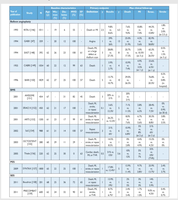
Myocardial revascularization has been subject to more rando-mized clinical trials (RCTs) than almost any other intervention (Figure 1). In order to inform the current Guidelines, this Task Force performed a systematic review of all RCTs performed since 1980, comparing head-to-head the different revascularization strategies—including CABG, balloon angioplasty, and PCI with bare-metal stents (BMS) or with various US Food and Drug Table 1: Classes of recommendations
Table 2: Levels of evidence
Level of evidence A
Data derived from multiple randomized clinical trials or meta-analyses.
Level of evidence B
Data derived from a single randomized clinical trial or large non-randomized studies.
Level of evidence C
Consensus of opinion of the experts and/ or small studies, retrospective studies, registries. ESC/ EA CT S GU IDELINES
Administration-approved drug-eluting stents (DES)—against medical treatment as well as different revascularization strategies, and retrieved 100 RCTs involving 93 553 patients with 262 090 patient-years of follow-up [4].
Formulation of the best possible revascularization approach, also taking into consideration the social and cultural context, will often require interaction between cardiologists and cardiac sur-geons, referring physicians, or other specialists as appropriate. Patients need help with taking informed decisions about their treatment and the most valuable advice will probably be provided to them by the‘Heart Team’ [5]. Recognizing the importance of the interaction between cardiologists and cardiac surgeons, the leadership of both the ESC and the EACTS has given this Joint Task Force, along with their respective Guideline Committees, and the reviewers of this document the mission to draft balanced, patient-centred, evidence-driven practice guidelines on myocardial revas-cularization. The respective Chairpersons of these two associations and CPG Chairperson were also given the task to adapt to the dec-laration of interest policy and to ensure that their Task Force members followed it throughout the development process of the Guidelines. In the event that any of the Task Force members had a potential conflict of interest to declare, he/she did not participate in thefinal decision of the Task Force on the given subject.
3. SCORES AND RISK STRATIFICATION
Myocardial revascularization in the elective setting is appropriate when the expected benefits, in terms of survival or health out-comes (symptoms, functional status, and/or quality of life), exceed the expected negative consequences of the procedure. Whether medical therapy, PCI, or CABG is preferred should depend on the risk–benefit ratios of these treatment strategies, weighting the risks of periprocedural death, myocardial infarction and stroke against improvements in health-related quality of life, as well as long-term freedom from death, myocardial infarction or repeat revasculari-zation. The Heart Team should take into consideration the coron-ary anatomy, disease, age and comorbidities, patient preference, and hospital/operator experience.
Numerous models have been developed for risk stratification, fo-cussing on anatomical complexity or clinical risk, and have demon-strated their value during decision-making [6]. Those models most frequently used in a clinical setting are summarized in the Tables of recommendation [risk models to assess short-term (in-hospital or 30-day) and medium-to-long-term (≥1 year) outcomes].
(1) The EuroSCORE predicts surgical mortality [7,8]. It is based on an old data set and has been shown to overestimate the risk of mortality, and should therefore no longer be used [9,10]. (2) The EuroSCORE II is an update of the logistic EuroSCORE model
and is derived from a more contemporary data set better reflecting current cardiac surgical practice [11]. Its value has been demonstrated in specific cohorts of patients undergoing CABG [12]. Compared with its original version, the EuroSCORE II may have a better ability to predict mortality [12–14].
(3) The Society of Thoracic Surgeons (STS) score is a risk-prediction model, validated in patients undergoing cardiac surgery, with a specific model for CABG surgery and combined CABG and valve surgery [15,16]. It can be used to predict in-hospital or 30-day mortality (whichever occurs last) and in-hospital morbidity.
(4) The SYNTAX score (Table3) was developed to grade the ana-tomical complexity of coronary lesions in patients with left main or three-vessel disease, and was found to be an inde-pendent predictor of long-term major adverse cardiac and cerebrovascular event (MACCE) in patients treated with PCI but not CABG [17,18]. It facilitates the selection of optimal treatment by identifying patients at highest risk of adverse events following PCI. The interobserver variability of the Synergy between Percutaneous Coronary Intervention with TAXUS and Cardiac Surgery (SYNTAX) score is significant [19] although development of non-invasive assessments may sim-plify calculation of the SYNTAX score [20].
(5) The National Cardiovascular Database Registry (NCDR CathPCI) risk score has been developed to predict risk in PCI patients and should only be used in this context [21].
(6) The age, creatinine, ejection fraction (ACEF) model is a simple score as it contains only three variables, and was developed using
1964 2014 1964 FIRST CABG PROCEDURES 1977 CORONARY ANGIOPLASTY 1980 ECSS n=768 1986 CORONARY STENTS 1993 ERACI n=127 1993 RITA n=1011 1995 MASS n=214 1995 CABRI n=1054 2000 SIMA n=123 1996 BARI n=1829 1997 FMS n=152 2002 SOS n=988 2001 AWESOME n=454 1994 GABI n=359 1994 EAST n=392 2010 CARDia n=510 2001 ERACI II n=450 2001 ARTS n=1205 2012 VA CARDS n=198 2012 FREEDOM n=1900 2009 SYNTAX n=1800 2009 LE MANS n=105 1997 RITA-2 n=1018 2007 MASS II n=611 EXCELn=2600 2007 COURAGE n=2287 1999 AVERT n=341 2006 OAT n=2166 2003 ALKK n=300 2007 SWISS-II n=201 2008 JSAP n=384 2009 BARI-2D n=384 2011 STICH n=1212 2012 FAME-2 n=888 ISCHEMIA n=8000 2001 TIME n=305 1984 VA n=686 1984 CASS n=780 2011 LEIPZIG LM n=201 2011 PRECOMBAS n=600
Revascularization vs. MT Balloon angioplasty vs. CABG BMS vs. CABG DES vs. CABG
BMS = bare-metal stent; CABG = coronary artery bypass grafting; DES = drug-eluting stent.
data from a cohort of surgical patients [22]. ACEF has also been validated to predict mortality in patients undergoing PCI [23]. (7) The clinical SYNTAX score is a combination of the ACEF and
SYNTAX scores. Originally established as an additive model,
the subsequent development of a logistic model has provided more tailored risk assessment [24].
(8) The SYNTAX II score is a combination of anatomical and clinical factors [age, creatinine clearance, left ventricular (LV) function, Table 3: Guide to calculate the SYNTAX score
Steps Variable assessed Description
Step 1 Dominance The weight of individual coronary segments varies according to coronary artery dominance (right or left). Co-dominance does not exist as an option in the SYNTAX score.
Step 2 Coronary segment The diseased coronary segment directly affects the score as each coronary segment is assigned a weight, depending on its location, ranging from 0.5 (i.e. posterolateral branch) to 6 (i.e. left main in case of left dominance).
Step 3 Diameter stenosis The score of each diseased coronary segment is multiplied by 2 in case of a stenosis 50–99% and by 5 in case of total occlusion.
In case of total occlusion, additional points will be added as follows: - Age >3 months or unknown
- Blunt stump - Bridging - First segment visible distally - Side branch at the occlusion
Step 4 Trifurcation lesion The presence of a trifurcation lesion adds additional points based on the number of diseased segments: - 1 segment +3
- 2 segments +4 - 3 segments +5 - 4 segments +6
Step 5 Bifurcation lesion The presence of a bifurcation lesion adds additional points based on the type of bifurcation according - Medina 1,0,0 or 0,1,0 or 1,1,0: add 1 additional point
- Medina 1,1,1 or 0,0,1 or 1,0,1 or 0,1,1: add 2 additional point
Additionally, the presence of a bifurcation angle <70° adds 1 additional point. Step 6 Aorto-ostial lesion The presence of aorto-ostial lesion segments adds 1 additional point.
Step 7 Severe tortuosity The presence of severe tortuosity proximal of the diseased segment adds 2 additional points. Step 8 Lesion length Lesion length >20 mm adds 1 additional point.
Step 9
Step 10 Thrombus The presence of thrombus adds 1 additional point.
Step 11 Diffuse disease/small vessels The presence of diffusely diseased and narrowed segments distal to the lesion (i.e. when at least 75% of the length of the segment distal to the lesion has a vessel diameter of <2 mm) adds 1 point per segment number.
+1 +1 +1
+1 per non visible segment +1 if <1.5 mm diameter
+1 if both <1.5 and ≥1.5 mm diameter +0 if ≥1.5 mm diameter (i.e. bifurcation lesion)
. [29]: ESC/ EA CT S GU IDELINES
Risk models to assess short-term (in-hospital or 30-day) outcomes Score Development cohort (patients, design) Patient inclusion Coronary procedures Number of variables
Outcome Recommendation Validation studies
Calculation Refa
Clinical Anatomical CABG PCI
STS Score n = 774 881 Multicentre 01/2006 – 12/2006 100% (i) CABG 40 2 In-hospital or 30-dayb mortality, and in-hospital morbidityc I B 5–10 http://riskcalc.sts. org 15,16 EuroSCORE II n =16 828 Multicentre 05/2010 – 07/2010 47% (i) CABG 18 0 In-hospital
mortality IIa B IIb C >10
www.euroscore.org /calc.html 11 ACEF n = 4557 Single-centre 2001 – 2003 - 3 0 In-hospital or 30-dayb mortality IIb C IIb C 5–10 [Age/ejection fraction (%)] + 1d 22 NCDR CathPCI 181 775 Multicentre 01/2004 – 03/2006 100% PCI 8 0 In-hospital mortality IIb B <5 - 21 EuroSCORE n =19 030 Multicentre 09/1995 – 11/1995 64% (i) CABG 17 0 Operative
mortality III B III C >50
www.euroscore.org /calcold.html 7, 8
ACEF = age, creatinine, ejection fraction; (i) CABG = (isolated) coronary artery bypass grafting; NCDR = National Cardiovascular Data Registry; PCI = percutaneous coronary intervention; STS = Society of Thoracic Surgeons.
aReferences.
bWhichever occurs last.
cPermanent stroke, renal failure, prolonged ventilation, deep sternal wound infection, re-operation, length of stay <6 or >14 days. dIf creatinine is >2 mg/dl.
Risk models to assess medium- to long-term (≥1 year) outcomes
Score Development cohort Patient inclusion Coronary procedures Number of variables
Outcome Recommendation Validation studies
Calculation Refa
Clinical Anatomical CABG PCI
SYNTAX None, expert opinion none - 0 11 (3 general, 8 per lesion) MACCE I B I B >50 www. syntaxscore.com 30 SYNTAX II 1800 Multicentre 03/2005 – 04/2007 50% CABG, 50% PCI 6 12 4-year
mortality IIa B IIa B <5 - 25
ASCERT CABG 174 506 Multicentre 01/2002 – 12/2007 100% (i) CABG 23 2 Mortality >2 years IIa B <5 - 27 ASCERT PCI 206 081 Multicentre 2004 – 2007 100% PCI 17 2 Mortality >1 year IIa B <5 - 28 Logistic Clinical SYNTAX 6 508 Multicentre 03/2005 – 04-2007 100% PCI 3 11 1-year MACE and mortality IIa B <5 - 24
ASCERT = American College of Cardiology Foundation–Society of Thoracic Surgeons Database Collaboration (ACCF–STS) on the comparative effectiveness of revascularization strategies; (i) CABG = (isolated) coronary artery bypass grafting; MACCE = major adverse cardiac and cerebrovascular events; PCI = percutaneous coronary intervention; SYNTAX = synergy between percutaneous coronary intervention with TAXUS and cardiac surgery.
a
gender, chronic obstructive pulmonary disease, and peripheral vascular disease] and predicts long-term mortality in patients with complex three-vessel or left main (LM) coronary artery disease (CAD) [25]. It was found to be superior to the convention-al SYNTAX score in guiding decision-making between CABG and PCI in the SYNTAX trial, and subsequently validated in the drug-eluting stent for left main coronary artery disease DELTA registry. (9) For the American College of Cardiology Foundation–Society of
Thoracic Surgeons Database Collaboration (ASCERT) study [26], two large datasets from the National Cardiovascular Data Registry (NCDR) and STS were used to develop several models to predict mortality at different time points following CABG and PCI [27,28]. Comparative analyses of these models are limited because avail-able studies have largely evaluated individual risk models in differ-ent patidiffer-ent populations, with differdiffer-ent outcome measures being reported at various time points, and most models are restricted to one type of revascularization. In addition, several important vari-ables, such as frailty, physical independence and porcelain aorta, are not incorporated in current risk scores. An ideal risk–benefit model enables comparison of the short-term benefits of PCI to the long-term benefits of CABG; however, even though risk models may provide useful information for predicting mortality and major adverse events, prediction of which patients will receive benefit in terms of quality of life is so far unavailable.
These limitations restrict the ability to recommend one specific risk model. It is also important to acknowledge that no risk score can accurately predict events in an individual patient. Moreover, limitations exist in all databases used to build risk models, and dif-ferences in definitions and variable content can affect the per-formance of risk scores when they are applied across differing populations. Ultimately, risk stratification should be used as a guide, while clinical judgement and multidisciplinary dialogue (The Heart Team) remain essential [25].
4. PROCESS FOR DECISION-MAKING AND
PATIENT INFORMATION
4.1 Patient information and informed consent
The process of medical decision-making and patient information is guided by the‘four principles’ approach to healthcare ethics: autonomy, beneficence, non-maleficence, and justice [31]. The informed consent process should not be regarded as a necessary legal requirement but as an opportunity to optimize decision-making. Patient-related factors, institutional factors and referral patterns may impact the decision-making process.Informed consent requires transparency, especially if there is controversy over various treatment options. Collaborative care requires the pre-conditions of communication, comprehension, and trust. Treatment decisions should not be based solely on re-search results and the physician’s appraisal of the patient’s circum-stances, since active patient participation in the decision-making process may yield better outcomes. Patients are subject to bias by labels when considering coronary revascularization [32], and patient preference may sometimes contradict evidentiary best practice. Patients may have limited understanding of their disease and sometimes unreasonable expectations with regard to the out-comes of a proposed intervention. As many as 68% of patients are not aware of an alternative revascularization strategy [33]. Short-term procedure-related and long-Short-term risks and benefits—such
as survival, relief of angina, quality of life, potential need for late re-intervention, and uncertainties associated with different treat-ment strategies—should be thoroughly discussed. Patients can only weigh this information in the light of their personal values and cultural background and must therefore have the time to reflect on the trade-offs imposed by the outcome estimates.
In order to seek a second opinion or to discuss thefindings and consequences with referring physicians, enough time should be allowed—up to several days, as required—between diagnostic catheterization and intervention. Patient information needs to be unbiased, evidence-based, up-to-date, reliable, accessible, rele-vant, and consistent with legal requirements. Consistent use of ter-minology, that the patient understands, is essential. A written patient information document is needed. These recommenda-tions pertain to patients in stable condition, for whom various treatment options exist and who can make a decision without the constraints of an urgent or emergency situation (Table4).
Anonymous treatment should be avoided. The patient has the right to obtain information on the level of expertise of the oper-ator, the workload of the centre and whether all treatment options including surgery are available on site. Patients considered for revascularization should also be clearly informed of the con-tinuing need for medical therapy, as well as lifestyle modification and other secondary prevention strategies (section 20).
4.2 Multidisciplinary decision-making
(Heart Team)
The Heart Team, made up of clinical or non-invasive cardiologists, cardiac surgeons and interventional cardiologists, provides a balanced, multidisciplinary decision-making process [5]. Add-itional input may be needed from other specialties involved in the care of the patient. The Heart Team should meet on a regular basis to analyse and interpret the available diagnostic evidence, put into context the clinical condition of the patient, determine the need—or otherwise—for an intervention and the likelihood of safe and effective revascularization with either PCI or CABG.Ad hoc meetings of the Heart Team should facilitate and support efficient clinical workflows.
The demand for an interdisciplinary approach is underlined by reports on (i) underuse of revascularization procedures in 18–40% of patients with CAD [34], and (ii) inappropriate use of revasculariza-tion strategies and a lack of case discussions [35]. The large variabil-ity between European countries in PCI-to-CABG ratios (ranging from 2.0 to 8.6 in 2007) has raised concerns regarding the appropri-ate selection of revascularization in Europe [36]. Rappropri-ates for the in-appropriate use of PCI (11–15%) or doubt over the appropriateness of PCI (40–50% [5,37] and, to a lesser degree for CABG (1–2% and 0–9%, respectively) are reported [5,38]. The increasing underuse of CABG is in part explained by PCI treatment in patients with indica-tions for surgery [39,40]. Multidisciplinary decision-making in a Heart Team can minimize specialty bias and prevent self-referral from interfering with optimal patient care [32,41].
Standard evidence-based, interdisciplinary, institutional proto-cols may be used for common case scenarios, to avoid the need for the systematic case-by-case review of all diagnostic angiograms, but complex cases should be discussed individually. In these cases, revascularization should not be performed at the time of diagnostic angiography, to allow sufficient time to assess all available informa-tion, and clearly explain and discuss thefindings with the patient [41]. The rationale for a decision and consensus on the optimal
ESC/ EA CT S GU IDELINES
revascularization treatment should be documented on the patient’s chart. In hospitals without a cardiac surgical unit or in an ambula-tory setting, protocols should be designed in collaboration with an expert interventional cardiologist and a cardiac surgeon. Decisions made by a Heart Team seem to be reproducible [42].
4.3 Timing of revascularization and ad hoc
percutaneous coronary intervention
Studies of patients scheduled for revascularization have revealed that considerable morbidity and mortality are associated with extended delay of treatment [43,44]. The waiting period for diag-nostic catheterization should therefore be minimal. Once the de-cision for revascularization has been reached after diagnostic coronary angiography, the Task Force recommends that patients with severe symptoms Canadian Cardiovascular Society (CCS) Class 3 and those with high-risk anatomy [left main disease or
equivalent; three-vessel disease or proximal left anterior descend-ing (LAD) or depressed ventricular function] preferably undergo revascularization (PCI or CABG) within 2 weeks. For all other patients with stable coronary artery disease (SCAD) and an indica-tion for revascularizaindica-tion, it is desirable to perform revasculariza-tion (PCI or CABG) within 6 weeks (Table4) [44].
Ad hoc PCI is defined as a therapeutic intervention performed within the same procedure as the diagnostic coronary angiography. Ad hoc PCI is convenient, associated with fewer access site compli-cations, and often cost-effective and safe [45]. In the USA, however, up to 30% of patients undergoingad hoc PCI are potential candi-dates for CABG [45]. Although this number may be lower in Europe [35], ad hoc PCI should not be applied as a default approach [45,46]. Ad hoc PCI in stable patients is only justified after adequate informa-tion given to the patient (see secinforma-tion 4.1) and if a full diagnostic work-up, including functional testing (section 5) is available. Institutional protocols developed by the Heart Team in accordance with current guidelines should define specific anatomical criteria Table 4: Multidisciplinary decision pathways, patient informed consent, and timing of intervention
ACS Multivessel SCAD SCAD with ad-hoc PCI
indication according to predefined Heart-Team
protocols
Shock STEMI NSTE-ACS
Multidisciplinary decision making
Not mandatory during the acute phase.
Mechanical circulatory support according to Heart-Team protocol.
Not mandatory during the acute phase.
Not mandatory during the acute phase. After stabilization recommended as in stable multivessel CAD.
Required. Not required.
Informed consent
Verbal witnessed informed consent or family consent if possible without delay.
Verbal witnessed informed consent unless written consent is legally required. Written informed consent.a
Written informed consent.a Written informed consent.a
Time to revascularization Emergency: no delay. Emergency: no delay. Urgency: within 24 hours if possible and no later than 72 hours.
For patients with severe symptoms (CCS 3) and for those with high– risk anatomy (left main disease or equivalent, three-vessel disease or proximal LAD or depressed ventricular function), revascularization (PCI or CABG) should be performed within 2 weeks.
For all other patients with SCAD, revascularization (PCI or CABG) should be performed within 6 weeks.
Ad hoc
Procedure Proceed with intervention based on best evidence/ availability. Non-culprit lesions treated according to institutional protocol or Heart Team decision. Proceed with intervention based on best evidence/ availability. Non-culprit lesions treated according to institutional protocol or Heart Team decision. Proceed with intervention based on best evidence/ availability. Non-culprit lesions treated according to institutional protocol or Heart Team decision.
Plan most appropriate intervention allowing enough time from diagnostic catheterization to intervention.
Proceed with intervention according to institutional
ACS = acute coronary syndromes; CABG = coronary artery bypass grafting; CCS = Canadian Cardiovascular Society; LAD = left anterior descending; NSTE-ACS = non—ST-segment elevation acute coronary syndrome; PCI = percutaneous coronary intervention; SCAD = stable coronary artery disease; STEMI = ST-segment elevation myocardial infarction.
aThis may not apply to countries that legally do not ask for written informed consent. ESC and EACTS advocate documentation of patient consent for all
and clinical subsets that may be—or should not be—treated ad hoc. Complex pathologies in stable patients, including lesions of the LM or proximal LAD and three-vessel disease, should in general not be treatedad hoc, but discussed by the Heart Team.
Recommendations for decision-making and patient infor-mation in the elective setting
Recommendations Classa Levelb Refc
It is recommended that patients undergoing coronary angiography are informed about benefits and risks as well as potential therapeutic consequences ahead of the procedure.
I C –
It is recommended that patients are adequately informed about short- and long-term benefits and risks of the revascularization procedure as well as treatment options. Enough time should be allowed for informed decision-making.
I C –
–
– It is recommended that institutional
protocols are developed by the Heart Team to implement the appropriate revascularization strategy in accordance with current guidelines. In case of PCI centres without on-site surgery, institutional protocols should be established with partner institutions providing cardiac surgery.
I C
It is recommended that patients for whom decision-making is complex or who are not covered by the institutional protocol are discussed by the Heart Team.
I C
PCI = percutaneous coronary intervention.
aClass of recommendation. bLevel of evidence. cReferences.
5. STRATEGIES FOR DIAGNOSIS: FUNCTIONAL
TESTING AND IMAGING
Exercise testing and cardiac imaging are used to confirm the diagno-sis of CAD, to document ischaemia in patients with stable symptoms, to risk-stratify patients, and to help choose treatment options and evaluate their efficacy as explained in detail in the ESC Guidelines on the management of stable coronary artery disease [47].
Another indication for non-invasive imaging before revasculari-zation is the detection of myocardial viability in patients with poor LV function.
5.1 Non-invasive tests
The documentation of ischaemia using functional testing is recom-mended in patients with suspected SCAD before elective invasive procedures, preferably using non-invasive testing before invasive
angiography. Although several tests can be used, it is important to avoid unnecessary diagnostic steps. The current evidence sup-porting the use of various tests for the detection of CAD is based on meta-analyses and multicentre studies, and using only anatomical evaluation of invasive coronary angiography as the reference stand-ard [47]. The risks of exercise, pharmacological stressors, contrast agents, invasive procedures, and cumulative ionizing radiation must be weighed against the risk of disease or delayed diagnosis [48].
Multi-detector computed tomography (MDCT) can detect cor-onary atherosclerosis and stenoses and is reliable for ruling out sig-nificant CAD in patients with low-to-moderate probability of CAD [49]. The tests for detection of ischaemia are based on either reduc-tion of perfusion or inducreduc-tion of ischaemic wall moreduc-tion abnormal-ities during exercise or pharmacological stress. The best-established stress imaging techniques are echocardiography and perfusion scintigraphy. Both may be used in combination with exercise stress or pharmacological stress. Newer stress imaging techniques also include stress magnetic resonance imaging (MRI), positron emission tomography (PET), and combined approaches. The term ‘hybrid imaging’ refers to imaging systems in which two modalities [MDCT and PET; MDCT and single photon emission computed tomog-raphy (SPECT)] are combined in the same scanner, allowing both studies to be performed in a single imaging session. Ischaemia imaging has been regarded the most appropriate in patients with intermediate pre-test probability (15–85%) of significant CAD [47], while in asymptomatic patients or in those with low or high pre-test probability, the tests are generally not recommended. More detailed information about the imaging tests in the detection of CAD are available in the ESC Guidelines on the management of SCA [47] and in the Web addenda.
5.2 Invasive tests
Invasive coronary angiography has been regarded as the reference standard for the detection and the assessment of the severity of CAD but, as an invasive procedure, it is associated with specific procedure-related adverse events. Even experienced interventional cardiologists cannot, without functional information, accurately predict the significance of many intermediate stenoses on the basis of visual assessment or quantitative coronary angiography. When non-invasive stress imaging is contraindicated, non-diagnostic, or unavailable, the measurement of fractional flow reserve (FFR) or coronaryflow reserve is helpful during diagnostic coronary angiog-raphy [50]. Deferral of PCI or CABG in patients with FFR >0.80 appears safe [51–53]. Fractional flow reserve measurement is indi-cated for the assessment of the functional consequences of moder-ate coronary stenoses. FFR-guided PCI with medical therapy has been shown to decrease the need for urgent revascularization com-pared with the best available medical therapy alone [54].
5.3 Detection of myocardial viability
Non-invasive assessment of myocardial viability has been used to guide the management of patients with chronic ischaemic systolic LV dysfunction. Multiple imaging techniques, including PET, SPECT, and dobutamine stress echocardiography, have been eval-uated for assessment of viability and prediction of clinical outcome after myocardial revascularization [55]. In general, nuclear imaging techniques have a high sensitivity, whereas tech-niques evaluating contractile reserve have a somewhat lower
ESC/ EA CT S GU IDELINES
sensitivity but higher specificity. MRI has a high diagnostic accur-acy for assessing the transmural extent of myocardial scar tissue and can also assess contractile reserve, but its ability to detect via-bility and predict recovery of wall motion is no better than other imaging techniques. The differences in performance between the various imaging techniques are small, and experience and avail-ability commonly determine which technique is used. The evi-dence is mostly based on observational studies or meta-analyses. One RCT, relating to PET imaging, showed that patients with a sub-stantial amount of dysfunctional but viable myocardium are likely to benefit from myocardial revascularization [56].
6. REVASCULARIZATION FOR STABLE CORONARY
ARTERY DISEASE
6.1 Rationale for revascularization
Prior to revascularization, patients with SCAD must receive guideline-recommended medical treatment, due to its established benefits in terms of prognosis and symptom relief [47]. Revascularization, by either PCI or CABG, may be indicated in flow-limiting coronary stenoses to reduce myocardial ischaemia and its adverse clinical manifestations [85–87]. The indications for revascularization in patients with SCAD are persistence of symp-toms despite medical treatment and/or improvement of progno-sis [47]. Consequently, revascularization and medical therapy should be seen as complementary, rather than competitive treat-ment strategies. Specific evidence and recommendations for dia-betic patients are addressed in section 10.
Angina is associated with impaired quality of life, reduced physical endurance, mental depression, and recurrent hospitalizations and outpatient visits [88]. Revascularization by PCI or CABG more effect-ively relieves angina, reduces the use of anti-angina drugs, and improves exercise capacity and quality of life, compared with a strat-egy of medical therapy alone (Table2Web addenda) [54,89–96].
Ischaemia is of prognostic importance in patients with SCAD, particularly when occurring at low workload [97,98]. Revascularization relieves myocardial ischaemia more effectively than medical treatment alone [92,97,99,100]. The extent, location, and severity of coronary artery obstruction as assessed by coron-ary angiography or coroncoron-ary computed tomography (CT) angiog-raphy are important prognostic factors in addition to ischaemia and left ventricular function [101–103].
6.2 Evidence basis for revascularization
The evidence basis for revascularization with PCI and/or CABG, compared with medical treatment, is derived from several RCTs that are summarized in Table5. It is important to consider that the best current revascularization results achieved with PCI are with new-generation drug-eluting stents (DES) and for CABG with maximal use of arterial grafts. Although revascularization procedures are associated with the risk of biomarker-defined periprocedural myocardial infarction, several studies indicate that pre-PCI—but not post-PCI—biomarker elevations impact adversely on prognosis [104]. While spontaneous myocardial infarction has a well established adverse impact on prognosis and notably mortality, recent studies suggest that, compared with medical treatment, PCI is associated with a lower risk of spontaneous myocardial infarction [105]. Indications for diagnostic testing in patients with suspected CAD and stable symptoms
Asymptomatica Symptomatic b Low (<15%) Intermediate (15–85%) High (>85%)
Classc Leveld Classc Leveld Classc Leveld Classc Leveld Refe
Anatomical detection of CAD
Invasive angiography III A III A IIb A I A 50–52,54
CT angiographyf,g III B III C IIa A III B 57–62
Functional test
Stress echo III A III A I A III A 63–65
Nuclear imaging III A III A I A III A 60,66–70
Stress MRI III B III C I A III B 71–75
PET perfusion III B III C I A III B 67,69,70,76,77
Combined or hybrid imaging test
III C III C IIa B III B 78–83
CAD = coronary artery disease; CT = computed tomography; MRI = magnetic resonance imaging; PET = positron emission tomography.
a
Screening for silent (asymptomatic) myocardial ischaemia may be considered in selected high-risk patients, such as those with diabetes mellitus [84].
bPre-test probability of CAD. Low 0—15%; intermediate 15—85%; high >85% as assessed using the criteria based on ESC Guidelines of SCAD [47]. c
Class of recommendation.
dLevel of evidence. e
References.
fThis refers to CT angiography, not calcium scoring.
Table 5: Revascularization versus medical therapy
Year of
publication Study N
Baseline characteristics Primary endpoint Max clinical follow-up Age (y) Women (%) Diabetes (%) MVD (%) EF (%)
y Results y Death MI Revasc.
CABG 1982 ECSS [109] 768 <65c 0 - 100 >50c - - - 8 11.4% vs. 20.1%a - -1984 VA [110] 686 - - - 86 - - - - 18 70% vs. 67% 49% vs. 41% 41% vs. 62%d 1984 CASS [111] 780 51 10 9 73 - - - - 10 19.2% vs. 21.8% -8.9% vs. 36.9%e 2011 STICH[112] 1212 60 12 39 91 27 Death 4.7 36% vs. 41% 4.7 36% vs. 41% - -Balloon angioplasty 1997 RITA-2[89] 1018 - 18 9 40 - Death or MI 2.7 6.3% vs. 3.3%a 7 8.5% vs. 8.4% 6.3% vs. 4.5%d 27.2% vs. 35.4%d 1999 AVERT [113] 341 58 16 16 43 61 Cardiac death, cardiac arrest, MI, stroke, revascularization, or hospitalization due to angina 1.5 20.9% vs. 13.4%a 1.5 0.6% vs. 0.6%b 2.8% vs. 2.4%d 16% vs. 12%d 2003 [114]ALK 300 58 13 16 0 -MI, revascularization, or rehospitalization for severe angina
1 10% vs. 18% 4.7 4.0% vs. 11.2%a 6.7% vs. 7.9% 17% vs. 24% 2007 SWISSI-II [92] 201 55 12 11 - 57 Cardiac death, MI, or revascularization 10.2 28.1% vs. 63.8%a 10.2 6.3% vs. 21.0%a 11.5% vs. 38.1%a 27.1% vs. 43.8%a BMS/CABG 2001 TIME [90] 305 80 43 23 79 53 Death, MI, or hospitalization for ACS 0.5 19.0% vs. 49.3%a 1 11.1% vs. 8.1% - -2010 MASS-II[94] 611 60 31 29 100 67 Cardiac death, MI, or revascularization 1 6.4% (CABG) vs. 24.4% (BMS) vs. 14.3% (MT)a 10 25.1% (CABG) vs. 24.9% (PCI) vs. 31% (MT) 10.3% (CABG) vs. 13.3% (PCI) vs. 20.7 (MT)a 7.4% (CABG) vs. 41.9% (PCI) vs. 39.4 (MT)a BMS
2006 [115]OAT 2166 59 22 21 18 48 Death, MI, or NYHA IV heart failure 4 17.2% vs. 15.6% 4 9.1% vs. 9.4% 6.9% vs. 5.0% 18.4% vs. 22.0%a 2007 COURAGE [91] 2287 62 15 33 69 61 Death or MI 4.6 19.0% vs. 18.5% 4.6 7.6% vs. 8.3% 13.2% vs. 12.3% 21.1% vs. 32.6%a 2008 JSAP [116] 384 64 26 40 32 65
Death, ACS, stroke,
or emergency hospitalization 3.3 22.0% vs. 33.2%a 3.3 2.9% vs. 3.9% 1.6% vs. 3.8% 21.4% vs. 36.5%a DES 2012 FAME-2[54] 888 64 22 27 42 -Death, MI, or urgent revascularization 1 4.3% vs. 12.7%a 1 0.2% vs. 0.7% 3.4% vs. 3.2% 3.1% vs. 19.5%a
ACS = acute coronary syndromes; BMS = bare-metal stents; CABG = coronary artery bypass grafting; DES = drug-eluting stents; EF = ejection fraction; MI = myocardial infarction; MT = medical therapy; MV = multivessel; MVD = multivessel disease; NYHA = New York heart Association; Revasc = revascularization; y = years.
a
P<0.05;bCardiac death;cInclusion criteria;dNo statistical analyses performed;eRepeat CABG, excluding PCI.
Only trials with at least 100 patients per treatment arm were included. Age and ejection fraction are reported as means.
ESC/ EA CT S GU IDELINES