Frequency of Chromosome Healing and Interstitial Telomeres in 40 Cases of Constitutional Abnormalities
23
0
0
Texte intégral
(2) 2 Abstract Human telomeres play a major role in stabilizing chromosome ends and preventing fusions. Chromosomes bearing a broken end are rescued by the acquisition of a new telomeric cap without any subtelomeric sequences being present at the breakpoint, a process referred to as chromosome healing. Conversely, a loss of telomeric function or integrity can lead to the presence of interstitial telomeres at the junction site in translocations or ring chromosomes. In order to determine the frequency at which interstitial telomeres or chromosome healing events are observed in target chromosome abnormalities, we conducted a retrospective FISH study using pan-telomeric and chromosome-specific subtelomeric probes on archival material from 40 cases of terminal deletions, translocations or ring chromosomes. Of the 19 terminal deletions investigated, 17 were negative for the subtelomeric probe specific to the deleted arm despite being positive for the pantelomeric probe. These 17 cases were thus considered as been rescued through chromosome healing, suggesting that this process is frequent in terminal deletions. In addition, as two of these cases were inherited from a parent bearing the same deletion, chromosomes healed by this process are thus stable through mitosis and meiosis. Regarding the 13 cases of translocations and eight ring chromosomes, four and two cases respectively demonstrated pan-telomeric sequences at the interstitial junction point. Furthermore, two cases of translocations and one ring chromosome had both interstitial pan-telomeres and subtelomeres, whereas two other cases of ring chromosomes and one case of translocation only showed interstitial subtelomeres. Therefore, interstitial (sub)telomeric sequences in translocations and ring chromosomes are more common than previously thought, as we found a frequency of 43% in this study. Moreover, our results illustrate the necessity of performing FISH with both subtelomeric and pan-telomeric probes when investigating these rearrangements, as the breakpoints can be either in the distal part of the pantelomeres, or in between the two types of sequences.. Keywords: Chromosome healing, FISH, Interstitial telomeres, Ring chromosomes, Subtelomeres, Terminal deletions, Translocations..
(3) 3. Introduction Human telomeres are specialized chromatin structures present at chromosome ends. They are constituted of (TTAGGG)n DNA repeat sequences (Moyzis et al., 1988), are highly conserved among vertebrates (Meyne et al., 1989), and contain proteins forming a protective ‘t-loop’ structure capping the chromosome ends (Griffith et al., 1999). Telomeres are essential for chromosomes, as they prevent degradation or fusion, and enable the cell to distinguish intact chromosome ends from damaged DNA (Hackett et al., 2001). Furthermore, this structure is necessary for the complete replication of chromosome ends with the help of telomerase, a specialized cellular reverse transcriptase enzyme (Morin, 1989). In humans, telomerase activity is developmentally regulated and is found in the placenta, liver, hematopoietic and germline cells (Melek and Shippen, 1996) as well as in most tumors (de Lange, 1995).. Broken chromosomes without telomeres will enter in a breakage-fusion-bridge (BFB) cycle if no healing occurs.. This cycle will cause chromosomal instability that can induce. formation of dicentric, ring or fusion chromosomes (Hackett et al., 2001) and could lead to terminal inverted duplications (Ballif et al., 2003). Healing of broken ends can occur through two general pathways, ensuring the acquisition of a new telomeric cap and stabilizing the deleted chromosome. First, direct addition of telomeric sequences onto the broken end can be achieved through a telomerase-mediated de novo telomere addition (Flint et al., 1994; Varley et al., 2000; Wilkie et al., 1990) or a telomerase-independent recombination-based mechanism (Neumann and Reddel, 2002; Varley et al., 2002). Second, telomeres can also be retrieved from another location through a mechanism called telomere capture, in which subtelomeres and/or pan-telomeres from another chromosome are translocated at the broken end of the deleted chromosome (Bosco and Haber, 1998; Meltzer et al., 1993; Ning et al., 1998). Although very similar to unbalanced subtelomeric translocations, telomere capture would occur only de novo, while unbalanced cryptic translocations would be inherited from a parent carrying a balanced translocation. On the other hand, some constitutional anomalies are generated when the telomeres loose their ability to prevent fusion. For example, although interstitial telomeric sequences can be detected in normal conditions, as indicated by the major telomeric repeat array found at 2q13 (Ijdo.
(4) 4. et al., 1991), most interstitial telomeres occur at the fusion point in telomeric translocations, jumping translocations or ring chromosomes (Reviewed in Tables 1 and 2).. In order to further study the frequency of chromosome healing and interstitial telomeres in human constitutional abnormalities, we conducted a retrospective study on cases taken from the 1991-2006 archives of a pediatric hospital, the Centre Hospitalier Universitaire (CHU) SainteJustine.. Materials and Methods Cytogenetics Searching for chromosome rearrangements possibly containing interstitial telomeres or rescued by telomere healing, we reviewed all cases of deletions, translocations and ring chromosomes seen at CHU Sainte-Justine between 1991 and 2006. We selected all deletion cases where a terminal deletion was apparent as well as all translocation and ring cases in which at least one breakpoint involved the terminal band, as evaluated at the karyotype with a minimal resolution of 500 bands. Therefore, a retrospective study was conducted on 40 cases: 19 terminal deletions, 13 translocations involving a terminal band and eight ring chromosomes (Table 3). Chromosome preparations and GTG-banding were previously done according to the previously described method (Lemieux et al., 1990). All but four cases were de novo abnormalities with normal parental karyotypes: cases 12 and 13 were inherited from the same mother, while cases 28 and 29 were inherited from a father and mother respectively. Parental rearrangements were not included in this study. Four cases from this cohort (cases 12, 13, 23, and 40) were previously published elsewhere (Boutouil et al., 1996; DesGroseilliers et al., 2006; Maranda et al., 2006). Karyotypes of all cases were done following the guidelines of ISCN (2005). The terminal band was used to designate the breakpoint when only interstitial subtelomeres were present, while pter or qter designate the breakpoint when both interstitial subtelomeres and pan-telomeres were present. In cases with interstitial pan-telomeres, but in which the presence of interstitial subtelomeres could be not assessed, the terminal band was used..
(5) 5. Fluorescence in situ hybridization The pan-telomeric peptide nucleic acid (PNA) probe [FITC-(C3TA2)3] from DAKO (Mississauga, On, Canada) recognizing the consensus sequence (TTAGGG)n of human pantelomeres was hybridized after minor modifications to the protocol of Vermeesh et al. (1997). Slides were counterstained with propidium iodide (PI) (Sigma-Aldrich Canada Ltd, Oakville, Canada) or DAPI (Roche Diagnostics, Laval, Canada), and bands were visualized as described by Lemieux et al. (1992), or by the reverse DAPI function of the Cytovision software (version, Applied Imaging, San Jose, CA). Subtelomeric probes [TelVysion 1p/1q, 2p/2q, 4p/4q, 5p/5q, 7p/7q, 8p/8q, 9p/9q, 11p/11q, 13q, 14q, 16p/16q, 18p/18q, 20p/20q, 21q, Xp/Yp, and Xq/Yq from Vysis (Downers Grove, USA), and 1p and 6p from AL Technologies (Arlington, USA)] were hybridized according to the company’s instructions. 30 metaphases were scored for each probe in order to ensure accuracy and reproducibility of results.. Results Terminal deletions Of the 19 cases of terminal deletions studied (Table 3), all showed a signal for the pantelomeric probe at the deleted end. 17 of them (89%) showed no signal with the arm-specific subtelomeric probe at the deleted arm (as shown for cases 7 and 12; Fig. 1A, B). Furthermore, in these 17 cases, the missing subtelomeric signal was not present on any other chromosomes, ruling out a balanced cryptic translocation. As for the two remaining cases (4 and 8), the presence of the arm-specific subtelomeric probe at the deleted end (Fig. 1C, D) prompted us to reconsider these deletions as interstitial and revise the breakpoints (Table 3).. Translocations and ring chromosomes Of the 13 translocations investigated with the pan-telomeric probe (Table 3), four (31%) showed interstitial pan-telomeric sequences at the fusion point (cases 20, 21, 22, and 23; Fig. 1E, H, I, K). Also, the presence of subtelomeres could be assessed at the junction point in cases 20 and 22 (Fig. 1G, I), while the unbalanced nature of these translocations could be confirmed by the presence of three signals of the 7q subtelomere in case 20 (Fig. 1F) and of the 14q subtelomere in.
(6) 6. case 22 (Fig. 1J). As for case 32, a balanced t(X;1), no interstitial pan-telomeres were detected by PNA-FISH (Fig. 1L), despite the fact that PNA-FISH probes allow detection of shorter pantelomeric sequences than DNA-FISH probes (Bolzan and Bianchi, 2006). A signal for the 1p subtelomere was present at the junction point on the der(1) as well as at the distal end of the der(X) (Fig. 1M), whereas the Xq subtelomere was only found at the distal end of the der(1) (Fig. 1N). This suggests that the breakpoint on 1p was within the subtelomeres, whereas the Xq breakpoint was proximal to the subtelomeres. Unfortunately, FISH with subtelomeric probes could not be performed on any other cases because no more archival material was available.. For the eight cases of ring chromosomes (Table 3), only two (25%) presented interstitial pan-telomeric sequences (cases 33 and 34; Fig. 1O, P), and three cases (37,5%) had interstitial subtelomeres.. Indeed, FISH with subtelomeric probes revealed the presence of interstitial. subtelomeres in cases 34 (Fig. 1Q), 35 and 40. Unfortunately, such analyses could not be performed on the remaining cases due to lack of archival material.. Discussion Chromosome healing in terminal deletions Of the 19 deletion cases studied, 17 lacked the specific subtelomeric region of the deleted arm, despite the presence of pan-telomeres at this same deleted end (Table 3). In the two remaining patients (cases 4 and 8), presence of the arm-specific subtelomere and pan-telomeres at the deleted end indicated that these deletions were interstitial rather than terminal. Consequently, our results suggest that the 17 cases of terminal deletions were subjected to chromosome healing after chromosome breakage. Furthermore, as two of these deletions (cases 12 and 13) were inherited from a parent, this implies that such chromosome healing is stable. Although we did not detect any translocations implicating the deleted chromosomes even after high-resolution analyses, we cannot rule out telomere capture or other unbalanced cryptic translocations as means to repair broken ends in our cohort of patients, as FISH with all the subtelomeric probes could not be performed due to lack of archival material. However, the incidence of telomere capture or unbalanced cryptic translocations was found to be relatively low in the literature. Indeed, Marinescu et al. (1999) found an unbalanced cryptic translocation.
(7) 7. inherited from the mother in only one of 104 patients carrying a 5p terminal deletion, while Davies et al. (2003) reported that three cases out of 16 terminal deletions were cryptic unbalanced translocations (two of which were de novo). More recently, a study by Ravnan et al. (2006), on a population suffering from idiopathic mental retardation or developmental delay, reported that 30.4% of all cryptic subtelomeric rearrangements were unbalanced translocations/telomere capture events. However, as 24% of these were true unbalanced translocations inherited from a parent carrying a balanced form of the translocation, telomere capture events could therefore represent less than 23% of all subtelomeric rearrangements found in that population (Ravnan et al., 2006). Thus, chromosome healing, i.e. direct addition of telomeric sequences onto the broken end, is the most probable mechanism by which the 17 terminal deletions we report here were stabilized.. Interstitial (sub)telomeres in translocations and ring chromosomes So far, approximately 30 cases of interstitial telomeres have been reported in the literature, either in stable or jumping translocations, or telomeric associations (Table 1). Reports of translocated chromosomes with interstitial telomeres by Park et al. (1992) and Rossi et al. (1993) initially suggested that telomeric translocations were common, as respectively 4/9 and 7/8 translocations presented interstitial pan-telomeres. However, in 1995, the work of Rivera et al. suggested that telomeric translocations were in fact less common, as no interstitial telomeres were found among 16 patients. As for ring chromosomes, approximately 20 cases with interstitial telomeres have been reported in the literature (Table 2). In particular, Flejter et al. (1996) reported a case with a familial r(19) which presented interstitial pan-telomeric sequences, while Canevini et al. (1998) reported that 2 out of 3 cases of r(20) had both subtelomeric and telomeric interstitial sequences (Table 2). On the other hand, as part of a study of patients with 22q13 deletions, Luciani et al. (2003) tested 17 cases of r(22) and found no interstitial telomeres. That being said, most of the previous reports of interstitial telomeres did not investigate the presence of interstitial subtelomeres, especially in cases of translocations (Tables 1 and 2). Since then, several studies have reported the combined hybridization of pan-telomeric and subtelomeric probes on patients presenting chromosomal abnormalities, mostly ring chromosomes (Tables 1 and 2). In particular, Sigurdardottir et al. (1999) and Mignon-Ravix et al. (2007) each published a case where interstitial subtelomeres were present, but not pan-telomeres (Tables 1 and 2). Altogether, this supports the.
(8) 8. notion that interstitial (sub)telomeres in ring chromosomes and translocations are more frequent than previously thought. In our cohort of 21 patients with a translocation or a ring chromosome, four translocations and two ring chromosomes had interstitial telomeres (cases 20, 21, 22; 23, 33 and 34), while one translocation and two ring chromosomes (cases 32, 35 and 40) had interstitial subtelomeres without interstitial detectable pan-telomeric sequences using a PNA-FISH probe (Table 3). These results would thus tend to indicate that the frequency of interstitial (sub)telomeres is not as low as reported, given a combined frequency of 43% in our cohort. Furthermore, the impossibility of performing FISH with subtelomeric probes in all cases of translocations and ring chromosomes might have led to an underestimation of the frequency of interstitial subtelomeres in our patients. In comparison, in their study of telomeric translocation involving chromosome 15, Mignon-Ravix et al. (2007) showed the presence of interstitial subtelomeres and/or pan-telomeres in four of the eight cases (50%) (Table 1).. Even though the frequency of interstitial telomeres in translocations and ring chromosomes is not precisely known, there is evidence that acrocentric chromosomes are more frequently implicated. Indeed, Rivera et al. (1999) reviewed 14 cases of jumping translocations and 12 cases of stable translocations published in the literature. Five of the eight jumping translocations and nine of the 12 stable translocations with interstitial telomeres implicated acrocentric chromosomes (Table 1). Reviewing the 15 constitutional telomeric associations, Huang et al. (2004) also found that 12 cases involved acrocentric chromosomes, three of which were positive for interstitial telomeres (Table 1). Our data is consistent with these observations, since five of the 13 stable translocations we report implicate acrocentrics (Table 3) and that three of these five cases were positive for interstitial telomeres (cases 21, 22 and 23). Although we were not able to rule out the possibility of de novo synthesis of telomeres in cases of interstitial (sub)telomeres in translocations and ring chromosomes, we do not believe that this mechanism could be implicated. Indeed, as ring formation or addition of foreign material through translocation would be means to stabilize the broken ends, there would have been no further need for other healing mechanisms. Furthermore, in all three cases (20, 22 and 34) with interstitial pan-telomeres where subtelomeric FISH was performed, a signal was present at the.
(9) 9. junction point, indicating that the breakpoint was not in the subtelomeres, but in the distal part of the telomeric TTAGGG repeats. The exact mechanism behind the presence of interstitial telomeres in constitutional cases is still unknown. In fact, interstitial telomeres are mostly studied in cancer cells or cell lines, as well as in cells exposed to radiation or with mutations of genes implicated in the maintenance of genomic stability (reviewed in Bolzan and Bianchi, 2006). In constitutional cases of translocations, such as those reported here, deficiency of proteins implicated in the capping, maintenance or replication of telomeres could have been responsible for the telomeric loss of function in parental gametes, leading to the abnormality observed in their offspring. For example, single nucleotide polymorphisms (SNPs) in DNA-PKcs, a gene implicated in DNA repair, lead to the presence of uncapped telomeres and radiation-induced telomere-double-strand break (telomere-DSB) fusions as a result of reduced abundance and activity of the protein (Williams et al., 2009). Also, a simple break in the (sub)telomeric region, followed by the fusion of this broken end with another broken chromosome end could be another mechanism leading to interstitial telomeres in translocations. As for ring chromosomes, intrachromosomal tethering of the p and q arms, as observed in interphasic nuclei for chromosomes 3, 4, 5, 6, 7, 9, 10, 12, 17, 18, and 20 (Daniel and St Heaps, 2004), could explain the presence of both subtelomeric p- and q-arm probes, as well as pan-telomeric probe, in these rearrangements.. Our study indicates that the frequency at which interstitial telomeres occur in ring chromosomes and telomeric translocations might not be as low as previously thought. This discrepancy is possibly due to the lack of subtelomeric FISH in many reports. Additionally, our data further illustrate the necessity of performing FISH with both subtelomeric and pan-telomeric probes in each case, even when one of the probes yields a negative result, as breakpoints may be located between the two types of sequences. Our results also confirm that telomeric translocations frequently implicate acrocentric chromosomes. As for terminal deletions, our data suggest that these rearrangements are stabilized through the acquisition of telomeric sequences, probably by chromosome healing, as suggested by the literature. Finally, it also indicates that these stabilization mechanisms are efficient in humans and generate chromosomes that are stable enough to be transmitted to offsprings..
(10) 10. Acknowledgments We would like to thank N. St-Jules, L. Montreuil, S. St-Amand and D. Lachance for their helpful technical skills..
(11) 11 Table 1. Karyotype and FISH results for previously reported cases of translocations with interstitial (sub)telomeres. Reference [Case]. Devriendt et al., 1997. Karyotype(s). Interstitial pan-. Interstitial sub-. telomeric signal. telomeric signal. 46,XX,der(14)t(14;15)(qter;q24)[120]/46,XX,der(4)t(4;15)(qter;q24)[25]/. der(4): +. nd. 46,XX,der(16)t(15;16)(q24;pter)[3]/46,XX[59]. der(14): + der(16): nd. Gross et al., 1996. 45,XX,der(15)t(15;22)(pter;q11.2),-22/45,XX,der(8)t(8;22)(qter;q11.2),-22/. +. nd. 45,XX,der(7)t(7;22)(pter;q11.2),-22/45,XX,-22 Huang et al., 2004 [Proband]. 46,X,tas(Y;15)(qter;pter)[39]/45,X[10]/46,XY[1]. +. Yp+. Huang et al., 2004 [Father]. 46,X,tas(Y;19)(qter;pter)[63]/45,X[7]. +. Yq+, 19p+. Jewett et al., 1998. 46,XX,der(8)t(8;15;15)(qter;q24;q24),der(15)t(7;15)(p22;q24)[51]/. der(6): +. nd. 46,XX,der(8)t(8;15)(qter;q24),der(15)t(7;15)(p22;q24)[49]/. der(8): +. 46,XX,der(12)t(12;15)(qter;q24),der(15)t(7;15)(p22;q24)[7]/. der(12): +. 46,XX,der(6)t(6;15)(pter;q24),der(15)t(7;15)(p22;q24)[5]/ 46,XX,der(8)t(8;15;15;15)(qter;q24;24;24),der(15)t(7;15)(p22;q24)[4]/ 46,XX,der(8)t(8;15)(pter;q24),der(15)t(7;15)(p22;q24)[1] Josifova et al., 2006 [Proband]. 45,XY,der(8)t(8;15)(pter;q11.2),-15. +. 8p+. Josifova et al., 2006 [Father]. 45,XY,der(2)t(2;15)(pter;q11.2),-15. +. 2p+. Lefort et al., 1997. 46,XY,t(1;2)(pter;p12)/46,XY,t(2;12)(p12;qter)/46,XY,t(2;5)(p12;qter)/46,XY,t(2;6)(p12;qter). +. nd. Mignon-Ravix et al., 2007 [1]. 45,XY,der(3)t(3;15)(qter;q14),-15. +. nd. Mignon-Ravix et al., 2007 [4]. 45,XX,der(10)t(10;15)(qter;q14),-15. +. nd. Mignon-Ravix et al., 2007 [6]. 45,XX,der(13)t(13;15)(pter;q13),-15. +. nd. Mignon-Ravix et al., 2007 [7]. 45,XY,der(13)t(13;15)(q34;q15),-15. -. 13q+. Park et al., 1992 [1]. 45,XX,-15,der(19)t(15;19)(q13;pter),/45,XX,der(8)t(8;15)(pter;q13),-15/. der(1): +. nd. 45,XX,der(1)t(1;15)(qter;q13),-15. der(8): + der(19): +. Park et al., 1992 [2]. 45,XY,der(5)t(5;15)(qter;q13),-15. +. nd. Park et al., 1992 [3]. 46,XX,der(2)t(2;17)(qter;q23). +. nd. Park et al., 1992 [5-daughter]. 46,XX,der(1)t(1;13 or 14 or 21 or 22)(qter;q?11)pat. +. nd. Park et al., 1992 [5-father]. 46,XY,der(1)t(1;13 or 14 or 21 or 22)(qter;q?11). +. nd. Petit et al., 1998. 45,XX,der(15)t(13;15)(q12.2;pter),-13/45,XY,der(15)t(13;15)(q12.1;qter),-13. +. nd. Qu et al., 1994. 45,X?,der(2)t(2;21)(pter;q21),-21. +. nd. Reddy and Murphy, 2000. 46,XY,der(19)t(9;19)(q10;pter),–9,+i(9)(q10)[100]/. der(8): +. 46,XY,der(8)t(8;9)(pter;q10),–9,+i(9)(q10)[4]. der(19): +. nd.
(12) 12 Reeve et al., 1993. 45,XX,der(12)t(12;15)(qter;q13),-15. +. nd. Rivera et al., 1999. 46,XX,der(8)t(8;12)(pter;p10),-12,i(12)(p10). +. nd. Rossi et al., 1993 [1]. 46,XX,tas(17;22)(p13;q13). +. nd. Rossi et al., 1993 [2]. 45,XX,der(5)t(5;15)(qter;q13),-15. +. nd. Rossi et al., 1993 [3]. 45,XX,-15,der(18)t(15;18)(q13;qter),/45,X,der(X)t(X;15)(qter;q13),-15. der(18): +. nd. Rossi et al., 1993 [4]. 45,XX,der(12)t(12;15)(qter;q11.1),-15. +. nd. Rossi et al., 1993 [5]. 45,XY,der(9)t(9;15)(qter;q11.1),-15. +. nd. Rossi et al., 1993 [6]. 45,XX,der(2t(2;22)(qter;q11.1),-22. +. nd. Rossi et al., 1993 [7]. 45,XY,der(5)t(5;13)(qter;q10),-13/46,XY,der(5)t(5;13)(qter;q10),-13,i(13)(q10). +. nd. Vermeesch et al., 1997. 46,XY,der(18)t(4;18)(qter;q23)/46,XY,der(18)t(4;18)(qter;p11). +. nd. Zahed et al., 2004. 46,XY,der(8)t(8;18)(qter;p11.2),r(8)(p11.1q23)[111]/46,XY,r(18)(p11.1p23)[34]/. der(8): +. nd. 46,XY,r(18)(p11.1q23),der(20)t(18;20)(p11.2;qter)[15]. der(20): +. der(X): +. r(18): nd: not done Karyotypes were revised to follow the guidelines of ISCN (2005).
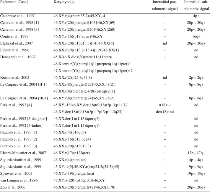
(13) 13. Table 2. Karyotype and FISH results for previously reported cases of ring chromosomes with interstitial (sub)telomeres. Reference [Case]. Karyotype(s). Interstitial pan-. Interstitial sub-. telomeric signal. telomeric signal. Calabrese et al., 1997. 46,XY,r(4)(pterq35.2)/45,XY,-4. +. 4p+. Canevini et al., 1998 [1]. 46,XY,r(20)(pterqter)[105]/46,XY[69]. +. 20p+, 20q+. Canevini et al., 1998 [3]. 46,XY,r(20)(pterqter)[20]/46,XY[260]. +. 20p+, 20q+. Conte et al., 1997. 46,XY,r(16)(p13.3qter)/46,XY. +. 16q+. Elghezal et al., 2007. 46,XX,r(20)(p13q13.3)[14]/46,XX[6]. nd. 20p+, 20q+. Flejter et al., 1996. 46,XX,r(19)(p13.2q13.4)[119]/46,XX[1]. +. nd. Henegariu et al., 1997. 45,X/46,X,dic r(Y)(pterq11q11pter)/. +. nd. 46,X,tetra r(Y)(pterq11q11pterpterq11q11pter)/ 47,X,tetra r(Y)(pterq11q11pterpterq11q11pter)x2 Kosho et al., 2005. 46,XX,r(2)(p25.3q37.3). nd. 2p+, 2q+. Le Caignec et al., 2004 [II-1]. 46,XX,r(8)(pterqter)[22]/45,XX,–8[2]/. +. 8p+, 8q+. +. 8p+, 8q+. r(18): +. nd. 47,XX,r(8)(pterqter),+r(8)(pterqter)[1] Le Caignec et al., 2004 [III-1]. 46,XY,r(8)(pterqter)[24]/45,XY,–8[2]. Park et al., 1992 [4]. 45,XY,-18/46,XY,der(18)t(9;18)(?p13;p11.3)/ 46,XY,der(18)r(9;18)(?p11?p13;p11.3q23). Park et al., 1992 [5-daughter]. der(18): nd. 46,XX,der(1)t(1;15)(qter;q?). +. nd. Park et al., 1992 [5-father]. 46,XY,der(1)t(1;15)(qter;q?). +. nd. Pezzolo et al., 1993 [1]. 46,XX,r(4)(p16q35). +. nd. Pezzolo et al., 1993 [2]. 46,XX,r(16)(p13.3q24). +. nd. Pezzolo et al., 1993 [3]. 46,XX,r(20)(p13q13.3). +. nd. Ricard-Mousnier et al., 2007. 46,XY,r(17)(p1?3qter). +. 17p-, 17q+. Sigurdardottir et al., 1999. 46,XX,r(4)(pterqter). +. 4p+, 4q+. Sigurdardottir et al., 1999. 45,XY,-9[5]/46,XY,r(9)(p24.3q34.3)[45]. -. 9p+, 9q+. Speevak et al., 2003. 46,XY,r(19)(pterqter)mat. +. 19p+, 19q+. van Langen et al., 1996. 47,XY,+r(20)(p13q13.3)/46,XY. +. nd. Zou et al., 2006. 46,XX,r(20)(pterqter)[42]/46,XX[178]. +. 20p+, 20q+. nd : not done Karyotypes were revised to follow the guidelines of ISCN (2005).
(14) 14. Table 3. Karyotype and FISH results for 40 cases of deletions, translocations and ring chromosomes. Case Karyotype(s) Deletions. Pan-telomeric probe. Subtelomeric probe(s). Deleted end. Deleted end. 1. 46,XX,del(1)(q42.2). +. 1q-. 2. 46,XY,del(4)(p16.1). +. 4p-. 3. 46,XY,del(4)(p16.3). +. 4p-. 4. 46,XX,del(4)(p16.3p16.3). +. 4p+. 5. 46,XX,del(4)(q34). +. 4q-. 6. 46,XY,del(5)(p13.2). +. 5p-. 7. 46,XX,del(5)(p15.2). +. 5p-. 8. 46,XY,del(6)(p23p25). +. 6p+. 9. 46,XX,der(8)del(8)(p23.1)inv dup(8)(p21.2p23.1). +. 8p-. 10. 46,XY,del(11)(q23.3). +. 11q-. 11. 46,XX,der(16)del(16)(p13.3)inv(16)(p13.2p13.3). +. 16p-. 12. a. 46,XX,del(18)(p11.2)mat. +. 18p-. 13. a. 46,XX,del(18)(p11.2)mat. +. 18p-. 14. 46,XX,del(18)(q21.31). +. 18q-. 15. 46,XY,del(18)(q21.32)[8]/46,XX[31]. +. 18q-. 16. 46,XY,del(18)(p11.21). +. 18p-. 17. 46,XY,del(21)(q22). +. 21q-. 18. 46,X,del(X)(p11.22). +. Xp-. 19. 46,X,del(X)(q26.2). +. Xq-. Interstitial signal. Interstitial signal. Translocations 20. 46,XY,der(9)t(7;9)(q32.3;qter). +. der(9): 9q+. 21. 45,XY,der(20)t(15;20)(q14;pter). +. nd. 22. 46,XY,der(21)t(14;21)(q24;qter)[25]/46,XY[5]. +. der(21): 21q+. 23b. 45,X,tas(Y;13)(p11.31;p11.2)[62]/46,XY[7]. +. nd. 24. 46,XX,t(1;7)(q44;q21.1). -. nd. 25. 46,XX,t(1;7)(q32.1;q36.1). -. nd. 26. 46,XX,t(2;2)(q37.3;p23). -. nd. 27. 46,XX,t(2;5)(q21.1;q35.3). -. der(5): 5q-. 28. 46,XX,t(4;15)(p16.3;q12)pat. -. nd. 29. 46,XY,der(5)t(5;9)(p15.31;q21.2)mat. -. der(5): 5p-. 30. 46,XX,t(7;11)(q11.21;q25). -. der(11): 11p-. 31. 46,XX,der(13)t(2;13)(q33;q34). -. nd. 32. 46,X,t(X;1)(q22;p36.3). -. der(1): 1p+. Interstitial signal. Interstitial signal. Ring chromosomes.
(15) 15. 33. 46,XX,r(4)(p16q35). +. nd. 34. 46,XX,r(20)(pterqter)[45]/46,XY[90]. +. 20p+, 20q+. 35. 46,XY,r(5)(p15.31q35.3)[41]/47,XY,r(5)(p15.31q35.3)x2[1]/. -. 5p-, 5q+. 45,XY,-5[2]/46,XY[1] 36. 46,XY,r(18)(p11.3q23). -. nd. 37. 46,XY,r(13)(p13q34)[100]/46,XY,r(13)(p13q13)[21]/. -. 13q-. 46,XY,dicr(13)(p13q34q34p13)[3]/45,XY,-13[8] 38. 46,XX,dicr(21)(p13q22.3q22.3p13)[32]/45,XX,-21[2]. -. 21q-. 39. 46,XX,r(22)(p13q13.3)[85]/47,XX,r(22)(p13q13.3)x2[2]/. -. nd. -. Yp-, Yq+. 45,XX,-22[3]/46,XX[17] 40. c. 46,X,dic r(Y)(p11.3q12q12p11.3)[92]/ 46,X,tetra r(Y)(p11.3q12q12p11.3p11.3q12q12p11.3)[8]/ 46,X,tetra r(Y)x2[1]/46,X,del dic r(Y)(p11q12)[4]/45,X[158]. nd: could not be performed due to lack of archival material a. previously published by Maranda et al. (2006). b. previously published by Boutouil et al. (1996). c. previously published by DesGroseilliers et al. (2006).
(16) 16. Legend to Figure 1. Fig. 1: Partial metaphases showing fluorescence in situ hybridization (FISH) with either pantelomeric or specific subtelomeric probes. FISH chromosomes were counterstained with either propidium iodide (red) or DAPI (blue). Arrowheads indicate the presence or absence of an interstitial signal with the pan-telomeric on the abnormal chromosomes; arrows show the presence or absence of the subtelomeric signal. (A) Case 7: absence of the 5p subtelomere on the deleted chromosome 5. (B) Case 12: absence of the 18p subtelomere on the deleted chromosome 18. (C) Case 4: presence of the 4p subtelomere on the deleted chromosome 4. (D) Case 8: presence of the 6p subtelomere on the deleted chromosome 6. Case 20: (E) presence of interstitial pan-telomeres at the junction site of the der(9)t(7;9); (F) presence of the 7q subtelomere in terminal position on the der(9); and (G) presence of the subtelomere 9q at the junction site on the der(9). (H) Case 21: presence of interstitial pan-telomeres at the junction site of the der(20)t(15;20). Case 22: (I) presence of interstitial pan-telomeres and of the 21q subtelomere at the junction site of the der(21)t(14;21); (J) and presence of the subtelomere 14q in terminal position on the der(21). (K) Case 23: presence of interstitial pan-telomeres at the junction site of the tas(Y;13). Case 32: (L) absence of interstitial pan-telomeres at the junction site of both the der(1) and der(X) of a t(X;1); (M) presence of the subtelomere 1p at the junction site of the der(1) and in terminal position on the der(X); (N) and presence of the subtelomere Xq in terminal position on the der(1) only. (O) Case 33: presence of interstitial pan-telomeres on the r(4). Case 34: (P) presence of interstitial pan-telomeres on the r(20); (Q) and presence of the subtelomeres 20p and 20q on the r(20)..
(17) 17.
(18) 18. References. Ballif BC, Yu W, Shaw CA, Kashork CD, Shaffer LG: Monosomy 1p36 breakpoint junctions suggest pre-meiotic breakage-fusion-bridge cycles are involved in generating terminal deletions. Hum Mol Genet 12:2153-2165 (2003). Bolzan AD, Bianchi MS: Telomeres, interstitial telomeric repeat sequences, and chromosomal aberrations. Mutat Res 612:189-214 (2006). Bosco G, Haber JE: Chromosome break-induced DNA replication leads to nonreciprocal translocations and telomere capture. Genetics 150:1037-1047 (1998). Boutouil M, Fetni R, Qu J, Dallaire L, Richer C-L, Lemieux N: Fragile site and interstitial telomere repeat sequences at the fusion point of a de novo (Y;13) translocation. Hum Genet 98:323-327 (1996). Calabrese G, Giannotti A, Mingarelli R, Di Gilio MC, Piemontese MR, Palka G: Two newborns with chromosome 4 imbalances: Deletion 4q33->q35 and ring r(4)(pterq35.2-qter). Clin Genet 51:264-267 (1997). Canevini MP, Sgro V, Zuffardi O, Canger R, Carrozzo R, Rossi E, Ledbetter D, Minicucci F, Vignoli A, Piazzini A, Guidolin L, Saltarelli A, dalla Bernardina B: Chromosome 20 ring: A chromosomal disorder associated with a particular electroclinical pattern. Epilepsia 39:942-951 (1998). Conte RA, Kleyman SM, Kharode C, Verma RS: Delineation of a ring chromosome 16 by the FISH-technique: A case report with review. Clin Genet 51:196-199 (1997). Daniel A, St Heaps L: Chromosome loops arising from intrachromosomal tethering of telomeres occur at high frequency in G1 (non-cycling) mitotic cells: Implications for telomere capture. Cell Chromosome 3:3 (2004) at http://www.cellandchromosome.com/content/3/1/3. Davies AF, Kirby TL, Docherty Z, Ogilvie CM: Characterization of terminal chromosome anomalies using multisubtelomere FISH. Am J Med Genet A 120A:483-489 (2003). de Lange T: Telomere dynamics and genome instability in human cancer: Telomeres, pp 265-293 (Cold Spring Harbor Laboratory Press, 1995)..
(19) 19. DesGroseilliers M, Fortin F, Lafreniere AM, Brochu P, Lemyre E, Lemieux N: Dynamic increase of a 45,X cell line in a patient with multicentric ring Y chromosomes. Cytogenet Genome Res 115:90-93 (2006). Devriendt K, Petit P, Matthijs G, Vermeesch JR, Holvoet M, De Muelenaere A, Marynen P, Cassiman JJ, Fryns JP: Trisomy 15 rescue with jumping translocation of distal 15q in Prader-Willi syndrome. J Med Genet 34:395-399 (1997). Elghezal H, Hannachi H, Mougou S, Kammoun H, Triki C, Saad A: Ring chromosome 20 syndrome without deletions of the subtelomeric and CHRNA4-KCNQ2 genes loci. Eur J Med Genet 50:441-445 (2007). Flejter WL, Finlinson D, Root S, Nguyen W, Brothman AR, Viskochil D: Familial ring (19) chromosome mosaicism: Case report and review. Am J Med Genet 66:276-280 (1996). Flint J, Craddock CF, Villegas A, Bentley DP, Williams HJ, Galanello R, Cao A, Wood WG, Ayyub H, Higgs DR: Healing of broken human chromosomes by the addition of telomeric repeats. Am J Hum Genet 55:505-512 (1994). Griffith JD, Comeau L, Rosenfield S, Stansel RM, Bianchi A, Moss H, de Lange T: Mammalian telomeres end in a large duplex loop. Cell 97:503-514 (1999). Gross SJ, Tharapel AT, Phillips OP, Shulman LP, Pivnick EK, Park VM: A jumping robertsonian translocation: A molecular and cytogenetic study. Hum Genet 98:291-296 (1996). Hackett JA, Feldser DM, Greider CW: Telomere dysfunction increases mutation rate and genomic instability. Cell 106:275-286 (2001). Henegariu O, Pescovitz OH, Vance GH, Verbrugge J, Heerema NA: A case with mosaic di-, tetra-, and octacentric ring Y chromosomes. Am J Med Genet 71:426-429 (1997). Huang B, Martin CL, Sandlin CJ, Wang S, Ledbetter DH: Mitotic and meiotic instability of a telomere association involving the Y chromosome. Am J Med Genet A 129A:120-123 (2004). Ijdo JW, Baldini A, Ward DC, Reeders ST, Wells RA: Origin of human chromosome 2: An ancestral telomere-telomere fusion. Proc Natl Acad Sci U S A 88:9051-9055 (1991). ISCN (2005): An international system for human cytogenetic nomenclature. (S. Karger, Basel, 2005)..
(20) 20. Jewett T, Marnane D, Stewart W, Hayworth-Hodge R, Finklea L, Klinepeter K, Rao PN, Pettenati MJ: Jumping translocation with partial duplications and triplications of chromosomes 7 and 15. Clin Genet 53:415-420 (1998). Josifova DJ, Mazzaschi R, Ballard T, Ogilvie CM, Splitt M: A constitutional telomeric translocation showing meiotic instability. Am J Med Genet A 140:1228-1233 (2006). Kosho T, Matsushima K, Sahashi T, Mitsui N, Fukushima Y, Sobajima H, Ohashi H: "Ring syndrome" involving chromosome 2 confirmed by FISH analysis using chromosomespecific subtelomeric probes. Genet Couns 16:65-70 (2005). Le Caignec C, Boceno M, Jacquemont S, Nguyen The Tich S, Rival JM, David A: Inherited ring chromosome 8 without loss of subtelomeric sequences. Ann Genet 47:289-296 (2004). Lefort G, Blanchet P, Chaze A, Sarda P, Demaille J, Pellestor F: PRINS labeling reveals interstitial telomeric sequences in a constitutional jumping translocation: 47th Annual Meeting of the American Society of Human Genetics, p A131 (American Journal of Human Genetics, Baltimore, MD 1997). Lemieux N, Drouin R, Richer CL: High-resolution dynamic and morphological G-bandings (GBG and GTG): A comparative study. Hum Genet 85:261-266 (1990). Lemieux N, Dutrillaux B, Viegas-Pequignot E: A simple method for simultaneous R- or Gbanding and fluorescence in situ hybridization of small single-copy genes. Cytogenet Cell Genet 59:311-312 (1992). Luciani JJ, de Mas P, Depetris D, Mignon-Ravix C, Bottani A, Prieur M, Jonveaux P, Philippe A, Bourrouillou G, de Martinville B, Delobel B, Vallee L, Croquette MF, Mattei MG: Telomeric 22q13 deletions resulting from rings, simple deletions, and translocations: Cytogenetic, molecular, and clinical analyses of 32 new observations. J Med Genet 40:690696 (2003). Maranda B, Lemieux N, Lemyre E: Familial deletion 18p syndrome: Case report. BMC Med Genet 7:60 (2006) at http://www.biomedcentral.com/1471-2350/7/60. Marinescu RC, Johnson EI, Grady D, Chen X-N, Overhauser J: FISH analysis of terminal deletions in patients diagnosed with Cri-du-Chat syndrome. Clin Genet 56:282-288 (1999). Melek M, Shippen DE: Chromosome healing: Spontaneous and programmed de novo telomere formation by telomerase. Bioessays 18:301-308 (1996)..
(21) 21. Meltzer PS, Guan X-Y, Trent JM: Telomere capture stabilizes chromosome breakage. Nat Genet 4:252-255 (1993). Meyne J, Ratliff RL, Moyzis RK: Conservation of the human telomere sequence (TTAGGG)n among vertebrates. Proc Natl Acad Sci U S A 86:7049-7053 (1989). Mignon-Ravix C, Depetris D, Luciani JJ, Cuoco C, Krajewska-Walasek M, Missirian C, Collignon P, Delobel B, Croquette MF, Moncla A, Kroisel PM, Mattei MG: Recurrent rearrangements in the proximal 15q11-q14 region: A new breakpoint cluster specific to unbalanced translocations. Eur J Hum Genet 15:432-440 (2007). Morin GB: The human telomere terminal transferase enzyme is a ribonucleoprotein that synthesizes TTAGGG repeats. Cell 59:521-529 (1989). Moyzis RK, Buckingham JM, Cram LS, Dani M, Deaven LL, jones MD, Meyne J, Ratliff RL, Wu J-R: A highly conserved repetitive DNA sequence, (TTAGGG)n, present at the telomeres of human chromosomes. Proc Natl Acad Sci U S A 85:6622-6626 (1988). Neumann AA, Reddel RR: Telomere maintenance and cancer -- look, no telomerase. Nat Rev Cancer 2:879-884 (2002). Ning Y, Liang JC, Nagarajan L, Schrock E, Ried T: Characterization of 5q deletions by subtelomeric probes and spectral karyotyping. Cancer Genet Cytogenet 103:170-172 (1998). Park VM, Gustashaw KM, Wathen TM: The presence of interstitial telomeric sequences in constitutional chromosome abnormalities. Am J Hum Genet 50:914-923 (1992). Petit P, Devriendt K, Vermeesch JR, De Cock P, Fryns JP: Unusual de novo t(13;15)(q12.1;p13) translocation leading to complex mosaicism including jumping translocation. Ann Genet 41:22-26 (1998). Pezzolo A, Gimelli G, Cohen A, Lavaggetto A, Romano C, Fogu G, Zuffardi O: Presence of telomeric and subtelomeric sequences at the fusion points of ring chromosomes indicates that the ring syndrome is caused by ring instability. Hum Genet 92:23-27 (1993). Qu J, Dallaire L, Fetni R, Lemieux N, Richer CL: Repetitive telomeric sequences in chromosomal translocations involving chromosome 21: 44th Annual Meeting of the American Society of Human Genetics pA115 (American Journal of Human Genetics, New Orleans, LA 1994)..
(22) 22. Ravnan JB, Tepperberg JH, Papenhausen P, Lamb AN, Hedrick J, Eash D, Ledbetter DH, Martin CL: Subtelomere FISH analysis of 11 688 cases: An evaluation of the frequency and pattern of subtelomere rearrangements in individuals with developmental disabilities. J Med Genet 43:478-489 (2006). Reddy KS, Murphy T: Fusion of 9 beta-satellite and telomere (TTAGGG)(n) sequences results in a jumping translocation. Hum Genet 107:268-275 (2000). Reeve A, Norman A, Sinclair P, Whittington-Smith R, Hamey Y, Donnai D, Read A: True telomeric translocation in a baby with the Prader-Willi phenotype. Am J Med Genet 47:1-6 (1993). Ricard-Mousnier B, N'Guyen S, Dubas F, Pouplard F, Guichet A: Ring chromosome 17 epilepsy may resemble that of ring chromosome 20 syndrome. Epileptic Disord 9:327-331 (2007). Rivera H, Sitch FL, Crolla JA: Telomeric translocations are uncommon. Genet Couns 6:343-347 (1995). Rivera H, Vasquez AI, Perea FJ: Centromere-telomere (12;8p) fusion, telomeric 12q translocation, and i(12p) trisomy. Clin Genet 55:122-126 (1999). Rossi E, Floridia G, Casali M, Danesino C, Chiumello G, Bernardi F, Magnani I, Papi L, Mura M, Zuffardi O: Types, stability, and phenotypic consequences of chromosome rearrangements leading to interstitial telomeric sequences. J Med Genet 30:926-931 (1993). Sigurdardottir S, Goodman BK, Rutberg J, Thomas GH, Jabs EW, Geraghty MT: Clinical, cytogenetic, and fluorescence in situ hybridization findings in two cases of "Complete ring" syndrome. Am J Med Genet 87:384-390 (1999). Speevak MD, Smart C, Unwin L, Bell M, Farrell SA: Molecular characterization of an inherited ring (19) demonstrating ring opening. Am J Med Genet A 121A:141-145 (2003). van Langen IM, Otter MA, Aronson DC, Overweg-Plandsoen WC, Hennekam RC, Leschot NJ, Hoovers JM: Supernumerary ring chromosome 20 characterized by fluorescence in situ hybridization. Clin Genet 49:49-53 (1996). Varley H, Di S, Scherer SW, Royle NJ: Characterization of terminal deletions at 7q32 and 22q13.3 healed by de novo telomere addition. Am J Hum Genet 67:610-622 (2000). Varley H, Pickett HA, Foxon JL, Reddel RR, Royle NJ: Molecular characterization of intertelomere and intra-telomere mutations in human ALT cells. Nat Genet 30:301-305 (2002)..
(23) 23. Vermeesch JR, Petit P, Speleman F, Devriendt K, Fryns JP, Marynen P: Interstitial telomeric sequences at the junction site of a jumping translocation. Hum Genet 99:735-737 (1997). Williams ES, Klingler R, Ponnaiya B, Hardt T, Schrock E, Lees-Miller SP, Meek K, Ullrich RL, Bailey SM: Telomere dysfunction and DNA-PKcs deficiency: Characterization and consequence. Cancer Res 69:2100-2107 (2009). Wilkie AO, Lamb J, Harris PC, Finney RD, Higgs DR: A truncated human chromosome 16 associated with alpha thalassaemia is stabilized by addition of telomeric repeat (TTAGGG)n. Nature 346:868-871 (1990). Zahed L, Oreibi G, Azar C, Salti I: Ring chromosome 18q and jumping translocation 18p in an adult male with hypergonadotrophic hypogonadism. Am J Med Genet A 129A:25-28 (2004). Zou YS, Van Dyke DL, Thorland EC, Chhabra HS, Michels VV, Keefe JG, Lega MA, Feely MA, Uphoff TS, Jalal SM: Mosaic ring 20 with no detectable deletion by FISH analysis: Characteristic seizure disorder and literature review. Am J Med Genet A 140:1696-1706 (2006)..
(24)
Figure

Documents relatifs
Poznyak, “Unified Lyapunov function for a finite- time stability analysis of relay second-order sliding mode control systems,” IMA Journal of Mathematical Control and Information,
/ La version de cette publication peut être l’une des suivantes : la version prépublication de l’auteur, la version acceptée du manuscrit ou la version de l’éditeur. Access
Then, we wondered whether overexpression of Pot1 rescues the telomere length defect of D223Y mutant since both proteins compete for ssDNA binding at telomeres and overexpression of
Nous avons ´ egalement montr´ e que chez les souris Cx3cr1 −/− les M φ/CMs s’accumulent spontan´ ement dans l’espace sous-r´ etinien en l’ab- sence de d´ eg´ en´
In summary, in our study, PUC occurred in 32% of dogs with CKD and was subclinical in 92% of these cases. This high frequency of PUC also was found in dogs with IRIS stage 1
Et occupe la place permettant de renaitre Il prône l’abandon de toutes les affaires Il faut laisser sa mère un faut laisser son père Pour consacrer à Dieu notre temps sur la terre
The inversion has no significant effect on reproductive performance of sows sired by heterozygous boars. Owing to the very low number of homozygous carriers
The frequencies of various types of heteroploid embryos, embryos in which one or more cells contained a structural aberration and embryos in which all the chromo-. somes
