Université de Montréal
Acute inactivation of the contralesional hemisphere for longer
durations improves recovery after cortical injury
Par
Babak Khoshkrood Mansoori
Département de physiologie, Université de MontréalFaculté de Médecine
Mémoire présenté à la Faculté de Médecine en vue de l’obtention du grade de Maîtrise
en sciences neurologiques
September 2013
ii
Advisor: Numa Dancause PT, PhD
Thesis Committee: Allan M. Smith Dorothy Barthélémy
iii
Résumé
Au cours des dernières années, des méthodes non-invasives de stimulations permettant de moduler l’excitabilité des neurones suivant des lésions du système nerveux central ont été développées. Ces méthodes sont maintenant couramment utilisées pour étudier l’effet de l’inhibition du cortex contralésionnel sur la récupération motrice à la suite d’un accident vasculocérébral (AVC). Bien que plusieurs de ces études rapportent des résultats prometteurs, les paramètres permettant une récupération optimale demeurent encore inconnus.
Chez les patients victimes d'un AVC, il est difficile de débuter les traitements rapidement et d'initier l’inhibition dans les heures suivant la lésion. L'impact de ce délai est toujours inconnu. De plus, aucune étude n’a jusqu’à maintenant évalué l’effet de la durée de l’inhibition sur la récupération du membre parétique. Dans le laboratoire du Dr Numa Dancause, nous avons utilisé un modèle bien établi de lésion ischémique chez le rat pour explorer ces questions. Nos objectifs étaient d’évaluer 1) si une inactivation de l’hémisphère contralésionnel initiée dans les heures qui suivent la lésion peut favoriser la récupération et 2) l’effet de la durée de
l’inactivation sur la récupération du membre parétique.
Suite à une lésion dans le cortex moteur induite par injections d’un vasoconstricteur, nous avons inactivé l’hémisphère contralésionnel à l’aide d’une pompe osmotique assurant l’infusion continue d’un agoniste du GABA (Muscimol). Dans différents groupes expérimentaux, nous avons inactivé l’hémisphère contralésionnel pour une durée de 3, 7 et 14 jours suivant la lésion. Dans un autre groupe, le Muscimol a été infusé pour 14 jours mais à un débit moindre de façon à pouvoir étudier le lien entre la fonction du membre non-parétique et la récupération du membre parétique. Les données comportementales de ces groupes ont été comparées à celles d’animaux ayant récupéré de façon spontanée d'une lésion similaire.
iv
Nos résultats indiquent que l’augmentation de la durée de l’inactivation (de 3 à 14 jours) accélère la récupération du membre parétique. De plus, les deux groupes ayant reçu une
inactivation d'une durée de 14 jours ont montré une plus grande récupération fonctionnelle que le groupe n’ayant pas reçu d’inactivation de l’hémisphère contralésionnel, le groupe contrôle. Nos résultats suggèrent donc que l’inactivation de l’hémisphère contralésionnel initiée dans les heures suivant la lésion favorise la récupération du membre parétique.
La durée d’inhibition la plus efficace (14 jours) dans notre modèle animal est beaucoup plus longues que celles utilisées jusqu’à maintenant chez l’homme. Bien qu’il soit difficile d’extrapoler la durée idéale à utiliser chez les patients à partir de nos données, nos résultats suggèrent que des traitements de plus longue durée pourraient être bénéfiques.
Finalement, un message clair ressort de nos études sur la récupération fonctionnelle après un AVC: dans le développement de traitements basés sur l’inhibition de l’hémisphère
contralésionnel, la durée de l’inactivation est un facteur clef à considérer.
Mots-clés: accident vasculocérébral, AVC, avant-bras, contralésionnel; cortex; inhibition; inactivation; lésion; main; rat; récupération motrice
v
Abstract
With the introduction of non-invasive brain stimulation methods aimed at modulating the
excitability of cortical areas after stroke, many groups are intensively investigating the effects of inhibition of the contralesional hemisphere on functional recovery. Although the reported results of these studies are very promising, limitations of enrolling acute stroke patients as well as technical difficult of establishing continuous inhibition protocols have left several open ended questions regarding the treatment parameters and patient selection. For example, the efficacy of inhibition treatment in acute setting after stroke and the effect of treatment duration are two questions that are virtually unexplored.
Therefore, in the laboratory of Prof. Numa Dancause, we took advantage of a well established rodent
model of cortical ischemic lesion to gain direct and objective insight about the importance of contralesional inactivation on motor recovery of the paretic limb. Using an Endothelin-1 rodent model of ischemic cortical lesion, we pharmacologically inactivated the contralesional
hemisphere with a GABA agonist (Muscimol). By doing so we were interested in the effect of early treatment when contralesional inactivation is initiated rapidly after the lesion.
Early after induction of cortical ischemic lesion, the contralesional hemisphere was inactivated with continuous infusion of the Muscimol for 3, 7 or 14 days in three different groups of
animals. In a fourth group, Muscimol was infused at slower rate for 14 days to provide additional insights on the relation between the effects of inactivation on the non-paretic forelimb behavior and the recovery of the paretic forelimb. We included a group of animals with spontaneous recovery that received no inactivation after lesion.
vi
Our results indicated that increasing inactivation duration (from 3 to 14 days) accelerated the recovery of grasping function. Both groups with 14 days of inactivation had similar recovery profiles and performed better than animals that spontaneously recovered. In fact, the duration of inactivation, not the intensity, correlated with the better functional outcomes.
Our results support early contralesional inactivation to improve recovery of the paretic forelimb after cortical lesion. Moreover, based on our results, the duration of inactivation is the most important factor to correlate with the functional outcomes. Therefore, by providing precise temporal and behavioral evidence, our results provide a window of opportunity for the
researchers in which the current gap in our understanding of the clinical efficacy of
contralesional inhibition in acute phase after stroke can be approached with more confidence.
Keywords: contralesional; cortex; forelimb; hand; inhibition; inactivation; lesion; stroke; rat; recovery
vii
Table of Contents
Résumé ... iii Abstract ... v LIST OF FIGURES ... ix LIST OF TABLES ... ix LIST OF ABBREVIATIONS ... x ACKNOWLEDGEMENTS ... xii CHAPTER I. ...1 General introduction ...1Interhemispheric inhibition in health and after stroke ... 2
Possible mechanism of hyperexcitability of contralesional hemisphere after stroke ... 3
Contralesional inhibition after stroke ... 6
Limitations of human studies: major questions to be answered ... 9
The effect of lesion size ... 9
The choice of inhibition: what cortical circuits are engaged? ... 10
The optimal time to deliver the intervention ... 11
CHAPTER II. Manuscript submission ... 13
Cover Page: ... 14 Summary: ... 15 Introduction: ... 17 Methods: ... 18 Results: ... 28 Discussion: ... 33 References: ... 41
Chapter III. General Discussion ... 45
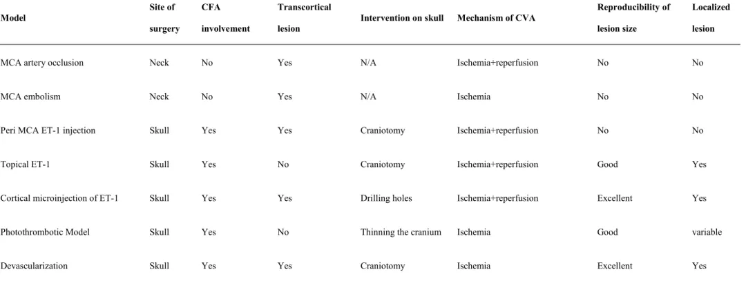
Modeling human stroke in rats ... 45
The current rat models of stroke ... 46
Middle Cerebral Artery occlusion (MCAo) models ... 46
Photothrombosis ... 47
viii
Reversible contralesional inactivation with GABA ... 51
The effects of contralesional inactivation on the grasping function of the paretic forelimb ... 55
Conclusions ... 58
Future direction ... 59
References: ... 61
Figures ... 71
ix LIST OF FIGURES
Figure 1. Experimental design. ... 71
Figure 2. Spread of muscimol infusion in different experimental groups. ... 73
Figure 3. Responses to intracortical microstimulation following sustained muscimol infusion. ... 74
Figure 4. Histological evaluation of the tissue surrounding the cannula in the contralesional hemisphere. ... 75
Figure 5. Histological evaluation of the endothelin-1 lesion size. ... 76
Figure 6. Side effects of contralesional inactivation. ... 77
Figure 7. Non-paretic forelimb grasping performance on the Montoya test. ... 78
Figure 8. The effect of inactivation on spontaneous use of forelimbs. ... 79
Figure 9. Effects of inactivation on the recovery of the paretic forelimb. ... 80
Figure 10. Correlation analyses of factors potentially involved in the recovery of the paretic forelimb. ... 81
LIST OF TABLES Table 1 - Animal groups and inactivation protocols ... 83
x LIST OF ABBREVIATIONS
Group 14D
Group with 14 days of inhibition, 23 Group 14Dslow
Group with 14 days of inhibition at slow rate, 24
Group 3D
Group with 3 days of inhibition, 22 Group 7D
Group with 7 days of inhibition, 22 BOLD signal
Blood oxygenation dependent signal, 8 CFA
Caudal forelimb area, 18 CL
Contralesional, 4 CS
Conditioning stimulus, 4 cTBS
Continous theta burst stimulation, 10 DMSO
Dimethyl sulfoxide, 19 ECA
External carotid artery, 35 ET1
Endotheline 1, 35 FDI
First dorsal interosseous, 4 fMRI
Functional magnetic resonance imaging, 6 GABA
Gamma aminobutyric acid, 4 ICA
Internal carotid artery, 35 ICF Intracortical facilitation, 3 ICMS Intracortical microstimulation, 19 ISI Interstimulus interval, 4 M1
Primary motor cortex, 6 MCA
Middle cerebral artery, 35 MCAo
Middle cerebral artery occlusion, 35 MT
Motor thresold, 4 RFA
Rostral forelimb area, 19 rTMS
Repetitive transcranial magnetic stimulation, 6
SICI
Short-interval intracortical inhibition, 3 tDCS
Transcranial direct current stimulation, 7 TMS
Transcranial magnetic stimulation, 3 TS
xi
xii
ACKNOWLEDGEMENTS
Throughout my graduate studies I have been blessed to be around remarkable individuals who helped me in various aspects of my development. Clearly my acknowledgement must start with my advisor and mentor, Prof. Numa Dancause who was brave enough to take a chance on me two and a half years ago. I will forever be influenced by his excellent guidance. Numa has shown me how to be a compassionate and successful mentor while establishing and maintaining a top-level research laboratory. He is, and will continue to be, my role model as I progress in my clinical-academic career.
I would like to offer my most sincere thanks to Dr. Stephan Quessy who, with his work ethic and passion, kept to his promise that graduate studies was about science, hard work, fun and excitement. Additionally, I owe Numa and Stephan and extra thank you for reminding me that there is world of wonder going on outside the department.
My Friends and colleagues in GRSNC have been the greatest source of support. Thanks to Joan Deffeys, Marina Martinez and Pascal Fortier for their analytical prowess and statistical solutions for the analysis of the data. I owe a special thanks to Boris Tovykine with whom it was a pleasure to start and finish a dreadful ICMS session shoulder to shoulder. I appreciate the excellence of Loyda Jean-Charles’s contribution in my project, and I would like to offer a thank you to Lawrence Morse, Minh-thi Nguyen and Amin Nasri. I enjoyed every second of my stay in the collaborative atmosphere of Numa’s lab, thanks to all the members of this lab including the ones mentioned above as well as Melvin Dea, Eleonore Serrano and Adjia Hamadjida.
I’d like to thank Rejean Dubuc’s lab for generously sharing their equipment and technical knowledge with me throughout my Master’s studies.
xiii
Thanks are certainly due to all the great people in the GRSNC and Faculty of Medicine specifically Drs. Allan Smith, Serge Rossignol, Dorothy Barthélémy, Jannic Boehm and Richard Robitaille who never hesitated to treat me as a student of their own to provide guidance and help for my present and future endeavors.
CHAPTER I.
General introduction
Stroke is the leading cause of disability worldwide (Go et al., 2013). Morbidity and mortality with acute stroke is substantial (Hartmann et al., 2001) however in long term, some of the initial deficits of the survived patients are subject to various degrees of recovery (Langhorne et al., 2009). Depending on the underlying cause of the stroke, up to sixty percent of stroke patients will still suffer from significant functional deficits a year after stroke incidence (Petty et al., 2000). With 700,000 cases per year in north America, this signifies that one person dies from stroke every three minutes and two are left with various degrees of permanent disability (Feigin et al., 2009).
The recent improvements in the management of acute stroke are reflected in the substantial increase of stroke survivors, many of whom show persistent neurological deficits in terms of paresis, aphasia, apraxia or neglect (Langhorne et al., 2009). Therefore, investigating the
reparative events in the brain after stroke, neuroprotective therapies and developing strategies to augment the efficacy of rehabilitative techniques is the priority of stroke research.
In the last few years, several approaches to increase adaptive plasticity and recovery after stroke have been proposed and are currently being tested. In particular, many groups are
intensively investigating the effects of inhibition of the contralesional hemisphere on functional recovery (Hummel and Cohen, 2006). The rationale behind the treatment strategy used in most studies is the concept of post-stroke interhemispheric imbalance (Liepert et al., 2000; Nowak et al., 2009). According to this hypothesis, increased activity in the contralesional hemisphere after stroke exerts an augmented inhibitory influence on the ipsilesional hemisphere. By doing so, it
General introduction
2 interferes with function and adaptive plasticity in the ipsilesional hemisphere and hence the recovery of the paretic arm. Therefore, one proposed method of reducing interhemispheric imbalance is to inhibit the contralesional hemisphere.
Interhemispheric inhibition in health and after stroke
In normal conditions, both hemispheres are engaged in a mutual transcallosal
interhemispheric inhibition which contributes to the functional coupling of the two hemispheres at the onset and during performance of voluntary movements (Jeeves et al., 1988; Ferbert et al., 1992; Gerloff et al., 1998; Di Lazzaro et al., 1999; Grefkes et al., 2008). It has been shown that when one hemisphere is inhibited in healthy subjects with low frequency transcranial stimulation methods, the excitability of contralateral hemisphere is augmented (Plewnia et al., 2003;
Schambra et al., 2003; Kim et al., 2004), the performance of the ipsilateral hand is increased (Kobayashi et al., 2004; Dafotakis et al., 2008) and the interhemispheric inhibition from contralateral hemisphere to the inhibited hemisphere is enhanced significantly (Lang et al., 2004).
The transcallosal imbalance after stroke follows a similar principle. A substantial body of animal and human studies has documented increased excitability of contralesional sensorimotor brain areas after stroke (Liepert et al., 2000; Marshall et al., 2000; Abo et al., 2001; Manganotti et al., 2002; Wittenberg et al., 2007) because of the decreased inhibitory transcallosal inputs from ipsilesional to the contralesional (intact) hemisphere. The disinhibited contralesional cortex (Traversa et al., 1998; Liepert et al., 2000; Cicinelli et al., 2003; Rossini et al., 2003) exerts further inhibitory transcallosal inputs into the ipsilesional hemisphere (Murase et al., 2004), which possibly contributes to the loss of function in addition to that of the damage to corticospinal fibers (Nowak et al., 2009). The magnitude of increased excitability in
General introduction
3 contralesional hemisphere (Liepert et al., 2000; Manganotti et al., 2002, 2008; Nardone and Tezzon, 2002a; Shimizu et al., 2002) and the interhemispheric inhibition from contralesional to ipsilesional hemisphere (Murase et al., 2004; Duque et al., 2005) is found to be proportional to the motor impairment in stroke patients.
Possible mechanism of hyperexcitability of contralesional hemisphere after stroke
The increased neuronal activity of the contralesional hemisphere occurs in a very short time (< 1 hour) after cortical damage (Mohajerani et al., 2011). In this short time, the post-stroke rewiring and experience cannot not be cause of the enhanced contralesional activity. This effect is most probably a result of immediate circuit loss or de-afferentaion of glutamatergic
transcallosal fibers from the ipsilesional to the contralesional hemisphere
(Buchkremer-Ratzmann and Witte, 1997). In fact, if the lesion spares the cortical layer V (from which the main callosal output arises) there will be no subsequent contralesional hyperexcitability.
In the contralesional hemisphere of acute and subacute stroke patients with cortical and subcortical lesions, the balance of excitatory and inhibitory activity is shifted towards an increase of excitatory activity (a lower threshold for activation of excitatory interneurons) due to
decreased potency of inhibitory circuits (Bütefisch et al., 2003a). In human stroke subjects, experimental paired pulse stimulation paradigms with non-invasive stimulus patterns to measure short-interval intracortical inhibition (SICI) and intracortical facilitation (ICF) has been widely used to study the changes of the potency of inhibitory and excitatory circuits at the cortical level. SICI is a type of intracortical inhibition that can be studied by paired TMS. The protocol to measure SICI involves a sub-threshold conditioning stimulus followed by a supra-threshold test stimulus applied at the same cortical site with one coil (Kujirai et al., 1993). The test response is
General introduction
4 typically inhibited at inter-stimulus intervals (ISI) of 1-6 ms. There is considerable evidence that SICI occurs within the cortex itself rather than in sub-cortical structures and reflects the potency of GABAergic inhibitory circuits (Kujirai et al., 1993; Chen et al., 1998; Di Lazzaro et al., 1998). ICF is tested using protocols similar to SICI with a sub-threshold conditioning stimulus followed by a supra-threshold test stimulus that is also applied at the same cortical site with one coil (Kujirai et al., 1993). Facilitation occurs at an ISI of 8-30 ms. Similar to SICI, ICF occurs at the cortical level rather than in subcortical structures (Ziemann et al., 1996b; Nakamura et al., 1997) and appears to be mediated by separate neuronal populations from SICI (Chen et al., 1998; Ashby et al., 1999; Strafella and Paus, 2001).
For example, Bütefisch and collaborators documented a marked hyperexcitability of contralesional hemisphere in terms of decreased SICI after paired-pulse stimulation of
contralesional hemisphere (Bütefisch et al., 2003a). In this experiment, they used a paired pulse paradigm with a single TMS coil where conditioning stimulus (CS) of about 80% of motor threshold (MT) was delivered 2 ms before a test stimulus (TS) with intensity of 110% of MT at the cortical area that would elicit a maximal response in first dorsal interosseous muscle (FDI) of the non-paretic hand. The diminished SICI they found was attributed to GABA down regulation (Ziemann et al., 1996a, 1996b; Ashby et al., 1999). Moreover, Bütefisch and collaborators (2003a) demonstrated decreased threshold for excitation of excitatory circuits of the
contralesional hemisphere. For the latter experiment, they used a paired pulse paradigm with a constant ISI of 2 ms, while the CS varied from 20% to 100 % of MT and TS was fixed at 110% of MT. They observed that in CL hemisphere of stroke patients, the excitation threshold was decreased to less than 50-60% of MT compared to normal subject where the threshold is about 70% of MT. So all in all, the new electrophysiological evidence in human stroke survivors has
General introduction
5 demonstrated a hyperexcitability of CL hemisphere, which is linked to the down regulation of GABA.
Contralesional GABA-Receptor down regulation has been demonstrated by more direct molecular studies as well. Qü and collaborators, in a series of experiments using
autoradiography, investigated the density of [3H] muscimol binding sites to GABA-A receptors in ipsi- and contralateral hemisphere in two animal models of stroke 7 days after the lesion in mice (Qü et al., 1998b), and rats (Qü et al., 1998a). In both experiments, they observed a drastic decrease of the [3H] muscimol binding in contralesional hemisphere. In the latter study, in addition to the autoradiographic evidence of [3H] muscimol binding in contralesional
hemisphere, they used electrophysiology on 400 µm rat brain slices 7 days after the lesion. In the cortex of contralesional hemisphere, they recorded field potentials from layer 2 and stimulated the layer 6 in a pattern that activates GABA mediated inhibitory circuits. They observed a reduction of GABA mediated inhibition in CL hemisphere that correlated with the diminished density of [3H] muscimol binding (autoradiography) in these animals.
Similarly, Lee and Yamashita (Lee et al., 2011) showed a decrease in expression of GABAA R-ɑ1 subunits in the contralesional hemisphere for at least two weeks after a traumatic
brain injury (TBI) in the primary somatosensory cortex of mice. This study used real-time reverse-transcriptase PCR to look at the gene expression in terms of mRNA of various GABAA
-Receptor subunits. The results demonstrated a decreased expression of ɑ1 and ɣ2 subunits in CL hemisphere as soon as 7 days after injury. Next, they investigated the temporal down-regulation of GABAA R-ɑ1 protein in CL hemisphere by immunohistochemical staining: At 2 weeks after
the injury the expression of GABAA R-ɑ1 was decreased in all cortical layers and this effect
General introduction
6 correlation between the downregulation of GABA and upregulation of activity dependent gene (Alivin-1) in the CL hemisphere. Hence, the down-regulation of GABA could be associated with increased neuronal activity in CL hemisphere.
Contralesional inhibition after stroke
The precise role of lesion induced changes in the contralesional cortex in functional reorganization and recovery after stroke is yet to be determined, but at least in small cortical lesions, recent studies have suggested a detrimental role for increased excitability of homotopic contralesional cortex (Liepert et al., 2000; Neumann-Haefelin and Witte, 2000; Nardone and Tezzon, 2002b; Duque et al., 2005; Manganotti et al., 2008). In the current literature, this phenomenon has been widely attributed to abnormally increased interhemispheric inhibition from contralesional to ipsilesional primary motor cortex (Murase et al. 2004; Duque et al. 2005) after stroke.
Consistent with the model of interhemispheric inhibition, new lines of investigation have provided insight about the importance of decreasing the excitability of contralesional hemisphere in promoting recovery after stroke. Support for this hypothesis is provided by fMRI connectivity studies that demonstrated that improvements of motor function after low frequency repetitive transcranial magnetic stimulation (rTMS) over contralesional M1 is associated with a reduction of interhemispheric inhibition between the contralesional and ipsilesional primary motor cortices (Grefkes et al., 2010).
TMS and transcranial direct current stimulation (tDCS) methods have been the mainstay of the above-mentioned experiments to change the excitability of contralesional hemisphere. TMS is a well-established non-invasive method in brain research to interfere with and measure the
General introduction
7 cortical activity (Dimyan and Cohen, 2010). A very short magnetic pulse introduced via the scalp induces an electric field in the underlying brain that activates the axons and/or neurons. When applied as repetitive rTMS in a train of several thousand pulses, the TMS can be used to change the excitability of cortical neurons for minutes to several hours depending on the stimulation frequency and duration (Rothwell, 1997; Di Lazzaro et al., 2004). The exact neurobiological mechanisms of the sustained effect of rTMS in humans in not clear yet, but it has been shown to resemble long term depression (Di Lazzaro et al., 2004).
In humans, reducing the excitability of primary motor cortex of the contralesional
hemisphere by means of low-frequency (≤ 1Hz) rTMS is shown to improve the function of the paretic hand in sub-acute and chronic stages of stroke (Hummel and Cohen, 2006). Recent evidence suggest that the neuromodulatory interventions by means of low frequency rTMS are particularly more effective when applied on a regular basis (Conforto et al., 2011; Avenanti et al., 2012). For example, in a sham controlled study, Avenanti and collaborators found that inhibiting the contralesional hemisphere with daily sessions of rTMS in the span of 10 days resulted in a significant increase in excitability of ipsilesional M1(Avenanti et al., 2012). The intervention in Avenanti’s study caused a significant decrease in interhemispheric inhibition to the ipsilesional cortex in terms of decreased ipsilesional silent period (Avanzino et al., 2007) meaning that the observed effect was mediated by transcallosal rather than corticospinal pathways (Boroojerdi et al., 1996; Meyer et al., 1998). The behavioral improvements after the aforementioned intervention, as indicated by grip force and dexterity, persisted for three months.
Cathodal (i.e. inhibitory) tDCS has also been widely used in proof-of-principle studies to reduce the excitability of contralesional hemisphere after stroke (Fregni et al., 2005; Hummel et al., 2005; Sparing et al., 2009). Despite the fundamental neuromodulatory difference between the
General introduction
8 tDCS and rTMS, the functional outcomes following the contralesional hemisphere inhibition at least in mild to moderate hand impairment seems to be similar (Hummel and Cohen, 2006). In tDCS, a small transcranial current (1-2 mA) is applied via surface electrodes. Because of the electrical resistance of SCALP and cranium, this current is not sufficient enough to induce action potentials in cortical neurons. Thus, in contrast to TMS, it does not cause a muscle twitch when used. tDCS alters the level of intrinsic postsynaptic activity by causing a shift in the membrane potential. Depending on the direction of the electrical flow, a bi-directional shift in the
membrane potential can be achieved. Hence tDCS can be used to increase (anodal stimulation) or decrease (cathodal stimulation) the excitability of neurons underneath the stimulation electrode (Nitsche and Paulus, 2000).
Zimmerman and collaborators recently showed that applying cathodal tDCS on the contralesional hemisphere significantly improves learning of a new finger sequence in stroke patients (Zimerman et al., 2012). By employing a paired pulse paradigm on the ipsilesional M1, they associated the improvements in the motor performance to decreased potency of inhibitory circuits in the ipsilesional M1. This implies that interhemispheric modulation of GABAergic circuits in ipsilesional hemisphere is responsible for the behavioral effect following
contralesional inhibition. Likewise, similar to rTMS studies, the behavioral gain achieved by contralesional cathodal tDCS correlates with significant movement related fMRI activity in ipsilesional cortex in moderately impaired stroke patients (Stagg et al., 2012). In other words, the ipsilesional increase in the M1 BOLD signal following the contralesional inhibition is associated with better motor performance. This is perfectly in line with a very recent meta-analysis (Rehme et al., 2012) that has demonstrated that irrespective of the method of contralesional inhibition,
General introduction
9 restoration of neuronal activity in ipsilesional primary motor cortex is the most critical factor that underlies motor recovery after stroke.
Limitations of human studies: major questions to be answered
The effect of lesion size
The studies reviewed above strongly suggest that non-invasive contralesional brain
stimulation can become an important treatment approach in neurorehabilitation after stroke. This claim is supported by functional imaging studies that cast light on the pathophysiological
disturbances in cortical networks after stroke. According to these studies, persistent overactivity of contralesional areas is often observed in patients with less successful recovery of function, a finding that is typically associated with severe corticospinal tract lesions (Ward et al., 2007; Schaechter et al., 2008).
Thus, the initial impairment and the lesion size seem to be a critical factor in treatment response after contralesional hemisphere inhibition. Nair and collaborators recently applied cathodal tDCS on the contralesional M1 for 5 days in a group of chronic stroke patients with severe impairments of upper extremity (Nair et al., 2011). The authors discovered that in severely paretic patients, tDCS was able to effectively reduce the contralesional overactivity, however the ipsilesional activity was increased only in a few patients and was not significantly different from that of sham stimulation. Similar negative findings have been attributed in part to ceiling effect (Malcolm et al., 2007) meaning that the amount of improvement achievable in severely impaired patients might be limited because of the extensive circuit loss.
In addition to the effect of lesion size, the previous studies have left open questions regarding the importance of inter-subject differences in stimulation sensitivity (Hamada et al.,
General introduction
10 2012), lesion location and time after stroke (Hesse et al., 2011), and duration or number of treatment sessions necessary to achieve a sustained effect (Bestmann et al., 2004, 2010; Sarfeld et al., 2012). None of these possible parameters, however, has been effectively tested in a well controlled study (Khedr and Fetoh, 2010), which makes drawing a unanimous conclusion for proper patient selection virtually impossible.
The choice of inhibition: what cortical circuits are engaged?
A subset of studies is reporting no additional benefit over motor training with non-invasive brain stimulation methods other than low frequency rTMS or tDCS. For example, in two recent studies, Talelli and collaborators used continuous theta burst stimulation (cTBS) to inhibit the contralesional primary motor cortex of chronic stroke patients in single sessions (Talelli et al., 2007) or multiple sessions in combination with physical therapy for two weeks (Talelli et al., 2012). Compared to sham stimulation, patients demonstrated no additional benefit of
contralesional inhibition in terms of grip force or skilled hand movements.
One possible mechanism for these contradictory results can be the difference of low frequency rTMS and cTBS in engaging the GABAergic interneurons in the stimulated cortex (Grefkes and Fink, 2012). Recent evidence from animal models suggest that cTBS and low frequency rTMS have selective effects on certain classes of cortical interneurons (Trippe et al., 2009). Low frequency rTMS induces a steady increase of the activity dependent proteins of the cortical inhibitory interneurons possibly leading to sustained but gradual increase of secretion and reuptake of GABA at the synaptic level, however, cTBS causes strong activation of inhibitory neurons leading to immediate but not sustained GABA release (Trippe et al., 2009; Benali et al., 2011). The latter animal evidence is supported by a recent study using magnetic
General introduction
11 resonance spectroscopy that demonstrated robust but transient increase of cortical GABA content following cTBS in human motor cortex (Stagg et al., 2009).
As mentioned earlier in this chapter, down regulation of GABAergic system in the
contralesional hemisphere seems to underlie the transcallosal imbalance after stroke. In this light, do the contradictory results from human studies simply reflect the differences of the various non-invasive inhibitory modalities in potentiating the GABAergic system?
The optimal time to deliver the intervention
With the introduction of non-invasive brain stimulation methods aimed at modulating the excitability of cortical areas after stroke, understanding the post-stroke adaptive and maladaptive events has become very critical to optimize the neuromodulatory interventions and proper patient selection. The evolution of activation in the sensory-motor cortex from early contralesional activity to late ipsilesional activity suggests that a dynamic bihemispheric reorganization of motor networks occurs during recovery from hemiparesis (Marshall et al., 2000). Therefore the immediate phase after stroke, when the transcallosal imbalance is at its maximal strength, seems to be a critical window for contralesional inhibition. Nonetheless, few studies have tested the effect of contralesional inhibition very early after stroke (Conforto et al., 2011), none of which has included more than one or two patients with less than one week after the ischemic attack. Instead, a rapidly growing number of studies using diverse protocols of inhibition of the contralesional cortex are implementing non invasive stimulation methods in subacute and chronic stages after cerebral ischemic attacks when the evidence of transcallosal imbalance gradually subside (Cramer, 2008).
General introduction
12 While the human studies might have the limitations of enrolling acute stroke patients in proof of principle studies or implement comprehensive selection criteria, the well-established animal stroke models provide a window of opportunity in which the effect of contralesional inhibition can be investigated in a well controlled experimental environment. Using such a model, we used a pharmacological method of inactivation using a GABA agonist (Muscimol) to gain direct and objective insight about the importance of potentiating the GABA system in contralesional hemisphere on motor recovery of the paretic limb. In particular, we were interested in the effect of early treatment when contralesional inactivation is initiated rapidly after the lesion, the period during which increased activity in the contralesional hemisphere is at its highest.
Mansucript submission. Journal of experimental Neurology
CHAPTER II. Manuscript submission
The following manuscript is resubmitted to the journal of Experimental Neurology in November 2013.
Mansucript submission. Journal of experimental Neurology
Cover Page:
Acute inactivation of the contralesional hemisphere for longer durations improves recovery after cortical injury
Babak K. Mansoori1,2 MD, Loyda Jean-Charles1,2 MSc, Boris Touvykine1,2 MSc, Aihua Liu3 PhD, Stephan Quessy1 PhD, Numa Dancause1,2 PhD
(The authors information is removed from the submission cover page according to the regulations of Canadian government to protect the personal information)
Mansucript submission. Journal of experimental Neurology
15 Running title: Acute contralesional inactivation after lesion
Summary:
A rapidly growing number of studies using inhibition of the contralesional hemisphere after stroke are reporting improvement in motor performance of the paretic hand. These studies have used different treatment onset time, duration and non-invasive methods of inhibition. Whereas these results are encouraging, several questions regarding the mechanisms of inhibition and the most effective treatment parameters are currently unanswered. In the present study, we used a rat model of cortical ischemic lesion to study the effects of GABA-mediated inactivation on motor recovery. In particular, we were interested in understanding better the effect of inactivation duration when it is initiated within hours following a cortical lesion. Cortical lesions were induced with endothelin-1 microinjections. The contralesional hemisphere was inactivated with continuous infusion of the GABA-A agonist Muscimol for 3, 7 or 14 days in three different groups of animals. In a fourth group, Muscimol was infused at slower rate for 14 days to provide additional insights on the relation between the effects of inactivation on the non-paretic forelimb behavior and the recovery of the paretic forelimb. In spontaneously recovered animals, the lesion caused a sustained bias to use the non-paretic forelimb and long-lasting grasping deficits with the paretic forelimb. Animals in which the contralesional hemisphere was inactivated for 3 and 7 days did not show such bias to use their non-paretic forelimb and inactivation of 14 days resulted in a bias to use the paretic forelimb. In contrast, infusion at a slower rate for 14 days did not caused this bias to use the paretic forelimb. Increasing inactivation duration also accelerated the recovery of grasping function. Both groups with 14 days of inactivation had similar recovery profiles and performed better than animals that spontaneously recovered. Whereas the plateau performance of the paretic forelimb correlated with the duration of contralesional inactivation, it was not correlated with the spontaneous use of the forelimbs or with grasping performance of the
Mansucript submission. Journal of experimental Neurology
16 non-paretic hand. Our results support that contralesional inactivation initiated within hours after a cortical lesion can improve recovery of the paretic forelimb. In our model, increasing the duration of the inactivation improved motor outcomes but the spontaneous use and motor performance of the non-paretic forelimb had no impact on recovery of the paretic forelimb.
Highlights:
GABA-mediated inactivation of the contralesional hemisphere can improve recovery Inactivation initiated within hours after cortical lesions can improve recovery
Inactivating the contralesional hemisphere for longer duration has better outcomes Inactivation duration correlates better with recovery than non-paretic limb behavior
Keywords: contralesional; cortex; forelimb; hand; inhibition; inactivation; lesion; stroke; rat; recovery
Mansucript submission. Journal of experimental Neurology
17 Introduction:
Several studies have shown that following a lesion, there an increase of cortical excitability in the contralesional hemisphere (Meyer et al., 1985; Mohajerani et al., 2011; Sakatani et al., 1990). The hyperexcitability in the contralesional cortex (Buchkremer-Ratzmann et al., 1996) is
associated with a diminution of GABAergic inhibition (Witte and Stoll, 1997) and a reduction of GABA-A receptor binding (Lee et al., 2011; Qu et al., 1998), suggesting that it may be related to a decreased potency of the inhibitory GABAergic system. Longitudinal imaging studies in
humans have shown that the contralesional activity is typically maximal early after the injury and progressively diminishes with time and recovery (Carey et al., 2006; Jaillard et al., 2005;
Marshall et al., 2000).
In the last few years, several modulatory approaches using non-invasive stimulation techniques to favor adaptive plasticity and recovery after stroke have been proposed and are currently being tested. In particular, many groups are intensively investigating the effects of inhibition of the contralesional hemisphere on behavioral recovery (Hummel and Cohen, 2006). The rationale behind the treatment strategy used in most studies is based on the concept of interhemispheric imbalance (Liepert et al., 2000; Nowak et al., 2009). According to this
hypothesis, hyperexcitability in the contralesional hemisphere results in an augmented inhibitory influence on the ipsilesional hemisphere. In this manner, the contralesional hemisphere would interfere with function and adaptive plasticity in the ipsilesional hemisphere and with recovery of the paretic arm. However, this hypothesis is far from being universally accepted.
While some studies using protocols of inhibition of the contralesional cortex with non-invasive stimulation techniques show improvement in motor performance of the paretic hand (Khedr et al., 2009; Nowak et al., 2008; Takeuchi et al., 2005), inhibition of contralesional areas
Mansucript submission. Journal of experimental Neurology
18 with atypically high activity in chronic stroke patients was also shown to interfere with
performance of the paretic hand (Johansen-Berg et al., 2002; Lotze et al., 2006). To date, only a few studies have used multiple treatments sessions, many of them with different treatment duration and onset (Boggio et al., 2007; Fregni et al., 2006; Khedr et al., 2009). Therefore, the effect of inhibition duration on behavioral recovery is virtually unexplored. No study has yet tested the effect of contralesional inhibition initiated within hours following the lesion, when the interhemispheric imbalance should be maximal. Moreover, the mechanisms through which non-invasive stimulation methods can induce cortical inhibition and to what extent they act through GABA are not fully understood, leaving open the question if potentiating GABA-mediated inhibition of the contralesional hemisphere can improve recovery.
To provide some insight on these issues, we used a well-established method of inactivation consisting of continuous infusion of the GABA-A agonist Muscimol (Martin, 1991) in a rat model of ischemic cortical lesion. In particular, our objectives were to confirm that GABA-mediated inactivation and very early inactivation could favor recovery, and to study the effect of duration of contralesional inactivation on motor outcomes. These data increase our
understanding of the basic interactions between inactivation of the contralesional hemisphere and recovery and may provide useful cues for the development of treatments based on contralesional inhibition after stroke.
Methods:
Animals
A total of 53 Sprague-Dawley rats (Charles River, QC, CA) of approximately 2 months of age and weighing 250-300 grams were included in the study. All animals were housed in solitary standard Plexiglas cages with reverse day-night cycle (7am-7pm). They were handled only
Mansucript submission. Journal of experimental Neurology
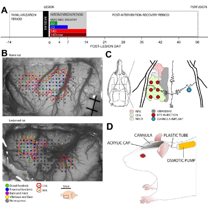
19 during the dark cycle. Upon their arrival at our facility, animals were familiarized with banana food pellets in the Montoya staircase test (Biernaskie and Corbett, 2001; Montoya et al., 1991) for 10 work-days (Figure 1). To incite reaching behavior in the Montoya test, food access was carefully monitored during the two weeks of familiarization. For each rat, the daily food minimum corresponded to 5% of its body weight. Rats had free access to 85% of their daily minimum in the home cage. They could obtain more food to surpass the 100% value in the Montoya staircase apparatus. On any given day, if the animal did not attain its daily minimum in the Montoya, additional food was supplied in its home cage to reach the daily minimum.
Baseline data were collected at the end of the familiarization period. Spontaneous use of forelimbs in exploratory behavior was documented with the cylinder wall test (Schallert et al., 2000) and grasping function with the Montoya Staircase test (Montoya et al., 1991). Animals that performed above the inclusion criteria (see Montoya Staircase test below) were randomly assigned to an experimental group. During the post-lesion period, food was restricted for 12-14h prior to each behavioral testing session and animals were given free access to food after testing. The weight of the animals was recorded daily during the 2 weeks prior to the lesion and weekly after the lesion. If an animal lost more than 10% of its original body weight at any point during the experiment, it was excluded. Two animals from Group 14D were excluded from the study during the recovery period because of weight loss and seizures. Animals had ad libitum access to water at all times. Our experimental protocol followed the guidelines of the Canadian Council on Animal Care and was approved by the Comité de Déontologie de l'Expérimentation sur les Animaux of the Université de Montréal.
Mansucript submission. Journal of experimental Neurology
20 To detect spontaneous asymmetrical use of forelimbs, rats were placed in a transparent cylinder of 19cm diameter and 33cm height for 3-30 minutes or until 60 touches to the cylinder wall was achieved (Schallert et al., 2000). Animals were videotaped from above using a high definition digital video camera (30 frames/second). The videos were analyzed frame-by-frame offline to count the use of paretic versus non-paretic limbs during vertical exploration of the cylinder wall. The forelimb asymmetry score was calculated using the following equation:
Montoya Staircase test: Grasping and retrieving performance
Rats were placed in a Plexiglas chamber (6-cm wide, 12-cm high and 30cm long) with a central platform (2.3-cm wide, 6-cm high and 19-cm long) that supports the weight, separating the right and left forelimbs (Biernaskie and Corbett, 2001; Montoya et al., 1991). A pair of staircases with seven steps on each side was loaded into the Plexiglas chamber on both sides of the central platform. Each step had a smooth well that can hold one to three standard 45mg banana flavored food pellets (Bioserve Inc., Frenchtown, NJ, USA). During the familiarization period, animals had a session of Montoya staircase in the morning and one in the afternoon, the two separated by 3 to 4 hours. In a session, the rats had 4 trials with each hand (8 trials per day in totals). Initially, for each trial, every well on one side of the staircase was filled with 3 food pellets and 15
minutes were given to retrieve the pellets. In the following days, the number of pellets in each well and the time provided was progressively tapered according to the performance of the rat. However, by the 8th day, only one pellet per well and three minutes per trial were given to all rats. On the 9th and 10th days of the familiarization period, the performance in terms of the
Mansucript submission. Journal of experimental Neurology
21 number of eaten pellets was recorded and used to establish if the animal reached our inclusion criteria. To be included in the study, rats needed to eat 4 out of 7 pellets in 3 of the 4 trials on both days with one of the two arms. Based on these criteria, 9 animals were excluded from the study. The ischemic lesion was induced in the cortex contralateral to the arm with the best performance score.
Surgical Procedures
All surgical procedures were conducted aseptically. Anesthesia was induced with Ketamine (80mg/kg, intra-peritoneal) and maintained with isoflurane (~2% in 100% oxygen) delivered via a custom-made facial mask adapted to our stereotaxic frame. Pulse rate and oxygen saturation were monitored and documented during the surgery. A self-regulating heating blanket (Harvard Apparatus, Holliston, MA) was used to maintain body temperature during the surgery. A midline incision was made to expose the skull and neck muscles. A small incision was made between C0 and C1 to release cerebrospinal fluid of the cisterna magna and reduce intracranial pressure.
In order to confirm the location of the motor areas of 3-month-old Sprague Dawley rats, we conducted intracortical microstimulation (ICMS) mapping experiments in naïve animals (n=3) (Figure 1B). Following craniotomy and durectomy, anesthesia was switched to ketamine hydrochloride (~10mg/kg/10 minutes; intraperitoneal) for the collection of electrophysiological data. A glass coated tungsten microelectrode (~1 MΩ) was used for electrical stimulation applied at a depth of ~1600μm. Stimulation consisted of a 40ms train of 13 monophasic cathodal pulses of 200 µs delivered at 350 Hz from an electrically isolated, constant current stimulator
(Dancause et al., 2006; Nudo et al., 1992; Nudo et al., 1996). Pulse trains were repeated at 1 Hz intervals; current was ≤100 µA. Microelectrode interpenetration distances were ~333µm.
Mansucript submission. Journal of experimental Neurology
22 Stimulations were done to identify the caudal and rostral forelimb areas (CFA and RFA). The two areas were typically separated with neck and vibrissa representations. Based on ICMS maps, we selected the stereotaxic coordinates for six injections of endothelin-1 (ET-1) as in Fang and collaborators (Fang et al., 2010) (Figure 1C). ICMS mapping was conducted in additional animals (n=3) at the end of the recovery period (day 60 after the lesion) and confirmed that the lesion destroyed large portion of the CFA and appeared to leave the RFA intact (Figure 1B).
To induce the cortical lesion, six 0.6-mm holes were drilled over the caudal forelimb area (CFA) based on stereotaxic coordinates (+1.5, +0.5, -0.5mm anteroposterior, +2.5, +3.5mm mediolateral to bregma; Figure 1B) (Fang et al., 2010). Using a 1µl Hamilton syringe, a microinjection of endothelin-1 (ET1) (EMD chemicals, CA, USA) was made in each hole at a depth of 1.5 mm in the cortex (0.33µl, 0.3 µg/µl, 3nl/s). Following each injection of endothelin-1 (ET-1), the holes were sealed with bone wax. After the lesion induction, an additional 0.6-mm hole was drilled over the center of the contralesional CFA (+0.5mm anterior and +3mm lateral to bregma). A cannula (0.36mm, brain infusion kit 1; Alzet, CA, USA) was implanted at the depth of 1.5mm below the cortical surface and secured in place with acrylic cement (Figure 1D). The cannula was connected to an osmotic pump filled with Muscimol (10mM, Tocris Bioscience, Bristol, UK), a non-toxic GABA-A agonist. Muscimol was chosen because it is a well-established and simple way to reliably inactivate neural tissue for sustained periods without causing damage (Martin, 1991; Martin et al., 1999; Reiter and Stryker, 1988). Neck muscles were then glued back with dermal adhesive and the skin sutured. After the surgery, animals received a regimen of pain, anti-inflammatory and antibiotics medication and their recovery was closely followed.
Mansucript submission. Journal of experimental Neurology
23
Experimental groups
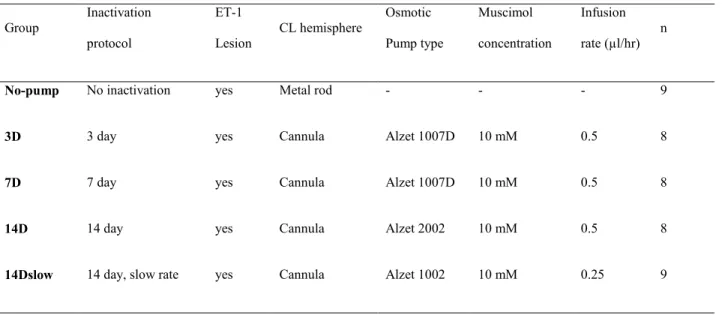
In 3 different experimental groups (Table 1), the contralesional hemisphere was inactivated for 3, 7 or 14 days with infusion of Muscimol (10mM) at a rate of 0.5µl/hour (Groups 3D, 7D and 14D) beginning within 2 hours following the lesion. An additional group (Group 14Dslow) also
received 14 days of Muscimol infusion, but delivered at a slower rate (0.25µl/hour). Because no pump model could provide infusion of 3 days, the plastic tube between the pump and the cannula was cut and sealed in a minor procedure 72 hours after the lesion in Group 3D.
In control animals (Group Lesionno pump), to reproduce the mechanical damage caused by
the cannula, a stainless steel rod of the same diameter as the cannula was lowered at a depth of 1.5mm and immobilized in acrylic. In additional control rats (n=3), a pump infusing saline (0.5µl/hour) for 14 days was implanted after the cortical lesion. No statistical difference of performance on the Montoya staircase tests was found between the two control groups (One way ANOVA paretic hand F=0.10; p=0.76; non-paretic hand F=0.23 p=0.64). The absence of
behavioral effects from saline infusion is in line with previous publications using comparable infusion methods in the motor cortex (Martin et al., 2000). On post-lesion day 56, the animal was killed, the brain fixed, cryoprotected and cut coronally for anatomical reconstruction and
estimation of the lesion size (MicroBrightField, VT, USA).
Reversible inactivation using Muscimol infusion of long duration
The effects of chronic brain inactivation using GABA-A agonist Muscimol on neural activity and behavior have been carefully documented (Hata and Stryker, 1994; Martin, 1991; Martin and Ghez, 1999; Reiter and Stryker, 1988). In kittens, recording of neural activity in the visual cortex following infusion with osmotic pumps for 8 to 11 days with parameters similar to the ones we
Mansucript submission. Journal of experimental Neurology
24 used (0.5µl/h of 10Mm) showed that inactivation extended over a radius of 1 to 3.5mm (Reiter and Stryker, 1988). To confirm that our injection protocol resulted in comparable spread, we filled the osmotic pump with Fluorophore-conjugated Muscimol molecule (BODIPY® TMR-X conjugate; Molecular probes). We infused fluorescent Muscimol at 0.5µl/h for 3 days in one animal (Group 3D; Figure 2A), for 7 days (Group 7D; Figure 2B) in one animal and for 13 days (Group 14D; Figure 2C) in one animal. Whereas the pump model infuses for 14 days, we chose to perfuse on day 13 to insure the infusion was still at its maximum. Finally we infused
fluorescent Muscimol at 0.25µl/hour in one animal for 13 days (Group 14Dslow; Figure 2D).
Following the infusion period, animals were perfused, the tissue processed and images acquired according to an established protocol using this product (Allen et al., 2008). The radius for each animal was an average of the maximal radius found on a coronal section and the AP radius calculated from the span of sections showing the fluorescence. We found that Groups 3D and 7D had identical diffusion radius (1.2mm). Group 14Dslow had the smallest (1.1mm) and Group 14D the largest radius (1.9mm) of Muscimol diffusion. However, these differences are small and their significance questionable considering the variability of the diffusion radius reported by others (Reiter and Stryker, 1988). Thus, analyses and conclusions in the present set of experiments are based on the behavioral effect of the inactivation on the non-paretic forelimb
and not on the rate of infusion or hypothetical radius of inactivation.
To show that in our model, Muscimol effectively induced a reversible contralesional inactivation and did not cause permanent damage to the contralesional hemisphere, we have conducted a series of controls. First, we conducted ICMS mapping in the contralesional
hemisphere at the end of the recovery period in one animal of Group Lesionno pump, Group 7D and
Mansucript submission. Journal of experimental Neurology
25 surrounding the cannula using normal current intensity and could not differentiate the responses in the animals that received Muscimol from the Lesionno pump or from naïve animals. We also
visually inspected the Nissl stained tissue around the cannula in all of our animals and found no differences between experimental groups (Figure 4). To further confirm this finding, we
calculated the size of the lesion made by cannula in the contralesional hemisphere in Nissl-stained sections for Group 14D and 14Dslow, the two groups with Muscimol infusion of the longest duration, and compared it to the lesions made by the metal rod in the Group Lesionno pump. In the contralesional hemisphere, the cannula made a small mechanical lesion in all groups. On average for Groups Lesionno pump, 14D and 14Dslow, the lesion made by the cannula was
0.15mm3, corresponding to 1.6% of the volume of the ET-1 lesion. A one-way ANOVA between the three groups found no significant difference (F=2.15, p=0.16) and, combining Groups
14D+14Dslow and comparing the lesion size to the Group Lesionno pumpalso did not show any difference (t-test; equal variance not assumed: F=0.88, p=0.40).
Altogether, these data support that Muscimol transiently inactivated the contralesional hemisphere and did not result in any permanent damage in our experimental groups. Thus, whereas there is permanent damage to the contralesional hemisphere, it was a small mechanical damage caused by the insertion of the cannula that was of identical size across experimental groups and controlled for in our Groups Lesionno pump.
Histology and anatomical reconstruction
At the end of the experiment, animals were given a lethal dose of sodium pentobarbital and were transcardially perfused with heparinized 0.1 M phosphate buffered saline (PBS) followed by 4% paraformaldehyde in 0.1 M PBS. Brains were extracted and post-fixed in 4% paraformaldehyde.
Mansucript submission. Journal of experimental Neurology
26 The brains were cryoprotected in a solution of 20% sucrose and 2% dimethyl sulfoxide (DMSO) over-night followed by 20% sucrose for 48 hours and quickly frozen at -55°C utilizing methyl butane and stored at -80°C (Brocard et al., 2010). Coronal sections were cut with a cryostat (40µm thickness). One out of six sections were Nissl stained and used for analysis of the lesion size and location. The lesion size was calculated with a software (MicroBrightField, Colchester, VT, USA) using the following formula:
Statistical Analysis of experimental results
All values are reported as mean ± SEM, unless specified, and significance was considered at p < 0.05. Lesion volume between groups of animals was compared with a one-way ANOVA. For the Cylinder and Montoya tests, two repeated measures ANOVAs were conducted using animal group, time and group x time as factors and Tukey-HSD for post-hoc testing. Pre-lesion baseline data and post-intervention recovery period data (day 21 to 56 post-lesion) were included in this analysis. Statistical analysis during the intervention period was impossible because behavioral data could not be collected in all animals (see Results).
Mixed modeling, adjusting for correlations between individual measurements over time, was conducted to identify the plateau performance of the paretic limb on the Montoya test. Regressions were used to investigate how different factors correlated with this plateau
Mansucript submission. Journal of experimental Neurology
27 individual, observations taken close in time tend to be more highly correlated than observations taken far apart in time, was assumed in the analysis. To avoid making unfounded assumptions on the shape of the effect of time, we treated time as a categorical variable in the analysis.
Regression results report the Spearman’s correlation coefficient and t-test on the slope of the distribution. Statistical significance for the t-test was adjusted according to the Bonferroni correction factor (p < 0.017).
Mansucript submission. Journal of experimental Neurology
28 Results:
Cortical lesions
The average ET-1 induced lesion volume was 6.8±2.5mm3 (Mean ± standard deviation). As reported by others using a similar ET-1 lesion protocol (Fang et al., 2010), ischemic lesions destroyed all cortical layers and there was no significant difference of lesion size between experimental groups (F=0.206, P = 0.892) (Figure 5).
General behavior during the intervention period
During the infusion of Muscimol, rats were typically much less active. Some rats made no movement in the Montoya staircase tests (Figure 6). In Group 3D, some rats were still inactive at day 7 after the lesion, showing that the reversal of the effects of inactivation took more than four days in some cases. Group 7D and 14D were more severely affected. More than half of the animals in these groups were inactive in the Montoya staircase in the first week during infusion of Muscimol. The side effects of Muscimol became problematic with time for some animals of Group 14D, and two rats from this group had to be excluded (see Methods). Using a slower rate of infusion (Group 14Dslow) diminished the inactivity caused by infusion of Muscimol. In fact,
the general behavior of animals from Group 14Dslow was the least affected by Muscimol delivery,
supporting that the rate of infusion had a greater influence on these deleterious effects than the duration of inactivation.
The effect of inactivation on grasping performance of the non-paretic forelimb
The animals receiving Muscimol showed signs of impairments of grasping performance with the non-paretic forelimb in the Montoya test, confirming the effectiveness of the pharmacological
Mansucript submission. Journal of experimental Neurology
29 agent. The analysis of the performance of the non-paretic forelimb in the Montoya test was done on the baseline and the post-intervention recovery period to includes all animals in each group. The ANOVA showed a significant effect of group (F=9.237, P<0.001), time (F=49.244, P<0.001), and time x group (F=11.034, P<0.001). The pre-lesion performance was similar between all groups. In Group Lesionno pump, the ET-1 lesion and the mechanical damage caused
by the metal rod did not decrease the performance of the non-paretic forelimb to a significant level (Figure 7). In Groups 3D and 7D, whereas inactivation affected grasping during the intervention period, performance was not statistically different from pre-lesion baseline or from Group Lesionno pump in the post-intervention recovery period. The effect of inactivation on the
non-paretic forelimb function of animals in the Group 14D was strikingly different. Grasping performance was significantly lower than pre-lesion baseline and from Group Lesionno pump until
the 35th day after the lesion. Thus 14 days of inactivation resulted in behavioral impairments of the non-paretic forelimb that lasted more than 2 weeks after the end of infusion. Using a slower infusion rate for 14 days in Group 14Dslow did not affect the level of impairments on grasping
performance caused by the inactivation. Together with results from Figure 6, these data show that a slower rate of infusion had less detrimental effects on the general state of the animals but still had a comparable impact on the function of the non-paretic forelimb.
The effect of inactivation on spontaneous use of forelimbs
Rats also reared less often during the infusion of Muscimol. This was particularly true for animals in Group 7D and 14D and less in Group 3D and 14Dslow (missing data points in Figure 8). We found that animals either did no touch the cylinder wall at all or when they did explore, we obtained a large number of touches. In only 5 rats, we obtained one data collection session
Mansucript submission. Journal of experimental Neurology
30
with less than 55 touches, but more than 22. We considered that more than 20 touches were sufficient to provide an average for that session and thus included that session in the group average. Four of these 5 sessions were during the infusion of Muscimol and thus, were not included in the statistical analyses. In all other data collection sessions (n=457), we obtained more than 55 touches. Thus, the laterality scores obtained for each rat, in any session, are robust.
Analysis of the spontaneous use of the forelimbs in the cylinder test was done for the post-intervention recovery period and includes all animals in each group. The ANOVA showed a significant effect of group (F=32.549, P<0.001) and group x time (F=4.338, p<0.001). After the cortical lesion, animals in Group Lesionno pump used their non-paretic forelimb more often
throughout the post-intervention recovery period (Figure 8). Thus, the lesion created a persistent bias for the use of the non-paretic forelimb. Following inactivation of short duration (Group 3D), there was an initial bias toward the use of the non-paretic forelimb. However, the animals
recovered symmetrical use of their forelimbs at day 28. Inactivation of longer duration caused an initial bias to use the paretic forelimb. Whereas animals in Groups 7D returned to symmetrical use of their forelimb by day 28, the bias for the paretic forearm persisted until day 35 in Group 14D. The laterality index of Group 14D was different from Lesionno pump group throughout
recovery. The use of a slower infusion rate for 14 days in Group 14Dslow still resulted in a bias to
use the paretic forelimb on day 21. However, the animals returned to a symmetrical forelimb use during spontaneous exploration on day 28 and their laterality index was not different from Group Lesionno pump, much like for Groups 3D and 7D.
The effect of inactivation on post-intervention recovery of the paretic forelimb
In the Montoya staircase test, grasping performance of the paretic forelimb showed a significant effect of group (F=6.016, P<0.001), time (F=49.528, P<0.001) and time x group (F=5.739,
Mansucript submission. Journal of experimental Neurology
31 P<0.001). Pre-lesion performance was similar among all experimental groups (Figure 9). The Group Lesionno pump had significant deficits until the 56th day after the lesion. Inactivation of the
contralesional hemisphere initially worsened the paretic forelimb function during the
intervention period but then improved it in the post-intervention recovery period. Groups 3D and 7D recovered faster from the lesion than Group Lesionno pump. They reached pre-lesion
performance at day 35 and 28, respectively, and Group 7D transiently performed better than Group Lesionno pump on day 28. Group 14D had significant deficits and poorer performance than
other groups on day 21. However, by day 28 and for the rest of the post-intervention recovery period, these animals had no deficits and performed significantly better than Group Lesionno pump.
Grasping performance of Group 14Dslow was similar to other groups and better than Group 14D
on day 21. This group performed better than Group Lesionno pump from day 28 to 49. Thus, the
beneficial effects of inactivation on recovery of the paretic forelimb were mostly preserved when a slower infusion rate was used to inactivate the contralesional hemisphere for 14 days. These results suggest that the beneficial effect of contralesional inactivation depends mainly on the duration of the inactivation and not the rate of infusion.
The effect of post-intervention behavior and inactivation duration on final level of recovery
We found that plateau performance on the Montoya test for the paretic limb was reached at day 7 for Group Lesionno pump, day 21 for Group 3D, day 28 for Group 7D and day 35 for Groups 14D
and 14Dslow. Thus, from day 35 to day 56, all groups had a stable level of performance. For each
animal, we calculated the average Montoya score between days 35 to 56 to obtain a ‘plateau performance score’. Three regressions were conducted to study factors potentially correlating with the plateau performance score.
Mansucript submission. Journal of experimental Neurology
32 First, we performed a regression between the asymmetry score on the Cylinder test at day 21 and the plateau performance score for each animal (Figure 10A). A positive correlation between these factors would suggest that increased spontaneous use of the paretic forelimb at the beginning of the post-intervention recovery period results in better recovery of the paretic
forelimb. We found a weak correlation between the two factors (R = 0.28) and a non-significant slope of the distribution (p = 0.13).
Second, we performed a regression analysis between the Montoya score of non-paretic forelimb at day 21 and the plateau performance score of the paretic forelimb (Figure 10B). Here, a negative correlation between these factors suggests that poor performance of the non-paretic forelimb at the beginning of the post-intervention period results in better recovery of the paretic forelimb. We found a weak negative correlation between the two factors (R = -0.12) and a non-significant slope of the distribution (p = 0.58).
A third regression was conducted between the inactivation duration value (0, 3, 7, 14) and the plateau performance score to test how well the inactivation duration correlated with the level of recovery (Figure 10C). A positive correlation between these factors suggests that a longer duration of inactivation results in better recovery of the paretic forelimb. The correlation between the two factors was higher than other variables tested (R = 0.52) and the slope of the distribution was highly significant (p = 0.0004). Overall, the correlation analyses suggest that the level of recovery was weakly influenced by the spontaneous use of forelimbs or on grasping function of the non-paretic forelimb after inactivation of the contralesional hemisphere. In contrast, the duration of inactivation had a great impact.
Mansucript submission. Journal of experimental Neurology
33 Discussion:
We used a rat model of cortical lesions to study the basic interaction between GABA-mediated inactivation of the contralesional hemisphere and behavioral recovery. Inactivation was initiated rapidly following the lesion and was maintained for increasing durations in different groups of animals. During the Muscimol delivery period, inactivation resulted in general inactivity of the animals, impaired the use of the non-paretic forelimb and worsened the function of the paretic forelimb. In the post-intervention recovery period, the adverse effects of inactivation
progressively reverted. Rats with inactivation of the contralesional hemisphere recovered their grasping skills faster than untreated animals and the time of recovery was shorter for animals with inactivation of longer duration. In comparison to Lesionno pump group the grasping function
of the paretic forelimb showed a tendency to be greater following inactivation of longer durations. In rats with 14 days of inactivation, this trend reached significance four weeks after the lesion and remained significant for the rest of the experiment. Using a slower infusion rate to inhibit the contralesional hemisphere for 14 days diminished the deleterious effect on the general behavior during inactivation while still preserving most of the beneficial effects on chronic recovery. Final recovery scores were correlated to the duration of inactivation but not to the spontaneous use of the forelimbs or the function of the non-paretic forelimb.
In our model, we found that acute inactivation of the contralesional hemisphere can favor recovery of the paretic forelimb. Inactivation of longer duration results in more pronounced and sustained recovery of function. Final recovery is more affected by the duration of inactivation than the effects of inactivation on general behavior or on the non-paretic forelimb.