Université de Montréal
$hape-keeping elements in tRNA
par
Ekaterina Zagriadskaia
Département de Biochimie
Faculté de Médecine
Thèse présentée à la Faculté des études supérieures
en
vue
de l’obtention du grade de Ph.D. en Biochimie
Decembre 2003
\jD
o
L)0
Université
(III
de Montréal
Direction des bibliothèques
AVIS
L’auteur a autorisé l’Université de Montréal à reproduire et diffuser, en totalité ou en partie, par quelque moyen que ce soit et sur quelque support que ce soit, et exclusivement à des fins non lucratives d’enseignement et de
recherche, des copies de ce mémoire ou de cette thèse.
L’auteur et les coauteurs le cas échéant conservent la propriété du droit
d’auteur et des droits moraux qui protègent ce document. Ni la thèse ou le
mémoire, ni des extraits substantiels de ce document, ne doivent être imprimés ou autrement reproduits sans l’autorisation de l’auteur.
Afin de se conformer à la Loi canadienne sur la protection des renseignements personnels, quelques formulaires secondaires, coordonnées
ou signatures intégrées au texte ont pu être enlevés de ce document. Bien
que cela ait pu affecter la pagination, il n’y a aucun contenu manquant.
NOTICE
The author cf this thesis or dissertation has granted a nonexciusive license allowing Université de Montréal to reproduce and publish the document, in part or in whole, and in any format,_soteLy for noncommercial educational and
research purposes.
The author and co-authors if applicable retain copyright ownership and moral
rights in this document. Neither the whole thesis or dissertation, nor
substantial extracts from it, may be printed or otherwise reproduced without the author’s permission.
In compliance with the Canadian Privacy Act some supporting forms, contact information or signatures may have been removed from the document. While this may affect the document page count, it does not represent any loss of
Université de Montréal
Faculté des études supérieures
Cette thèse intitulée:
$hape-keeping elements in
tRNA
présentée par
Ekaterina Zagriadskai a
A été évaluée par un jury composé des personnes suivantes:
Président-rapporteur
Directeur de recherche
Membre du jury
Examinateur externe
Représentant du doyen de la FES
Dr. Luis Rokeach
Dr. Serguei Chteinberg
Dr. belle Pelletier
Dr. WilÏiam McClain
Dr. Elliot Drobetsky
111
$UMMARY
RNA molecuies fulfihi important functions in living celis. They serve as messengers in the decoding of genetic information from DNA into proteins, and also carry out some catalytic functions. C atalytic R NAs n eed to assume u niquetertiary structures to b ecome
active. Tertiary structure of RNA is determined by its nucleotide sequence, however, the mies that govem RNA folding are flot welÏ understood. hi the present study, the structural scaffold oftransfer RNA (tRNA) was used to investigate how this particular RNA molecule forms and maintains its tertiary structure, and how the structure influences the fiinction.
Transfer RNAs function in protein biosynthesis by matching amino acids to the corresponding codons on mRNA. Ail tRNAs have universal tertiary structure known as the ‘L-shape’, which is essential for their function. Conserved tertiary interactions are involved in the formation of the L-shape, many of which concentrate in the ‘elbow’ region, formed by interaction of the D- and T-loops of tRNA. I have investigated sequence and structural requirements for this region of tRNA. For this, I created a tRNA gene library, where several positions in the D- and T-loops were randomized, and screened this Iibrary in vivo for the presence of functional suppressor tRNAs. Screening revealed the importance of a particular tertiary interaction, reverse-Hoogsteen (RH) base pair between nucleotides 54 and 58 in the T-loop, for the tRNA structure, while other conserved tertiary interactions in the elbow region tumed to be more tolerant of changes and could be replaced by alternative sets of interactions without destroying tRNA function. To find out the exact role of the R}{ base pair, I constructed the second tRNA gene library. Screening of this library showed, that the RH base pair is involved into formation of the L-shaped structure by providing correct juxtaposition of the two helicai domains of tRNA. Only those molecules that were able to
maintain the L-shape were functional.
My resuits contribute to the understanding of the role of different types of tertiary interactions in the formation of RNA structure and demonstrate that combinatorial approach can be successfuily used in vivo to investigate the principles ofRNA folding.
RÉSUMÉ
Les ARNs jouent un rôle important dans les cellules vivantes. Ils servent comme
messagers dans le décodage de l’information génétique et ils ont aussi des fonctions catalytiques. L’activité catalitique de l’ARN nécessite la formation d’une structure tertiaire unique. Cette structure tertiaire est déterminée par la séquence primaire de l’ARN, mais les règles qui gèrent ce repliment sont encore mal connues. Dans le travail présenté, nous avons utilisé l’ARN de transfert (ARNt) comme échafaudage pour étudier comment cet ARN particulier forme et maintient sa structure tertiaire, et comment cette dernière influence la fonction de l’ARNt.
Les ARNt fonctionnent dans la synthèse protéique. Leur rôle est d’apporter sur le ribosome l’acide aminé correspondant au codon de l’ARN messager. Tous les ARNt5 ont une structure tertiaire commune appelée ‘la forme en L’. Cette structure est maintenue par de nombreuses interactions tertiaires, dont plusieurs se concentrent dans la région du ‘coude’ de l’ARNt, formé par l’interaction entre les boucles D et T. Nous avons étudié les exigences imposées sur la séquence et la structure de cette région de l’ARNt. Pour ce faire, nous avons construit une bibliothèque des gènes d’ARNt où plusieurs positions dans les boucles D et T ont été rendues aléatoires. Cette bibliothèque a été criblée in vivo pour sélectionner des ARNts suppresseurs. Ce criblage a révélé l’importance d’une interaction tertiaire e n p articulier, so it 1 a p aire d e b ases detype ‘reverse-Hoogsteen’ (RH) e ntre 1 es nucléotides 54 et 5$ dans la boucle T, pour la structure et la fonction de l’ARNt, tandis que d’autres interactions tertiaires dans la région ‘coude’ se sont avérées plus variables et pouvaient être remplacées par des interactions alternatives. Pour trouver le rôle exact de la paire de bases RH, nous avons construit une deuxième bibliothèque des gènes des ARNt5. Le criblage de cette bibliothèque a montré que la paire de bases RH est impliquée dans la formation de la structure ‘en L’ par son rôle essentiel dans l’arrimage correct des deux domaines hélicoïdals de l’ARNt. Seulement les molécules capables d’adopter la structure
‘en L’ étaient fonctionnelles.
Ces résultats contribuent à la compréhension du rôle des différentes interactions tertiaires dans la formation de la structure de l’ARN et ils démontrent que l’approche utilisant des bibliothèques des gènes peut être très utile pour l’étude des principes du repliment de l’ARN.
V
TABLE 0F CONTENTS
Page
Summaiy iii
Résumé iv
Table of contents y
List of tables vii
List of figures viii
List of abbreviations x
Acknowledgments xi
Introduction: tRNA— a treasury of stereochemical information 1
Discovery oftRNA I
Cloverleaf secondary structure 2
L-shape of yeast tRNAe 2
tRNA structure in complexes with proteins 6
2-fonnoftRNA 8
Crystal structure ofthe tRNA-like domain oftmRNA 10
MitochondrialtRNAs 12
Detailed structure ofthe DT (elbow) region 14 Problems addressed in the thesis and an overview ofthe
experimental rnethod 18
Chapter 1. Article: Importance ofthe reverse Hoogsteen base pair
54-58 for tRNA function 21
Abstract 22
Introduction 22
Materials and Methods 23
Results 25 Discussion 29 Acknowledgernents 33 References 34 Legends to figures 37 Table 39 figures 40
Chapter 2. Article: Keeping the tRNA L-shape with the help ofthe
reverse-Hoosteen base pair 54-5 8 43
Abstract 44
Introduction 44
Materials and Methods 45
Resuits 47 Discussion M Acknowledgements 56 References 56 Legends to figures 57 Table 60 Figures 61
Chapter 3. Article: Specific and non-specific purine trap in the
T-loop of normal and suppressor tRNAs 63
Abstract 64
Introduction 64
Materials and Methods 67
Resuits 67 Discussion 75 Acknowledgements 79 References 80 Legends to figures 82 Table 86 Figures 87 Discussion 9
Conservation ofthe L-shape oftRNA 91
Importance of the reverse-Hoogsteen base pair for tRNA
structure and function 92
Alternative structures ofthe DT region oftRNA 93
Advantages of combinatorial rnethod 94
Conclusions 96
vii
LIST 0F TABLES
page
Chapter 1
Table I. The nucleotide sequences and the 3-galactosidase
activity ofthe selected tRNA clones 39 Chapter 2
Table I. Sequences ofthe D-and T-loops and suppressor activities
oftRNAs selected from the K- and M-libraries 60 Chapter 3
LIST 0F FIGURES
Introduction page
Figure 1. Cloverleaf and the L-shape oftRNA 3
Figure 2. The scheme of the L-shape oftRNA’ 4 Figure 3. Structure of yeasttRNA’ in its free form and compÏexed
to the aspartyl-tRNA synthetase 7
Figure 4. Comparison ofX-form and L-formoftRNA’’1 9 Figure 5. Structure ofthe tRNA-like domain oftmRNA 11 Figure 6. Stereo view ofa ‘boomerang’ model oftRNAGcUftom
chimpanzee 13
Figure 7. Structure ofthe DT region from yeasttRNAm 15
Chapter 1
Figure 1. The standard tRNA L-form 40 Figure 2. Construction ofthetRNAgene library 40 Figure 3. Northern blot showing the presence in the cytosol
and the level of aminoacylation of some suppressor tRNAs 41 Figure 4. Juxtaposition of the basesin RH-GA, RH-GG and other
alternative base pair candidates for replacement ofRH-UA 41 Figure 5. The model ofthe structure ofthe DT region for clone K31
superimposed on the corresponding region in the yeasttRNAPu1e 42 Chapter 2
Figure 1. A conventional representation of the DT region in the standard 61 tRNA structure
Figure 2. Design ofthe two combinatorial tRNA gene libraries 61 Figure 3. The aminoacylation levels of selected tRNA clones 61 Figure 4. Comparison ofthe DT region structure in the tRNAs selected
from the K- and M-libraries 62 Chapter 3
Figure 1. The structure ofthe DT region in the context of the tRNA L-form 87 Figure 2. Design ofthe combinatorial tRNA gene library 87 Figure 3. Comparison of the DT region structure in the Type I and Type II
tRNAs 8$
ix Figure 5. Comparison of the two reverse-Hoogsteen base pairs existing
in Type II tRNAs 89
Figure 6. Comparison of the non-specific purine trap (Type II) with the
AA-AA motif 90
LI$T 0F ABBREVIATIONS
Standard three-letter code is used for ailarnino acids
5S, 16S and 23S 5S, 16S and 23S subunits ofbacterial ribosome
À
angstrom(F10m)DNA deoxyribonucleic acid
RNA ribonucleic acid
mRNA messenger RNA
rRNA ribosomal RNA
tRNA transfer RNA
tRNAtGcU tRNA serine with anticodon GCU
mit tRNA mitochondrialtRNA
tmRNA transfer-messengerRNA
RH reverse-Hoogsteen base pair
ArcTGT archaeosinetRNA—guanine transglycosylase AspRS aspartyl-tRNA synthetase
G1nRS glutamyl-tRNA synthetase PheRS phenylalanyl-tRNA synthetase SerRS seryl-tRNA synthetase
RNase P ribonuclease P
EF-Tu elongation factor Tu
Y pyrimidine base R purine base P pseudouridine base A adenine base C cytosine base G guanine base T thymine base U uracil base
xi
ACKNOWLEDGMENTS
This w ork w ould n ever be d one w ithout h elp and s upport from my a dvisor, D r. S ergey Steinberg. His understanding was really remarkable. I also got a lot of tecirnical help at the beginning o f t his p roj ect from g raduate s tudent Cesar G ornez. T hanks t o C esar, S andra, Matthieu, Geneviève and Félix for the great tirne we had together at the lab. I’m also grateful to Prof. Léa Brakier-Gingras for discussions about my project and for advice on writing the thesis. Many department members were very helpftil during the time of my studies, especially Mme Syvie Beauchemin and Mme Denise Lessard. Thanks to Dominic Dulude and Martin Baril for their help with some experiments. I appreciate the support and encouragement from my farnily during the tirne I was writing this thesis. Finally, I’m grateful to Prof. Shana Kelley from Boston College, who allowed me to work in her lab while I was wnting my thesis.
INTRODUCTION
Transfer RNA— a treasury of stereochemical information
Discovery of tRNA
Transfer RNA (tRNA) is probably one of the rnost studied classes of biological macromolecules. Almost 50 years ofresearch have assembled an enormous amount ofboth functional and structural information about tRNAs. However, there are stiil questions waiting to be answered about them.
In 1955 Francis Crick formulated the “adaptor hypothesis” about the existence of adaptor molecules (possibly RNAs) in protein biosynthesis, which carry enzymatically attached amino acids and can specifically recognize codons on mRNA (Crick, 1966). In 1957-58 Paul Zamecnik, Mahïon Hoagland, Robert Holley and colleagues discovered the requirement for “soluble” RNA in order to incorporate amino acids into proteins (Hoagland
et ai, 1958; Holley, 1957). This RNA was later called transfer RNA. Transfer RNAs are
adaptor molecules that translate genetic code from messenger RNA into amino acid sequence. Thus, each tRNA has two functional centers: the anticodon, which recognizes the codon on mRNA during translation, and the acceptor 3’ end, where the cognate amino acid
is attached to tRNA by the enzyme aminoacyl-tRNA synthetase. During translation,
anticodon and acceptor end of tRNA interact with different subunits of the ribosome, and tRNA is involved in a complex series of movements and reactions on the ribosome, including translocation from A to P to E sites, decoding and peptidyl transfer. Besides ribosorne and arninoacyl-tRNA synthetases, tRNA interacts with a lot of other factors during its life cycle: 5’- and 3’- processing enzymes, rnodifying enzymes, CCA-adding enzyme and elongation or initiation factors.
Sorne functions of tRNA not related to protein synthesis have been discovered. For example, tRNAs serves as a primer for reverse transcription in the HW virus. Some plant
RNA viruses have tRNA-like structures at the 3’ ends of their genomes, which play the same role. Transfer RNAs are also involved in cell wall biosynthesis, as well as chlorophyli and heme biosynthesis, and in regulation of expression of some aminoacyl-tRNA synthetases (Soll and RajBhandary, 1995).
2
Cloverleaf secondary structure
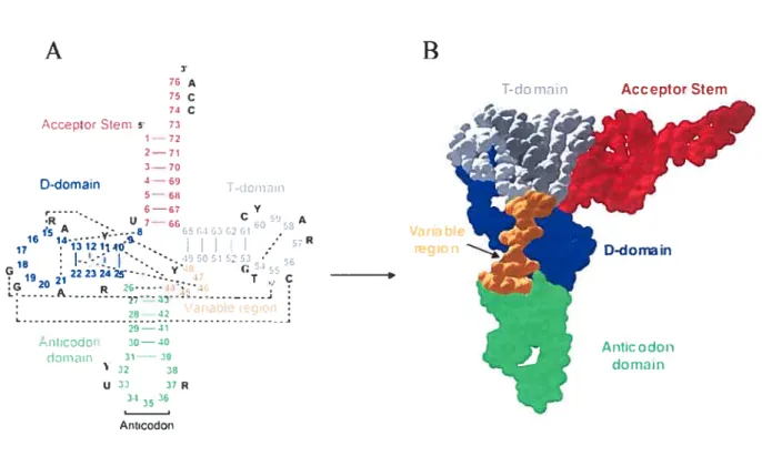
In 1965 Holley and co-workers (HolÏey et ai, 1965) rnanaged to isolate and sequence the first tRNA, yeast tRNAA1. They proposed three alternative secondary
structures for this moiecule, one of them being the “cloverleaf’. Later it became evident that c loverleaf s tructure is c ommon t o ail p rokaryotic, a rchaeai a nd e ukaryotic c ytosolic tRNAs. Cioverleaf (Fig. lA, page 3) consists of four stems: acceptor, D (for dihydrouridine), anticodon and T (for ribothymidine). Three stems are capped with loops, called D-, T- and anticodon loops, while the acceptor stem is formed by the 5’ and 3’ ends of the molecule. There is an unpaired CCA sequence at the very 3’ end of ail tRNAs, which is n ecessary for arninoacylation. T he 1 ength o f the a cceptor, T- and a nticodon stems, as
well as of T- and anticodon ioops, is conserved in ail tRNAs, while D- stem can vary from 3 to 4 base pairs, and the D-loop can have 7 to 11 nucleotides. Between anticodon and T-stems there is a region of variable length. In most tRNAs it is 4-5 bases long and is called variable loop, whule in tRNAs leucine, serine and prokaryotic tRNA tyrosine it is rnuch longer (10-24 bases) and forms a base paired stem with loop, called variable arm. There are more than 20 conserved or semi-conserved (restricted to purines or pyrirnidines) residues in tRNAs, which are mostly 1 ocated outside the stems (Dirheimere t ai, 1995). It has been realized early that these nucleotides may be involved in the long-range interactions and formation of tertiary structure. However, modeling attempts were not able to predict the correct tertiary fold of tRNA, though some important details, like coaxial stacking of stems or tertiary base pair between nucleotides 15 and 48 were predicted correctly (Levitt, 1969).
L-shape of yeast tRNAme
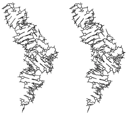
The first crystal structure of a tRNA at atomic resolution, that of yeasttRNA, was published in 1974 independently by the groups of Alexander Rich (Suddath et ai, 1974)
and Aaron Klug (Robertus et ai, 1974). They were later refined to about 2.5
À
resolution, and recently both structures were re-deterrnined using modem crystallographic methods with a resolution under 2À
(Shi and Moore, 2000; Jovine et ai, 2000). Though two structures resuit from different crystal forms, orthorhombic and monoclinic, the structures are alrnost identical. So, in 1974 it became clear, that tRNA had the shape of letter L (Fig. lB, page 3), where one side of L was formed by coaxial stacking of the anticodon and D-stems and the other side - by stacking of acceptor and T-stems (Fig.2, page 4).A
B
T-do moin Acceptor Stem
74 C AccelitorStem 5 73 1—72 2—71 is A ,..-.-8 3 f4 302fl . VaribIe 16 14- .S. . 17 13121140. .-.• region -18 -;«5051 52 19 21 2223z4Z-.::. c 2I13 , .antaote 1eiict - 41 Antcodon 30—40 do,v-iin 31— 39 32 38 u 33 37R 3) 36 Ancodon
figure 1. Cloverleaf and the L-shape oftRNA. A. Cloverleaf secondary structure of a canonical tRNA. Tertiary interactions are shown by dashed unes. Several conserved and modified nucleotides are shown: P stands for pseudouridine, T for ribothymidine, R for purine, Y for pyrimidine. B. A space fihling representation of the tertiary L-shaped structure ofyeasttRNA’.Domains are colored the same way as in A.
D-domain
Antic adoIl
4
Figure 2. The scheme of the L-shape oftRNA. Nucleotides are represented by
rectangles. Tertiary interactions in the core and the elbow region are shown as paired or overlapping rectangles. The anticodon loop is oriented to the left; CCA end is at the bottom. The elbow region is boxed, with T-loop nucleotides in black and D-loop nucleotides crosshatched. Nucleotides of the anticodon loop, nucleotide 47 and unstacked nucleotides ofthe D-loop are not shown.
anticodon stem I D-stem I
(n (D B n) o o (D -c C (n (D B 5, 3’
However, D- and anticodon stems are not perfectly coaxial, as there is a kink of about 26° between them. Two sides of L are about the same length (60À) and aimost perpendicular to each other. Two functional centers of tRNA, the anticodon and acceptor terminus, are iocated at the two ends of the molecule. Acceptor end is exposed into solution, while ail three nucieotides of the anticodon are stacked onto each other in the conformation suitable for recognition ofthe codon onrnRNA.
The distance between the anticodon and the acceptor end, calÏed ‘primary axis’, is about 80À. The corner, or the ‘elbow’ of L is formed by the T-ioop which interacts with the D-loop through formation of two base pairs, G18-P55 and G19-C56 (elbow region is boxed on Fig.2). Other long-range interactions help to make the ‘core’ ofthe molecuie, aiso calied ‘extended stem’, because it consists of base triples fonned by base pairs ofthe D-stem and nucieotides from the connector regions (nucleotides 8, 9 between the acceptor and D- stems and nucleotides 45-48 ofthe variable ioop, see Fig 2). There are four base triples and a reverse-Watson-Crick base pair 1 5-48 in the core region. In addition, stacking interactions are very important for the formation of tertiary structure: 71 out of 76 nucleotides participate in stacking, even if they are flot in the helices.
For a long time the tRNA1c structure was the only one known for an RNA molecuie, and was a source of valuable information on the structure of RNA heiices, stacking, non-canonical base pairs, long-range interactions and sugar-phosphate backbone conformations. For this reason it was considered ‘a treasury of stereochemical information’ (Saenger, 1984).
Three other crystal structures of tRNA were solved later: yeast tRNA’ (Moras et
ai, 1980), yeast initiatortRNAMet (Schevitz et ai, 1979) and E.coti initiator tR1\TAt (Woo et ai, 1980). All of them were later refined to atomic resolution. They ail conserve the L shape and have overail s tructures very sirnilar to that of yeast tRNA’. However, some smaller differences exist. For example, tRNA’, which has been crystalÏized in two different forms, A and B, lias a bigger angle between the two sides of L (about 110°, form A is shown in Fig. 3b, page 7). Also, D- and anticodon stems are more coaxial. The variable ioop has four nucleotides instead of five, so residues 46 and 48 have siightiy different positions, which induces different environrnent for base triple 8-14-21 and bases 9 and 45 (Westhof et aÏ, 1985). Also, the anticodons of the two neighboring tRNA moiecules
in the crystai pair with each other (they are self-compÏementary), which stabilizes the
6
exist in the B-form (B is the form obtained at lower temperature), while in the A-form it is present oniy in a fraction ofmolecules. Ail other interactions are the same as intRNA’.
Structures of initiator tRNAs also reveai some peculiarities that can be associated with their function in initiation, but not elongation, of protein synthesis (Basavappa and Sigler, 1991).
tRNAstructure in complexes with proteins
The structure of tRNA bound to its cognate aminoacyl-tRNA synthetase (GÏnRS) was the first tRNA-protein complex to be solved in 1989 (Rould etai). Today, structures of 11 other tRNAs bound to their synthetases are known (for tRNAs Leu, Val, 11e, Arg, Phe, Asp, Cys, Tyr, Pro, Thrand Ser; in some ofthem tRNAs and synthetases are from different species, but form active complexes). Also, structures of tRNA complexes with elongation factor EF-Tu (Nissen et al 1995) and several modification enzymes are known (Ishitani et
ai, 2003). Finally, ribosome crystal structures include mimics of parts oftRNAs or whole
tRNAs (Yusupov et ai, 2001; Ban et al 2000; Wirnberlyet ai, 2000). These structures give lots of information about tRNA function. One can also see the changes in tRNA structure induced by protein binding. It is impossible to discuss ah these structures in detail, so I will mention only a few of them. As a rule, with only one exception known today, tRNA in these complexes preserves its L-shape, however, protein binding usuaÏly induces local structural changes in tRNA.
For example, yeast tRNAM1 bound to its synthetase, AspRS (Ruff et ai, 1991), keeps the L-shape, but is nonetheless deformed compared to its free form (Fig.3, page 7). The anticodon arm is bent by as much as 20° towards the inside of the L, the anticodon loop is unfolded, with the anticodon bases unstacked in order to interact with the protein. The enzyme also interacts with the end of acceptor stem from the major groove side, which in RNA is much less accessible than the minor groove, by making it wider to accommodate protein side chains. The enzyme also specifically recognizes the G10-U25 wobble pair in the D- stem. However, most of the close contacts between protein and tRNA in this and other complexes are made with the backbone oftRNA, flot the bases.
In tRNA°’°-GlnRS complex (Rould et ai, 1989) the anticodon stem is extended by two non-canonical base pairs, U32-P38 and U33-A37, causing the anticodon to unstack, with its bases pointing into opposite directions and bound in different protein pockets.
A
B
7•
& ?1 ‘r’S•
fig. 3. Structure of yeast tRNA’ in its free form (B) and complexed with the aminoacyl-tRNA synthetase (A, protein flot shown). Anticodon loop is at the bottom, 3’ end is oriented to the lefi. Differences between the two structures concem the anticodon arm, CCA end and D-T ioop interaction.
Adapted from color plate 7 in Soli and RajBhandary (eds), tRNA:structure, biosynthesis andfunction, 1995.
$ The first base pair of the acceptor stem is disntpted by the protein. Unlike AspRS, G1nRS interacts with the acceptor stem from the more accessible minor groove side. It also recognizes base GlO in the D-stem. A peculiarity of the tRNA is that it has a loosely
packed core compared to yeast tRNA’: nucleotide 46, instead of making a triple with base pair 13-22, is bulged out, and nucleotide 45 takes its place instead of interacting with base pair 10-25. This resuits in a ‘hole’ at the place usually occupied by nucleotide 45. Not surprisingly, the replacement of variable ioop in this tRNA by a shorter one leads to the more compact core and increases the stability of the tRNA-synthetase complex (Bullock et ai, 2000).
Another interesting example is the tRNAt SerR$ complex (Biou et ai, 1994). Since tRNASet has a long variable ami, several reanangements are needed in the core ofthe tRNA at the place of the variable arrn attachment. This attachment is mediated by two
additional nucleotides in the D-loop ofthis tRNA, 20a and 20b. The variable ami protmdes
from the body of the molecule at an angle of about 45 degrees to the plane of the L and serves as the major identity element for recognition by its cognate synthetase. The rest of the tRNAt molecule is normal, including the standard interactions between the T- and D loops.
From this and other examples we can see that the L-shape oftRNA is universal, but flexible enough to allow tRNA to interact with many proteins, while keeping its general tertiary fold.
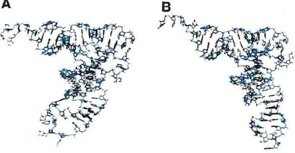
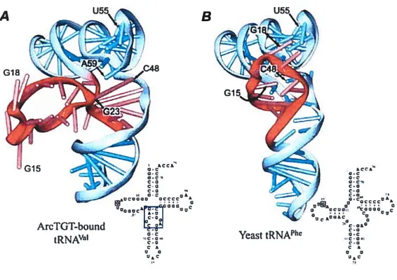
-form of tRNA
The only example known to date of a tRNA-protein complex, where the L-shape of tRNA is not maintained, is the recent structure ofthe modifying enzyme archaeosine tRNA — guanine transglycosylase (ArcTGT) bound to tRNA’ (Ishitani et aÏ, 2003). This is also the first detennined structure of a tRNA modification enzyme bound to a full-length tRNA. ArcTGT modifies nucleotide G15 into archaeosine in many archaeal tRNAs. In the L shape, G15 forms tertiary base pair with nucleotide 48 and as a resuit is buried deep inside the tRNA core. The crystal structure shows, that in order to reach G15, the enzyme completely unfolds the D-ami of tRNA. However, this tRNA is not cornpletely denatured, but forrns an alternative structure, narned 2-form (Fig. 4, page 9). While nucleotides 8 to 22 ofthe D-ami protrude, the rest ofthe D-stem (nucleotides 23 to 26) forms a new helical
B
Figure 4. Comparison of ?-form (A) and L-form (B) oftRNA. Sugar-phosphate backbone is represented as a ribbon, bases are shown as short sticks, Watson-Crick base pairs— as long sticks. The D-arm is colored in red. Corresponding secondary structures are shown for both tRNA forms. From Ishitaniet ai, 2003.
kCCA’ o-c o-c o-c c-Q c-o c-o O-C 0* Uc0000 acccc k Q c 0 3 ArcTGT-bound
tRNA’ Yeast tRNA’
Ace.. Q-c O-c o-c c-o c-o C-o Q-c fl oJ o CCCC & GO0 oc*00kC0 0g c-Ok0 c-o O-c c-o. c-o C k o o
10
structure with the nucleotides of the variable ioop, which authors eau ‘DV helix’. This helix is stacked on top of the anticodon stem. The acceptor/T domain remains folded and stacks to the last base pair 23-48 ofthe DV helix instead ofthe canonical base pair 15-48. Unfolded D-arm is extensively contactcd by protein, which helps to position the G1 5 correctly for the catalysis.
It is possible, that ?-forrn may be common for tRNA binding to several
modification enzymes that modify any ofthe core nucleotides 8, 9, 10, 13 or 22.
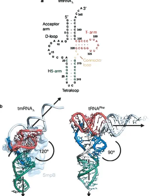
Crystal structure of the tRNA-like domain of tmRNA
One more recent crystal structure, that of tRNA-like domain of the transfer messenger RNA (tmRNA) bound to the SmpB protein (Gutmaim et ai, 2003), is another example of variability, which can be achieved within a tRNA-like foid.
tmRNA is a hybrid bacterial RNA that combines functions of both transfer and messenger RNAs. It rescues hbosomes stalled at the ends oftnmcated mRNAs lacking stop codons. tmRNA is recognized by alanyl-tRNA synthetase, charged with alanine and delivered by elongation factor Tu to the A-site on the ribosome together with SmpB. Then, the incompletely synthesized polypeptide chain is transferred from the P-site tRNA to the alanyl-tmRNA, and the ribosome resurnes translation, switching to the open reading frame of 10 arnino acids encoded by the mRNA region of the tmRNA. Termination takes place normally at the stop codon on tmRNA, and the added peptide serves as a degradation tag for the truncated protein.
The tRNA-Iike domain of tmRNA has canonicat acceptor and T-stems and T-loop (fig. 5a, page 11). However, the D-an-n is replaced by a loop, which does not have any base pairing. Helix 5 (H5) is an equivalent of the anticodon stem, but it is longer (with 8 base pairs) and ends with internai loop, afier which the rest of the moiecule follows. In the crystal structure, this loop was replaced by a tetraloop, thus cutting off the tRNA-like domain from the rest ofthe tmRNA.
The crystal structure shows, that the tRNA-like domain adopts an open L-shaped conformation, with the singÏe-stranded D-loop bound by SmpB protein. However, this structure is significantly different from conventional L-shape (fig. 5b). First, the angle between the two helical domains is much larger than in canonicai tRNA. Second, the T-arm
a ImRNA1 C345 5 A G-C Acceptor - C G u an-n G.C340 5G - C i-arrn D-toop AAAG CCCCC A 10 Q 32UGCGCG C A15 c uu U 325 UCQA . 2QC - G iCCtC G - C 1$ fr-cp H5-atmg C -u G UC TetraloDp
Fig. 5. Structure ofthe tRNA-like domain oftmRNA (from Gutmann et ai, 2003). a Cloverleaf-like secondary structure ofthe tRNA-like domain ofthe tmRNA.
b Comparison of the tertiary structure of the tRNA-like domain of the tmRNA with SmpB protein (lefi) and the yeasttRNA’ (right). Color code of different domains is the same as in a. In tmRNA, the poorly ordered acceptor stem and five nucleotides in the D-loop were modeled (shown in grey). tmRNA has a significantly larger angle between acceptor and anticodon stems (1200) than tRI’JA’ (90°), while its T-arm is rotated approximately 900
around its helical axis, compared to tRNA’.
b tmRNA,
/
tRNA1e12 relative to the rest of the molecule. Iii this conformation, T-loop cannot interact with the D loop. The latter is stabilized by contacts with SmpB. Due to rotation of the T-arm, the H5
arm of tmRNA points in the direction, almost orthogonal to that of the anticodon stem in the tRNA, when the two structures are superimposed by their T-stems. T his is certainly related to the functional differences between tmRNA and tRNA: tmRNA does flot participate in codon-anticodon interaction, instead, it needs to accommodate the rest of its molecule and the SmpB protein, avoiding their collision with ribosomal subunits. This can be achieved due to the rotation of the T-arm. Thus, the structure of tmRNA is adapted to its unique function in translation. However, it is flot clear from this crystal structure, given the absence of interactions between the T- and D-loops, why tmRNA has conserved the canonical tRNA sequence of the T-loop and the GG dinucleoitide in the D-loop, i.e., elements, which normally interact with each other. Maybe, tmRNA can adopt an alternative conformation (in the absence of SmpB), where the D- and T-Ïoops interact with each other as in normal tRNA.
Mitochondrial tRNAs
Many animal mitochondrial tRNAs (mit tRNAs) present unusual primary and secondary structures (Helm et ai, 2000). Almost any given mit tRNA, except those from plants, has some ‘weird’ features, like the absence of a conserved nucleotide, mismatches in the stems, or, in the extreme case, the lack of the entire D- or T- domain (in mit tRNAs from nematoda worms). Mit t RNAs are usually shorter than canonical tRNAs and have higher A-U base pair contents, which makes them less stable. Shortening usually affects D and T-loops and the D-stem. How this influences the tertiary structure of such tRNAs is not completely understood. No crystal structure of a mit tRNA is known at present. However, modeling and structural probing resuits suggest that mit tRNAs manage to preserve the overall tRNA fold by means of different structural compensations and alternative interactions. For example, many mit tRNAsGcu lack the D-domain, which should substantially influence their tertiary structure. However, these tRNAs usually have longer anticodon stems (at least 9 base pairs), and structural modeling showed, that they can be folded into ‘boomerang’ structures (Fig. 6, page 13) with the bigger angle between acceptor and anticodon domains compared to the L- shape, but with a distance between the anticodon and the CCA end close to that in canonical tRNAs (Steinberg et aï, 1994b).
figure 6. Stereo view of a ‘boomerang’ model of tRITAGcu from chimpanzee (Steinberg et ai, 1994b). The angle between the two helical domains is much larger than in canonical tRNA, but the distance between the anticodon and acceptor end is similar to standard.
14
This finding suggests that alternative tRNA structures are possible, as long as some crucial pararneters required for the tRNA function are conserved. Mit tRNAs may represent the molecules that fulfiul minimal requirements for functioning in protein synthesis.
More generally, there is a correlation between the lengths of the D- and anticodon stems and the number of connector nucleotides in atypical mit tRNAs, which allows molecules with longer anticodon and shorter D-stems to fold into the same L-shaped tertiary structure, while the point of cormection with the acceptor/T dornain can be moved along the anticodonlD domain Jike in a zipper (Steinberg and Cedergren, 1994a). The general rule established from the analysis of atypical mit tRNAs states that the acceptor/D dornain shotild contain 12 layers of stacked nticleotides to ensure its stacking with nucleotide 59 from the T-loop (Steinberg et ai, 1997). Stacked layers can be made flot only by Watson- Crick base pairs, but also by rnismatch pairs and even by single unpaired nucleotides intercalated into a helical stem. There is also a restriction on the minimal length of the two coimector regions in mit tRNAs: for N base pairs in the anticodon stem (N can vary from 5 to 10), the minimal length ofconnector 1 is 8-N and that of coimector 2 is 9-N nucleotides.
Detaïled structure of the DT (elbow) region
My thesis research was focused on the structure of the elbow region of tRNA. This region, which we also call the DT region, is where many tertiary interactions concentrate and where the two helical dornains corne together and interact through the D- and T-loops (sec Fig. 2, page 4). It is known from the crystal structures oftRNA that two base pairs are formed between D- and T-loops: G1$-P55 (non-Watson-Crick) and G19-C56 (Watson Crick), w ith t he b ase o f G 18 from t he D -loop b eing i ntercalated i nto the T -loop (Fig.7, page 15). There is an unusually large ‘gap’ between nucleotides 57 and 5$, where G1$ can
fit. The dinucleotide 59-60 is bulged out frorn the T-loop and stacks to the tertiary base pair
15-48, the last layer ofthe D-domain. There is a reverse-Hoogsteen (RH) base pair between bases T54 and A5$ in the T-loop, which is believed to stabilize the unique conformation of the T-loop, through its stacking to the base pair 53-61 of the T-stem and stabilization of hydrogen bond network from phosphate of nucleotide 60 to the amino group of C61 and ribose 58 (Rornby et ai, 1987). Conformation of the T-loop is also characterized by a U tum (sharp tum in the sugar-phosphate backbone) between F55 and
fig.7. Structure of the DT region from yeast tRNA (Shi and Moore, 2000, PDB entry YEHZ). RNA backbone is traced as a ribbon. D-loop is colored red; the last base pair of the T-stem, G53-C61, and the T-loop are blue, with the reverse-Hoogsteen base pair 154-A5$ in cyan; nucÏeotide 4$ is green.
ç
L’
16 C56. Ail these interactions are believed to be common to ail canonical tRNAs, since the nucleotide sequence of this region is very conserved. T-loop has the sequencc T54P55C56R57A58N59Y60 (with base A58 ofien methylated) and is always closed by a G53-C61 base pair, while the D-loop has a conserved dinucleotide GG. The rest ofthe D-loop, as well as the position of the GG sequence, vary from one tRNA to another, but since the D-loop is flexible, it is believed that GG can be aiways arranged to interact with P55 and C56.
Among canonical tRNAs, only eukaryotic initiator tRNAMet has some deviations
from the standard in the structure of the T-loop. Thus, in yeast initiator tRNAMet T54 is replaced by adenine, nucleotide 55 is not modified to pseudouridine, and there is an adenine instead of usual dihydrouridine at position 20 in the D-loop. However, the crystal structure shows only minor differences from the canonical structure. Gi 9-C56 and Gis U55 interactions are conserved, with G18 intercalating between bases 57 and 58, and the base pair A54-A58 is formed. An additional feature of yeast tRNAMet is that A20 interacts
with base 57, unlike the canonical case. Such subtie differences in the structure of the T loop may help distinguish initiator tRNA from elongator ones. Animal mitochondrial tRNAs do not aiways conserve canonical interactions between the T- and D-loops (for example, in mammalian mitochondria, only three tRNAs have them ail; Helm et ai, 2000). However, this does flot mean that the T- and D- loops do flot interact with each other in these tRNAs. It only means that they should have a different pattem of interaction.
It is believed that the T- loop oftRNA has its own intrinsic conformation, which is further stabilized by interactions with the D- loop (contrary to the D- loop, which does not have any particular conformation by itselO. This is supported by studies in solution (Romby et ai, 1987), as well as by recent crystal structures of tRNA complexes with pseudouridine synthase and ArcTGT.
Pseudouridine synthase is an enzynie that modifies the base of U55 in the T-loop into pseudouridine. This modification is highly c onserved in ail canonical tRNAs. Since P55 is involved in a tertiary interaction with GiS from the D-loop, it is flot readily accessible for the modification enzyme. Crystal structure of this enzyme complexed to the T-stem-loop RNA fragment reveals that upon binding to the pseudouridine synthase P55 is flipped out from the T-loop into the protein binding pocket (Hoang and Ferre-D’Amare, 2001). In such conformation, formation of base pairs G18-P55 and G19-C56 is not
possible. How this affects the L-shape is unknown, since only a fragment of tRNA was present in the complex. However, the structure of the T-Ioop is very similar to that in folded tRNA (even in the absence of modified bases): the reverse-Hoogsteen base pair T54-A58 is formed and nucleotides 59 and 60 are bulged. The differences exist in positions of bases 55, 56 and 57, which are extensively contacted by the enzyme, but this can resuit from ‘unfolding’, induced by protein binding. The authors conclude that the enzyme recognizes and binds a pre-formed T-loop structure.
In the ArcTGT compiex described above (Ishitani et ai, 2003), T-loop does flot make any contact with the D-loop, since the D-arm is unfolded. Stiil, it almost perfectly preserves its folded conformation (Fig.4), with the only exception of nucleotide 57, which is buiged out. Remarkably, nucleotide 59 from the buige, which is normaliy stacked to the tertiary base pair 15-48, stili makes a stacking interaction, though now it is with base pair 23-48 ofthe newiy formed DV helix.
However, t he s tructure of t lie T -loop e an b e r earranged u pon p rotein b inding, a s seen in the Thermus thermophiius tRNA-PheRS complex (Goidgur et ai, 1997). In this compiex, the synthetase makes contacts with the base pair 19-56 and with ‘P55. As a resuit, the reverse-Hoogsteen base pair T54-A58 is broken and instead A58 intercalates between T54 and ‘P55. G18 stili makes a hydrogen bond with q’55, but is not intercaiated between nucleotides 57 and 58, stacking oniy on G57. The reananged structure is stabilized by interaction ofnucleotide U59 with U16 from the D-loop.
Numerous examples of T-loop conformation have been recently identified in the ribosomal RNAs (one case in 5$, seven in 16$, and fifteen in 23$; Lee et ai, 2003), as weil as in the $ domain of RNase P (two cases; Krasilnikov and Mondragon, 2003b). They ail share the following structurai features: a non-canonicai base pair, three nucleotides in between, having similai conformation in ail motifs, and bulged nucleotides or a sharp tum of the backbone at the 3’ end of the motif. Interestingly, though a non-canonicai base pair is in most cases a reverse-Hoogsteen U-A, as in tRNA, other base pairs are possible, like reverse-Hoogsteen G-G or C-A. Bulged nucleotides at the 3’ end, as well as the nucleotides in between the base pair are often used to make tertiary contacts with other parts of the molecuie. Thus, T-ioop motif appears to be a stable, wide spread RNA motif suitabie for providing long-range interactions in RNA.
The DT region plays a major structural role in the tRNA: it maintains the perpendicular arrangement ofthe two helical domains. This structural role ofthe DT region
1$ is extensively discussed in the following chapters. However, this region is involved in the tRNA function through scveral other ways, being important for maturation of the 5’ and 3’ ends (Levinger et ctl 1995 and 199$), recognition by some aminoacyl-tRNA synthetases (Puglisi et aÏ, 1993; Du and Wang, 2003) and CCA-addition to the 3’ end ofthe tRNA (Li et ai, 1996).
Problems addressed in the thesis and an overview of the experimental method
In the literature review I have tried to illustrate two major principles: first, that it is important for tRNA to preserve its global tertiary foid, and second, that this goal can be achieved by different ways, giving a possibility for the existence of alternative stntctures. My thesis research has been focused on further investigation of these two aspects of tRNA structure.
Analysis of known RNA structures (not limited to tRNA) reveals that there are many c ommon structural motifs widely used in RNA folding. Often, structurally similar motives share the same consensus sequence, like GNRA tetraloops, but there are also numerous exampies of similar structures forrned by very different sequences, as in the case of the T-ioop motif mentioned above. Anyway, it is assumed that similar to protein structure, the tertiary structure of RNA is determined by its nucleotide sequence. However, predicting RNA tertiary foid from its primary sequence is presently an extremeiy challenging task, because the general rules of RNA folding are not well understood. We believe that unveiling these mies is an important scientific problem, soiving which can help us better understand RNA structure and functions. We try to find such niles using tRNA as a model structural scaffold.
Transfer RNA is a good model system to study RNA structure for several reasons. First, its canonical L-shape structure is well known. Second, there is a lot of functional and structural information accumulated about tRNA itself aid its complexes with other macromolecules. Third, several thousand tRNA sequences are known, both of canonical as well as n on-canonical (mostly m itochondrial) t RNAs. F ourth, t RNA g enes are r eiatively short and can be easiiy manipulated, including complete chemical synthesis. Finally, a large number of experimental systems lias been developed to study different aspects of tRNA function, both in vitro andin vivo.
One powerful approacli, which has proved useful, is the combinatorial approach. Initially, it has been developed in vitro and is known as SELEX (reviewed by Wilson and Szostak, 1999). It permitted to select RNA molecules with various binding propcrties (aptamers) from large pools of partially randomized sequences, called combinatorial libraries. Reverse transcription and PCR were used to amplify selected molecules, thus the pool could 5e enriched over several rounds of selection. Later this method was adapted for selection of RNA with catalytic activities (Lorsch and Szostak, 1996; Lee et ai, 2000; Murakarni et ai, 2003). hi vitro combinatorial approach has been successfully applied to tRNA, rnostly for studying aminoacylation determinants (Peterson et ai, 1993; Asahara et ai, 1998; reviews by Vortier et aI, 2001, and by Baskerville et ai, 1998). However, combinatorial approach has flot been used in vivo to study tRNA until recently. Our lab was the first to perfonn such experiments using the amber suppressor tRNA system, which proved to be an efficient tool for investigating the nues of tRNA structure formation (Bourdeau et aÏ, 1998). Since then, several more studies tising combinatorial tRNA libraries in vivo have been reported (Choi etai, 2003).
The combinatorial approach lias several advantages over directed and random mutagenesis: first, mucli larger number of sequences can be screened simultaneously (in vivo limit is about 108 sequences due to ceil transformation efficiency); second, no starting hypothesis about the role of particular nucleotides is required, instead, a whole region of a molecule can be targeted. In other words, the whole sequence and structural space, available for RNA witli a given function, can be explored by this method.
The system that I have used allows selection of tRNA molecules that are functional as suppressor tRNAs in protein synthesis in vivo. Thus, selected tRNAs should be functional at each particular step of their metabolism, i.e. synthesis, processing, aminoacylation, translation. Compared to in vitro studies, in vivo selection criteria are usually more numerous and more strict, and they should allow to establish general rules imposed on tRNA sequence and structure in living celis.
In the present work, I applied this method to elucidate general rules that govem formation of an important region of tRNA molecule - the DT (elbow) region. Two different
combinatorial libraries of tRNA genes containing randomized nucleotide positions in the D- and T- loops have been screened (they are referred to as K- and M-Iibraries). Analysis of selected suppressor tRNAs allowed us to identify what elements are primarily responsible for the folding of this region. They include the reverse-Hoogsteen base pair
54-20 58 in the T-Ioop and the stacking ofthe T-ioop buige 59-60 to the D-domain oftRNA. Our
approach also proved efficient in selecting sequences that have alternative types of interaction between the D- and T-loops. Our resuits contribute to the understanding of the role of different types of tertiary interactions in the formation of RNA structure and demonstrate that combinatorial approach can be successfttÏÏy used in vivo to investigate the principles ofRNA folding.
Cliapter 1. Article:
Importance of the reverse lloogsteen base pair
54-5$
for
tRNA
function
Ekaterina I. Zagryadskaya, Feux R. Doyon and Sergey V. Steinberg
Département de Biochimie, Université de Montréal, Montréal, Québec H3C 3J7, Canada NucleicAcids Research, 2003, vol. 31, no. 14, pp. 3946-3953
©
Oxford University Press, 2003Contribution of each author:
Ekaterina Zagriadskaia: developed detailed experimental design and did ail experirnents, participated in data analysis and preparation ofmanuscript and figures
Feux Doyon : did structural modeling, participated in preparation of figures
Sergey Steinberg: developed general experimental design, analyzed resuits, participated in structural modeling and preparation of manuscript and figures
22
ABSTRACT
To elucidate the general constraints imposed on the structure of the D and T-loops in functional tRNAs, active suppressor tRNAs were selected in vivo from a combinatorial tRNA gene library in which several nucleotide positions of these loops were randomized. Analysis of the nucleotide sequences of the selected clones demonstrates that among the randornized nucleotides, the most conservative are nucleotides 54 and 58 in the T-loop. li rnost cases, they make combination U54-A5$, which allows the formation of the normal reverse-Hoogsteen base pair. Surprisingly, other clones have either combination G54-A5$ or G54-G58. However, molecular modeling shows that these purine-purine base pairs can mimic very cÏosely the reverse-Hoogsteen base pair U-A, and thus can replace it in the T loop of a functional tRNA. This places the reverse-Hoogsteen base pair 54-5 8 as one of the most important structural aspects of the tRNA functionality. We suggest that the mai or role of this base pair is to preserve the conformation of dinucleotide 59-60, and through this, to maintain the general architecture ofthe tRNA L-form.
INTRODUCTION
One ofthe rnost conservative elernents in the tRNA tertiary structure is the region at the outer corner of the tRNA L-form, where the T-loop interacts with the D-loop. This region, which we will henceforth cali the DT region, is cornprised of the whole T-loop, the first base pair ofthe T-stem 53-61, and nucleotides 18 and 19 ofthe D-loop, which interact respectively with nucleotides 55 and 56 ofthe T-loop (Fig.1). Out of 11 nucleotides ofthe DT region, only three, 57, 59 and 60, show a limited variability: 57 is always a purine, whule 59 and 60 are pyrimidines in most cases (1). The other eight nucleotides of this region are invariable in cytosolic tRNAs. The DT region is involved in several important tRNA functions. First, it plays a major role in maintaining the perpendicular arrangement of the two helical domains called the L-forrn, which provides the proper juxtaposition of thetwo functional c enters, t lie a cceptor t erminus a nd t he a nticodon. A lso, tlis r egion j s
important for correct and efficient maturation of the termini of the molecule (2-4). finally, it harbors recognition elements for interaction with different tRNA-binding enzymes, including some aminoacyl-tRNA synthetases (5-10).
The tertiary stmcture of the DT region is of special interest and lias been the subject of a number of studies (11-14). The presence of such elements as the U-turn between P55
and C56, the unusual non-Watson-Crick base pairs T54-A58 and G18-P55, the mutuai intercalation of fragments 57-58 and 18-19, the buiging of mtcleotides 59-60, and the interaction of phosphate 60 with the amino group ofC6 1 makes this region one of the most
structuraily diversified in the whole tRNA. This diversity raises questions conceming the foie piayed by each of these elements in the structure of the DT region and of the whole molecule and the limits within which these elements could be modified without destroying tRNA structure and function. These questions becorne even more important in view of the recent finding that ribosomal RNA also contains motifs resembling the structure of the DT region (15). Thus, eiucidation of the role and the sequence requirernents for formation of the elements constituting this region in such a reiativeiy smali molecule as tRNA wouid contribute t o u nderstanding o f s tructure-function r eiationships j n o ther R NAs a nd R NA protein complexes, inciuding the ribosome. To address this probiem, we undertook here an analysis of the generai constraints imposed on the structures of the D and T-ioops in a tRNA functioning in vivo. For this, we selected suppressor tRNAs from a specialiy designed combinatorial tRNA iibrary in which a number of positions in the D and T-loops were randornized. Analysis of the nucieotide sequences of the successfui tRNA clones sheds light on the role ofparticular elements ofthe DI region in the global tRNA structure.
MATERIALS AND METHODS Strains
The Ecoli strains TOP1O (Invitrogen) and XAC-1 (F’ tacI373tacZuii8am proB/V AQac-proB)ii nalA rifargEam ara) were used respectiveiy for cioning and selection ofthe suppressor tRNAs. XAC-1 strain contains amber mutations in genes lacZ and argE (16).
Construction ofthe combbtatoriat tibraiy aitd setection ofsuppressor tRNAs
The tempiate oligonucleotide coding for the combinatoriai tRNA iibrary (Fig. 2) was synthesized at BioCorp Inc (Montreal, Canada), amplified by PCR to produce the
doubie-stranded DNA using prirners 5’GCGAATTCGGGGCTATA3’ and 5’GACTGCAGTGGTGGAGT3’, and cioned into piasmid pGFTB-1 using EcoRI and FstI restriction sites, as described previousiy (17). This plasmid provides a constitutive high level expression of a cloned tRNA gene (18). Ail enzymes were from New Engiand Bioiabs. 0f 20 ti of the ligation mixture, 5 cl were electroporated into the competent
24 TOP1O cells, providing 4.5x 10⁶ colonies, i.e. about a quarter ofthe sequence complexity of the library. The plasmid DNA was recovered using the Qiafilter Midiprep kit (Qiagen) and then transformed into the competent XAC-1 cells. The positive clones were selected as blue colonies when grown on the LB-agar containing ampicillin (100 ig/ml) and X-Gal (200 tl ofthe 20 mg/ml solution spread on top of each 150x15 mm plate). The plasmid DNA of these clones was extracted and retransformed into the XAC-1 ceils to confirm the dependence of the phenotype on the presence of the plasmid. The ability of the selected tRNAs to suppress the nonsense mutation in gene argE was checked by plating the retransformed )ckC-1 celis on the minimal A media without arginine.
Seqttencbtg
Sequencing of the selected tRNA genes was performed on the LI-COR DNA sequencing system (Département de Biochimie, Université de Montréal) using primers 5’-GCTTCTTTGAGCGAACGATCAAAAATAAGT-3’ and 5’-GGGTTTTCCCAGTCACGACGTTGTAAAACG-3’ 1 abeled at 5’ w ith &Dye 800 (LI-COR Biosciences).
Meastireinents ofthe /3-gatactosidase activity
3-galactosidase activity of clones with suppressor tRNA genes was determined as dcscribed by Miller (19) using ovemight cultures grown in the A medium containing 0.4% glucose and 1 mM MgSO4 to an A600 of 0.8-0.9. The values were obtained by averaging the measurements from three independent cultures and calculated as a percentage of the activity ofthe control tRNAMa5+.
Preseitce of tue sttppressor tRNAsin titecytosot and their antinoacytation tevet
To preserve the aminoacylated form of the tRNAs, the total cellular RNA was extracted under acidic conditions, as described previously (17). To obtain the deacylated tRNA, 4 jig of the total RNA was mixed with 1.5 pi of 0.5 M Tris (pH 9.0), incubated for 30 min at 37°C, and deposited on the acid polyacrylamide gel (6.5 % polyacrylamide, 8M urea, 0.1 M sodium acetate) together with the untreated fraction. The gel was run for 24 hours at 300 V at 4°C in 0.1 M sodium acetate, after which the part ofthe gel around the xylene cyanol dye was transferred by electroblotting to a Hybond-N nylon membrane
(Arnersham). The membrane was hybridized with two radiolabeled DNA probes, one complementary to region 26-44 of the suppressor tRNAs consisting of the anticodon stem and loop, and the other to region 34-5 3 of the E.coli 5$ rRNA. The 5$ rRNA probe was used to monitor the amount of total RNA in each sample. The hybridization was performed ovemight at 37°C in [7% SDS, 0.25 M Na2HPO4 (pH 7.4), lmM EDTA (pH 8.0), 1% BSA] using the Robbins hybridization incubator.
Computer modetiitg
Prelirninary modeling was done interactively, using lnsightlllDiscover package (Version 2000, Acceirys Inc., San Diego, CA). The X-ray structure of the yeast tRNA’ (20) was used as a starting conformation. The presumed structures of RH-GA or RH-GG were appended to the T-stem replacing base pair U54-A58. The other randomized nucleotides were arranged in a way to resemble the structure ofthe DT region in the normal tRNAs and, at the same tirne, to provide a reasonable system of H-bonds and base-base stacking interactions. E acli ni odel w as s ubmitted t o u nrestrained e nergy minimization jn vacuurn using the AMBER forcefield (21) until an energy minimum was reached. Visualizations were done in a Silicon Graphics 02 computer.
RESULTS
Tue tibraiy design
The library was built from E.coli tRNA’’uGc as a scaffold(f ig. 2). The choice was
determined by the fact that the most important tRNA identity element for the cognate
alanyl-tRNA synthetase, the G3-U70 base pair, was located in the acceptor stem, that is neither at the DT region, norin the anticodon, the sites that were modified in this study (22, 23). This would minimize the role of interaction with a particular aminoacyl-tRNA synthetase as a factor in the tRNA selection. To enable the tRNA5 to recognize the amber stop codon UAG, the anticodon TGC in the gene was repiaced by CTA. Ail five nucleotides ofregion 54-58 ofthe T-loop, which were known to be involved in conserved interactions either within the loop or with nucleotides of the D-loop, were fully randomized. Correspondingly, four nucleotides 16-19 of the D-loop, which could be involved in interactions with the T-loop, were also fully randomized. To prevent nucleotide G20 from substituting either G18 or G19 in their interactions with the nucleotides ofthe
T-26 loop, it was replaced by T20. To stimulate the formation of alternative structural pattems in the DT region, we added one and two nucleotides to the randomized regions of the T and D-loop, respectively. Thus, in the design, the T-loop contained eight nucleotides, one more than in the standard tRNA structure, whule the D-loop had ten nucleotides, which is flot unprecedented for the cytosolic tRNAs (1). Each loop had six randomized positions, providing for the total sequence complexity of a library of 1.7 x i07 variants.
C’toning aitd setection offiwctionat clones
The tRNA gene Iibrary was synthesized chemically, amplified by PCR, and cloned into the pGFIB-1 plasmid, as described previously (17). The selection of active suppressor tRNA clones was done in the XAC-1 strain of E.coÏi, which had nonsense amber mutations
in genes tacZ and argE. A successful suppression of the first mutation in presence of
5-bromo-4-chloro-3 -indolyl 3-D-galactopyranoside (X-Gal) provides blue colonies, which was used for the primary identification of functional tRNA clones. Out of 3 x iO clones screened, several dozens positive clones were selected, whose suppressor activity was confirmed by a subsequent retransformation and by a suppression ofthe second mutation in
gene argE that converts the arginine-dependent ceils into prototrophs. The 3-galactosidase
activity w as evaluated q uantitatively for each clone a nd c ornpared to that o fthe c ontrol
tRIAAa+. The latter tRNA vas derived from the normal tRNA’ by changing the
anticodon from UGC to CUA, and cloned in the same plasmid as the other suppressor tRNAs.
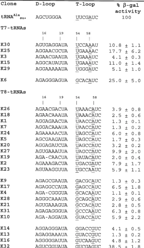
The nucleotide sequences ofthe selected tRNA clones, as deduced from their genes,
are presented in Table I. Only the sequences ofthose clones whose activity was at least 1%
of the control are given. Comparison to the original design revealed six clones with a nucleotide deletion in the T-loop and three clones with a deletion in the D-loop, providing respectively for a seven-member T-loop or a nine-member D-loop. h two clones, K25 and K30, mutations affected the non-randomized part of the T-loop, deleting respectively U59 and C60. No other mutation outside the randomized regions was found. For eight clones arbitrarily chosen from Table I, the in vivo level of the suppressor tRNA and of its arninoacylated form was determined by acid polyacrylarnide gel electrophoresis followed by hybridization with a specific probe complementary to the anticodon stem and loop. For
compared to that oftRNA’+ (Fig. 3). For each clone, most of the tRNA was found in the aminoacylated form.
A natysis oft!te n ucteotide sequeit ces
In the experiments described above, on average, only one in every 1,000 clones showed a detectable level of the nonsense suppression activity. This indicated that the nucleotide sequence space available for the DT region was rather small. A systematic analysis of the sequences of the selected clones could help to reveal the rules imposed on the structure ofthis region in functional tRNAs.
We started the analysis with the “quasi-normal” clones, those that had the normal seven nucleotides in the T-loop. Henceforth, we will eau such molecules T7-tRNAs, in contrast to T8-tRNAs having eight nucleotides in this loop. Analysis showed that in T7-tRNAs, the fifth position of the T-loop was always occupied by A and was the only invariable position in both randomized regions (Table I). The second most conservative nucleotide was the first one of the T-loop, which in all sequences except K6 was U. The presence of U and A respectively in the f irst and the fifth position of the T-loop would allow the formation of the reverse-Hoogsteen base pair U54-A5$ (RH-UA), as in the normal tRNAs. Although in the normal tRNAs U54 is aiways modified to T, it is not yet kriown whether it is the case in the selected tRNAs. On the other hand, because this modification does not interfere with the ability of the base to form H-bonds, its absence wotild not affect the formation of base pair U54-A5$. Another conservative feature consistent with wild-type tRNA is the presence of a purine in position 57 of ah but one T7-tRNAs. O ther r andomized n ucleotides, i ncluding ail s ix n ucleotides i n t he D -loop, w ere essentially more diversified and did flot seem to provide for a common structural pattem.
Among T8-tRNAs, half of the sequences (11 out of 22) also contained U in the first position ofthe T-loop (Table I). If this U plays the same role as it does in T7-tRNAs, there should be an A few nucleotides later that is able to form RH-UA with this U. GeneralÏy, this A could occupy either the fifth or the sixth position of the T-loop, depending on the position of the additional eighth nucleotide. Analysis showed that in those T8-tRNAs whose T-loop started with U, the only other conservative nucleotide was the A occupying the sixth position ofthe same ioop. Thus, the formation ofRH-UA ofthesetwo nucleotides would place the additional nucleotide in the region between them.
28
In ail other T$-tRNAs, the first position ofthe T-loop was occupied by G (Table I).
If this G plays a structural roie analogous to that piayed by U, its possible partner would occupy either the fifth or the sixth position of the T-ioop. None of the two positions were conserved in these sequences: the fifth nucieotide was aliowed to be either C or A, while the sixth was either A or G. To explore the abilities of both the fiflh and the sixth nucleotides to pair with the first G, we iooked for possible arrangements of three different dinucleotide combinations, GC, GA and GG that would be close to the arrangement of U and A in RH-UA. for GC, we did not find any satisfactory arrangement. However, for both combinations G A a nU G G w e found arrangements t hat are p resented in Fig. 4. Inthese arrangements, the G t hat i s e quivaient to U j n RH-UA d onates t wo h ydrogen a toms for
formation of H-bonds with atom N7 of the other purine. This purine can be either A or G.
In the latter case, an additional H-bond can be formed between N2-H and 06 of the first
and the second G, respectiveiy. The two arrangements GA and GG are superimposable in the sense that if one overiaps the positions of the giycosidic bonds of the first nucleotides, the glycosidic bonds of the second nucleotides in both arrangements would occupy about the same position. In the same sense, these two arrangements are fairly close to RH-UA. Accommodation of any of these arrangements based on the standard RH-UA wouid require
a shift and rotation of one of the bases by oniy 1.5
À
and 200, respectiveiy. Therefore, areplacement of RH-UA in the T-Ioop by either GA or GG would require only reiatively minor changes in the position of the neighboring nucleotides. To reflect the cioseness of these GA and GG arrangements to RH-UA, we will call them RH-GA and RH-GG, respectiveiy.
Further anaiysis revealed few additional nucleotide combinations iike CA and AA
seen in Fig. 4 that could also be arranged relativeiy closely to RH-UA while having a
reasonable system of H-bonds. Stili, ail these additional combinations were more distant
from R H-UAthan R H-GA or R H-GG a nd therefore, their incorporation j nto the T -loop instead of RH-UA would cause greater changes in the conformation of the whoie DT region. This latter aspect was expected to render these combinations iess preferable in this place than RH-GA or RH-GG.
The fact that GG and GA can be accommodated close to RH-UA, while GC camiot, makes the sixth rather that the fifth nucleotide of the T-ioop in T8-tRNAs the most probable partner to form a base pair with the G occupying the first position ofthis ioop. As
were mucli more diversified and did flot seem to provide for a common structural pattem. Finaiiy we can consider K6, the only T7-tRNA clone, whose T-ioop starts with G rather than with U. This clone has also A in the fifth position of the T-loop, which would aliow these G and A to forrn RH-GA, analogous to RH-UA existing in all other T7-tRNAs.
from this analysis, a clear picture emerges: in all selected tRNAs the first and the last randomized positions of the T-loop are aiways able to form a RH base pair, i.e. either RH-UA, RH-GA or RH-GG. The last randomized position is either the fifth in the T7-tRNAs o r t he s ixth j n t he T 8-T7-tRNAs. T he r egion b etween the f irst a nd the 1 ast p osition varies in length and sequence and does not seem to have a common pattem.
Modeting ofthe tRNA stritctttres
To confirm that the exchange ofRH-UA for either RH-GA or RH-GG in the T-loop did not cause any steric problem, we modeled the structure of the DT region for several clones having either RH-GA or RH-GG. After unrestrained energy minimization, the bases constituting the RH base pair always retained their juxtapositions and the inter-base H-bonds, as one can see in the example of the model for clone K31 (Fig. 5). Comparison of the models with the structure of the yeast tRNAP1e (20) showed that the whole region that
inciuded the T-stem, the RH base pair (RH-GA or RH-GG in the models and RH-UA in tRNA), as weli as nucleotides 59 and 60 was superimposable in ail structures.
DISCUSSION
The resuits presented here show that a tRNA couid be functional even if the structure of its DT region is substantially rnodified compared to the standard. Although for ail selected clones the efficiency of the nonsense codon suppression was lower than for the
tRNAAa+, it was strong enough to provide a level of the f3-gaiactosidase synthesis
sufficient to change the color of the colonies in the presence of X-Gal and to aliow ceil growth without extemal arginine. Additional examination of severai clones showed that suppressor tRNAs had a detectabie in vivo levei and existed mainly in the aminoacylated fonn.
The nucleotide sequences ofthe selected suppressor tRNAs demonstrated a range of diversity neyer seen in the natural cytosoiic tRNAs. In spite of this, the selected tRNAs constituted only a tiny fraction of the whole tRNA gene library, which implied the
30
existence of strong constraints imposed on the structure of functional tRNAs. b elucidate these constraints, we undertook a comparative analysis of the nucleotide sequences of the seiected tRNAs. It may be a littie surprising that among the seiected clones there were no clones having the wild-type sequence pattem. On the other hand, the wild-type sequence G18-G19-. .-U54-U55-C56-R57-A58 is expected to appear on average only once in $ 000 clones. Moreover, this probability can easily get beyond the technically detectable level if some additionaÏ requirements are imposed on the identity of the nucleotides ftanking the conservative dinucleotide Gi 8-Gi 9 in the D-ioop and on the additional eighth nucleotide in the T-loop. for most of the randomized nucleotides, our analysis did not find any obvious common pattem. The oniy exception consisted of the first and the last randomized positions
in the T-loop, which were aiways abie either to form RH-UA, analogous to base pair
U54-A58 in the normal tRNA5, or to mimic it closely via formation of RH-GA or R}J-GG. Modeling experiments showed that a repiacement of RH-UA with either RH-GA or RH GG did not cause any major rearrangement in the conformation of the DT region and provided for stable, sterically reasonabie tRNA structures. Because such an RH base pair can be formed in ail seiected tRNAs, its existence is judged to be one ofthe most important requirements irnposed on the structure of the DT region in a functional tRNA. In fact, this requirement has been the oniy one satisfied in ail seiected tRNAs, which allows us to conclude that the preservation of an RH base pair in the I-loop is more important for the tRNA function than that of other universai elements including inter-loop base pairs Gis-P55 and G19-C56.
Different expianations of the importance of the RH base pair for the tRNA function can be suggested. for example, this base pair couid be involved in a specific, vitally important interaction with a protein or other factor and thus should be preserved as such. However, a specific interaction like this would probabÏy not tolerate an exchange of RI-I UA for either RH-GA or RH-GG, because the juxtaposition of the gÏycosidic bonds and therefore, the conformation ofthe backbone in the two latter base pairs, however close it is
to that in RH-UA, is stiil notably different. Moreover, the three base pairs have different
chemical groups exposed on the surface and thus can hardly be recognized by the same factor. In another, more probable explanation, an RH base pair is needed to stabilize a particular conformation of a neighboring region in the tRNA structure and thus to enable this region to serve its function. We do not expect this region to inciude the top of the T