Published in: Acta Neuropathologica (2006), vol. 112, iss. 6, pp. 651-664. Status: Postprint (Author’s version)
Treating gliomas with glucocorticoids: from bedside to bench
Caroline Piette1, Carine Munaut1, Jean-Michel Foidart1, Manuel Deprez2
1Laboratoire de Biologie des Tumeurs et du Développement, Université de Liège, Liège, Belgium
2Laboratoire de Neuropathologie, Université de Liège, Tour de Pathologie B23/1, CHU Sart Tilman, 4000 Liège, Belgium
Abstract Glucocorticoids are used in the treatment of gliomas to decrease tumour-associated oedema and to
reduce the risk of acute encephalopathy associated with radiotherapy. However, the mechanisms by which glucocorticoids work are still largely unknown. In this paper, we survey the experimental and clinical evidence for the effects of glucocorticoids on tumour cell proliferation, apoptosis and sensitivity to chemotherapy, angiogenesis and vascular permeability. We then review current guidelines on the choice of molecule, dose and duration of glucocorticoid treatment for gliomas.
Keywords: glucocorticosteroid ; glioma ; cell culture ; oedema ; angiogenesis
Introduction
Since 1961, glucocorticoids have been used in the treatment of gliomas as they markedly improve clinical status and reduce surgical mortality and morbidity [27]. Their beneficial effects are attributed to a reduction of tumour-associated oedema. Glucocorticoids are therefore among the first drugs given to patients presenting with glioma and clinical or radiological signs of cerebral oedema, resulting in a dramatic, albeit transient, improvement of the patient's condition [25]. Preventive corticotherapy is also used to reduce the risk of acute encephalopathy associated with radiotherapy [25] although this indication has been challenged [75]. Interestingly, glucocorticoids interfere with chemotherapy and seem to reduce its haematopoietic toxicity [72]. As
glucocorticoids reduce the permeability of the capillary bed inside the tumour [80], they may also prevent access of chemotherapeutic drugs to the tumour [98]. Despite their widespread clinical use, glucocorticoids operate by mechanisms which are still largely unknown. In this context, the number of experimental studies on
glucocorticoids and gliomas appears surprisingly small with only 222 reports found on PubMed when searching for glucocorticoids and gliomas over the last 15 years (www.ncbi.nlm.nih.gov/entrez).
In this paper, we survey the basic mechanisms of action of glucocorticoids and the main pathways involved. We examine the evidence from in vitro experiments, dealing mainly with glioma cell proliferation. We also review the experimental studies on animal models pertaining to the control of cerebral oedema. We then review the information gained from clinical and neuro-imaging studies in patients with gliomas. Finally, we present the current indications and recommendations for the therapeutic use of glucocorticoids in this context.
Basic mechanisms of action of glucocorticoids Molecular mechanisms
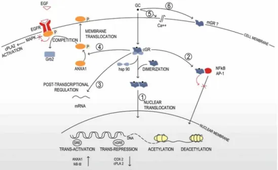
The mechanisms of action of glucocorticoids have been best studied in inflammatory diseases, such as asthma or rheumatoid arthritis [37]. Currently, glucocorticoid effects are classified as genomic or non-genomic (Fig. 1). Genomic effects are characterized by sensitivity to transcription and transduction inhibitors [86] and operate through three main mechanisms: trans-activation (which accounts for most major secondary effects of glucocorticoids); trans-repression, mediation of therapeutic anti-inflammatory effects; and post-transcriptional regulation. Non-genomic mechanisms drive more rapid effects such as activation of signalling cascades [51]. The genomic effects of glucocorticoids are mediated by a cytoplasmic glucocorticoid receptor (GR). Multiple isoforms of GR are generated by alternative RNA splicing, alternative translation initiation of the mRNA and post-translational modifications. GR-β is the best-characterized splice variant. It does not bind to
glucocorticoids, but binds to DNA, potentially interfering with the action of glucocorticoids [105]. The
inactivated GR is bound to a protein complex, including two molecules of 90 kDa heat shock protein (hsp90), a 59 kDa immunophilin protein, and various other inhibitory proteins. Glucocorticoids are thought to diffuse freely across the cell membrane because of their lipophilicity. After glucocorticoid-GR binding, hsp90 dissociates, exposing two nuclear localization signals [5].
Activation of gene transcription, or trans-activation, occurs after nuclear translocation of the glucocorticoid-GR complex, which forms a homodimer and binds to DNA at consensus sites named glucocorticoid responsive elements (GRE), inducing an enhancement of gene transcription. However, several genes have been shown to be trans-activated by glucocorticoids in the absence of a GRE consensus sequence in their promoter region. Target genes include, for example, annexin A1 (ANXA1), an inhibitor of the cytoplasmic phospholipase A2 (cPLA2),
IκB-α, an inhibitor of nuclear factor-κB (NF-κB) (reviewed in [5]) and mitogen-activated protein kinase phosphatase-1 (MKP-1), which inhibits ERK, JNK and p38 mitogen-activated protein kinase (MAPK) activities [74]. Glucocorticoids can also indirectly enhance gene transcription by inducing acetylation of the core histones of the nucleosomes. This leads to the unwinding of DNA, allowing the transcription factors to gain access to their binding sites and leading to enhanced gene transcription (reviewed in Hayashi [37]).
Fig. 1 Molecular mechanisms of glucocorticoids. Genomic mechanisms: glucocorticoids diffuse freely across the cell membrane. In the cytoplasm, the inactivated GR is linked to a protein complex, including two molecules of 90 kDa heat shock protein (hsp90). After glucocorticoid-GR binding, the protein complex dissociates, exposing two nuclear localization signals. The activated GR translocates into the nucleus as a homodimer. Activated GR may either enhance gene transcription via direct transactivation and acetylation, or decrease gene transcription via direct trans-repression and deacetylation (1). Activated GR may inhibit the activity of AP-1 or NF-κB by direct protein-protein interaction (indirect trans-repression) (2). Activated GR may exert post-transcriptional gene regulation via alterations of the mRNA turnover or translation (3). Non-genomic mechanisms: activated GR induces phosphorylation and activation of ANXA1. Activated ANXA1 translocates to the cell membrane and inhibits the EGF-dependent activation of cPLA2 by blocking the recruitment of Grb2 to the activated EGFR (4). Glucocorticoids induce alterations of the physicochemical properties of the cell membrane (5). Some non-genomic effects of glucocorticoids are thought to be mediated by a non-classical membrane-associated GR (6)
It is currently admitted that the major anti-inflammatory effects of glucocorticoids are linked to direct and indirect repression of the transcription of key genes of the inflammatory and immune response. Direct repression of gene transcription (trans-repression) occurs when the homodimeric-activated GR binds to negative
glucocorticoid responsive elements (nGRE), "composite GREs" or less well-defined regions of DNA regulating the transcription of target genes, as demonstrated for cyclooxygenase 2 (COX2) and cPLA2 (reviewed in Barnes [5]). Indirect repression of gene transcription results from inhibition of the activity of pro-inflammatory
transcription factors, such as activator protein-1 (AP-1) and NF-κB [56]. The negative regulation of AP-1 and NF-κB seems to be a key mechanism of glucocorticoid anti-inflammatory effects and occurs at different levels. First, activated GR may bind to AP-1 or NF-κB via a direct protein-protein interaction, resulting in mutual repression of the transcriptional activity [55]. Glucocorticoids may also antagonize the tumour necrosis factor alpha (TNF-α)-induced phosphorylation and activation of AP-1 [31]. Finally, the glucocorticoid-GR complex, probably through binding to a co-repressor molecule, may deacetylate histones, increasing DNA coiling and thus preventing the binding of transcription factors and leading to gene repression (reviewed in Hayashi [37]). Post-transcriptional gene regulation is included among the genomic effects of glucocorticoids because it affects gene expression and is sensitive to protein synthesis inhibitors (reviewed in Stellato [86]). This mechanism consists of alterations of the mRNA turnover or translation and has been demonstrated for IL-1α, IL-6, IL-8, IFN-β and granulocyte-macrophage colony-stimulating factor (GM-CSF) [86] and TNF [74].
The classical genomic modes of action do not explain some of the rapid effects of glucocorticoids. These rapid effects are mediated by non-genomic pathways and are insensitive to inhibitors of transcription and of protein synthesis (reviewed in Falkenstein [22] and Stellato [86]). Some of these effects do not require GR transduction and are based on alterations of the physicochemical properties of the cell membrane that modify levels of intracellular calcium [94]. Other rapid effects of glucocorticoids involve activation of the GR. This is the case in the A549 human adenocarcinoma cell line, where, after epidermal growth factor receptor (EGFR) activation,
Published in: Acta Neuropathologica (2006), vol. 112, iss. 6, pp. 651-664. Status: Postprint (Author’s version)
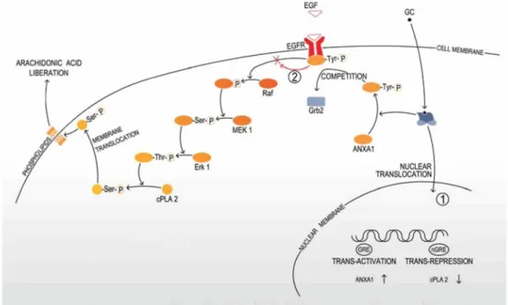
dexamethasone inhibits the MAPK-dependent activation of cPLA2 and the subsequent release of arachidonic acid (Fig. 2). This effect is mediated through dexamethasone-induced phosphorylation of ANXA1 which blocks the recruitment of the growth factor receptor-bound protein 2 (Grb2) to the activated EGF receptor (EGFR) [17]. This may be particularly relevant in gliomas where aberrant EGFR signalling has been recognized as an
important step in malignant progression [62]. In a human folliculo-stellate cell line, dexamethasone has been shown to induce a rapid serine phosphorylation and membrane translocation of ANXA1 via a mechanism that requires MAPK, phosphatidylinositol 3-kinase and calcium-dependent PKC pathways [83]. Finally, some non-genomic effects of glucocorticoids are thought to be mediated by a non-classical membrane-associated GR. So far, two putative membrane receptors have been identified: a 63 kD acidic glycoprotein found in the amphibian
Taricha granulosa [65] and a modified form of the classical GR, which could be linked to
glucocorticoid-induced lysis of lymphoma cells and in thymic involution and apoptosis [28, 29].
Glucocorticoid receptor-mediated effects can be experimentally modulated by RU486 (mifepristone), a GR antagonist which also shows anti-progesterone activity [11]. RU486 binds to GR with high affinity and impedes the dissociation of the protein complex, subsequently decreasing or slowing the formation of the activated receptor. Once linked to GR, RU486 may bind to DNA but is unable to directly modulate gene transcription. RU486 may also modulate the glucocorticoid-induced indirect repression of gene transcription. Interestingly, RU486 has been shown to possess GR agonistic activities depending on the intervention of other signalling pathways [11].
Fig. 2 Glucocorticoid inhibition of the MAPK-dependent release of arachidonic acid. The recruitment of the adapter protein Grb2 to the activated EGFR initiates the activation of the kinase Raf, MAPK/Erk kinase (MEK) and Erk. Activated Erk
phosphorylates specific serine residues within cPLA2, leading to membrane translocation of the enzyme and arachidonic acid liberation. Glucocorticoids inhibit the arachidonic acid liberation through genomic mechanisms (1), by direct trans-activation of ANXA1 and direct trans-repression of cPLA2 and GR-dependent non-genomic mechanisms (2) by inducing rapid phosphorylation of ANXA1, which blocks the recruitment of Grb2 to the activated EGFR. Tyr tyrosine, Ser serine, Thr threonine
Glucocorticoid-mediated apoptosis and cell survival
Glucocorticoids are able to influence cell survival in a type-specific manner and may either induce apoptosis (as in lymphoid cells, bone, hippocampus, eosinophils, fibroblasts and some small cell lung cancer cells), or promote cell survival (in erythroblasts, neutrophils, mammary gland, liver and fibroblasts) [77].
The mechanisms by which glucocorticoids induce apoptosis are not yet fully understood (reviewed in Schmidt [77] and Greenstein [35]). It is well established that their pro-apoptotic activity is strictly dependent upon the presence of GR. Nevertheless, it is still unclear if glucocorticoids act through transactivation, trans-repression or both. In an effort to identify glucocorticoid-targeted genes involved in apoptosis, Schmidt et al. [77] did a bioinformatic meta-analysis of eight expression microarray datasets and divided the candidate pro-apoptotic genes into three groups. The first group included genes directly involved in cell death (e.g. IκB-α and GILZ, an AP-1 inhibitor) and survival (e.g. the proto-oncogene c-myc). The second group comprised genes whose regulation might lead to cellular distress and subsequent activation of the apoptotic machinery (e.g. thioredoxin
interacting protein, which contributes to the oxidative stress). The last group was formed by genes that are not involved in cell death, such as genes counteracting the apoptotic response (e.g. receptors for TGFβ and for IL-7). Parallel to these direct transcriptional effects, indirect trans-repression of NF-κB or AP-1 is thought to be an important pathway of glucocorticoid-induced apoptosis.
In contrast, glucocorticoids promote survival in several cell types. They induce the proliferation of chicken erythroid progenitors, an effect which may be due to a GR-dependent transcriptional activation of c-Myb, a major protein of haematopoiesis [99]. Glucocorticoids also enhance neutrophil survival, probably via up-regulation of the expression of leukotriene B4 Receptor-1 (BLT1), leading to enhanced cell response to the
antiapoptotic effects of leukotriene B4 [85]. Glucocorticoids inhibit involution and programmed cell death of
mouse mammary gland, by inducing impairment of AP-1 activity [23]. In human granulosa cells, glucocorticoids almost completely inhibit the apoptosis triggered by serum deprivation, cyclic adenosine 3',5'-monophosphate (cAMP) and activation of the tumour suppressor gene p53, by up-regulating the antiapoptotic protein Bcl-2. Interestingly, the same authors showed that in myeloid leukaemia cells, glucocorticoids induced apoptosis via down-regulation of Bcl-2 [76]. Costas et al. [14] showed that glucocorticoids protect fibroblasts from TNF-induced apoptosis, by a pathway that does not involve NF-κB trans-repression. In rat hepatoma cells, Evans-Storms et al. [21] demonstrated that glucocorticoids suppress apoptosis— induced by serum deprivation—by increasing NF-κB activity in a GR-dependent manner. Finally, in thymocytes, Jondal et al. [43] suggested that basal levels of glucocorticoids might promote cell growth while high levels might induce apoptosis.
In the brain, glucocorticoids may also reduce neuron survival by amplifying glutamate excito-toxicity. Indeed, glucocorticoids have been shown to alter glutamate uptake by modulating the levels of excitatory amino acid transporters (EAAT) on astrocytes and microglia through TNF-α inhibition [41].
Therefore, it appears that depending on cell type, glucocorticoids can either induce apoptosis or promote cell survival. This paradoxical effect is probably related to cell-specific patterns of transcription factors, co-activators, co-repressors and panels of GR isoforms.
Effects of glucocorticoids on glioma: evidence from the laboratory Glioma cell proliferation: in vitro studies
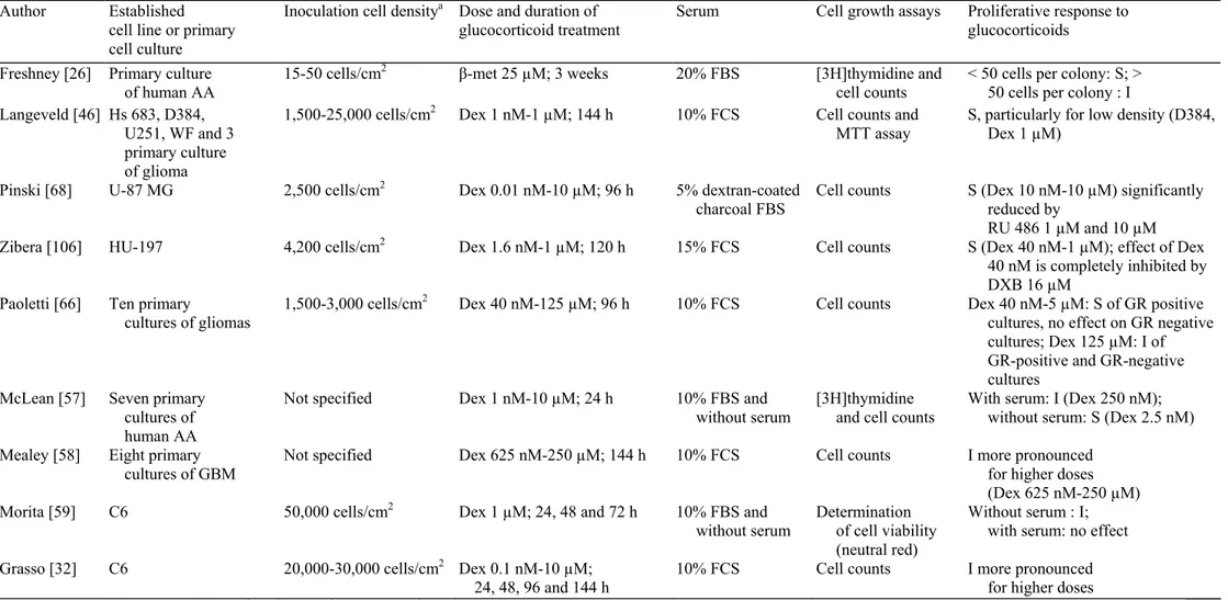
The proliferative activity of glucocorticoids on glioma cell lines has been studied for more than 30 years and both stimulatory [46, 57, 66, 68, 106] and inhibitory [30, 44, 57, 59, 66] effects have been observed. A careful survey of these reports suggests that at least some of the discrepancies between studies result from the use of non-comparable experimental conditions, most notably regarding cell line type, cell culture density,
glucocorticoid dose and culture medium (presence or absence of serum) (Table 1).
Cell density in glioma culture markedly affects the response to glucocorticoids as first demonstrated by Freshney et al. [26]. Using a primary culture derived from a human anaplastic astrocytoma in a 20% serum medium, they found that low-density cultures (below 50 cells per colony) proliferate in response to betamethasone at 25 µM, whilst cell proliferation is inhibited at higher cell density. This suggests that cell-cell interactions alter the proliferative response of glioma cells to glucocorticoids. Using dexamethasone at 1 µM in a D384 human cell line 10 % serum model, Langeveld et al. [46] made a similar observation and noticed that the proliferative effect of glucocorticoids decreases with higher inoculation cell density. They suggested that increasing cellular density could lower GR expression and consequently reduce the growth stimulatory effects of glucocorticoids.
Unfortunately, the authors could not confirm their hypothesis as the number of cells necessary to perform receptor-binding studies could only be obtained from confluent monolayers [46]. Interestingly though, in a model of rat C6 cell line cultured in the presence of serum, Vielkind et al. [96] showed that GR level was influenced by cell density and was higher in areas where cells had reached confluency. In short, it seems that glucocorticoids have stimulatory effects on cell proliferation at low cell density but tend to become inhibitory at higher cell density, possibly in relation to modifications of GR expression.
The dose at which glucocorticoids are given also orients the cell culture response towards proliferation or inhibition. Glucocorticoid concentrations used in experiments range from 0.01 nM to 250 µM. Both stimulatory and inhibitory effects have been described. In primary cultures of human glioblastoma and rat C6 glioma cell lines, dexamethasone exerts an inhibitory effect at high concentrations [58, 32]. When exposed to
dexamethasone or methylprednisolone at doses of 250 µM, glioma cells develop significant toxic manifestations (swelling and vacuolization of the cytoplasm and cell processes with disruption of the cell membranes) while their cytology is not altered at concentrations between 0.625 and 6.25 µM [58]. Accordingly, Paoletti et al. [66] also observed an inhibition of primary glioma cultures at dexamethasone 125 µM, produced by GR-independent mechanisms, such as membrane alterations. By contrast, at low concentrations, dexamethasone usually has a proliferative effect in cultures supplemented with serum. D384 cell line grown in a 10% serum medium proliferates in response to dexamethasone. This stimulatory effect is maximal at 0.1 µM and disappears for
P Status:
ublished in: Acta Neuropathologica (2006), vol. 112, iss. 6, pp. 651-664. Postprint (Author’s version)
concentrations below 0.1 nM [46]. Using U-87 MG cells complemented with serum, Pinski et al. [68] observed that dexamethasone stimulates cell proliferation for concentrations between 10 nM and 10 µM, with a maximal effect at 10 µM. Thus, it appears that in the presence of serum, dexamethasone exerts a physiological dose-dependent proliferative effect on glioma cultures for concentrations between 0.1 nM and 10 µM; concentration above 125 µM result in cell growth inhibition due to non-specific cytotoxic effects.
The proliferative response to glucocorticoids also depends on the expression of GR in the cell culture. GR has been detected both in glioma cell lines [61] and in human GBM samples [20]. Its level of expression depends on the cellular density and is higher in areas of cell confluency [96]. Using the U-87 MG cell line in the presence of serum, Pinski et al. [68] demonstrated that the proliferative response to dexamethasone between 10 nM and 10 µM is significantly reduced in the presence of the GR antagonist RU 486, at concentrations of 1 µM and 10 µM. Zibera et al. [106] obtained similar results with the HU-197 cell line in the presence of serum. They showed that the 17 beta-carboxamide steroid DXB, a GR antagonist that competes with dexamethasone for binding to the GR but does not trigger the glucocorticoid effect, used at 16 µM, completely inhibits the proliferative effect of dexamethasone at 40 nM. Paoletti et al. [66] characterized the GR status of nine primary glioma cell cultures grown in the presence of serum and obtained five GR-positive and four GR-negative cultures. They showed that dexamethasone in concentrations ranging from 40 nM to 5 µM stimulates the proliferation of four of the five positive cultures, while dexamethasone at the same concentrations does not influence the growth of the GR-negative cultures. Using dexamethasone at 125 µM, they observed a significant inhibition of all the cultures, independently of their GR status. In summary, it appears that the presence of GR is necessary [68, 106] but not sufficient [66] to explain the proliferative effects of dexamethasone below 10 µM. On the other hand, the inhibitory effects of dexamethasone above 125 µM do not seem mediated by GR, but rather by aspecific mechanisms, such as membrane alterations [66].
Two studies have specifically addressed the influence of serum complementation or deprivation on the response of cell culture to glucocorticoids. Using dexamethasone at 1 nM on a primary culture of anaplastic astrocytoma, McLean et al. [57] obtained an inhibition of astrocytoma cell proliferation in the presence of 10% foetal bovine serum (FBS). By contrast, glucocorticoids restored the proliferation of astrocytoma cells which had been markedly reduced by serum deprivation. In a more recent paper, Morita [59] made the opposite observation and reported that in serum-free cultures dexamethasone at 1µM inhibits the proliferation of C6 rat cells, but has no effect in the presence of 10% FBS. In this model, dexamethasone is thought to exacerbate the cytotoxic effect of serum deprivation which depends on the activation of GR [59].
Several authors have studied in vitro how dexamethasone interacts with drugs used for chemotherapy. In 1977, using rat C6 glioma cells, Grasso et al. [33] showed that the growth inhibition induced by the antineoplastic agent BCNU was accentuated when cells were pre-treated by dexamethasone 1 µM but not when both drugs were given in combination. In 1997, Weller et al. [98] studied the in vitro interactions between dexamethasone and several anti-neoplastic agents (ACNU, VM-26, vincristine, cytarabine, methotrexate and adriamycin) in a T98G and a LN-229 serum-free model. Dexamethasone was added 24 h prior to anti-neoplastic agents and maintained for 72 h. The authors showed that dexamethasone (maximal effect at 100 nM) attenuates cytotoxic and growth inhibitory effects of all chemotherapy drugs. Nevertheless, the withdrawal of dexamethasone after 7 days of pre-treatment resulted in significant restoration of sensitivity to most drugs tested. Using T98G and C6 glioma cells, Wolff et al. [100, 101] showed that dexamethasone at 1µM induces partial resistance to
cisplatinium at 50 µM for 72 h, by a mechanism involving the GR. More recently, dexamethasone has been shown to protect glioblastoma U87MG [18] and T98G cell lines [87] against temozolomide-induced apoptosis. In addition, dexamethasone may interfere with suicide gene therapy as in the Herpes Simplex Virus thymidine kinase gene (HSV-tk) model [73]. In conclusion, in vitro studies suggest that dexamethasone antagonizes the cytotoxic and anti-proliferative effects of several anti-neoplastic agents. This observation highlights the importance of timing in combined anti-tumour chemotherapy.
Table 1 Glioma cell proliferation: in vitro studies
Author Established
cell line or primary cell culture
Inoculation cell densitya Dose and duration of
glucocorticoid treatment Serum Cell growth assays Proliferative response to glucocorticoids Freshney [26] Primary culture
of human AA 15-50 cells/cm
2 β-met 25 µM; 3 weeks 20% FBS [3H]thymidine and
cell counts < 50 cells per colony: S; > 50 cells per colony : I Langeveld [46] Hs 683, D384,
U251, WF and 3 primary culture of glioma
1,500-25,000 cells/cm2 Dex 1 nM-1 µM; 144 h 10% FCS Cell counts and
MTT assay
S, particularly for low density (D384, Dex 1 µM)
Pinski [68] U-87 MG 2,500 cells/cm2 Dex 0.01 nM-10 µM; 96 h 5% dextran-coated
charcoal FBS
Cell counts S (Dex 10 nM-10 µM) significantly reduced by
RU 486 1 µM and 10 µM
Zibera [106] HU-197 4,200 cells/cm2 Dex 1.6 nM-1 µM; 120 h 15% FCS Cell counts S (Dex 40 nM-1 µM); effect of Dex
40 nM is completely inhibited by DXB 16 µM
Paoletti [66] Ten primary
cultures of gliomas 1,500-3,000 cells/cm
2 Dex 40 nM-125 µM; 96 h 10% FCS Cell counts Dex 40 nM-5 µM: S of GR positive
cultures, no effect on GR negative cultures; Dex 125 µM: I of GR-positive and GR-negative cultures
McLean [57] Seven primary cultures of human AA
Not specified Dex 1 nM-10 µM; 24 h 10% FBS and
without serum [3H]thymidine and cell counts With serum: I (Dex 250 nM); without serum: S (Dex 2.5 nM) Mealey [58] Eight primary
cultures of GBM Not specified Dex 625 nM-250 µM; 144 h 10% FCS Cell counts I more pronounced for higher doses (Dex 625 nM-250 µM)
Morita [59] C6 50,000 cells/cm2 Dex 1 µM; 24, 48 and 72 h 10% FBS and
without serum Determination of cell viability (neutral red)
Without serum : I; with serum: no effect
Grasso [32] C6 20,000-30,000 cells/cm2 Dex 0.1 nM-10 µM;
24, 48, 96 and 144 h
10% FCS Cell counts I more pronounced
for higher doses
a Surface used for conversion: 96-well, 0.32 cm2; 24-well, 2 cm2; 6-well, 9.6 cm2
MTT 3-(4,5-dimethylthiazol-2-yl)-2,5-diphenyltetrazolium bromide, AA anaplastic astrocytoma, GBM glioblastoma, Dex: dexamethasone, β-met: betamethasone, S stimulation of cell proliferation, I inhibition of cell proliferation
Published in: Acta Neuropathologica (2006), vol. 112, iss. 6, pp. 651-664. Status: Postprint (Author’s version)
Tumour growth: in vivo experiments
Several in vivo studies have shown that tumour xenografts tend to regress in mice treated by glucocorticoids, improving survival [36, 90, 102, 104]. Histology of the xenografts suggests that glucocorticoids may inhibit angiogenesis. Wright et al. [104] used a mixed rat glioma cell line induced by intravenous injections of n-nitrosomethylurea and injected a cell suspension in the flank of 25 albino Wistar rats. They administered methylprednisolone acetate at 100 mg/kg twice weekly from the day of transplantation. None of the 13 rats receiving dexamethasone developed the neoplasm, while all 12 of the controls produced tumour. Tamargo et al. [90] generated subcutaneous gliomas in Fischer 344 rats by implantation of a 2-4 mm3 piece of 9L gliosarcoma
and studied the effect of local treatment by cortisone acetate on tumour growth. After 14 days, they observed a significant inhibition of tumour growth in animals treated by cortisone acetate. In parallel, they studied the effect of cortisone given locally on angiogenesis in VX2 carcinomas implanted in 60 rabbit corneas. After 21 days, they obtained a significant inhibition of angiogenesis by cortisone acetate, suggesting a vascular component in the tumour growth inhibition induced by glucocorticoids. Guerin [36] and Wolff [102] injected 9L and C6 rat glioma cells into the left putamen of syngenic rats. Two days after implantation, they administered
dexamethasone at 3 mg/kg/day and obtained a significant inhibition of tumour growth associated with a reduction of vascular density and an enhancement of survival. Using a rat intra-cerebral 9L gliosarcoma model, several authors [60] demonstrated that intra-peritoneal injections of dexamethasone (1.5 mg/kg repeated twice to 3 mg/kg repeated five times) on day 10-15 after inoculation significantly decreases tumour volume and
diminishes, in vessels larger than 25 µm, the tumour vessel dilatation associated with neoplastic angiogenesis [4].
Finally, Pinski et al. [68] investigated the influence of RU486, a GR antagonist, on the growth of U-87 MG cells in nude mice. They showed that daily injection of RU486 at 0.1 or 0.5 mg/animal significantly inhibited tumour growth at 4 weeks. However, this result should be interpreted with caution since RU486 has been show to present GR agonist effects in some circumstances (see above).
In mice, dexamethasone pre-treatment enhances the anti-tumour effects and reduces the hemato-toxicity of several chemotherapeutic agents, as demonstrated by two in vivo studies [71, 97]. In C3H/HeJ mice treated with carboplatin (600 mg/m2), Rinehart et al. [72] showed that dexamethasone pre-treatment (3 or 7 days, at 0.5
mg/day) significantly reduced mortality and induced less severe granulocytes and platelet nadirs. In nude mice with subcutaneous U-87 MG glioma xenografts, Wang et al. [97] demonstrated that dexamethasone pre-treatment (0.1 mg/day for 5 days) significantly increased the therapeutic effects of combined therapy with gemcitabine and carboplatin.
Tumour-associated oedema: in vivo models
In 1967, Klatzo [45] clarified the pathophysiology of brain oedema and distinguished the "cytotoxic" and the "vasogenic" types of oedema. "Cytotoxic" oedema results from a disturbance of cell metabolism, due, for example, to ischaemic or anoxic conditions. "Vasogenic" oedema develops in case of blood-brain-barrier damage, such as in brain tumour progression.
Animal studies have helped to clarify the pathophysiology of tumour-associated oedema [39] and have shown that it has both "vasogenic" and "cytotoxic" components.
The vasogenic component is due to an increased micro-vascular permeability of the brain and blood-tumour barrier leading to the extravasation of whole plasma, even at distance from the blood-tumour. For this reason, the term "tumour-associated" oedema is now preferred to that of "peritumoural oedema". Several mechanisms contribute to the vasogenic oedema [19]. Firstly, malignant gliomas produce molecular factors that increase the permeability of the capillary bed in the surrounding brain tissue and that are likely to be the source of tumour-associated oedema [63]. Among them, vascular permeability factor (VPF) is of particular interest, its in vivo activity being inhibited by dexamethasone through a pathway implicating de novo synthesis of a polypeptide, maybe ANXA1 [15, 16]. Secondly, newly formed micro-vessels within the tumour present characteristic features that contrast with the normal architecture of the blood-brain barrier: widened intercellular junctions,
discontinuous tight junctions, membranous fenestrations, non-contiguous basement membranes, active micropinocytosis and paucity of mitochondria (M.W. Brightman and T.S. Reese, unpublished data [9, 64]). Finally, immunological factors and inflammatory processes have also been implicated, such as prostaglandins, arachidonic acid [93] and leukotrienes [79], although there is no consensus on role of the latter [92].
The cytotoxic component is induced by the generation of oedematogenous metabolites by tumour cells,
including lactate, produced by aerobic glycolysis [70]. Their osmotic activity explains the protein-free cytotoxic component of the brain oedema.
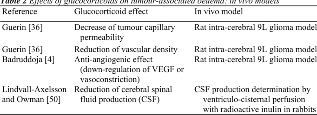
Experimental models of gliomas confirmed that tumour-associated oedema responds favourably to local or systemic corticotherapy (Table 2) [91]. Matsuoka and Hossmann [54] produced brain gliomas in nine cats by implantation of RG2 cells and injected, after 2-3 weeks, a single dose of 10 mg dexamethasone intramuscularly. In the treated group, they observed lower peritumoural white matter water content, electrolyte shifts and vascular
resistance than in the control group. Furthermore, cerebral blood flow was conserved while it was decreased in untreated group. Using a rat intra-cerebral 9L glioma model, Guerin et al. [36] studied the effect of
glucocorticoids on vascular permeability and vascular density. Two days after tumour implantation,
dexamethasone at 3 mg/kg was administered daily by intra-peritoneal injections. Glucocorticoid therapy reduced vascular permeability as demonstrated by the lower content of Evans blue in treated tumours. It also significantly reduced vascular density as shown by immunostaining for laminin. Using the same model, others [4] assessed anti-angiogenic effects of dexamethasone by magnetic resonance imaging (MRI), cerebral blood volume maps and microscopic evaluation, and demonstrated that glucocorticoid diminishes, in vessels larger than 25 µm, the tumour vessel dilatation associated with neoplastic angiogenesis. Authors suggest that this effect could be linked either to glucocorticoid-induced down-regulation of VEGF or to glucocorticoid-induced vasoconstriction. Working with C6 glioma xenografts in rodents and doses of dexamethasone in the clinical range (0.22 mg/kg/day), Swaroop et al. showed that glucocorticoids decrease tumour capillary permeability without modifying the local cerebral blood flow. They suggested that these effects are mediated by a reduction of the inducible nitric oxide synthetase (iNOS) expression within and around the tumour [78, 88, 89]. In rabbits, Lindvall-Axelsson and Owman [50] showed that betamethasone administered daily for 5 days significantly reduces cerebral spinal fluid production. Finally, Chang et al. [13] studied the effect of ANXA1 on the resolution of peritumoural oedema. They produced intra-cerebral tumours by implantation of C6 cells in Wistar rats. They showed that the cortical and tumoural water content was decreased by dexamethasone but not by ANXA1, suggesting that glucocorticoids do not exert their anti-oedema properties through induction of ANXA1. Table 2 Effects of glucocorticoids on tumour-associated oedema: in vivo models
Reference Glucocorticoid effect In vivo model
Guerin [36] Decrease of tumour capillary permeability
Rat intra-cerebral 9L glioma model Guerin [36] Reduction of vascular density Rat intra-cerebral 9L glioma model Badruddoja [4] Anti-angiogenic effect
(down-regulation of VEGF or vasoconstriction)
Rat intra-cerebral 9L glioma model Lindvall-Axelsson
and Owman [50] Reduction of cerebral spinal fluid production (CSF) CSF production determination by ventriculo-cisternal perfusion with radioactive inulin in rabbits
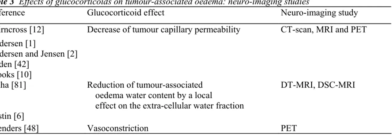
Glucocorticoids in human gliomas: clinical and neuro-imaging studies
The development of brain imaging studies—CT-scan, positron emission tomography (PET) and MRI—have led to a better understanding of the effects of glucocorticoids on tumour-associated oedema (Table 3).
CT-scan studies are based on the analysis of midline shift, ventricular compression, oedema, contrast
enhancement intensity and area, before and after glucocorticoid administration. These parameters are improved after glucocorticoid treatment in most patients. Maximum effect occurs within 2 weeks. The contrast
enhancement within tumours is reduced in patients treated with glucocorticoids suggesting a partial restoration of blood-brain barrier integrity [12].
Positron emission tomography makes it possible to non-invasively measure cerebral blood flow, cerebral blood volume, blood to tumour transport rate constant and metabolic processes, such as oxygen utilization. This technique demonstrated that glucocorticoids exert a direct effect on cerebral vessels and cause vasoconstriction as suggested by the reduction of regional cerebral blood flow (rCBF) and regional cerebral blood volume (rCB V) and enhancement of oxygen extraction without variation in oxygen utilization [48]. Furthermore, Jarden et al. [42] have demonstrated that glucocorticoids reduce tumour capillary permeability within 6 h after a single pharmacological dose.
Magnetic resonance imaging is a good tool for studying brain oedema, because the relaxation time 71 (longitudinal relaxation time) [53] and T2 (transverse relaxation time) are positively correlated to the water content of the tissue studied [1]. Derived techniques, such as the use of Gd-DTPA, 99Tc-DTPA [3], quantitative
dynamic susceptibility contrast perfusion MRI (DSC-MRI) [47] or diffusion tensor MRI (DT-MRI) [52, 81] provide an even more accurate means of studying brain oedema. Bell et al., using a mean T1 determination, found that glucocorticoids had no significant effects on water content and concluded that the clinical improvement observed in all of their patients could not be explained by any effect of glucocorticoids on the extent of brain oedema [8]. Nevertheless, the highest value in the histogram of T1 ("super-oedema") represents a more sensitive monitoring of changes in tumour-associated oedema because of its high heterogeneity. Using this technique, Andersen et al. [1] showed a significant glucocorticoid effect on brain oedema, and postulated that glucocorticoids act on the leaky blood-brain barrier within the tumour. The reduction of capillary
trans-Published in: Acta Neuropathologica (2006), vol. 112, iss. 6, pp. 651-664. Status: Postprint (Author’s version)
endothelial diffusion of Gd-DTPA in gliomas observed after glucocorticoid treatment tends to confirm this hypothesis [2]. DT-MRI and DSC-MRI studies demonstrated that glucocorticoids act by locally reducing the extra-cellular water fraction in tumour-associated brain oedema [81] and subsequently increasing perfusion in the oedematous brain [6]. Quantitative MRI can therefore measure steroid treatment response or failure in patients with malignant gliomas [82].
In short, early brain imaging studies [8] did not find any detectable decrease of tumour-associated oedema following glucocorticoids, but attributed the clinical benefit of glucocorticoids to vasoconstriction [7]. However, the development of more accurate techniques (PET, "super-oedema" determination, Gd-DTPA and DT-MRI) clearly demonstrated that glucocorticoids reduce tumour-associated oedema by a direct effect on tumour capillary permeability [1, 2, 42] and related this action to the rapid clinical improvement following glucocorticoid therapy.
Table 3 Effects of glucocorticoids on tumour-associated oedema: neuro-imaging studies
Reference Glucocorticoid effect Neuro-imaging study
Cairncross [12] Decrease of tumour capillary permeability CT-scan, MRI and PET
Andersen [1]
Andersen and Jensen [2] Jarden [42]
Brooks [10]
Sinha [81] Reduction of tumour-associated
oedema water content by a local effect on the extra-cellular water fraction
DT-MRI, DSC-MRI Bastin [6]
Leenders [48] Vasoconstriction PET
Recommendation and guidelines for corticotherapy in patients with gliomas
Although glucocorticoids are widely used in the treatment of patients with intracranial gliomas, no systematic review or meta-analysis has ever been conducted. The few published randomized trials deal with metastatic brain tumours [38, 95, 103]. In consequence, the indications, choice of glucocorticoids, dose and duration of treatment are still empirical and vary according to institutional practices.
In 2003, Sarin and Murthy [75] proposed a practical guideline for the use of glucocorticoids as medical decompressive therapy for primary and metastatic intracranial tumours. They recommend corticotherapy for patients who have intracranial tumours with symptoms of raised intracranial pressure or rapidly progressive focal neurological deficits. Corticotherapy is not recommended if the patient has seizures, or disturbances of higher mental functions, or for prophylactic use during cranial irradiation. The daily dose (6, 12 or 24 mg
dexamethasone, given in two fractionated doses) is based on the severity of headache or vomiting and the presence of focal deficit or altered consciousness. Doses up to 100 mg of dexamethasone or 2,000 mg of methylprednisolone have been proposed in the absence of improvement [49, 69]. Nevertheless, in view of major secondary effects, Sarin and Murthy [75] recommend a maximal dose of 24 mg dexamethasone outside clinical trial settings. The treatment should be stopped in the absence of improvement after 48 h. In case of improvement or stabilization, dexamethasone should be tapered every 48 h. Glucocorticoids are generally given from
diagnosis until radiotherapy is completed, a period of about 8-12 weeks [40]. High-dose steroid use and radiotherapy are sufficient to induce severe immuno-suppression in patients with primary brain cancer and prophylactic antibiotherapy has been advocated for patients with low CD4 counts [40].
Evaluation of the Human Corticotropin-Releasing Factor (hCRF) as an alternative to dexamethasone is currently in progress in a phase III study ("A Phase III Randomized, Double-Blind Study Comparing Human
Corticotropin-Releasing Factor to Dexamethasone for Control of Symptoms Associated with Peritumoral Brain Edema in Patients with Primary Malignant Glioma"). The purpose of this study is to examine the safety and effectiveness of hCRF compared to dexamethasone in patients with primary malignant glioma who require increased dexamethasone doses to control symptoms of tumour-associated oedema (http:// www.clinicaltrials.gov, identifier NCT00226668, sponsored by Neurobiological Technologies). To the best of our knowledge, only one trial has studied the use of glucocorticoids alone as a potential
chemotherapeutic agent. In this randomized controlled trial, Green et al. [34] compared the survival of patients with supratentorial malignant gliomas treated with carmustine (BCNU) or procarbazine or high-dose
methylprednisolone alone or BCNU plus high-dose methylprednisolone, in addition to radiotherapy. Patients treated with BCNU or procarbazine had a significantly longer survival than patients receiving high-dose methylprednisolone. Furthermore, the combination of BCNU plus high-dose methylprednisolone tended to be
less effective than BCNU alone in patients with poor prognosis.
Other trials included glucocorticoids as part of a larger chemotherapeutic regimen. Pendergrass et al. [67] studied the "eight-drugs-in-one-day" (eight-in-one) regimen—comprising high-dose methylprednisolone, vincristine, hydroxyurea, procarbazine, CCNU, cisplatin, cytosine arabinoside and either cyclophosphamide or
dacarbazine—in the treatment of 107 children with recurrent or incompletely resected brain tumours. Methylprednisolone was included for anti-emetic, anti-oedema and possible anti-tumour effects. After two cycles of chemotherapy and within a 4- to 6-week interval, 3 of the 10 patients with recurrent grade III and IV gliomas had an objective response including two complete responses. Seven of 17 patients with newly diagnosed grade III and IV gliomas had an objective response including three complete responses. In 1976, the Children's Cancer Study Group initiated a randomized clinical trial, CCG-943, to study the effectiveness of
chemotherapy—prednisone, CCNU and vincristine—in the treatment of 58 children with high-grade
astrocytoma [84]. Prednisone was included to minimize undesirable neurological reactions to drug therapy and for the possibility that its effects on cell membrane permeability might enhance the action of the other
chemotherapeutic agents. The addition of chemotherapy to surgery and radiotherapy significantly prolonged survival and event-free survival of children with supratentorial or cerebellar high-grade astrocytoma. In a phase III trial, Finlay et al. [24] compared the eight-in-one regimen with prednisone, CCNU and vincristine in the treatment of children with high grade astrocytomas and showed that the eight-in-one chemotherapy provides no benefit to the three-drug regimen used in the CCG-943 study.
Conclusions
Glucocorticoids have been used for more than 40 years in the treatment of intracranial gliomas. However, little is known about the precise mechanisms by which they affect tumour growth, reduce tumour-associated oedema and improve patients' clinical status. Interpretation of experimental data is difficult due to the high variability of tested parameters and experimental conditions. Furthermore, there have been surprisingly few clinical studies on the use of glucocorticoids in this context. However, in view of their widespread use in patients with gliomas and their multiple interactions with other treatments, elucidating the exact mechanisms of glucocorticoid action in gliomas should be a research priority.
Acknowledgments
Caroline Piette is Research Fellow at the Fond National de la Recherche Scientifique (FC 66846). Carine Munaut is Research Associate at the Fond National de la Recherche Scientifique. This work was supported by grants from the University of Liège (Grant C-02/82) and from the Centre Anti-Cancéreux Près l'Université de Liège.
References
1. Andersen C, Haselgrove JC, Doenstrup S, Astrup J, Gyldensted C (1993) Resorption of peritumoural oedema in cerebral gliomas during dexamethasone treatment evaluated by NMR relaxation time imaging. Acta Neurochir (Wien) 122:218-224
2. Andersen C, Jensen FT (1998) Differences in blood-tumour-barrier leakage of human intracranial tumours: quantitative monitoring of vasogenic oedema and its response to glucocorticoid treatment. Acta Neurochir (Wien) 140:919-924
3. Andersen C, Taagehoj JF, Muhler A, Rehling M (1996) Approximation of arterial input curve data in MRI estimation of cerebral blood-tumor-barrier leakage: comparison between Gd-DTPA and 99mTc-DTPA input curves. Magn Reson Imaging 14:235-241
4. Badruddoja MA, Krouwer HG, Rand SD, Rebro KJ, Pathak AP, Schmainda KM (2003) Antiangiogenic effects of dexamethasone in 9L gliosarcoma assessed by MRI cerebral blood volume maps. Neuro-oncology 5:235-243
5. Barnes PJ (1998) Anti-inflammatory actions of glucocorticoids: molecular mechanisms. Clin Sci (Lond) 94:557-572
6. Bastin ME, Carpenter TK, Armitage PA, Sinha S, Wardlaw JM, Whittle IR (2006) Effects of dexamethasone on cerebral perfusion and water diffusion in patients with high-grade glioma. AJNR Am J Neuroradiol 27:402-408
7. Beaney RP, Leenders KL, Brooks DJ (1987) Effects of dexamethasone in brain tumour patients. Lancet 1:571-572
8. Bell BA, Smith MA, Kean DM, McGhee CN, MacDonald HL, Miller JD, Barnett GH, Tocher JL, Douglas RH, Best JJ (1987) Brain water measured by magnetic resonance imaging. Correlation with direct estimation and changes after mannitol and dexamethasone. Lancet 1:66-69
9. Brightman MW, Reese TS (1969) Junctions between intimately apposed cell membranes in the vertebrate brain. J Cell Biol 40:648-677 10. Brooks DJ, Beaney RP, Lammertsma AA, Leenders KL, Horlock PL, Kensett MJ, Marshall J, Thomas DG, Jones T (1984) Quantitative measurement of blood-brain barrier permeability using rubidium-82 and positron emission tomography. J Cereb Blood Flow Metab 4:535-545
11. Cadepond F, Ulmann A, Baulieu EE (1997) RU486 (mifepristone), mechanisms of action and clinical uses. Annu Rev Med 48:129-156 12. Cairncross JG, Macdonald DR, Pexman JH, Ives FJ (1988) Steroid-induced CT changes in patients with recurrent malignant glioma. Neurology 38:724-726
13. Chang CC, Shinonaga M, Kuwabara T, Mima T, Shigeno T (1990) Effect of recombinant human lipocortin I on brain oedema in a rat glioma model. Acta Neurochir Suppl (Wien) 51:145-147
Published in: Acta Neuropathologica (2006), vol. 112, iss. 6, pp. 651-664. Status: Postprint (Author’s version)
14. Costas MA, Muller IL, Holsboer F, Arzt E (2000) Transrepression of NF-kappaB is not required for glucocorticoidmediated protection of TNF-alpha-induced apoptosis on fibroblasts. Biochim Biophys Acta 1499:122-129
15. Criscuolo GR, Lelkes PI, Rotrosen D, Oldfield EH (1989) Cytosolic calcium changes in endothelial cells induced by a protein product of human gliomas containing vascular permeability factor activity. J Neurosurg 71:884-891
16. Criscuolo GR, Merrill MJ, Oldfield EH (1988) Further characterization of malignant glioma-derived vascular permeability factor. J Neurosurg 69:254-262
17. Croxtall JD, Choudhury Q, Flower RJ (2000) Glucocorticoids act within minutes to inhibit recruitment of signalling factors to activated EGF receptors through a receptordependent, transcription-independent mechanism. Br J Pharmacol 130:289-298
18. Das A, Banik NL, Patel SJ, Ray SK (2004) Dexamethasone protected human glioblastoma U87MG cells from temozolomide induced apoptosis by maintaining Bax:Bcl-2 ratio and preventing proteolytic activities. Mol Cancer 3:36
19. Del Maestro RF, Megyesi JF, Farrell CL (1990) Mechanisms of tumor-associated edema: a review. Can J Neurol Sci 17:177-183 20. Ellemann K, Christensen L, Gjerris F, Briand P, Kruse-Larsen C (1988) Glucocorticoid receptors in glioblastoma multiforme: a new approach to antineoplastic glucocorticoid therapy. Acta Neurochir (Wien) 93:6-9
21. Evans-Storms RB, Cidlowski JA (2000) Delineation of an antiapoptotic action of glucocorticoids in hepatoma cells: the role of nuclear factor-kappaB. Endocrinology 141:1854-1862
22. Falkenstein E, Tillmann HC, Christ M, Feuring M, Wehling M (2000) Multiple actions of steroid hormones—a focus on rapid, nongenomic effects. Pharmacol Rev 52:513-556
23. Feng Z, Marti A, Jehn B, Altermatt HJ, Chicaiza G, Jaggi R (1995) Glucocorticoid and progesterone inhibit involution and programmed cell death in the mouse mammary gland. J Cell Biol 131:1095-1103
24. Finlay JL, Boyett JM, Yates AJ, Wisoff JH, Milstein JM, Ge-yer JR, Bertolone SJ, McGuire P, Cherlow JM, Tefft M (1995)
Randomized phase III trial in childhood high-grade astrocytoma comparing vincristine, lomustine, and prednisone with the eight-drugs-in-1-day regimen. Childrens Cancer Group. J Clin Oncol 13:112-123
25. Frappaz D, Chinot O, Bataillard A, Ben Hassel M, Capelle L, Chanalet S, Chatel M, Figarella-Branger D, Guegan Y, Guyotat J, Hoang-Xuan K, Jouanneau E, Keime-Guibert F, Laforet C, Linassier C, Loiseau H, Maire IP, Menei P, Rousmans S, Sanson M, Sunyach MP (2003) Summary version of the Standards, Options and Recommendations for the management of adult patients with intracranial glioma (2002). Br J Cancer 89(Suppl 1):S73-S83
26. Freshney RI, Sherry A, Hassanzadah M, Freshney M, Crilly P, Morgan D (1980) Control of cell proliferation in human glioma by glucocorticoids. Br J Cancer 41:857-866
27. Galicich JH, French LA, Melby JC (1961) Use of dexamethasone in treatment of cerebral edema associated with brain tumors. J Lancet 81:46-53
28. Gametchu B (1987) Glucocorticoid receptor-like antigen in lymphoma cell membranes: correlation to cell lysis. Science 236:456-461 29. Gametchu B, Chen F, Sackey F, Powell C, Watson CS (1999) Plasma membrane-resident glucocorticoid receptors in rodent lymphoma and human leukemia models. Steroids 64:107-119
30. Gibelli N, Zibera C, Butti G, Assietti R, Sica G, Scerrati M, Iacopino F, Roselli R, Paoletti P, Robustelli della CG (1989) Hormonal modulation of brain tumour growth: a cell culture study. Acta Neurochir (Wien) 101:129-133
31. Gonzalez MV, Jimenez B, Berciano MT, Gonzalez-Sancho JM, Caelles C, Lafarga M, Munoz A (2000) Glucocorticoids antagonize AP-1 by inhibiting the activation/phosphorylation of JNK without affecting its subcellular distribution. J Cell Biol AP-150:AP-1AP-199-AP-1208
32. Grasso RJ, Johnson CE (1977) Dose-response relationships between glucocorticoids and growth inhibition in rat glioma monolayer cultures. Proc Soc Exp Biol Med 154:238-241
33. Grasso RJ, Johnson CE, Boler RK, Moore NA (1977) Combined growth-inhibitory responses and ultrastructural alterations produced by 1,3-bis(2-chloroethyl)-1-nitrosourea and dexamethasone in rat glioma cell cultures. Cancer Res 37:585-594
34. Green SB, Byar DP, Walker MD, Pistenmaa DA, Alexander E Jr, Batzdorf U, Brooks WH, Hunt WE, Mealey J Jr, Odom GL, Paoletti P, Ransohoff J, Robertson JT, Selker RG, Shapiro WR, Smith KR Jr, Wilson CB, Strike TA (1983) Comparisons of carmustine, procarbazine, and high-dose methylprednisolone as additions to surgery and radiotherapy for the treatment of malignant glioma. Cancer Treat Rep 67:121-132
35. Greenstein S, Ghias K, Krett NL, Rosen ST (2002) Mechanisms of glucocorticoid-mediated apoptosis in hematological malignancies. Clin Cancer Res 8:1681-1694
36. Guerin C, Wolff JE, Laterra J, Drewes LR, Brem H, Goldstein GW (1992) Vascular differentiation and glucose transporter expression in rat gliomas: effects of steroids. Ann Neurol 31:481-487
37. Hayashi R, Wada H, Ito K, Adcock IM (2004) Effects of glucocorticoids on gene transcription. Eur J Pharmacol 500:51-62
38. Horton J, Baxter DH, Olson KB (1971) The management of metastases to the brain by irradiation and corticosteroids. Am J Roentgenol Radium Ther Nucl Med 111:334-336
39. Hossmann KA (1989) The pathophysiology of experimental brain edema. Neurosurg Rev 12:263-280
40. Hughes MA, Parisi M, Grossman S, Kleinberg L (2005) Primary brain tumors treated with steroids and radiotherapy: low CD4 counts and risk of infection. Int J Radiat Oncol Biol Phys 62:1423-1426
41. Jacobsson J, Persson M, Hansson E, Ronnback L (2006) Corticosterone inhibits expression of the microglial glutamate transporter GLT-1 in vitro. Neuroscience GLT-139:475-483
42. Jarden JO, Dhawan V, Moeller JR, Strother SC, Rottenberg DA (1989) The time course of steroid action on brain and blood-to-tumor transport of 82Rb: a positron emission tomographic study. Ann Neurol 25:239-245
43. Jondal M, Pazirandeh A, Okret S (2004) Different roles for glucocorticoids in thymocyte homeostasis? Trends Immunol 25:595-600 44. Kaup B, Schindler I, Knupfer H, Schlenzka A, Preiss R, Knupfer MM (2001) Time-dependent inhibition of glioblastoma cell proliferation by dexamethasone. J Neurooncol 51:105-110
45. Klatzo I (1967) Presidental address. Neuropathological aspects of brain edema. J Neuropathol Exp Neurol 26:1-14
46. Langeveld CH, Vanwaas MP, Stoof JC, Sutanto W, Dekloet ER, Wolbers JG, Heimans JJ (1992) Implication of glucocorticoid receptors in the stimulation of human glioma cell-proliferation by dexamethasone. J Neurosci Res 31:524-531
47. Law M, Yang S, Babb JS, Knopp EA, Golfinos JG, Zagzag D, Johnson G (2004) Comparison of cerebral blood volume and vascular permeability from dynamic susceptibility contrast-enhanced perfusion MR imaging with glioma grade. AJNR Am J Neuroradiol 25:746-755 48. Leenders KL, Beaney RP, Brooks DJ, Lammertsma AA, Heather JD, McKenzie CG (1985) Dexamethasone treatment of brain tumor patients: effects on regional cerebral blood flow, blood volume, and oxygen utilization. Neurology 35:1610-1616
49. Lieberman A, LeBrun Y, Glass P, Goodgold A, Lux W, Wise A, Ransohoff J (1977) Use of high dose corticosteroids in patients with inoperable brain tumours. J Neurol Neurosurg Psychiatry 40:678-682
50. Lindvall-Axelsson M, Owman C (1990) Actions of sex steroids and corticosteroids on rabbit choroid plexus as shown by changes in transport capacity and rate of cerebrospinal fluid formation. Neurol Res 12:181-186
51. Losel R, Wehling M (2003) Nongenomic actions of steroid hormones. Nat Rev Mol Cell Biol 4:46-56
52. Lu S, Ahn D, Johnson G, Cha S (2003) Peritumoral diffusion tensor imaging of high-grade gliomas and metastatic brain tumors. AJNR Am J Neuroradiol 24:937-941
53. MacDonald HL, Bell BA, Smith MA, Kean DM, Tocher JL, Douglas RH, Miller JD, Best JJ (1986) Correlation of human NMR T1 values measured in vivo and brain water content. Br J Radiol 59:355-357
54. Matsuoka Y, Hossmann KA (1981) Corticosteroid therapy of experimental tumour oedema. Neurosurg Rev 4:185-190
55. McEwan IJ, Wright AP, Gustafsson JA (1997) Mechanism of gene expression by the glucocorticoid receptor: role of protein-protein interactions. Bioessays 19:153-160
56. McKay LI, Cidlowski JA (1999) Molecular control of immune/inflammatory responses: interactions between nuclear factor-kappa B and steroid receptor-signaling pathways. Endocr Rev 20:435-459
57. McLean JS, Frame MC, Freshney RI, Vaughan PF, Mackie AE, Singer I (1986) Phenotypic modification of human glioma and non-small cell lung carcinoma by glucocorticoids and other agents. Anticancer Res 6:1101-1106
58. Mealey J Jr, Chen TT, Schanz GP (1971) Effects of dexamethasone and methylprednisolone on cell cultures of human glioblastomas. J Neurosurg 34:324-334
59. Morita K, Ishimura K, Tsuruo Y, Wong DL (1999) Dexamethasone enhances serum deprivation-induced necrotic death of rat C6 glioma cells through activation of glucocorticoid receptors. Brain Res 816:309-316
60. Morris GM, Micca PL, Coderre JA (2004) The effect of dexamethasone on the uptake of p-boronophenylalanine in the rat brain and intracranial 9L gliosarcoma. Appl Radiat Isot 61:917-921
61. Moutsatsou P, Kazazoglou T, Fleischer-Lambropoulos H, Psarra AM, Tsiapara A, Sekeris CE, Stefanis C, Vernadakis A (2000) Expression of the glucocorticoid receptor in early and late passage C-6 glioma cells and in normal astrocytes derived from aged mouse cerebral hemispheres. Int J Dev Neurosci 18:329-335
62. Nathoo N, Goldlust S, Vogelbaum MA (2004) Epidermal growth factor receptor antagonists: novel therapy for the treatment of high-grade gliomas. Neurosurgery 54:1480-1488
63. Ohnishi T, Sher PB, Posner IB, Shapiro WR (1990) Capillary permeability factor secreted by malignant brain tumor. Role in peritumoral brain edema and possible mechanism for anti-edema effect of glucocorticoids. J Neurosurg 72:245-251
64. Oldendorf WH, Cornford ME, Brown WJ (1977) The large apparent work capability of the blood-brain barrier: a study of the mitochondrial content of capillary endothelial cells in brain and other tissues of the rat. Ann Neurol 1:409-417
65. Orchinik M, Murray TF, Moore FL (1991) A corticosteroid receptor in neuronal membranes. Science 252:1848-1851
66. Paoletti P, Butti G, Zibera C, Scerrati M, Gibelli N, Roselli R, Magrassi L, Sica G, Rossi G, Dellacuna GR (1990) Characteristics and biological role of steroid-hormone receptors in neuroepithelial tumors. J Neurosurg 73:736-742
67. Pendergrass TW, Milstein JM, Geyer JR, Mulne AF, Kosnik EJ, Morris JD, Heideman RL, Ruymann FB, Stuntz JT, Bleyer WA (1987) Eight drugs in one day chemotherapy for brain tumors: experience in 107 children and rationale for preradiation chemotherapy. J Clin Oncol 5:1221-1231
68. Pinski J, Halmos G, Shirahige Y, Wittliff JL, Schally AV (1993) Inhibition of growth of the human malignant glioma cell line (U87MG) by the steroid hormone antagonist RU486. J Clin Endocrinol Metab 77:1388-1392
69. Renaudin J, Fewer D, Wilson CB, Boldrey EB, Calogero J, Enot KJ (1973) Dose dependency of decadron in patients with partially excised brain tumors. J Neurosurg 39:302-305
70. Rhodes CG, Wise RJ, Gibbs JM, Frackowiak RS, Hatazawa J, Palmer AJ, Thomas DG, Jones T (1983) In vivo disturbance of the oxidative metabolism of glucose in human cerebral gliomas. Ann Neurol 14:614-626
71. Rinehart J, Keville L, Measel J, Spiekerman AM, Burke K (1995) Corticosteroid alteration of carboplatin-induced hematopoietic toxicity in a murine model. Blood 86:4493-4499
72. Rinehart J, Keville L, Neidhart J, Wong L, DiNunno L, Kin-ney P, Aberle M, Tadlock L, Cloud G (2003) Hematopoietic protection by dexamethasone or granulocyte-macrophage colony-stimulating factor (GM-CSF) in patients treated with carboplatin and ifosfamide. Am J Clin Oncol 26:448-458
Published in: Acta Neuropathologica (2006), vol. 112, iss. 6, pp. 651-664. Status: Postprint (Author’s version)
73. Robe PA, Nguyen-Khac M, Jolois O, Rogister B, Merville MP, Bours V (2005) Dexamethasone inhibits the HSV-tk/ ganciclovir bystander effect in malignant glioma cells. BMC Cancer 5:32
74. Roger T, Chanson AL, Knaup-Reymond M, Calandra T (2005) Macrophage migration inhibitory factor promotes innate immune responses by suppressing glucocorticoid-induced expression of mitogen-activated protein kinase phosphatase-1. Eur J Immunol 35:3405-3413
75. Sarin R, Murthy V (2003) Medical decompressive therapy for primary and metastatic intracranial tumours. Lancet Neurol 2:357-365 76. Sasson R, Tajima K, Amsterdam A (2001) Glucocorticoids protect against apoptosis induced by serum deprivation, cyclic adenosine 3',5'-monophosphate and p53 activation in immortalized human granulosa cells involvement of Bcl-2. Endocrinology 142:802-811 77. Schmidt S, Rainer J, Ploner C, Presul E, Riml S, Kofler R (2004) Glucocorticoid-induced apoptosis and glucocorticoid resistance: molecular mechanisms and clinical relevance. Cell Death Differ ll(Suppl 1):S45-S55
78. Shinoda J, McLaughlin KE, Bell HS, Swaroop GR, Yamaguchi S, Holmes MC, Whittle IR (2003) Molecular mechanisms underlying dexamethasone inhibition of iNOS expression and activity in C6 glioma cells. Glia 42:68-76
79. Shinonaga M, Chang CC, Kuwabara T (1990) Relation between macrophage infiltrates and peritumoral edema. Adv Neurol 52:475-481 80. Siegal T, Soti F, Biegon A, Pop E, Brewster ME (1997) Effect of a chemical delivery system for dexamethasone (Dex-CDS) on peritumoral edema in an experimental brain tumor model. Pharm Res 14:672-675
81. Sinha S, Bastin ME, Wardlaw JM, Armitage PA, Whittle IR (2004) Effects of dexamethasone on peritumoural oedematous brain: a DT-MRI study. J Neurol Neurosurg Psychiatry 75:1632-1635
82. Sinha S, Bastin ME, Whittle IR (2003) Rapid clinical deterioration in a patient with multi-focal glioma despite corticosteroid therapy: a quantitative MRI study. Br J Neurosurg 17:537-540
83. Solito E, Mulla A, Morris JF, Christian HC, Flower RJ, Buckingham JC (2003) Dexamethasone induces rapid serine-phosphorylation and membrane translocation of annexin 1 in a human folliculostellate cell line via a novel nongenomic mechanism involving the glucocorticoid receptor, protein kinase C, phosphatidylinositol 3-kinase, and mitogen-activated protein kinase. Endocrinology 144:1164— 1174
84. Sposto R, Ertel IJ, Jenkin RD, Boesel CP, Venes JL, Ortega JA, Evans AE, Wara W, Hammond D (1989) The effectiveness of chemotherapy for treatment of high grade astrocytoma in children: results of a randomized trial. A report from the Childrens Cancer Study Group. J Neurooncol 7:165-177
85. Stankova J, Turcotte S, Harris J, Rola-Pleszczynski M (2002) Modulation of leukotriene B4 receptor-1 expression by dexamethasone: potential mechanism for enhanced neutrophil survival. J Immunol 168:3570-3576
86. Stellato C (2004) Post-transcriptional and nongenomic effects of glucocorticoids. Proc Am Thorac Soc 1:255-263
87. Sur P, Sribnick EA, Patel SJ, Ray SK, Banik NL (2005) Dexamethasone decreases temozolomide-induced apoptosis in human gliobastoma T98G cells. Glia 50:160-167
88. Swaroop GR, Malcolm GP, Kelly PA, Ritchie I, Whittle IR (1998) Effects of nitric oxide modulation on tumour blood flow and microvascular permeability in C6 glioma. Neuroreport 9:2577-2581
89. Swaroops GR, Kelly PA, Holmes MC, Shinoda J, Whittle IR (2001) The effects of dexamethasone therapy on permeability, blood flow and iNOS expression in experimental glioma. J Clin Neurosci 8:35-39
90. Tamargo RJ, Leong KW, Brem H (1990) Growth inhibition of the 9L glioma using polymers to release heparin and cortisone acetate. J Neurooncol 9:131-138
91. Tamargo RJ, Sills AK Jr, Reinhard CS, Pinn ML, Long DM, Brem H (1991) Interstitial delivery of dexamethasone in the brain for the reduction of peritumoral edema. J Neurosurg 74:956-961
92. Unterberg A, Schmidt W, Wahl M, Ellis EF, Marmarou A, Baethmann A (1991) Evidence against leukotrienes as mediators of brain edema. J Neurosurg 74:773-780
93. Unterberg A, Wahl M, Hammersen F, Baethmann A (1987) Permeability and vasomotor response ol cerebral vessels during exposure to arachidonic acid. Acta Neuropathol (Berl) 73:209-219
94. Urbach V, Walsh DE, Mainprice B, Bousquet J, Harvey BJ (2002) Rapid non-genomic inhibition ol ATP-induced Cl-secretion by dexamethasone in human bronchial epithelium. J Physiol 545:869-878
95. Vecht CJ, Hovestadt A, Verbiest HB, van Vliet JJ, van Putten WL (1994) Dose-effect relationship ol dexamethasone on Karnolsky performance in metastatic brain tumors:a randomized study ol doses ol 4, 8, and 16 mg per day. Neurology 44:675-680
96. Vielkind U, Walencewicz A, Levine JM, Bohn MC (1990) Type-II glucocorticoid receptors are expressed in oligodendrocytes and astrocytes. J Neurosci Res 27:360-373
97. Wang H, Li M, Rinehart JJ, Zhang R (2004) Pretreatment with dexamethasone increases antitumor activity ol carbopla-tin and gemcitabine in mice bearing human cancer xenogralts: in vivo activity, pharmacokinetics, and clinical implications lor cancer chemotherapy. Clin Cancer Res 10:1633-1644
98. Weller M, Schmidt C, Roth W, Dichgans J (1997) Chemotherapy ol human malignant glioma: prevention ol efficacy by dexamethasone? Neurology 48:1704-1709
99. Wessely O, Deiner EM, Beug H, von Lindern M (1997) The glucocorticoid receptor is a key regulator ol the decision between sell-renewal and differentiation in erythroid progenitors. EMBO J 16:267-280
100. Wolff JE, Denecke J, Jurgens H (1996) Dexamethasone induces partial resistance to cisplatinum in C6 glioma cells. Anticancer Res 16:805-809
101. Wolff JE, Jurgens H (1994) Dexamethasone induced partial resistance to methotrexate in C6-glioma cells. Anticancer Res 14:1585-1588
102. Wolff JE, Molenkamp G, Hotfilder M, Laterra J (1997) Dexamethasone inhibits glioma-induced formation ol capillary like structures in vitro and angiogenesis in vivo. Klin Padiatr 209:275-277
103. Wollson AH, Snodgrass SM, Schwade JG, Markoe AM, Landy H, Feun LG, Sridhar KS, Brandon AH, Rodriguez M, Houdek PV (1994) The role ol steroids in the management ol metastatic carcinoma to the brain. A pilot prospective trial. Am J Clin Oncol 17:234-238 104. Wright RL, Shaumba B, Keller J (1969) The effect ol glucocorticosteroids on growth and metabolism ol experimental glial tumors. J Neurosurg 30:140-145
105. Zhou J, Cidlowski JA (2005) The human glucocorticoid receptor: one gene, multiple proteins and diverse responses. Steroids 70:407-417
106. Zibera C, Gibelli N, Butti G, Pedrazzoli P, Carbone M, Ma-grassi L, Robustelli della CG (1992) Prolilerative effect ol dexamethasone on a human glioblastoma cell line (HU 197) is mediated by glucocorticoid receptors. Anticancer Res 12:1571-1574