HAL Id: inserm-00663564
https://www.hal.inserm.fr/inserm-00663564
Submitted on 27 Jan 2012
HAL is a multi-disciplinary open access
archive for the deposit and dissemination of
sci-entific research documents, whether they are
pub-lished or not. The documents may come from
teaching and research institutions in France or
abroad, or from public or private research centers.
L’archive ouverte pluridisciplinaire HAL, est
destinée au dépôt et à la diffusion de documents
scientifiques de niveau recherche, publiés ou non,
émanant des établissements d’enseignement et de
recherche français ou étrangers, des laboratoires
publics ou privés.
Olivier Malard, Isabelle Guisle-Marsollier, Véronique Sébille-Rivain, Christian
Verger, Christian Géraut, Catherine Gratas-Rabbia-Ré
To cite this version:
Dominique Tripodi, Sylvia Quéméner, Karine Renaudin, Christophe Ferron, Olivier Malard, et al..
Gene expression profiling in sinonasal adenocarcinoma.. BMC Medical Genomics, BioMed Central,
2009, 2 (1), pp.65. �10.1186/1755-8794-2-65�. �inserm-00663564�
Open Access
Research article
Gene expression profiling in sinonasal adenocarcinoma
Dominique Tripodi*
†1,2, Sylvia Quéméner
†1, Karine Renaudin
3,4,
Christophe Ferron
5, Olivier Malard
5, Isabelle Guisle-Marsollier
6,
Véronique Sébille-Rivain
7, Christian Verger
8, Christian Géraut
2and
Catherine Gratas-Rabbia-Ré
1,9Address: 1Inserm, UMR 892, Nantes, F-44007, France; Université de Nantes, UFR Médecine et Techniques Médicales, Nantes, F-44000, France, 2Service de Médecine du Travail et des Risques Professionnels, CHU de Nantes, Nantes, F-44093, France, 3Service d'Anatomie Pathologique, CHU de Nantes, Nantes, F-44093, France, 4Université de Nantes, UFR Médecine et Techniques Médicales, EA Biométadys, Nantes, F-44093, France, 5Service ORL, CHU de Nantes, Nantes, F-44093, France, 6Université de Nantes, UFR Médecine et Techniques Médicales, Plateforme Puces à ADN-OGP, Nantes, F-44000, France, 7Université de Nantes, UFR Médecine et Techniques Médicales, Laboratoire de Biomathématiques-Biostatistiques, Nantes, F-44000, France, 8Consultation des Pathologies Professionnelles, CH Hôtel-Dieu, Rennes, F-35000, France and 9Service de Biochimie, CHU de Nantes, Nantes, F-44093, France
Email: Dominique Tripodi* - dominique.tripodi@chu-nantes.fr; Sylvia Quéméner - sylvia.quemener@chu-brest.fr;
Karine Renaudin - karine.renaudin@chu-nantes.fr; Christophe Ferron - christophe.ferron@chu-nantes.fr; Olivier Malard - olivier.malard@chu-nantes.fr; Isabelle Guisle-Marsollier - isabelle.guisle@nantes.inserm.fr; Véronique Sébille-Rivain - veronique.sebille@univ-olivier.malard@chu-nantes.fr;
Christian Verger - christian.verger@univ-rennes1.fr; Christian Géraut - christian.geraut@univ-nantes.fr; Catherine Gratas-Rabbia-Ré - catherine.gratas@chu-nantes.fr
* Corresponding author †Equal contributors
Abstract
Background: Sinonasal adenocarcinomas are uncommon tumors which develop in the ethmoid sinus after exposure to wood dust. Although the etiology of these tumors is well defined, very little is known about their molecular basis and no diagnostic tool exists for their early detection in high-risk workers. Methods: To identify genes involved in this disease, we performed gene expression profiling using cancer-dedicated microarrays, on nine matched samples of sinonasal adenocarcinomas and non-tumor sinusal tissue. Microarray results were validated by quantitative RT-PCR and immunohistochemistry on two additional sets of tumors.
Results: Among the genes with significant differential expression we selected LGALS4, ACS5, CLU, SRI and
CCT5 for further exploration. The overexpression of LGALS4, ACS5, SRI, CCT5 and the downregulation of CLU were confirmed by quantitative RT-PCR. Immunohistochemistry was performed for LGALS4 (Galectin 4), ACS5 (Acyl-CoA synthetase) and CLU (Clusterin) proteins: LGALS4 was highly up-regulated, particularly in the most differentiated tumors, while CLU was lost in all tumors. The expression of ACS5, was more heterogeneous and no correlation was observed with the tumor type.
Conclusion: Within our microarray study in sinonasal adenocarcinoma we identified two proteins, LGALS4 and CLU, that were significantly differentially expressed in tumors compared to normal tissue. A further evaluation on a new set of tissues, including precancerous stages and low grade tumors, is necessary to evaluate the possibility of using them as diagnostic markers.
Published: 10 November 2009
BMC Medical Genomics 2009, 2:65 doi:10.1186/1755-8794-2-65
Received: 28 November 2008 Accepted: 10 November 2009
This article is available from: http://www.biomedcentral.com/1755-8794/2/65 © 2009 Tripodi et al; licensee BioMed Central Ltd.
This is an Open Access article distributed under the terms of the Creative Commons Attribution License (http://creativecommons.org/licenses/by/2.0), which permits unrestricted use, distribution, and reproduction in any medium, provided the original work is properly cited.
Background
Sinonasal adenocarcinoma is a rare cancer which usually develops in the ethmoid sinuses. It mainly develops amongst 30 to 85 year old men, with a high frequency around 60. The incidence of this type of cancer was esti-mated by the IARC (International Agency for Research on Cancer) at 0.7/100 000 in China to 1.4/100 000 in USA and 1.5/100 000 in France, and it has been reported to account for 3% of head and neck tumors [1,2]. This cancer is recognized as an occupational cancer. In fact, it is well confirmed today that sinonasal adenocarcinoma is highly correlated with duration and level (3.5 mg/m3) of wood
dust exposure [3,4]. As such, woodworkers have very high risks of nasal cancer (Standard Mortality Ratio: 310, 95% CI, 160-560) [5,6]. Other suspected risk factors include exposure to leather dust [7,8], metals such as chromium or nickel [9,10], and formaldehyde, although the epide-miological data regarding this chemical are partly conflict-ing [4,11]. In contrast to most other head and neck cancers, alcohol and tobacco do not seem to be risk fac-tors [12]. Although the etiology of sinonasal adenocarci-noma is well-defined, its wood-related pathogenesis is not clearly understood [13]. From a morphological and his-topathological point of view, these tumors are mainly intestinal-type adenocarcinomas [14,15] and demon-strate characteristic changes, such as gland formation, seen in adenocarcinomas at other anatomic sites. The most common clinical symptoms (nosebleeding, rhinitis and nasal obstruction) are not specific and this explains the delay in the diagnosis and the frequency of advanced stages. The conventional treatment includes local surgery [16] associated with radiotherapy. The survival rate at 5 years is only about 50% and it is important to point out that secondary effects are considerable due to the location of these tumors [17]. Therefore, early detection and alter-native treatments are necessary. This requires, however, better knowledge of the molecular mechanisms involved in the development of these tumors. Although many reports on epidemiological studies and risk factors of sinonasal adenocarcinomas have been published, only a small number of reports have been made so far on their molecular biology. As reviewed recently by Llorente et al [13], several groups have proceeded with molecular stud-ies of sinonasal adenocarcinomas. However these focused on specific genes, such as ERBB1, CCND1, ERBB2, TP53, K-ras, COX-2 or APC, involved either in other head and neck tumors or in colorectal cancer because of morpho-logical similarities [13,18,19]. Two groups reported com-parative genomic hybridization in ethmoid sinus adenocarcinomas and revealed hot spots of chromosomal imbalances [20-22]. Global genetic modifications (micro-nuclei and chromosomal aberrations) were also found in buccal epithelial cells and blood lymphocytes of wood furniture workers [23]. The conclusion of all these
investi-gations is that ethmoid sinus adenocarcinomas have their own molecular development pathway.
Thus, to identify genes involved in this pathway, we pio-neered a gene expression profiling study of 9 sinonasal adenocarcinomas versus their matched normal tissue. We found 186 genes with significant differential expression. The further evaluation of several selected genes by reverse-transcription quantitative real-time-PCR (RT-qPCR) and immunohistochemistry (IHC), on two additional valida-tion samples, confirmed the microarray data. We have hereby opened up a new field of investigation into biomarkers of this tumor type and have identified two promising candidate genes: LGALS4 and CLU.
Methods
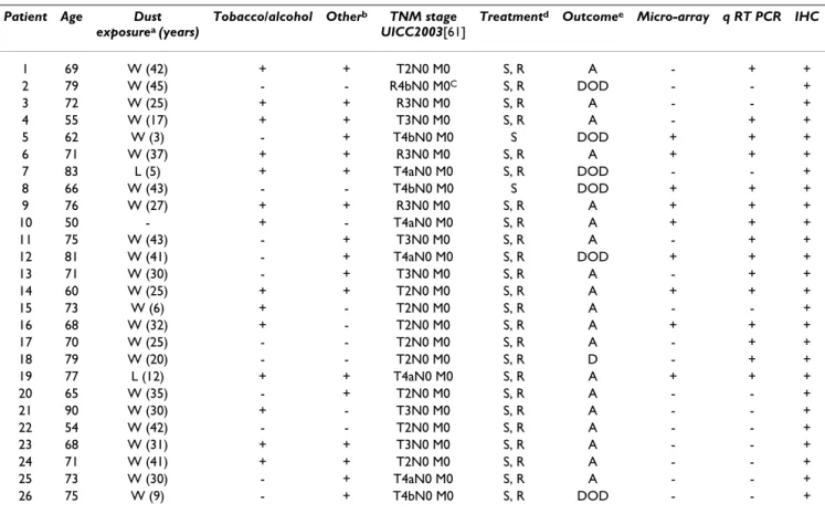
SubjectsOur study included 26 patients. A first set of 19 male patients undergoing surgery for ethmoid sinus adenocar-cinomas were initially included between 2004 and 2006. Following this, a second set of 7 patients whose samples were collected from 2006 to 2007 was used to complete the immunohistochemistry study.
This project was approved by the Clinical Board of the Centre Hospitalo-Universitaire of Nantes and all included patients provided written informed consent in accordance with French regulations and the Declaration of Helsinki. All patients answered a codified questionnaire regarding occupational exposures, addictive consumption and fam-ily history. Twenty three patients out of 26 were exposed to wood dust and most of them had other occupational exposures (such as solvents and pesticides) sometimes combined with tobacco and/or alcohol. Two patients were exposed to leather dust (P7, P19), whereas only one (P10) had no occupational exposure (Table 1). Patient ages ranged from 50 to 80 years with a mean age of 69 years. To date, six patients have died as a direct result of their disease (Table 1).
Tissue specimens
Two pieces of tissue samples were obtained from each patient undergoing surgery for ethmoidal adenocarci-noma: one from the tumor and one non-tumor sample obtained from the opposite sinus at 3 to 4 cm distance (herein referred to as "normal" tissue). All samples were immediately frozen and stored at -80°C. Remaining sur-gical resections of tumors and normal tissue were fixed in 10% formalin and embedded in paraffin before histolog-ical examination and diagnosis according to World Health Organization recommendations [24]. Two main types of sinonasal adenocarcinoma are recognized in the ethmoid sinus based on the histological similarity to ade-nocarcinoma of the intestine: Intestinal Type Adenocarci-noma (ITAC) and non-Intestinal Type AdenocarciAdenocarci-noma
(non-ITAC). ITAC can be further divided into five catego-ries [15,25]: the "papillary-type" (well-differentiated ade-nocarcinoma), the "colonic-type" (moderately-differentiated adenocarcinoma), the "solid-type" (poorly-differentiated adenocarcinoma), the "mucinous--type" and the "mixed--type" composed of a mixture of the pre-viously defined patterns. Non-ITAC are divided into low-grade and high-low-grade subtypes.
RNA extraction
On each matched normal and pathological tissue speci-men from patients P1 to P19, two RNA extractions were performed from about 40 frozen sections (10 μm thick) using a Total RNA and Protein Isolation kit (Macherey-Nagel, Düren, Germany) according to the manufacturer's instructions. For each sample, the first and last sections were stained with hemalun/phloxin to confirm the histol-ogy and to evaluate the percentage of tumor tissue. 10 samples had to be eliminated for microarray analysis because of necrosis or a too low percentage of
non-necrotic tumor tissue (less than 50%). Six out of these ten patients were included in the validation process by RT-qPCR as this technique is more sensitive than microarrays for identifying tumor cells within a sample. The other samples were completely excluded from the molecular analysis (Table 1).
The quantity and quality of each RNA were respectively evaluated with the NanoDrop® ND-1000 spectrophotom-eter (Nanodrop Technologies, Wilmington, DE) and the Agilent 2100 Bioanalyser (Agilent, Santa Clara, CA). The RNAs extracted were of good quality and the RNA integ-rity number (RIN) was >7.5 in all cases [26].
RNA amplification and microarray hybridization
Cancer-dedicated microarrays were prepared in-house (ADN-OGP- Microarray Platform Nantes, France) with methods previously described in detail [27,28] using 22,175 probe sets (50-mer oligonucleotides - MWG Bio-tech, Roissy, France) interrogating 6,864 genes involved in
Table 1: Summary of clinical data and use of tumor samples
Patient Age Dust
exposurea (years)
Tobacco/alcohol Otherb TNM stage
UICC2003[61]
Treatmentd Outcomee Micro-array q RT PCR IHC
1 69 W (42) + + T2N0 M0 S, R A - + + 2 79 W (45) - - R4bN0 M0C S, R DOD - - + 3 72 W (25) + + R3N0 M0 S, R A - - + 4 55 W (17) + + T3N0 M0 S, R A - + + 5 62 W (3) - + T4bN0 M0 S DOD + + + 6 71 W (37) + + R3N0 M0 S, R A + + + 7 83 L (5) + + T4aN0 M0 S, R DOD - - + 8 66 W (43) - - T4bN0 M0 S DOD + + + 9 76 W (27) + + R3N0 M0 S, R A + + + 10 50 - + - T4aN0 M0 S, R A + + + 11 75 W (43) - + T3N0 M0 S, R A - + + 12 81 W (41) - + T4aN0 M0 S, R DOD + + + 13 71 W (30) - + T3N0 M0 S, R A - + + 14 60 W (25) + + T2N0 M0 S, R A + + + 15 73 W (6) + - T2N0 M0 S, R A - - + 16 68 W (32) + - T2N0 M0 S, R A + + + 17 70 W (25) - - T2N0 M0 S, R A - + + 18 79 W (20) - - T2N0 M0 S, R D - + + 19 77 L (12) + + T4aN0 M0 S, R A + + + 20 65 W (35) - + T2N0 M0 S, R A - - + 21 90 W (30) + - T3N0 M0 S, R A - - + 22 54 W (42) - - T2N0 M0 S, R A - - + 23 68 W (31) + + T3N0 M0 S, R A - - + 24 71 W (41) + + T2N0 M0 S, R A - - + 25 73 W (30) - + T4aN0 M0 S, R A - - + 26 75 W (9) - + T4bN0 M0 S, R DOD - - + a: dust exposure: W = wood, L = leather
b: pesticides (xylophene), solvents (acetone, formaldehyde) c: R = recurrent tumor
d: treatment: S = surgery, R = radiotherapy post-surgery
different types of tumors. These microarrays therefore included triplicate probes for each gene, housekeeping genes and controls.
For microarray analysis one round of amplification was conducted on 500 ng total RNA using an Amino Allyl MessageAmp®II aRNA Amplification kit (Ambion, Austin,
TX) according to the manufacturer's instructions, and the quantity and quality of each amplified RNA (aRNA) were again evaluated. Microarrays were carried out in duplicate for both RNA extractions of each tissue except for two patients as not enough RNA was available. The targets were prepared by labeling with Cy3-dUTP aRNA from the tumor and normal tissues. In order to reduce individual variations, the reference was prepared by mixing an equal quantity of all normal tissues [29,30] and aliquots were then labeled with Cy5-dUTP (Amersham Biosciences, Pis-cataway, NJ). Each Cy3-dUTP sample was mixed with an equal amount of Cy5-dUTP reference sample and the mix-ture was applied to microarray slides for hybridization at 40°C for 16 h [27]. The slides were then washed twice at room temperature for 2 min with 2× SSC and 0.1% SDS, for 2 min with 1× SSC, and twice for 2 min with 0.2× SSC and scanned at 10 μm/pixel resolution by ScanAr-ray®ExpressHT (PerkinElmer Life Sciences, Boston, MA).
Microarray data analysis
Scanned signals were quantified from all microarrays by GenePix Pro software version 5.1 (Axon Instruments, Union City, CA) and consolidated expression values were performed by MADSCAN software in five steps [30,31]. The information was extracted from the features close to the background or saturated and normalization was per-formed by the rank invariant and lowest fitness method with spatial normalization. Outlier values were elimi-nated with the spots in triplicate and biological replicates. To identify genes differentially expressed in tumor sam-ples, a two-class comparison analysis by Significance Analysis of MicroArray (SAM) [32] was performed on data filtered by differences between normal and pathological tissue medians as previously described [30] and genes with differential expression were visualized using Cluster [33] and Tree view [31]. An unsupervised clustering was also performed with a hierarchical clustering algorithm [33] using the Pearson coefficient and Student test. The clusters of genes with the same regulation were function-ally annotated by GoMiner [34].
The data have been incorporated into the NCBI Gene Expression Omnibus (GEO) http:// www.ncbi.nlm.nih.gov/projects/geo/ and are accessible through GEO Series GPL 8957 and GSE 17433.
cDNA synthesis and real-time PCR (RT-qPCR)
To confirm the microarray data we performed quantita-tive RT-PCR on selected genes using the MX4000 system and the Brilliant SYBR Green QPCR Core Reagent Kit (Stratagene, La Jolla, CA). Initially, cDNA was prepared in 20 μl using 1 μg of DNase-treated total RNA and the SuperScript III Reverse Transcriptase System (Invitrogen, Carlsbad, CA). Following a 5 fold dilution, 2 μl of each sample were used for RT-qPCR with the different pairs of primers (Additional file 1: "Primers sequences"). The fol-lowing PCR cycle parameters were used: hot-start DNA polymerase activation 95°C for 10 min, 40 cycles with denaturation at 95°C for 30 sec, specific annealing tem-perature as indicated in "Additional file 1: Primer sequences" for 30 sec and extension at 72°C for 30 sec. Each reaction was run in duplicate. The threshold cycles, obtained from the MX4000 software, were averaged (SD<0.5). Relative expression of the target gene in the tumor versus matched normal tissue was calculated using the following equation described by Pfaffl [35], using the average Ct of three housekeeping genes: RPLPO (Ribos-omal Protein, Large, PO), UBC (Ubiquitin C) and β2M (beta-2 microglobulin):
Relative expression per patient and per gene:
GOI = gene of interest
HK = housekeeping gene (average of Ct of the three housekeeping genes).
Eff = efficiency of the RT-qPCR obtained from the stand-ard curve
Statistical significance was obtained using a pair-wise fixed reallocation randomization test using the REST soft-ware [36]. To insure specificity of the RT-qPCR, an agarose gel electrophoresis was initially performed to verify whether a single PCR product was generated and then a melting curve was performed at the end of each RT-qPCR. Linearity and efficiency of the RT-qPCR were checked for each gene with a standard curve of 4 logs prepared with Universal RNA (Stratagene-AGILENT, CA). Efficiency was >90% in all cases.
Immunohistochemical analysis
Protein expression of selected genes was assessed in depar-affinized 5-μm sections of normal and pathological for-malin-fixed tissue from 26 patients with sinonasal adenocarcinomas included in the study. The following antibodies were used: monoclonal antibody against human Clusterin (clone CLI-9, Alexis Corporation
R Eff GOI Ct Normal tissue - Ct tumoral tissue GOI
Eff HK
= ^( )
Lausen, Switzerland, 1:500 dilution), monoclonal anti-body against human Acyl CoA synthetase 5 (ACS5) (Abnova, Jhongli City, Taiwan 1:200 dilution at 4°C over-night), polyclonal antibody against Galectin-4 (T-20) (Santa Cruz, Heidelberg, Germany, 1:50 dilution). All specimens were submitted to heat-induced antigen retrieval and processed using the EnVision Detection Kit (DAKOCYTOMATION, Trappes, France), except for LGALS4 that was processed using ABC VECTASTAIN Elite ABC Kit (Burlingame, CA), with 3,3'-diaminobenzidine as chromatogen and a hematoxylin counterstain. In each experiment, negative controls were performed by omit-ting the primary antibody.
Results
Microarray analysis
Gene expression profiles of 9 ethmoid adenocarcinomas were examined using microarrays consisting of 6864 human genes involved in many types of cancers.
With the two-class comparison SAM, 186 genes were found to be significantly differentially expressed between ethmoid adenocarcinomas and normal sinonasal tissue. Among these 186 genes, 150 were up-regulated and 36 were down-regulated (Figure 1A and "Additional File 2: Genes with significant differential expression"). The top 59 genes (1< fold change < -1) are described in Table 2. The genes with the highest fold expression variation were selected for validation by RT-qPCR: LGALS4 (fold change: 3.6), ACS5 (fold change: 2.1), and CLU (fold change: -3.6). By unsupervised clustering (i.e. without any initial classification of the samples) 7 tumors out of 9 were sep-arated from normal samples (Figure 1B). However, 5 clus-ters of genes with differential expression between tumor and normal samples were revealed. Using GoMiner [34] the genes involved in metabolism and biosynthesis func-tions were found to be overexpressed, whereas those involved in transcription, angiogenesis, cellular signaling and mitochondrial functions were down-regulated. Based on this non-supervised analysis 2 more genes with high differential expression were selected for RT-qPCR analysis: SRI and CCT5. Involved in drug resistance, these genes also featured in the list of overexpressed genes obtained from the two-class comparison analysis, with a fold change of 1.5 and 0.9 respectively.
Relative expression level of selected genes
To validate the differential gene expression obtained by microarray analysis, quantitative PCR analysis of the selected genes was performed in matched sets of tumors and normal tissues. The patients used for microarray anal-ysis and 6 additional patients were included. As RNA from normal tissue was no longer available, we used the Ct average (SD<1Ct) of all normal tissues for P8 and P19
patients to calculate the relative expression level of each gene [35].
A significant differential expression in tumor tissue versus normal tissue was confirmed for all selected genes. The genes with the highest overexpression were LGALS4 with a mean ratio of 1309 (0.17-5993, p = 0.001), then ACS5 with a mean ratio of 9.48 (0.14-23.55, p = 0.001). P10 and P11 patients overexpressed neither LGALS4 nor ACS5. (Figure 2A-B). CLU was highly down-regulated in most of the tumors (mean ratio:0.044, 0.005-0.26, p = 0.001) (Figure 2C). Many isoforms of CLU have been described in the literature [37], and we quantified by RT-qPCR the main ones, i.e. the nuclear form (n-clu) and the cytosolic form (s-clu). Both were found to be down-regu-lated (data not shown). Regarding SRI and CCT5, their sig-nificant up regulation was confirmed (p = 0.0016 and p = 0.006 respectively) although the fold change was much lower ("Additional file 3: Relative expression of SRI and CCT5").
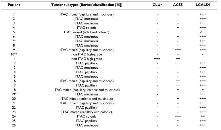
Immunohistochemical analysis of LGALS4, ACS5 and CLU To confirm the variation in expression of the selected genes at the protein level, we performed immunohisto-chemical analysis of matched normal sinonasal and tumor tissues from the 15 patients used for the molecular analysis as well as from an independent set of 11 other patients, using specific antibodies for LGALS4, ACS5 and CLU (Table 3). In the normal sinusal mucosa, these three markers were expressed by serous cells of the seromuci-nous glands present in the lamina propria. A weak and focal cytoplasmic staining of a small number of seromuci-nous glands was observed with the antibodies against LGALS4 and CLU while the staining was more intense and diffuse for ACS5 (Figure 3A-B-C). Among the 26 tumors analyzed, only 2 were high-grade non-ITAC and the oth-ers were ITAC: 5 "papillary-type" (well-differentiated ade-nocarcinoma), 2 "colonic-type" (moderately-differentiated adenocarcinoma), 9 "mucinous-type" ade-nocarcinoma and 8 mixed-type adeade-nocarcinoma (Table 3).
With the LGALS4 antibody the ITAC tumor cells displayed a strong cytoplasmic and membranous staining with an additional nuclear staining in the well-differentiated ade-nocarcinomas. Interestingly, in a mixed ITAC sample (P5) the poorly differentiated "solid-type" component showed no immunoreactivity for LGALS4 while the "colonic-type" component was positive (Table 3 and Figure 3D). Non-ITAC samples displayed no LGALS4 expression.
For ACS5, fifty percent of the tumor samples were negative while the remaining 50% showed a weak to strong cyto-plasmic staining without any correlation with the
histo-Table 2: Top 59 genes differentially expressed in sinonasal adenocarcinomas after two-class comparison analysis Accession
Number
Gene Symbol Gene annotation Fold change (log2)
NM_006149 LGALS4 Up-regulated genes
lectin, galactoside-binding, soluble, 4 (galectin 4)
3.084 NM_016234 ACS5 fatty-acid-Coenzyme A ligase, long-chain 5 2.621 NM_001845 COL4A1 collagen, type IV, alpha 1 1.779 NM_002228 JUN v-jun sarcoma virus 17 oncogene homolog (avian) 1.758 NM_001816 CEACAM8 carcinoembryonic antigen-related cell adhesion molecule 8 1.732 NM_001122 ADFP adipose differentiation-related protein 1.628 XM_067746 similar to 60 kDa heat shock protein, mitochondrialprecursor (Hsp60) (60 kDa
chaperonin) (CPN60) (Heat shock protein60) (HSP-60)
1.617 NM_004591 CCL20 chemokine (C-C motif) ligand 20 1.560 BC000097 TGFBI transforming growth factor, beta-induced, 68 kDa 1.493 NM_000393 COL5A2 collagen, type V, alpha 2 1.461 NM_003130 SRI sorcin 1.456 NM_001153 ANXA4 annexin A4 1.434 NM_005566 LDHA lactate dehydrogenase A 1.416 NM_005563 STMN1 stathmin 1/oncoprotein 18 1.414 NM_017958 PLEKHB2 pleckstrin homology domain containing, family B (evectins) member 2 1.409 XM_092196 similar to Cytochrome c, somatic (LOC164837), mRNA. 1.387 AF112214 MRPL13 mitochondrial ribosomal protein L13 1.370 AJ250915 HSPD1 heat shock 60 kDa protein 1 (chaperonin) 1.346 BC003623 YWHAZ tyrosine 3-monooxygenase/tryptophan 5-monooxygenase activation protein, zeta
polypeptide
1.342 NM_006111 ACAA2 acetyl-Coenzyme A acyltransferase 2 (mitochondrial 3-oxoacyl-Coenzyme A thiolase) 1.335 NM_021821 MRPS35 mitochondrial ribosomal protein S35 1.329 NM_002592 PCNA proliferating cell nuclear antigen 1.319 NM_001827 CKS2 CDC28 protein kinase regulatory subunit 2 1.278 AB062125 TPM3 tropomyosin 3 1.224 NM_016245 DHRS8 dehydrogenase/reductase (SDR family) member 8 1.206 NM_001226 CASP6 caspase 6, apoptosis-related cysteine protease 1.194 NM_004670 PAPSS2 3'-phosphoadenosine 5'-phosphosulfate synthase 2 1.172 XM_088293 similar to cytochrome c (LOC157317), mRNA. 1.164 NM_001428 ENO1 enolase 1, (alpha) 1.145 XM_060849 similar to cytochrome C, expressed in somatic tissues(LOC128146), mRNA. 1.133 AF135381 CKLF chemokine-like factor 1.133 X84907 ENO1 enolase 1, (alpha) 1.121 NM_005720 ARPC1B actin related protein 2/3 complex, subunit 1B, 41 kDa 1.114 NM_021130 PPIA peptidylprolyl isomerase A (cyclophilin A) 1.110 NM_001288 CLIC1 chloride intracellular channel 1 1.095 BC015130 CYCS cytochrome c, somatic 1.081 NM_012255 XRN2 5'-3' exoribonuclease 2 1.068 M34664 HSPD1 heat shock 60 kDa protein 1 (chaperonin) 1.066 AF054185 PSMA7 proteasome (prosome, macropain) subunit, alpha type, 7 1.044 NM_006601 TEBP unactive progesterone receptor, 23 kD 1.037 AF136630 CBX3 chromobox homolog 3 (HP1 gamma homolog, Drosophila) 1.023 AF274941 CKS1B CDC28 protein kinase regulatory subunit 1B 1.013 AF320053 MYCN v-myc myelocytomatosis viral related oncogene, neuroblastoma derived (avian) 1.006
Down-regulated genes
NM_001831 CLU clusterin
(complement lysis inhibitor, SP-40,40, sulfated glycoprotein 2, testosterone-repressed prostate message 2, apolipoprotein J)
-3.606 NM_005978 S100A2 S100 calcium binding protein A2 -2.152 S68290 AKR1C1 aldo-keto reductase family 1, member C1 (dihydrodiol dehydrogenase 1; 20-alpha
(3-alpha)-hydroxysteroid dehydrogenase)
-2.102 NM_003713 PPAP2B phosphatidic acid phosphatase type 2B -1.966 AB000889 PPAP2B phosphatidic acid phosphatase type 2B -1.936 NM_001321 CSRP2 cysteine and glycine-rich protein 2 -1.916 NM_006485
/
FBLN1 fibulin 1 -1.799 AF007162 CRYAB crystallin, alpha B -1.590 NM_002825 PTN pleiotrophin (heparin binding growth factor 8, neurite growth-promoting factor 1) -1.405
logical type or with the differentiation of the tumor (Table 3 and Figure 3E). In contrast to normal mucosa, CLU was found to be absent in tumors except in one high-grade non-ITAC
tumor (Patient P11) where there was a diffuse cytoplas-mic staining (Table 3 and Figure 3F).
NM_001063 TF transferrin -1.256 NM_004186 SEMA3F sema domain, immunoglobulin domain (Ig), short basic domain, secreted, (semaphorin) 3F -1.246 NM_000424 KRT5 keratin 5 (epidermolysis bullosa simplex, Dowling-Meara/Kobner/Weber-Cockayne types) -1.207 AF059617 PLK2 polo-like kinase 2 (Drosophila) -1.186 NM_005596 NFIB nuclear factor I/B -1.149 NM_006206 PDGFRA platelet-derived growth factor receptor, alpha polypeptide -1.090 NM_005900 MADH1 MAD, mothers against decapentaplegic homolog 1 (Drosophila) -1.047
Table 2: Top 59 genes differentially expressed in sinonasal adenocarcinomas after two-class comparison analysis (Continued)
Heat map of the two-class comparison (A) and unsupervised (B) analysis
Figure 1
Heat map of the two-class comparison (A) and unsupervised (B) analysis. Expression levels are color coded with red, green, black and gray, corresponding to an increase, decrease or no change in gene expression, or missing data, respec-tively.
Discussion
Ethmoid carcinomas are uncommon tumors recognized as an occupational disease amongst woodworkers. Cur-rent treatment with surgery and radiotherapy is unsatisfac-tory given the 50% survival at 5 years and the serious side effects. To better understand the molecular events involved in this tumor and to identify potentially novel markers we pioneered a gene expression profiling study of 9 sinonasal adenocarcinomas.
This study, using dedicated-microarrays containing 6864 genes previously known to be involved in cancer, allowed us to select 5 genes (LGALS4, ACS5, CLU, SRI and CCT5) with significant differential expression between tumors and normal tissue. We confirmed by RT-qPCR the overex-pression of LGALS4, ACS5, SRI, CCT5 and the down-reg-ulation of CLU. By IHC on an independent set of patients, we focused our interest on the genes with the highest dif-ferential expression i.e. LGALS4, ACS5 and CLU, and con-firmed the results at the protein level for LGALS4 and CLU.
The LGALS4 gene codes for the Galectin 4 protein [38]. Galectins constitute a family of proteins containing carbo-hydrate recognition domains (CRD) with high affinity for β galactosides. Their complete physiological functions are not known but they have been reported to be involved in inflammation, apoptosis, cell adhesion and cell growth. LGALS4 in particular has been detected in normal epithe-lial cells of the oral esophagus, and in the intestinal mucosa [39,40]. In tumors, LGALS4 expression increases in liver, gastric, breast cancer and mucinous epithelial ovarian cancer whereas it is down-regulated in colon ade-nocarcinoma [41-43]. The presence of two binding sites for c-Rel, a subunit of NFκ-B, and the experimental data obtained with transgenic mice for c-Rel, suggest that LGALS4 could be a downstream component of the NFκ-B pathway, known to be involved in the regulation of tumorogenesis [44,45]. In cancer cell lines LGALS4 is expressed in highly differentiated cell lines which form polarized monolayers while undifferentiated cell lines do not express LGALS4 but Galectin1 [38,42]. In our series of ethmoid adenocarcinoma, the LGALS4 is the gene with the highest differential expression and our IHC data are in accordance with the literature, given that we found that LGALS4 is overexpressed in all ethmoid tumors except the high-grade non ITAC tumors which are poorly differenti-ated. LGALS4 expression seems to be correlated to both histological type and the differentiation status of the ade-nocarcinoma. This trend was confirmed by the P5 case where LGALS4 was overexpressed only in the "colonic-type" component and not in the poorly differentiated "solid-type" component of the tumor. For patient 6 (P6) we observed a strong overexpression of LGALS4 by IHC, which contrasts with the relative expression obtained by RT-qPCR (fold change 0.45). We therefore hypothesize that, in this "mucinous-type" ITAC containing numerous mucin lakes, the RNA extracted from the tissue was not representative of the tumor.
The highly conserved gene CLU (apolipoproteinJ, sulfated glycoprotein 2), codes for Clusterin, a sulfated glycopro-tein with chaperone activity found in numerous tissues and body fluids. CLU has been reported as being involved in many biological functions such as DNA repair, cell cycle regulation and apoptosis [37,46]. CLU is described as being overexpressed in several types of cancers includ-ing colon, breast and lung cancer [37], yet a down-regula-tion has been found in esophageal squamous cell carcinoma, in some pancreatic, prostate or colon cancers and in HPV-negative squamous cell carcinoma of the head and neck [37,46,47], suggesting a pro-survival or a pro-apoptotic function. The recent description of several isoforms, including the nuclear form (n-CLU) and the cytoplasmic or secreted form (s-CLU), might help to resolve these apparent contradictions and to define the
Relative expression levels of LGALS4, ACS5, CLU, in tumors versus matched normal sinusal tissue as determined by RT-qPCR
Figure 2
Relative expression levels of LGALS4, ACS5, CLU, in tumors versus matched normal sinusal tissue as determined by RT-qPCR. Fold change was calculated according to the equation described in the Materials and Methods with normalization against the average of three housekeeping genes, RPLPO, β2 microglobulin, and ubiquitin C. *tumor tissue versus average of all normal sinusal tissues (cf. RT-qPCR Results for details).
CLU p=0.001 0 0,1 0,2 0,3 17 18 14 10 9 5 13 4 1 16 19 12 8 6 11 LGALS4 p=0.001 0 1000 2000 3000 4000 5000 6000 10 11 6 4 18 17 19 14 8 12 5 1 16 9 13
Relative expression leve
l * * * * A C ACS5 p=0.001 0 5 10 15 20 25 10 11 19*8* 4 6 17 14 13 18 9 1 16 5 12 B Patients
cellular functions of Clusterin as well as its potential use as a biomarker [48-50].
In our series of ethmoid tumors, CLU was highly down-regulated at the RNA level. Although the level of Clusterin detected by IHC in normal tissue was rather low, we con-firmed the down-regulation of the protein except in one case (P11). This patient was also the one whose tumor sample showed the least down-regulation of CLU by RT-qPCR. This case is of interest because the patient was exposed to wood and, in contrast with most of the cases reported in the literature, he presented a non-ITAC tumor. The absence of Clusterin in ethmoid tumors suggests a pro-apoptotic function in normal ethmoidal tissue, possi-bly in response to DNA damage caused by wood dust, or other occupational exposures. It is useful to note that CLU is localized on chromosome 8p21-p12 [51]. In fact, by comparative genomic hybridization, Ariza et al. found losses on 8p21 in about 50% of patients with sinonasal adenocarcinomas [20]. This feature was confirmed by the study of Korinth et al. who reported a loss of 8p in 61% of
cases [21] in a series of 42 patients. We do not know the cytogenetics of our tumors but it would be worthwhile ascertaining whether the down-regulation of CLU in the tumors studied here is due to deletion on chromosome 8p or if other mechanisms such as epigenetic regulation
occur on the CLU gene.
ACS5, Acyl coenzyme A synthetase 5 (FACL5, E.C. 6.2.1.3.), is one isoform of the ACSs, key proteins in lipid metabolism via the activation of fatty acids in acylCoA thioesters. These esters are the metabolites for oxidation, elongation and desaturation of fatty acids as well as for the synthesis of complex lipids. ACS5 is essential for lipid metabolism but it might also play a role in intermediate metabolism and regulation of gene expression [52]. This gene has been well characterized in the small intestine mucosa by Gassler et al [53,54]. ACS5 is expressed in the enterocytes from the villus tip but not in the crypts and it could be involved in the differentiation and maintenance of crypt-villus axis, by inducing TRAIL apoptosis in apical villi of the mucosa. Within the context of tumorogenesis,
Table 3: LGALS4, ACS5 and CLU expression in 26 sinonasal adenocarcinomas (IHC analysis).
Patient Tumor subtypes (Barnes'classification [25]) CLUa ACS5 LGALS4
1 ITAC mixed (papillary and mucinous) - - +++ 2 ITAC mucinous - - +++ 3 ITAC mucinous - - +++ 4 ITAC colonic - + +++ 5 ITAC mixed (solid and colonic) - ++ -/++ 6 ITAC mucinous - + +++ 7* ITAC mucinous - - +++ 8 ITAC mucinous - - +++ 9 ITAC mixed (papillary and mucinous) - +++ +++ 10** non ITAC high-grade - -
-11 non ITAC high-grade +++ ++ -12 ITAC papillary - +++ +++ 13 ITAC mucinous - - +++ 14 ITAC papillary - - +++ 15 ITAC mucinous - - +++ 16 ITAC mixed (papillary and mucinous) - ++ +++ 17 ITAC papillary - ++ +++ 18 ITAC mixed (papillary, colonic and mucinous) - + + 19* ITAC mucinous - + +++
20 ITAC mixed (colonic and mucinous) - + +++ 21 ITAC mixed (papillary and mucinous) - - +++ 22 ITAC papillary - - +++ 23 ITAC mixed (papillary and colonic) - - +++ 24 ITAC colonic - +++ ++ 25 ITAC papillary - + +++ 26 ITAC mucinous - - +++ * Patients exposed to leather dust
** No occupational exposure
a- Intensity of immunostaining in tumoral cells:
+++: positivity of 75 to 100% of cells with an strong staining.
++: positivity of 25 to 75% of cells with heterogenous weak to strong staining. +: focal and weak positivity of 1 to 25% of cells.
few reports have been published on ACS5. In adenoma and adenocarcinoma of the small intestine ACS5 expres-sion is decreased [54] while it is up-regulated in gliomas [55], in well-differentiated endometrioid adenocarcino-mas [56] and in certain colorectal adenocarcinoadenocarcino-mas [57]. The RT-qPCR data in our panel of tumors revealed an increase in the expression of ACS5 (p = 0.001), eventhough it has not been confirmed by IHC. Whereas some tumors expressed strong ACS5, others had com-pletely lost the expression of this molecule. Moreover, we could not find any correlation between ACS5 expression and histological type, differentiation or collateral expo-sures.
The other selected genes were not evaluated by immuno-histochemistry as their variation in expression was much lower and our primary goal was to find new markers for a better characterization of these tumors with a clear etiol-ogy. Nevertheless, we confirmed the transcriptional pro-filing obtained with the microarray by RT-qPCR.
SRI (Sorcin) and CCT5 (chaperonin-containing complexe peptide 1) are less known genes. Both code for multi-drug resistance proteins and might be involved in the cell detoxification [58,59]. These genes were slightly overex-pressed in our panel of tumors. This trend could be related
to the chemical or particle exposures of the patients. In fact, SRI has also been identified by Differential Display analysis as being overexpressed in oral cancer mediated by tobacco-chewing [60].
Conclusion
In conclusion, our transcriptomic study has enabled us to identify genes involved in sinonasal adenocarcinomas. The validation of microarray data by RT-qPCR and immu-nohistochemistry confirmed the significant alterations of LGALS4 and CLU expression. Because of the low inci-dence of these tumors we had a limited number of patients and only one without wood exposure, preventing any correlation between survival and wood exposure. Nevertheless, after validation using tissue microarrays in a large set of tumors, including pre-cancerous lesions and early stages, LGALS4 and CLU could be included in a panel of non invasive diagnostic/prognostic tests for the follow-up of woodworkers, to allow an earlier detection of lesions using a sinonasal smear.
Competing interests
The authors declare that they have no competing interests.
Authors' contributions
TD conceived the design of the study, performed the ques-tionnaire, the follow up of the patients and participated in the drafting of the paper. SQ participated in the tissue col-lection, performed the molecular and data analyses, and contributed to the drafting of the paper. KR performed the pathological diagnoses and the immunohistochemistry interpretation. CF, OM and CV participated in the tissue collection, IGM to the microarray study. VSR and CG con-tributed to the design of the study and the epidemiologi-cal questionnaire. CGRR participated in the study design, supervised the project and prepared the manuscript. All authors read and approved the final manuscript.
Additional material
Additional file 1
Primer sequences. Click here for file
[http://www.biomedcentral.com/content/supplementary/1755-8794-2-65-S1.doc]
Additional file 2
Genes with significant differential expression in sinonasal adenocar-cinomas, identified by two-class comparison.
Click here for file
[http://www.biomedcentral.com/content/supplementary/1755-8794-2-65-S2.doc]
Representative cases of LGALS4, CLU and ACS5 expression in matched normal mucosa (×100), and tumor tissue (×25)
Figure 3
Representative cases of LGALS4, CLU and ACS5 expression in matched normal mucosa (×100), and tumor tissue (×25). A-B-C: Normal sinusal mucosa immu-nostaining. (A-C): Weak and focal cytoplasmic staining of serous cells in a few seromucinous glands with LGALS4 and CLU. (A):Weak staining of respiratory epithelium with LGALS4. (B): Strong and diffuse immunostaining of serous cells with ACS5. D-E-F: Tumor immunostaining. (D): Poorly-differentiated "solid-type" component showing no immunore-activity for LGALS4 while the "colonic-type" component is positive in a mixed ITAC (patient 5). (E): Example of ACS5 expression in a "colonic-type" ITAC. (F): No immunoreactiv-ity for CLU in tumor samples (×100) except in one non-ITAC (Insert * (×25), Patient 11).
Acknowledgements
We thank Dr. Jean Léger and Dr. Rémi Houlgatte for their help with the microarray analysis, Marie-Thérèse Le Cabellec for the cryostat sections, and Cécile Deleine for the immunohistochemical staining. We are grateful to C. Beauvillain, F. Jégoux, and C. Roedlich for their critical review and helpful discussions during the preparation of the manuscript.
Grant support: La Ligue Contre le Cancer, comité Pays de la LOIRE; La Direction de la Recherche Clinique du Centre Hospitalo-Universitaire de Nantes.
References
1. Dulguerov P, Jacobsen MS, Allal AS, Lehmann W, Calcaterra T: Nasal
and paranasal sinus carcinoma: are we making progress? A series of 220 patients and a systematic review. Cancer 2001, 92(12):3012-3029.
2. Luce D, Leclerc A, Begin D, Demers PA, Gerin M, Orlowski E, Kogevinas M, Belli S, Bugel I, Bolm-Audorff U, Brinton LA, Comba P, Hardell L, Hayes RB, Magnani C, Merler E, Preston-Martin S, Vaughan TL, Zheng W, Boffetta P: Sinonasal cancer and occupational
exposures: a pooled analysis of 12 case-control studies.
Can-cer Causes Control 2002, 13(2):147-157.
3. Pesch B, Pierl CB, Gebel M, Gross I, Becker D, Johnen G, Rihs HP, Donhuijsen K, Lepentsiotis V, Meier M, Schulze J, Bruening T:
Occu-pational risks for adenocarcinoma of the nasal cavity and paranasal sinuses in the German wood industry. Occup Environ
Med 2007, 65(3):191-196.
4. IARC: Wood dust and formaldehyde. In Monogr Eval Carcinog
Risks Hum Volume 62. Edited by: IARC. Lyon: IARC; 1995.
5. Demers PA, Boffetta P, Kogevinas M, Blair A, Miller BA, Robinson CF, Roscoe RJ, Winter PD, Colin D, Matos E, et al.: Pooled reanalysis
of cancer mortality among five cohorts of workers in wood-related industries. Scand J Work Environ Health 1995, 21(3):179-190.
6. IARC: Cancer risk from occupational exposure to wood dust.
A pool analysis of epidemiological studies. Tech Rep 1998:30.
7. IARC: IARC Monogr Eval Carcinog Risks Hum. Leather
industries, boot and shoe manufacture and repair. In Overall
evaluations of carcinogenicity: an updating of IARC monographs Volume 1-42. Lyon: IARC; 1987:232-237.
8. Bonneterre V, Deschamps E, Persoons R, Bernardet C, Liaudy S, Mai-tre A, de Gaudemaris R: Sino-nasal cancer and exposure to
leather dust. Occup Med (Lond) 2007, 57(6):438-443.
9. Andersen A, Barlow L, Engeland A, Kjaerheim K, Lynge E, Pukkala E:
Work-related cancer in the Nordic countries. Scand J Work
Environ Health 1999, 25(Suppl 2):1-116.
10. Hernberg S, Westerholm P, Schultz-Larsen K, Degerth R, Kuosma E, Englund A, Engzell U, Hansen HS, Mutanen P: Nasal and sinonasal
cancer. Connection with occupational exposures in Den-mark, Finland and Sweden. Scan J Work Environ Health 1983, 9:315-326.
11. Hauptmann M, Lubin JH, Stewart PA, Hayes RB, Blair A: Mortality
from solid cancers among workers in formaldehyde indus-tries. Am J Epidemiol 2004, 159(12):1117-1130.
12. Hecht SS: Tobacco carcinogens, their biomarkers and
tobacco-induced cancer. Nat Rev Cancer 2003, 3(10):733-744.
13. Llorente JL, Perez-Escuredo J, Alvarez-Marcos C, Suarez C, Hermsen M: Genetic and clinical aspects of wood dust related
intesti-nal-type sinonasal adenocarcinoma: a review. Eur Arch
Otorhi-nolaryngol 2009, 266(1):1-7.
14. Barnes L, Johnson JT: Clinical and pathological considerations
in the evaluation of major head and neck specimens resected for cancer. Part II. Pathol Annu 1986, 21(Pt 2):83-110.
15. Kleinsasser O, Schroeder HG: Adenocarcinomas of the inner
nose after exposure to wood dust. Morphological findings and relationships between histopathology and clinical behav-ior in 79 cases. Arch Otorhinolaryngol 1988, 245(1):1-15.
16. Roux FX, Moussa R, Devaus B, Nataf F, Page P, Laccourreye O, Schwaab G, Brasnu D, Lacau Saint-Guily J: Subcranial
fronto-orbito-nasal approach for ethmoidal cancers surgical tech-niques and results. Surg Neurol 1999, 52(5):501-508.
17. Suarez C, Llorente JL, Fernandez De Leon R, Maseda E, Lopez A:
Prognostic factors in sinonasal tumors involving the anterior skull base. Head Neck 2004, 26(2):136-144.
18. Bornholdt J, Hansen J, Steiniche T, Dictor M, Antonsen A, Wolff H, Schlunssen V, Holmila R, Luce D, Vogel U, Husgafvel-Pursiainen K, Wallin H: K-ras mutations in sinonasal cancers in relation to
wood dust exposure. BMC Cancer 2008, 8:53.
19. Holmila R, Cyr D, Luce D, Heikkila P, Dictor M, Steiniche T, Stjernvall T, Bornholdt J, Wallin H, Wolff H, Husgafvel-Pursiainen K: COX-2
and p53 in human sinonasal cancer: COX-2 expression is associated with adenocarcinoma histology and wood-dust exposure. Int J Cancer 2008, 122(9):2154-2159.
20. Ariza M, Llorente JL, Alvarez-Marcas C, Baragano L, Salas A, Rod-riguez Prado N, Hermsen M, Suarez C, Sampedro A: Comparative
genomic hybridization in primary sinonasal adenocarcino-mas. Cancer 2004, 100(2):335-341.
21. Korinth D, Pacyna-Gengelbach M, Deutschmann N, Hattenberger S, Bockmuhl U, Dietel M, Schroeder HG, Donhuijsen K, Petersen I:
Chromosomal imbalances in wood dust-related adenocarci-nomas of the inner nose and their associations with patho-logical parameters. J Pathol 2005, 207(2):207-215.
22. Hermsen MA, Llorente JL, Perez-Escuredo J, Lopez F, Ylstra B, Alva-rez-Marcos C, Suarez C: Genome-wide analysis of genetic
changes in intestinal-type sinonasal adenocarcinoma. Head
Neck 2009, 31(3):290-297.
23. Rekhadevi PV, Mahboob M, Rahman MF, Grover P: Genetic
dam-age in wood dust-exposed workers. Mutdam-agenesis 2009, 24(1):59-65.
24. Franchi A, Santucci M, Wenig B: Adenocarcinoma. In World Health
Organization classification of tumors Pathology and genetics of head and neck tumours Edited by: Barnes L, Eveson J, Reichart P, Sidransky D.
Lyon: IARC; 2005:20-23.
25. Barnes L: Intestinal-type adenocarcinoma of the nasal cavity
and paranasal sinuses. Am J Surg Pathol 1986, 10(3):192-202.
26. Fleige S, Pfaffl MW: RNA integrity and the effect on the
real-time qRT-PCR performance. Mol Aspects Med 2006, 27(2-3):126-139.
27. Was H, Cichon T, Smolarczyk R, Rudnicka D, Stopa M, Chevalier C, Leger JJ, Lackowska B, Grochot A, Bojkowska K, Ratajska A, Kieda C, Szala S, Dulak J, Jozkowicz A: Overexpression of heme
oxygen-ase-1 in murine melanoma: increased proliferation and via-bility of tumor cells, decreased survival of mice. Am J Pathol
2006, 169(6):2181-2198.
28. Mori K, Berreur M, Blanchard F, Chevalier C, Guisle-Marsollier I, Masson M, Redini F, Heymann D: Receptor activator of nuclear
factor-kappaB ligand (RANKL) directly modulates the gene expression profile of RANK-positive Saos-2 human osteosa-rcoma cells. Oncol Rep 2007, 18(6):1365-1371.
29. Chelh I, Meunier B, Picard B, Reecy MJ, Chevalier C, Hocquette JF, Cassar-Malek I: Molecular profiles of Quadriceps muscle in
myostatin-null mice reveal PI3K and apoptotic pathways as myostatin targets. BMC Genomics 2009, 10:196.
30. Lamirault G, Gaborit N, Le Meur N, Chevalier C, Lande G, Demolombe S, Escande D, Nattel S, Leger JJ, Steenman M: Gene
expression profile associated with chronic atrial fibrillation and underlying valvular heart disease in man. J Mol Cell Cardiol
2006, 40(1):173-184.
31. Le Meur N, Lamirault G, Bihouee A, Steenman M, Bedrine-Ferran H, Teusan R, Ramstein G, Leger JJ: A dynamic, web-accessible
resource to process raw microarray scan data into
consoli-Additional file 3
Relative expression levels of SRI and CCT5 in tumors versus matched normal sinonasal tissue as determined by RT-qPCR. Fold change was
calculated according to the equation described in the Materials and Meth-ods with normalization against the average of three housekeeping genes,
RPLPO, β2 microglobulin, and ubiquitin C. *tumor tissue versus
aver-age of all normal sinonasal tissues (cf. RT-qPCR Results for detail).
Click here for file
[http://www.biomedcentral.com/content/supplementary/1755-8794-2-65-S3.ppt]
Publish with BioMed Central and every scientist can read your work free of charge "BioMed Central will be the most significant development for disseminating the results of biomedical researc h in our lifetime."
Sir Paul Nurse, Cancer Research UK Your research papers will be:
available free of charge to the entire biomedical community peer reviewed and published immediately upon acceptance cited in PubMed and archived on PubMed Central yours — you keep the copyright
Submit your manuscript here:
http://www.biomedcentral.com/info/publishing_adv.asp
BioMedcentral dated gene expression values: importance of replication.
Nucleic Acids Res 2004, 32(18):5349-5358.
32. Tusher VG, Tibshirani R, Chu G: Significance analysis of
micro-arrays applied to the ionizing radiation response. Proc Natl
Acad Sci USA 2001, 98(9):5116-5121.
33. Eisen MB, Spellman PT, Brown PO, Botstein D: Cluster analysis
and display of genome-wide expression patterns. Proc Natl
Acad Sci USA 1998, 95(25):14863-14868.
34. Zeeberg BR, Feng W, Wang G, Wang MD, Fojo AT, Sunshine M, Nar-asimhan S, Kane DW, Reinhold WC, Lababidi S, Bussey KJ, Riss J, Bar-rett JC, Weinstein JN: GoMiner: a resource for biological
interpretation of genomic and proteomic data. Genome Biol
2003, 4(4):R28.
35. Pfaffl MW: A new mathematical model for relative
quantifica-tion in real-time RT-PCR. Nucleic Acids Res 2001, 29(9):e45.
36. Pfaffl MW, Horgan GW, Dempfle L: Relative expression software
tool (REST) for group-wise comparison and statistical analy-sis of relative expression results in real-time PCR. Nucleic
Acids Res 2002, 30(9):e36.
37. Shannan B, Seifert M, Leskov K, Willis J, Boothman D, Tilgen W, Rei-chrath J: Challenge and promise: roles for clusterin in
patho-genesis, progression and therapy of cancer. Cell Death Differ
2006, 13(1):12-19.
38. Huflejt ME, Leffler H: Galectin-4 in normal tissues and cancer.
Glycoconj J 2004, 20(4):247-255.
39. Paclik D, Danese S, Berndt U, Wiedenmann B, Dignass A, Sturm A:
Galectin-4 controls intestinal inflammation by selective reg-ulation of peripheral and mucosal T cell apoptosis and cell cycle. PLoS ONE 2008, 3(7):e2629.
40. Paclik D, Lohse K, Wiedenmann B, Dignass AU, Sturm A:
Galectin-2 and -4, but not galectin-1, promote intestinal epithelial wound healing in vitro through a TGF-beta-independent mechanism. Inflamm Bowel Dis 2008, 14(10):1366-1372.
41. Duerr EM, Mizukami Y, Ng A, Xavier RJ, Kikuchi H, Deshpande V, Warshaw AL, Glickman J, Kulke MH, Chung DC: Defining
molecu-lar classifications and targets in gastroenteropancreatic neu-roendocrine tumors through DNA microarray analysis.
Endocr Relat Cancer 2008, 15(1):243-256.
42. Brule F van den, Califice S, Castronovo V: Expression of galectins
in cancer: a critical review. Glycoconj J 2004, 19(7-9):537-542.
43. Heinzelmann-Schwarz VA, Gardiner-Garden M, Henshall SM, Scurry JP, Scolyer RA, Smith AN, Bali A, Bergh P Vanden, Baron-Hay S, Scott C, Fink D, Hacker NF, Sutherland RL, O'Brien PM: A distinct
molecular profile associated with mucinous epithelial ovar-ian cancer. Br J Cancer 2006, 94(6):904-913.
44. Romieu-Mourez R, Kim DW, Shin SM, Demicco EG, Landesman-Bol-lag E, Seldin DC, Cardiff RD, Sonenshein GE: Mouse mammary
tumor virus c-rel transgenic mice develop mammary tumors. Mol Cell Biol 2003, 23(16):5738-5754.
45. Shen HM, Tergaonkar V: NFkappaB signaling in carcinogenesis
and as a potential molecular target for cancer therapy.
Apop-tosis 2009, 14(4):348-363.
46. Shannan B, Seifert M, Boothman DA, Tilgen W, Reichrath J:
Clus-terin and DNA repair: a new function in cancer for a key player in apoptosis and cell cycle control. J Mol Histol 2006, 37(5-7):183-188.
47. Martinez I, Wang J, Hobson KF, Ferris RL, Khan SA: Identification
of differentially expressed genes in positive and HPV-negative oropharyngeal squamous cell carcinomas. Eur J
Can-cer 2007, 43(2):415-432.
48. Trougakos IP, Djeu JY, Gonos ES, Boothman DA: Advances and
challenges in basic and translational research on clusterin.
Cancer Res 2009, 69(2):403-406.
49. Kevans D, Foley J, Tenniswood M, Sheahan K, Hyland J, O'Donoghue D, Mulcahy H, O'Sullivan J: High clusterin expression correlates
with a poor outcome in stage II colorectal cancers. Cancer
Epi-demiol Biomarkers Prev 2009, 18(2):393-399.
50. Sakai I, Miyake H, Takenaka A, Fujisawa M: Expression of potential
molecular markers in renal cell carcinoma: impact on clin-icopathological outcomes in patients undergoing radical nephrectomy. BJU Int 2009, 104(7):942-946.
51. Fink TM, Zimmer M, Tschopp J, Etienne J, Jenne DE, Lichter P:
Human clusterin (CLI) maps to 8p21 in proximity to the lipo-protein lipase (LPL) gene. Genomics 1993, 16(2):526-528.
52. Black PN, Faergeman NJ, DiRusso CC: Long-chain
acyl-CoA-dependent regulation of gene expression in bacteria, yeast and mammals. J Nutr 2000, 130(2S Suppl):305S-309S.
53. Gassler N, Kopitz J, Tehrani A, Ottenwalder B, Schnolzer M, Karten-beck J, Lyer S, Autschbach F, Poustka A, Otto HF, Mollenhauer J:
Expression of acyl-CoA synthetase 5 reflects the state of vil-lus architecture in human small intestine. J Pathol 2004, 202(2):188-196.
54. Gassler N, Schneider A, Kopitz J, Schnolzer M, Obermuller N, Kartenbeck J, Otto HF, Autschbach F: Impaired expression of
acyl-CoA-synthetase 5 in epithelial tumors of the small intes-tine. Hum Pathol 2003, 34(10):1048-1052.
55. Yamashita Y, Kumabe T, Cho YY, Watanabe M, Kawagishi J, Yoshim-oto T, Fujino T, Kang MJ, YamamYoshim-oto TT: Fatty acid induced
gli-oma cell growth is mediated by the acyl-CoA synthetase 5 gene located on chromosome 10q25.1-q25.2, a region fre-quently deleted in malignant gliomas. Oncogene 2000, 19(51):5919-5925.
56. Gassler N, Yang SH, Keith M, Helmke BM, Schirmacher P, Ober-muller N: Expression of acyl-CoA synthetase 5 in human
endometrium and in endometrioid adenocarcinomas.
His-topathology 2005, 47(5):501-507.
57. Gassler N, Herr I, Schneider A, Penzel R, Langbein L, Schirmacher P, Kopitz J: Impaired expression of acyl-CoA synthetase 5 in
spo-radic colorectal adenocarcinomas. J Pathol 2005,
207(3):295-300.
58. Ooe A, Kato K, Noguchi S: Possible involvement of CCT5,
RGS3, and YKT6 genes up-regulated in p53-mutated tumors in resistance to docetaxel in human breast cancers. Breast
Cancer Res Treat 2007, 101(3):305-315.
59. Zhou Y, Xu Y, Tan Y, Qi J, Xiao Y, Yang C, Zhu Z, Xiong D: Sorcin,
an important gene associated with multidrug-resistance in human leukemia cells. Leuk Res 2006, 30(4):469-476.
60. Nagpal JK, Das BR: Identification of differentially expressed
genes in tobacco chewing-mediated oral cancer by differen-tial display-polymerase chain reaction. Eur J Clin Invest 2007, 37(8):658-664.
61. Patel SG, Shah JP: TNM staging of cancers of the head and neck:
striving for uniformity among diversity. CA Cancer J Clin 2005, 55(4):242-258.
Pre-publication history
The pre-publication history for this paper can be accessed here: