HAL Id: hal-03011607
https://hal.archives-ouvertes.fr/hal-03011607
Submitted on 18 Nov 2020
HAL is a multi-disciplinary open access
archive for the deposit and dissemination of sci-entific research documents, whether they are pub-lished or not. The documents may come from teaching and research institutions in France or abroad, or from public or private research centers.
L’archive ouverte pluridisciplinaire HAL, est destinée au dépôt et à la diffusion de documents scientifiques de niveau recherche, publiés ou non, émanant des établissements d’enseignement et de recherche français ou étrangers, des laboratoires publics ou privés.
Structure of the agonist 12–HHT in its BLT2
receptor-bound state
Fabrice Giusti, Marina Casiraghi, Elodie Point, Marjorie Damian, Jutta
Rieger, Christel Le Bon, Alexandre Pozza, Karine Moncoq, Jean-Louis
Banères, Laurent Catoire
To cite this version:
Fabrice Giusti, Marina Casiraghi, Elodie Point, Marjorie Damian, Jutta Rieger, et al.. Structure of the agonist 12–HHT in its BLT2 receptor-bound state. Scientific Reports, Nature Publishing Group, 2020, 10 (1), �10.1038/s41598-020-59571-6�. �hal-03011607�
Structure of the agonist 12–HHT in its BLT2
receptor-bound state
E-mail:
Running header
Structure of the agonist 12–HHT in its BLT2
receptor-bound state
Fabrice Giusti,1,2 Marina Casiraghi,1,3 Elodie Point,1 Marjorie
Damian,4 Jutta Rieger,5 Christel Le Bon,1 Alexandre Pozza,1
Karine Moncoq,1 Jean-Louis Ban`eres,4 Laurent J. Catoire1,∗
1Laboratoire de Biologie Physico-Chimique des Prot´eines Membranaires, UMR 7099, CNRS/
Universit´e de Paris, Institut de Biologie Physico–Chimique (FRC 550), 13 rue Pierre et Marie
Curie, F–75005 Paris, France;2Present address: Institut de Chimie S´eparative de Marcoule, ICSM
UMR 5257, Site de Marcoule, Bˆatiment 426, BP 17171, F-30207 Bagnols sur C`eze Cedex, France;
3Present address: Department of Molecular and Cellular Physiology, Stanford University School
of Medicine, 279 Campus Drive, 94305 Stanford California, USA; 4Institut des Biomol´ecules Max
Mousseron (IBMM), UMR 5247 CNRS, Universit´e Montpellier, ENSCM, 15 av. Charles Flahault,
34093 Montpellier, France; 5Institut Parisien de Chimie Mol´eculaire, Sorbonne Universit´e, CNRS,
UMR 8232, Equipe Chimie des Polym`eres, 4 place Jussieu, 75252, Paris Cedex 05, France
Abstract
G Protein-Coupled receptors represent the main communicating pathway for signals from the outside to the inside of most of eukaryotic cells. They define the largest family of integral membrane receptors at the surface of the cells and constitute the main target of the current drugs on the market. The low affinity leukotriene receptor BLT2 is a receptor involved in pro- and anti-inflammatory pathways and can be activated by various unsaturated fatty acid compounds. We present here the NMR structure of the agonist 12–HHT in its BLT2-bound state and a model of interaction of the ligand with the receptor based on a conformational homology modeling associated with docking simulations. Put into perspective with the data obtained with leukotriene B4, our results illuminate the ligand selectivity of BLT2 and may help define new molecules to modulate the activity of this receptor.
Introduction
1 2
G protein-coupled receptors (GPCRs) are integral membrane proteins that allow the
sig-3
nal transduction from the outside to the inside of most of eukaryotic cells.1 These receptors
4
consist in a large family of proteins whose activities can be related to various ligands, from
5
small organic compounds like neurotransmitters or hormones, to lipids, peptides or proteins.
6
As such, they are key players in many biological processes and represent one of the most
com-7
mon target of clinical drugs.2,3 Signal transduction through GPCRs concentrates a cascade
8
of biological events that, if we exclude the constitutive activity, starts with the interaction of
9
an extracellular signaling molecule with these membrane proteins, and triggers, at the end,
10
a cellular response. This binding of a ligand onto its cognate receptor represents a
funda-11
mental stage in the activation process. To get a picture of this interaction at the atomic
12
scale is not trivial as the number of high-quality crystals in the presence of natural agonists
13
is limited.4–6
14
In addition to X-ray diffraction and cryo-electronic microscopy (cryo-EM),7–10 NMR
15
spectroscopy can bring important information regarding conformational and energy
land-16
scapes11–16 or, as shown here, on the structure of natural GPCR ligands in their receptor
17
bound-states. This technique can indeed provide a detailed description of the ligand in its
18
bound-state, at physiological temperature and with a native protein.17–25 Especially with
19
very flexible ligands, like those described in this study, NMR data can constitute the basic
20
input to subsequent X-ray- or cryo-EM-based molecular modeling of ligand/GPCR
com-21
plexes.
22
The leukotriene receptors 126 (BLT1) and 227–30 (BLT2) are cell surface GPCRs that
23
share 45% amino acid sequence identity in human and are involved in pro- and anti-inflammatory
24
pathways.31–34 They were initially named high (BLT1) and low leukotriene B4 (LTB4)
25
(BLT2) receptors as the equilibrium dissociation constant (Kd) values of LTB4 in the pres-26
ence of membrane fractions transfected by either BLT1 or BLT2 is 20-fold weaker in the
case of BLT2 compared to BLT1 transfected HEK 293 cells (i.e. ∼1 nM and ∼20 nM for
28
BLT1 and BLT2, respectively).30 BLT1 receptor is essentially expressed in leukocytes and
29
lymphocytes and is mainly activated by the LTB435 which is a strong potent lipid
inflam-30
matory mediator. By contrast, BLT2 is expressed in various tissues and has been shown to
31
bind to different arachidonic acid metabolites with moderate affinities, including LTB4.36
32
In 2008, the heptadecanoid 12S–hydroxyheptadeca-5Z,8E,10E-trienoic acid37 (12–HHT)
33
was suggested to be the endogenous ligand of BLT2.38 In membrane fractions of Chinese
34
Hamster Ovary cells (CHO) transfected by BLT2, the half maximal inhibitory concentration
35
(IC50) and the half maximal effective concentration (EC50) values of 12–HHT are about one
36
order of magnitude lower than LTB4 while it does not bind to BLT1.38 The main source of
37
12–HHT comes as a reaction product of the conversion of prostaglandin H2 to thromboxane
38
A2 and malonyldialdehyde by the thromboxane synthase.39 Recent studies highlighted an
39
important activity of the 12–HHT/BLT2 axis in various pathologies, including inflammatory
40
and allergic diseases,38,40–42 wound healing43 and cancers.44–48
41
Here, we determined by NMR spectroscopy the three-dimensional (3D) structure of the
42
agonist 12–HHT associated with human BLT2. As observed with the LTB4 in the presence
43
of the same receptor,18 12–HHT adopts also a non-extended conformation. We propose
44
also a tentative model of interaction of 12–HHT with BLT2 based on X-ray crystal structure
45
conformational homology modeling and docking simulations, with the support of unequivocal
46 experimental data. 47 48 49 50
Methods
51 52
Sample preparations. The heptdadecanoid 12–HHT and the eicosanoid LTB4 were
53
obtained from Cayman Chemical, Ann Arbor, USA, as ethanolic solutions. The ethanol
54
was extensively evaporated under vacuum. Then the eicosanoids in excess respectively to
55
the receptor were directly dissolved by the NMR sample containing the receptor associated
56
with perDAPol in a 100%-D2O solution (20 mM Tris/HCl buffer pH 8, 100 mM NaCl) at
57
final concentrations of ∼120 and ∼140 µM of 12–HHT and LTB4, respectively. The
re-58
ceptor concentration was ∼15µM which gives rise to ligand/BLT2 molar ratio of 8 and ∼9
59
for 12–HHT/BLT2 and LTB4/BLT2. Considering a percentage of properly folded receptor
60
ranging from 50 to 70%,49 this means an effective ligand/receptor ratios of 15±4. Synthesis 61
of perDAPol was performed as already described50 and the overexpression, purification and
62
folding of perdeuterated human BLT2 receptor is detailed in Catoire et al. 2010.18
63 64
Site-directed mutagenesis. All mutations were introduced in the wild-type BLT2
65
receptor by PCR-mediated mutagenesis using the QuickChange multisite-directed
mutage-66
nesis kit (Stratagene) and the wild-type BLT2 construct as a template. Mutations were
67
confirmed by nucleotide sequencing.
68 69
Ligand binding assays. Agonist binding to the isolated BLT2 receptor was
mon-70
itored through ligand-dependent receptor-catalyzed G protein activation, as described in
71
Arcemisb´eh`ere et al (two different types of experiments were carried out to demonstrate
72
2010). Gαi2 and Gβ 1γ 2 were prepared as previously described.51 Briefly, agonist-dependent 73
functional coupling of the purified receptor to Gαi2β 1γ 2 was assessed through the rate of 74
GTPγS binding at increasing agonist concentrations determined by monitoring the relative
75
increase in the intrinsic fluorescence (λexcitation= 300 nm, λemission= 345 nm) of Gαi2(200 nM 76
of purified G protein) in the presence of BLT2 (20 nM) in buffer containing 10 mM MOPS,
pH 7.2, 130 mM NaCl, and 2 mM MgCl2 at 15◦C after the addition of 10 µM GTPγS. The 78
data were normalized to the fluorescence maximum obtained in the presence of saturating
79
concentrations in 12–HHT (10 µM).
80 81
NMR spectroscopy. All NMR experiments were conducted at 25◦C and 700 MHz on
82
a Bruker Avance spectrometer equipped with a cryoprobe. The dipolar interactions were
83
detected and collected in a transferred mode, i.e. in the presence of an excess of ligand over
84
the receptor thanks to a electrostatically-driven fast association and the perdeuteration of
85
the receptor which allow detection of transferred cross-relaxation for GPCR ligands with
86
equilibrium dissociation constants in the high-to-low nanomolar range (see the theoretical
87
and experimental demonstration in Catoire et al. 201119). The following parameters were 88
used for 2D NOESY experiments: 4 different mixing times (τm = 0.1 s, 0.2 s, 0.35 s, 0.5 s 89
in the study of 12–HHT and τm = 0.05 s, 0.1 s, 0.2 s, 0.5 s with the LTB4); data size = 90
256(t1) × 8,192(t2) complex points, t1max = 36.5 ms, t2max = 585 ms, 128 acquisitions per 91
increment, experiment time = 11.5 to 15.7 hours. Water suppression was conducted by using
92
an excitation sculpting scheme with gradients.52 Prior to Fourier Transform, the time
do-93
main signal was apodized by a square cosine in both dimensions. No baseline correction was
94
applied. 1H chemical shifts are referenced to H
2O (calibrated at 4.7 ppm at 25◦C). Chemical 95
shift assignments are based on COSY spectra from Catoire et al. 201018 and 2011.19 Data
96
processing and analyzing were performed with TOPSPIN software.
97 98
Structure calculations. 12–HHT and LTB4 pdb files were produced with
PRO-99
DRG.53Parameter and topology files were generated with XPLO2D (version 3.3.2).54
Struc-100
ture calculations were performed with the program ARIA (Ambiguous Restraints for
Itera-101
tive Assignment) (version 2.3)55 associated with CNS56 using standard protocols. For each
102
ligand, calculations were based on four sets of NOE data corresponding to four distinct τm 103
(see Tables S1 to S4 and S6 to S9 for 12–HHT and LTB4, respectively). A full relaxation
matrix treatment of NOE data has been applied in ARIA/CNS to take into account indirect
105
1H-1H cross-relaxation pathways.57,58 In the case of 12–HHT, only dipolar contacts
involv-106
ing H2, H3, and H17 with the other ligand protons were taken into account as these dipolar
107
restraints display the lowest level of non-specific binding contribution to the peak volumes
108
(unstructured parts in the ligand in the absence of the receptor). For LTB4, only protons at
109
both ends interacting with the other protons in the ligands, i.e. H2, H3, H4 and H16, H17,
110
H18, H19 and H20, were taken into account in the structure calculation. The structures
111
were drawn using the software PyMOL.
112 113
Homology modelling of receptors and ligand docking simulations.
Homol-114
ogy modelling of BLT2 based on X-ray crystal structures was performed with the software
115
Modeller (version 9.2).59–61 Several structures were tested, including the two mentionned in
116
this manuscript: BLT1 (pdb code 5x3362) and β2AR (pdb code 3p0g4). Docking simulations
117
of 12–HHT in human BLT2 receptor were subsequently performed with HADDOCK (version
118
2.2) taking as active residues S174ECL2 and R2707.35 only.
119 120 121 122
Results
123 124Structure of 12–HHT associated with BLT2 receptor.
125 126
The NMR study of 12–HHT in its receptor-bound state was realized in vitro in a
127
detergent-free solution63 following a method that has been already applied with the LTB4 in
128
the presence of the same receptor.18,19 Briefly, the heterologous human BLT2 receptor was
129
expressed in Escherichia coli in a 100%-D2O solution to inclusion bodies64,65 and was sub-130
sequently folded to its native state using amphipols.49,66 The NMR structure of 12–HHT is
based on the detection of dipolar interactions in the ligand through two-dimensional
homonu-132
clear 1H Nuclear Overhauser Effect SpectroscopY (NOESY) experiments.67 The dipolar
in-133
teractions were collected in a transferred mode in the presence of an excess of ligand over the
134
receptor. Indeed, it has been demonstrated that solution-state NMR can detect transferred
135
NOEs even with equilibrium dissociation constants below the micromolar range because of
136
i ) an inherent ultra-fast diffusive association of these negatively charged agonists onto a
137
highly positively charged extracellular surface, and ii ) the slowing down of the 1H-1H
cross-138
relaxation thanks to receptor perdeuteration.19 In order to improve the number and quality
139
of intra-ligand 1H-1H dipolar contacts, BLT2 was maintained soluble and stable in solution 140
associated with a perdeuterated amphipol named perDAPol.50 Compared to the pioneer
141
study of LTB4 associated with BLT2, perDAPol offered the possibility to observe
intra-142
aliphatic 1H dipolar interactions in the ligand (Figure 1) (for a comparative observation, see 143
Figure S1).
144
In the presence of BLT2 associated with either amphipols or nanodiscs, 12–HHT displays
145
a higher proportion of non-specific binding compared to LTB4 (see for instance
Supplemen-146
tary Figure S8 in Casiraghi et al. 201611). This is presumably due to a more hydrophobic
147
character which favours the interaction of the ligand with the belt of surfactant molecules
148
or lipids.11 To correctly assess the presence of specific intra-ligand dipolar interactions, the
149
NMR collection of constraints was based on a rigorous observation of specific intra-ligand1 H-150
1H dipolar interactions in the bound-state in the presence of a perdeuterated receptor.18,19In
151
the absence of the receptor, i.e. in the presence of perDAPol only, we observed the absence
152
and/or the presence of weak1H–1H dipolar interactions between aliphatic protons located at
153
both ends, i.e. from protons H2 to H4 on one side, and from H13 to H17 on the other side,
154
with the other1H in the ligand (Figures S2andS3). This indicates that both ligand ends are 155
not structured in the absence of the receptor. Moreover, non-specific 1H to 1H interactions 156
between the ligand and the surfactant can be observed in a 2D NOESY spectrum (e.g. the
157
regions squared with a green dashed line in Figure S2).
10 98115 6 12 7 2 4 3 13b 13a 14+15+16 17 TRIS H2O 10 9 6 5 11 8 12 7 2 4 3 13b 13a 17 TRIS H2O TRIS 14+15+16 1H 1H TRIS perDAPol 7 9 11 4 2 OH ζ β 13 µ λ κ 10 θ 8 3 α ι η 5 6 δ ε γ 14 15 16 17 ν ξ 12 1 O OH
Figure 1: Dipolar interactions in the 12–HHT/u-2H-wtBLT2/perDAPol sample observed
in a 2D NOESY spectrum (τm = 0.5 s, νH = 700 MHz, 25◦C, [12–HHT] = 120 µM,
[BLT2] = 15 µM). The corresponding 1D 1H spectrum is shown above the 2D spectrum
and a 1D spectrum of free 12–HHT in solution is displayed on the left side. Numbers refer to the protons annotated on the 12–HHT chemical structure indicated above the spectrum.
The NMR data corresponding to only specific interactions were collected at four different
159
Nuclear Overhauser Effect (NOE) mixing times τm, i.e. 0.1, 0.2, 0.35 and 0.5 s and integrated 160
for structure calculations (Tables S1 to S4). Dipolar interactions between H2, H3 and H17
161
with the other protons in the ligand were actually enough to obtain a converged set of
162
structures (Tables S1 toS4andFigure S4). This set of low energy conformers of 12–HHT in
163
its BLT2 bound-state is depicted in Figure 2 (with associated structural statistics gathered
164
in Table 1). In order to describe any conformational rearrangement of the structure of the
165
ligand upon binding to its receptor, a structure analysis of 12–HHT free in solution was
166
performed (Table S5) and is also indicated in Figure 2. As expected, in the free state,
167
structure calculation indicates that both aliphatic ends of the molecule are flexible with
168
the coexistence of various rotamers. However, compared to calculations conducted without
169
any experimental restraints, 12–HHT free in solution adopts preferential conformations at
170
the pentyl-end (carbon atoms 13 to 17), precluding any extended conformation along the
171
axis defined by the unsaturated bonds. Compared to the free-state in solution, the ligand
172
describes a well-constrained conformation in the presence of the receptor. In particular,
173
both ends, the carboxyl-end (1-carboxy-pent-4-ene-5-yl chain, carbons C1 to C6) and the
174
pentyl-end (carbon atoms 13 to 17), adopt a unique orientation respectively to the rigid core
175
of the molecule (dihedral angles ζ and κ in Figure 2).
176 177 178
Docking model of 12–HHT associated with BLT2 receptor.
179 180
The set of 20 low energy conformers depicted inFigure 2was further integrated in a model
181
of the BLT2 receptor to perform docking simulations using the software HADDOCK.68,69
182
The model of the BLT2 receptor using Modeller59–61 was based on the active state of the
183
β2 adrenergic receptor (β2AR) (pdb code 3p0g4) in spite a structure of BLT1 associated
184
with an inverse agonist being available in the protein databank (pdb code 5x3362). Using
7 9 11 4 2 OH ζ β 13 µ λ κ 10 θ 8 3 α ι η 5 6δ ε γ 14 15 16 17 ν ξ 12 1 O OH 4 3 5 6 1 2
FREE BOUND FREE
90
90 90
90
90 90
Figure 2: Three-dimensional structure of 12–HHT free in solution or bound to BLT2. (Top) Primary chemical structure of 12–HHT. The carbons are numbered from the carboxyl func-tion to the methyl group. Greek letters refer to some dihedral angles displayed at the bottom of the figure. (Middle) Six different views of two ensembles of 20 energy-minimized conform-ers (in white, hydrogen atoms; in red, oxygen atoms; carbon atoms are assigned a different color for each conformer). The red arrows indicate the transition from the free to the bound state for a same orientation of the diene located at the center of the molecule (carbons C8 to C11). (Bottom) Comparison of dihedral angles between the free and the bound states for the set of 20 conformers displayed above.
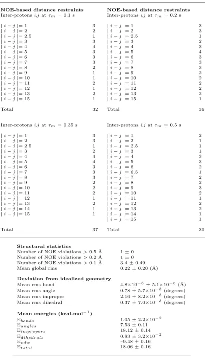
Table 1: Summary of structural constraints and structure statistics for a set of 20 structures of 12–HHT in the presence of BLT2 receptor. (in the case where the inter-protons distance indicated is not an integer, this means that magnetically not equivalent protons could not be distinguished in the NOESY spectrum. For instance, between H5 and H6, a dipolar interaction with H7 corresponds to an average inter-proton restraints of 2.5).
NOE-based distance restraints NOE-based distance restraints Inter-protons i,j at τm= 0.1 s Inter-protons i,j at τm= 0.2 s
| i − j |= 1 3 | i − j |= 1 3 | i − j |= 2 2 | i − j |= 2 3 | i − j |= 2.5 1 | i − j |= 2.5 1 | i − j |= 3 3 | i − j |= 3 2 | i − j |= 4 4 | i − j |= 4 3 | i − j |= 5 3 | i − j |= 5 4 | i − j |= 6 3 | i − j |= 6 3 | i − j |= 7 3 | i − j |= 7 3 | i − j |= 8 2 | i − j |= 8 3 | i − j |= 9 1 | i − j |= 9 2 | i − j |= 10 1 | i − j |= 10 2 | i − j |= 11 2 | i − j |= 11 2 | i − j |= 12 1 | i − j |= 12 2 | i − j |= 13 2 | i − j |= 13 2 | i − j |= 15 1 | i − j |= 15 1 Total 32 Total 36
Inter-protons i,j at τm= 0.35 s Inter-protons i,j at τm= 0.5 s | i − j |= 1 3 | i − j |= 1 2 | i − j |= 2 3 | i − j |= 2 1 | i − j |= 2.5 1 | i − j |= 2.5 1 | i − j |= 3 2 | i − j |= 3 1 | i − j |= 4 4 | i − j |= 4 3 | i − j |= 5 4 | i − j |= 5 3 | i − j |= 6 3 | i − j |= 6 2 | i − j |= 7 3 | i − j |= 6.5 1 | i − j |= 8 3 | i − j |= 7 2 | i − j |= 9 2 | i − j |= 8 2 | i − j |= 10 2 | i − j |= 9 3 | i − j |= 11 2 | i − j |= 10 2 | i − j |= 12 1 | i − j |= 11 1 | i − j |= 13 2 | i − j |= 12 2 | i − j |= 14 1 | i − j |= 13 2 | i − j |= 15 1 | i − j |= 14 1 | i − j |= 15 1 Total 37 Total 30 Structural statistics
Number of NOE violations > 0.5 ˚A 1 ± 0 Number of NOE violations > 0.2 ˚A 1 ± 0 Number of NOE violations > 0.1 ˚A 3.4 ± 0.49 Mean global rms 0.22 ± 0.20 (˚A) Deviation from idealized geometry
Mean rms bond 4.8×10⁻³± 5.1×10⁻⁵(˚A) Mean rms angle 0.78 ± 5.7×10⁻³(degrees) Mean rms improper 2.16 ± 8.2×10⁻³(degrees) Mean rms dihedral 0.37 ± 7.0×10⁻³(degrees) Mean energies (kcal.mol−1)
Ebonds 1.05 ± 2.2×10⁻² Eangles 7.53 ± 0.11 Eimpropers 18.12 ± 0.14 Edihedrals 0.83 ± 3.2×10⁻² Evdw –9.48 ± 0.16 Etotal 18.06 ± 0.16
BLT1 crystal structure in an inactive state does not allow the ligand 12–HHT to interact
186
with important amino acids in the BLT2 receptor that have been identified by site-directed
187
mutagenesis experiments associated with ligand binding assays (Figure 3A and Table 2).
188
In particular, residue S174 in the extra-cellular loop 2 (ECL2) BLT1-based BLT2 model is
189
located too far from the top of the ligand orthosteric pocket as the loop is in an open-lid
con-190
formation in the inactive state (Figure S5). To reproduce contacts between the ligand and
191
the receptor based on our binding studies, a conformational homology model was built. In
192
addition, the identity in amino acid sequence between BLT1 and BLT2 is only 45%, which
193
is mostly in the 7TM. Docking simulations were performed for each of the 20 NMR
con-194
formers based on two active residues identified by mutagenesis, S174 and R270 (Figure 3A).
195
Simulations with HADDOCK were started with 12–HHT NMR conformers well away from
196
the orthosteric site of β2AR-active-based BLT2 model, i.e. not partly positioned in the
197
orthosteric site.
198
All these simulations gave rise to a single cluster or a predominant cluster of structures
199
representing 97 to 100% of the water-refined models generated by HADDOCK. The
simula-200
tions proposed various possible orientations of the ligand in the orthosteric pocket, but only
201
one orientation depicted inFigure 4is compatible with site-directed mutagenesis experiments
202
associated with ligand binding assays (Figure 3AandTable 2) with a particular focus on the
203
two residues that establish hydrogen bonds with 12–HHT, i.e. S174ECL2 and R2707.35 (su-204
perscripts indicate residue numbering following the Ballesteros-Weinstein nomenclature70).
205
Indeed, that orientation shows an excellent agreement with these two single mutations, i.e.
206
S174ECL2A and R2707.35A, with EC50 values shifted from 21 nM (wild-type) to 295 nM for
207
S174ECL2A and 235 nM for R2707.35A. In that position, S174ECL2establishes hydrogen bonds 208
with the carboxylate group of 12-HHT and R2707.35 interacts with the hydroxyl moiety of 209
the ligand through hydrogen bonds as well (Figure 4). A similar position of the ligand that
210
came out from the simulations involves an additional hydrogen bond between the hydroxyl
211
group of the ligand and Q2677.32, but as no significant change in ligand binding could be 212
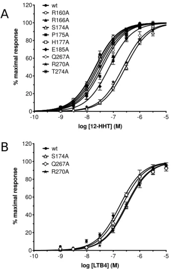
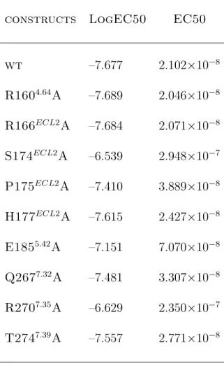
-10 -9 -8 -7 -6 -5 0 20 40 60 80 100 120 log [12-HHT] (M) % m ax im al re sp on se Q267A wt R160A R166A S174A P175A R270A T274A H177A E185A -10 -9 -8 -7 -6 -5 0 20 40 60 80 100 120 log [LTB4] (M) % m ax im al re sp on se S174A wt R270A Q267A
A
B
Figure 3: Gi protein activation catalyzed by the wild-type BLT2 receptor and its mutants
in the presence of increasing 12–HHT (A) or LTB4 (B) concentrations. Data are presented as the mean ± SEM of three experiments.
Table 2: EC50 values inferred from Gi protein activation catalyzed by the wild-type BLT2
receptor and its mutants in the presence of 12–HHT.
constructs LogEC50 EC50
wt –7.677 2.102×10⁻⁸ R1604.64A –7.689 2.046×10⁻⁸ R166ECL2A –7.684 2.071×10⁻⁸ S174ECL2A –6.539 2.948×10⁻⁷ P175ECL2A –7.410 3.889×10⁻⁸ H177ECL2A –7.615 2.427×10⁻⁸ E1855.42A –7.151 7.070×10⁻⁸ Q2677.32A –7.481 3.307×10⁻⁸ R2707.35A –6.629 2.350×10⁻⁷ T2747.39A –7.557 2.771×10⁻⁸
observed by introducing the mutation Q2677.32A (Figure 3A and Table 2), that orientation 213
was discarded.
214
The model of the interaction of 12–HHT NMR structure with a model of BLT2 based
215
on an active conformation of β2AR displays interactions with five secondary elements in
216
the receptor: 4 helices (II, III, VI and VII), which delineate the contours of the orthosteric
217
pocket, and one extra-cellular loop (ECL2) which plays a role of lid above the ligand pocket
218
(Figure 4). In addition to the two amino acids that establish hydrogen bonds with the ligand
219
(S174ECL2 and R2707.35), 10 other amino acids located at a distance ≤4˚A from the ligand
220
(depicted inFigure 4) show various weak interactions, including CH-to-π, CH-to-O,
NH-to-221
π, S-to-CH or N-to-CH proximities. The model of interaction depicted in Figure 4 is also
222
in accordance with some other neutral mutations that have been tested: first, residues that
223
are important for LTB4 binding to BLT1. R1604.64, which is highly conserved in both BLT1 224
and BLT2 receptors (Figure S6), has been identified to be crucial for LTB4 binding on BLT1
225
(residue R156 in BLT1) by potentially making a direct hydrogen bond with the carboxylate
226
head group.71Mutation of this residue to alanine results in a complete loss of LTB4 binding.71
227
Accordingly to our model, in which R1604.64 is located very far from 12–HHT (Figure S7), 228
R1604.64A mutant has no effect on 12–HHT binding (Figure 3A and Table 2). In the same 229
way, the E185A mutation did not significantly affect 12–HHT binding (Figure 3A) whereas
230
mutating this residue had a noticeable impact on LTB4 binding onto BLT1.71 Second, some
231
neutral mutations have been conducted. Just beside S174ECL2in ECL2, but not establishing 232
any interaction with the ligand, P175ECL2 and H177ECL2, which mutations to alanine do not
233
display a significant impact on ligand binding compared to the wild-type receptor. Another
234
residue in ECL2, which could possibly interact with the carboxyl function of the ligand,
235
R166ECL2, and an additional neutral mutation close to R2707.35, T2747.39A, do not impact 236
receptor ligand properties (Figure 3A and Table 2) in accordance with our model.
237 238 239
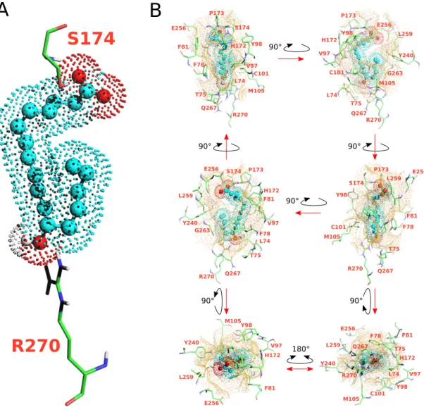
90° 90° 90° 90° 90° 90° 180° R270 Q267 T75 F78 L74 M105 C101 V97 Y98 F81 E256 P173 S174 H172 P173 H172 E256 L259 Y240 G263 M105 R270 Q267 T75 L74 C101 V97 Y98 E256 S174 P173 H172 F81 V97 F78 L74 T75 Q267 R270 G263 Y240 L259 S174 Y98 C101 M105 R270 P173 E256 F81 F78 L259 Q267 T75 Y240 M105Y98 V97 F81 H172 E256 L259 E256 F81 V97 F78 M105 C101 Y240 L74 H172 R270 T75 Y98 L259 Q267
A
B
R270
S174
Figure 4: Docking model of the NMR structure of 12–HHT in human BLT2 receptor (see HADDOCK structural statistics inTable S11). (A) represents the ligand in spheres and dots (hydrocarbon skeleton in cyan, oxygen atoms in red and the proton of the hydroxyle group in position 12 in white) double-locked at the top and the bottom of the orthosteric pocket by two hydrogen bonds with S174 and R270 residues. (B) represents six different views of the ligand in the orthosteric pocket of the receptor. The cavity of the ligand binding pocket is represented with a brown mesh surface at a maximum distance of 5˚A from the ligand. Amino acids delineating the pocket are indicated in orange.
Comparison of 12–HHT and LTB4 structures in their BLT2-bound states.
240 241
We present also in this study a new set of converged structures of LTB4 associated
242
with BLT2 in order to compare the bound structures of 12–HHT and LTB4 obtained under
243
identical conditions and procedures. Compared to the first calculation published in 2010,18
244
NMR data were collected at 700 MHz with a receptor associated with perDAPol50 instead
245
of DAPol and by using a softer methodology to remove the 1H signal of H2O to not affect 246
signal intensities from the ligand (see Material and Methods). As observed with 12–HHT,
247
in the absence of the receptor, both ends of the ligand are not structured, based on the
248
observation of intra-ligand 1H–to–1H dipolar interactions (Figure S8). Calculation based 249
on NMR data collected in the presence of BLT2 gives rise to a folded structure (Figure S9
250
and Table S10) similar to the previous published structure,18 but with an orientation of the
251
carboxyl-end (carbons 1 to 5) more loosely defined if we take into account an ensemble of 15
252
or 20 NMR structures (Figure S9and see dihedral ζ inFigure S10). If we try to coincide the
253
lowest energy conformers of LTB4 with the 12–HHT structure ensemble by superimposing
254
the most rigid part of the hydrocarbon skeletons, i.e. carbons 7 to 12, we find that, globally,
255
the fold of LTB4 is close to the 12–HHT structure in the presence of the same receptor
256
(Figure 5): the orientation of the carboxyl-end is similar, but not identical, the hydroxyl
257
group in position 12 points towards the same direction despite an opposite chirality of the
258
asymmetric carbon, and the methyl end for both ligands are quite close despite the LTB4
259
chain containing three more carbons. However, the two chains from carbon 12 –bearing
260
the hydroxyl group– to the methyl end display different orientations (see views 1 and 3 in
261
Figure 5). This region of these ligands is supposed to be located at the bottom of the pocket
262
of the receptor, based on the grafting of fluorescent probes at the carboxyl-end on the LTB4
263
for instance that does not affect the binding properties to BLT2.72
264
Attempts to get a model of LTB4 associated with BLT2 failed because we could not
265
identify clear mutants that impact significantly on the binding of LTB4 onto BLT2, and this
prevented us from getting a reasonable model of the ligand:receptor complex. Furthermore,
267
in contrast to 12–HHT, LTB4 is a very low-affinity ligand for BLT2, and this certainly
268
contributes to the fact that we could not get any satisfying model for this ligand.
269 270 271 272
Discussion
273 274Historically BLT2 was designated as the low-affinity LTB4 receptor, in contrast to BLT1,
275
with in cellulo Kd of ∼20 nM compared with ∼1 nM for BLT1.30 More recently, strong ev-276
idences led to the discovery of BLT2 endogenous agonist, 12–HHT,33,38,41 a non-eicosanoid
277
fatty acid compound which essentially comes from the conversion of prostaglandin H2 to
278
thromboxane A2. In cellulo measurements indicate a higher affinity of 12–HHT for BLT2
279
compared to LTB4, by about one order of magnitude.38 This was also observed by in vitro
280
binding measurements of LTB4 and 12–HHT onto a purified BLT2 receptor associated with
281
amphipols in solution, with Kdof ∼200 and ∼60 nM, respectively.19To be noted, the affinity 282
of the isolated receptor for its agonists is lower than that measured in cell systems. However,
283
high affinity can be recovered by associating the isolated receptor with its cognate G
pro-284
teins.51 Hence, the structures obtained here with the isolated BLT2 are likely signatures of
285
the low-affinity, uncoupled state of the receptor. A qualitative comparison of NMR NOESY
286
spectra clearly indicate an organization of both ends of 12–HHT in the presence of BLT2
287
(Figure S2and S3). Structure calculation confirmed that observation and led to a single set
288
of converged structures (Figure 2). The model proposed herein describes a ligand that is
289
double-locked in the receptor by two hydrogen bonds which are in accordance with
single-290
directed mutagenesis associated with ligand binding experiments (Figure 3A): one NH· · ·O
291
hydrogen bond between the OH moiety of the ligand and R2707.35 at the bottom of the
292
orthosteric pocket, and a stronger OH· · ·O hydrogen bond involving the COOH group of
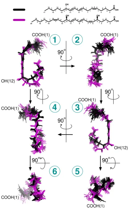
1 O OH 5 6 7 9 12 11 4 2 OH OH ζ β 13 14 15 ν 16 17 18 19 20 φ σ ρ ξ µ λ κ θ ε δ γ 10 8 3 α ι η 7 9 11 4 2 OH ζ β 13 µ λ κ θ 10 8 3 α ι η 5 6δ ε γ 14 15 16 17 ν ξ 12 1 O OH
1
2
4
3
5
6
OH(12) COOH(1) COOH(1) COOH(1) COOH(1) COOH(1) COOH(1) OH(12) 90 90 90 90 90 90Figure 5: Comparison of 12–HHT and LTB4 3D NMR structures associated with human BLT2. Six different views of superimposed ensembles of 20 energy-minimized conformers of 12–HHT (in black, from Figure 2), and the 7 lowest energy conformers of LTB4 (in purple, fromFigure S9). On Top are represented the chemical structures of the ligands.
12–HHT with S274ECL2, i.e. on the opposite side of the ligand pocket (Figure 4). These two 294
residues are highly conserved in BLT receptors (Figure S6), but interestingly, while BLT1
295
is almost activated by the LTB4 only, and not by 12–HHT, mutation of these two residues
296
does not affect the binding of LTB4 onto BLT2 (Figure 3B and Table 3). In complement to
297
measurements at equilibrium, in vitro off-rate constant measurements led to a bound time
298
3.6 times longer for 12–HHT than LTB4.19This also tends to suggest additional short range
299
interactions for 12–HHT compared with LTB4. A tentative superimposition of 12–HHT
300
and LTB4 in their BLT2-bound states described a similar fold, especially if we take into
301
account the 7 lowest energy structures obtained in the converged ensemble of structures of
302
LTB4 (Figure 5 and Table S10). However, several main features distinguish 12–HHT and
303
LTB4 than could explain these different binding properties: a shorter hydrocarbon chain for
304
12–HHT, with a double bond less, the absence of a hydroxyl group on position 5, and an
305
opposite chirality for the asymmetric carbone 12 (see top of Figure 5). In addition,
super-306
imposing the rigid core of these two ligands indicates noticeable differences, especially from
307
the asymmetric carbon 12 to the methyl end (Figure 5), a region which should interact with
308
the bottom of the orthosteric pocket.
309
Structures have revealed that a high percentage of identity between sub-families of class A
310
GPCRs can be observed for amino acids sculpting the orthosteric binding pocket in contrast
311
with the extracellular domains and membrane interface, which comprise the N-terminus end
312
and three extracellular loops and the top of the TMs. These regions display a higher
di-313
versity in both sequence and length.73 Experimental data indicate that ECLs are intimately
314
implicated in GPCR activation.74 Compilation of that information suggests a role of these
315
extracellular regions in GPCR signaling, including ligand binding and selectivity75 in
addi-316
tion to ligand efficacy, 76 allosteric modulations,e.g. 77 and constitutive activation.78 Our
317
model based on the crystallographic active state of the β2AR highlights the importance of
318
ECL2 as a lock above the orthosteric site which one residue, S174, display the strongest
in-319
teraction above all residues that interact with the ligand. Two additional residues, i.e. H172
Table 3: EC50 values inferred from Gi protein activation catalyzed by the wild-type BLT2
receptor and its mutants in the presence of LTB4.
constructs LogEC50 EC50
wt –7.677 2.102×10⁻⁸ R1604.64A –7.689 2.046×10⁻⁸ R166ECL2A –7.684 2.071×10⁻⁸ S174ECL2A –6.539 2.948×10⁻⁷ P175ECL2A –7.410 3.889×10⁻⁸ H177ECL2A –7.615 2.427×10⁻⁸ E1855.42A –7.151 7.070×10⁻⁸ Q2677.32A –7.481 3.307×10⁻⁸ R2707.35A –6.629 2.350×10⁻⁷ T2747.39A –7.557 2.771×10⁻⁸
and P173, associated with S174 define a hood above the ligand that probably contribute to
321
improve the residence time of the ligand to promote the binding of an intracellular
part-322
ner as equilibrium binding properties may not totally govern the activation of GPCRs. In
323
other words, non-equilibrium kinetics of the ligand binding event may also play an important
324
role.79
325
The method proposed in this study deserves to be improved as some imperfections could
326
introduce biases in both the structure calculation and also in the model. First of all, the
327
definition of parameter and topology files for organic compounds is not so trivial despite
328
the development of very efficient and convenient programs like PRODRG53 and XPLO2D54
329
that have been used in the present study. Structure calculations were based on the program
330
ARIA55 associated with CNS56 which contains a full relaxation matrix treatment of NOE
331
data to take into account indirect 1H–1H cross-relaxation pathways57,58 but does not take
332
into account the contribution of the chemical exchange of the ligand from the receptor in
333
the calculation. It would be interesting to include a matrix of exchange to properly gauge
334
the impact of the kof f –or conversely the residence time– of the ligand in the structure cal-335
culation. In the present study, the receptor is perdeuterated (98%) in order to limit the spin
336
diffusion into the ligand only, i.e. not relayed by protons of the protein, but the remaining
337
2% of protons in the receptor may slightly impact also the intra-ligand dipolar restraints
338
observed by NMR. We also tried to be as cautious as possible to use specific intra-dipolar
339
interactions only in the structure calculations, but this does not exclude some imperfections
340
in the approach. For all these reasons, we cannot exclude that both flexible ends of 12–HHT
341
(carbons 1 to 4 and 13 to 17) may display slightly different orientations compared to the set of
342
structures described herein. It should be noted that in the recent published structure of the
343
protaglandin E2 bound to EP3 receptor,80the ligand displays also a non-extended
conforma-344
tion in accordance with our results (Figure S11). In addition, docking simulations with the
345
set of conformers of free 12–HHT in solution (seeFigure 2) could not reproduce the contacts
346
observed between 12–HHT BLT2-bound structures and S174 and R270 (Figure S12).
ever, to help us to improve the method, NOE peak volumes for both 12–HHT and LTB4 are
348
available to the community inTables S1 toS4and Tables S6toS9, respectively. Ideally, the
349
experimental determination of a high-resolution structure of BLT2 receptor associated with
350
12–HHT would greatly help to adjust the approach detailed here. Other biophysical methods
351
like NMR chemical shift perturbation experiments with a specifically isotope-labeled BLT2
352
receptor, crosslinking and/or hydrogen/deuterium exchange associated with mass
spectrom-353
etry, and also molecular dynamics simulations could help to improve the model proposed in
354
the present study by determining some contact between the ligand and some amino acids of
355
the receptor. These methods have been used in the GPCR field to delineate ligand:receptor
356
contacts81–83and probe the changes in receptor conformation induced by the interaction with
357
the ligands.84 Overall, our data bring a first description of 12–HHT in its receptor-bound
358
state. This demonstrates the interest of a NMR-based approach to provide a description of
359
the structure and dynamics of natural ligands bound to unmodified receptors at physiological
360
temperatures, in complement to X-ray crystallography and cryoEM methods.
361 362 363 364 Acknowledgements 365 366
This work was supported by the Centre National de la Recherche Scientifique (CNRS),
367
Universit´e de Paris and Universit´es de Montpellier, the Agence Nationale de la Recherche
368
(ANR-17-CE11-0011), Laboratoire d’Excellence (LabEx) DYNAMO (ANR-11-LABX-0011)
369
and Equipements d’Excellence (EQUIPEX) CACSICE (ANR-11-EQPX-0008) from the French
370
Ministry of Research. The authors acknowledge access to the biomolecular NMR platform
371
of the IBPC that is supported by the CNRS, the Labex DYNAMO, the Equipex CACSICE
372
and the SESAME ˆIle-de-France.
373 374
375 376
Author Contributions
377 378
F.G. synthetized the perdeuterated amphipol; M.C., E.P., A.P. and L.J.C. performed the
379
production and purification of the receptor; M.D. and J.L.B performed single mutagenesis
380
experiments and ligand binding assays; F.G. and J.R. characterized the perdeuterated
am-381
phipol; C.L.B managed the NMR spectrometer and K.M. controlled structure calculations;
382
L.J.C. designed and supervised the project, performed the NMR sample preparations and
383
experiments; conducted structure calculations, molecular modeling and docking simulations,
384
and wrote the manuscript with editorial input from all authors.
385 386 387 Conflict of interest 388 389
The authors declare that they have no conflict of interest.
390
References
391
(1) Bockaert, J., & Pin, J.P. Molecular tinkering of G protein-coupled receptors: an
evo-392
lutionary success. EMBO J. 18, 1723-1729 (1999).
393
(2) Santos, R., Ursu, O., Gaulton, A., Bento, A.P., Donadi, R.S., Bologa, C.G., Karlsson,
394
A., Al-Lazikani, B., Hersey, A., Oprea, T.I., & Overington J.P. A comprehensive map
395
of molecular drug targets. Nat. Rev. Drug. Discov. 16, 19-34 (2017).
396
(3) Hauser, A.S., Attwood, M.M., Rask-Andersen, M., Schi¨oth, H.B., & Gloriam, D.E.
397
Trends in GPCR drug discovery : New agents, targets and indications. Nat. Rev.
398
Drug. Discov. 16, 829-842 (2017).
(4) Rasmussen, S.G., Choi, H.J., Fung, J.J., Pardon, E., Casarosa, P., Chae, P.S.,
De-400
vree, B.T., Rosenbaum, D.M., Thian, F.S., Kobilka, T.S., Schnapp, A., Konetzki, I.,
401
Sunahara, R.K., Gellman, S.H., Pautsch, A., Steyaert, J., Weis, W.I., & Kobilka, B.K.
402
Structure of a nanobody-stabilized active state of the β(2) adrenoceptor. Nature 469,
403
175-180 (2011).
404
(5) Ring, A.M., Manglik, A., Kruse, A.C., Enos, M.D., Weis, W.I., Garcia, K.C., &
405
Kobilka, B.K. Adrenaline-activated structure of 2-adrenoceptor stabilized by an
engi-406
neered nanobody. Nature 502, 575-579 (2013).
407
(6) Manglik, A., Kobilka, B.K., & Steyaert, J. Nanobodies to Study G Protein-Coupled
408
Receptor Structure and Function. Annu. Rev. Pharmacol. Toxicol. 57, 19-37 (2017).
409
(7) Weis, W.I., & Kobilka, B.K. The Molecular Basis of G Protein-Coupled Receptor
410
Activation. Annu. Rev. Biochem. 87, 897-919 (2018).
411
(8) Erlandson, S.C., McMahon, C., & Kruse, A.C. Structural Basis for G Protein-Coupled
412
Receptor Signaling. Annu. Rev. Biophys. 47, 1-18 (2018).
413
(9) Hilger, D., Masureel, M., & Kobilka, B.K. Structure and dynamics of GPCR signaling
414
complexes. Nat. Struct. Mol. Biol. 25, 4-12 (2018).
415
(10) Thal, D.M., Vuckovic, Z., Draper-Joyce, C.J., Liang, Y.L., Glukhova, A.,
Christopou-416
los, A., & Sexton, P.M. Recent advances in the determination of G protein-coupled
417
receptor structures. Curr. Opin. Struct. Biol. 51,28-34 (2018).
418
(11) Casiraghi, M., Damian, M., Lescop, E., Point, E., Moncoq, K., Morellet, N., Levy,
419
D., Marie, J., Guittet, E., Ban`eres, J.L., & Catoire L.J. Functional Modulation of a
420
G Protein-Coupled Receptor Conformational Landscape in a Lipid Bilayer. J. Am.
421
Chem. Soc. 138, 11170-11175 (2016).
(12) Casiraghi, M., Ban`eres J.L., & Catoire L.J. NMR Spectroscopy for the
Characteri-423
zation of GPCR Energy Landscapes. In: Topics in Medicinal Chemistry. Springer,
424
Berlin, Heidelberg, pp 1-26 (2017).
425
(13) Casiraghi, M., Damian, M., Lescop, E., Ban`eres J.L., & Catoire L.J. Illuminating
426
the Energy Landscape of GPCRs: The Key Contribution of Solution-State NMR
427
Associated with Escherichia coli as an Expression Host. Biochemistry 57, 2297-2307
428
(2018).
429
(14) Casiraghi, M., Point, E., Pozza, A., Moncoq, K., Ban`eres, J.L., & Catoire, LJ. NMR
430
analysis of GPCR conformational landscapes and dynamics. Mol. Cell. Endocrinol.
431
484, 69-77 (2019).
432
(15) Shimada, I., Ueda, T., Kofuku, Y., Eddy, M.T., & W¨uthrich, K. GPCR drug discovery:
433
integrating solution NMR data with crystal and cryo-EM structures. Nat. Rev. Drug.
434
Discov. 18, 59-82 (2019).
435
(16) Bostock, M.J., Solt, A.S., & Nietlispach, D. The role of NMR spectroscopy in mapping
436
the conformational landscape of GPCRs. Curr. Opin. Struct. Biol. 57, 145-156 (2019).
437
(17) Inooka, H., Ohtaki, T., Kitahara, O., Ikegami, T., Endo, S., Kitada, C., Ogi, K.,
438
Onda, H., Fujino, M., & Shirakawa, M. Conformation of a peptide ligand bound to
439
its G-protein coupled receptor. Nat. Struct. Biol. 8, 161-165 (2001).
440
(18) Catoire, L.J., Damian, M., Giusti, F., Martin, A., van Heijenoort, C., Popot, J.L.,
441
Guittet, E., & Ban`eres, J.L. Structure of a GPCR ligand in its receptor-bound state:
442
leukotriene B4 adopts a highly constrained conformation when associated to human
443
BLT2. J. Am. Chem. Soc. 132, 9049-9057 (2010).
444
(19) Catoire, L.J., Damian, M., Baaden, M., Guittet, E., & Ban`eres J.L.
Electrostatically-445
driven fast association and perdeuteration allow detection of transferred
relaxation for G protein-coupled receptor ligands with equilibrium dissociation
con-447
stants in the high-to-low nanomolar range. J. Biomol. NMR. 50, 191-195 (2011).
448
(20) O’Connor, C., White, K.L., Doncescu, N., Didenko, T., Roth, B.L., Czaplicki, G.,
449
Stevens, R.C., W¨uthrich, K., & Milon, A. NMR structure and dynamics of the agonist
450
dynorphin peptide bound to the human kappa opioid receptor. Proc. Natl. Acad. Sci.
451
U. S. A. 112, 11852-11857 (2015).
452
(21) Yong, K.J., Vaid, T.M., Shilling, P.J., Wu, F.J., Williams, L.M., Deluigi, M.,
453
Pl¨uckthun, A., Bathgate, R.A.D., Gooley, P.R., & Scott, D.J. Determinants of Ligand
454
Subtype-Selectivity at 1A-Adrenoceptor Revealed Using Saturation Transfer
Differ-455
ence (STD) NMR. ACS Chem. Biol. 13, 1090-1102 (2018).
456
(22) Brancaccio, D., Diana, D., Di Maro, S., Di Leva, F.S., Tomassi, S., Fattorusso,
457
R., Russo, L., Scala, S., Trotta, A.M., Portella, L., Novellino, E., Marinelli, L.,
458
& Carotenuto A. Ligand-Based NMR Study of C-X-C Chemokine Receptor Type
459
4 (CXCR4)-Ligand Interactions on Living Cancer Cells. J. Med. Chem. 61, 2910-2923
460
(2018).
461
(23) Chen, S., Lu, M., Liu, D., Yang, L., Yi, C., Ma, L., Zhang, H., Liu, Q., Frimurer,
462
T.M., Wang, M.W., Schwartz, T.W., Stevens, R.C., Wu, B., W¨uthrich, K., & Zhao
463
Q. Human substance P receptor binding mode of the antagonist drug aprepitant by
464
NMR and crystallography. Nat. Commun. 10, 638 (2019).
465
(24) Bender, B.J., Vortmeier, G., Ernicke, S., Bosse, M., Kaiser, A., Els-Heindl, S., Krug,
466
U., Beck-Sickinger, A., Meiler, J., & Huster, D. Structural Model of Ghrelin Bound
467
to its G Protein-Coupled Receptor. Structure 27, 537-544 (2019).
468
(25) Ferr´e, G., Louet, M., Saurel, O., Delort, B., Czaplicki, G., MKadmi, C., Damian, M.,
469
Renault, P., Cantel, S., Gavara, L., Demange, P., Marie, J., Fehrentz, J.A., Floquet,
the critical role of the octanoyl chain. Proc. Natl. Acad. Sci. U. S. A. 116, 17525-17530
472
(2019).
473
(26) Yokomizo, T., Izumi, T., Chang, K., Takuwa, Y., & Shimizu, T. A G-protein-coupled
474
receptor for leukotriene B4 that mediates chemotaxis. Nature 387, 620-624 (1997).
475
(27) Kamohara, M., Takasaki, J., Matsumoto, M., Saito, T., Ohishi, T., Ishii, H., &
Fu-476
ruichi, K. Molecular cloning and characterization of another leukotriene B4 receptor.
477
J. Biol. Chem. 275, 27000-27004 (2000).
478
(28) Tryselius, Y., Nilsson, N.E., Kotarsky, K., Olde, B., & Owman, C. Cloning and
char-479
acterization of cDNA encoding a novel human leukotriene B(4) receptor. Biochem.
480
Biophys. Res. Commun. 274, 377-382 (2000).
481
(29) Wang, S., Gustafson, E., Pang, L., Qiao, X., Behan, J., Maguire, M., Bayne, M.,
482
& Laz, T. A novel hepatointestinal leukotriene B4 receptor. Cloning and functional
483
characterization. J. Biol. Chem. 275, 40686-40694 (2000).
484
(30) Yokomizo, T., Kato, K., Terawaki, K., Izumi, T., & Shimizu, T. A second leukotriene
485
B(4) receptor, BLT2. A new therapeutic target in inflammation and immunological
486
disorders. J. Exp. Med. 192, 421-432 (2000).
487
(31) Izumi. T., Yokomizo, T., Obinata, H., Ogasawara, H., & Shimizu T. Leukotriene
488
Receptors: Classification, Gene Expression, and Signal Transduction. J. Biochem.
489
132, 1-6 (2002).
490
(32) Tager, A.M., & Lustera, A.D. BLT1 and BLT2: the leukotriene B4 receptors.
491
Prostaglandins Leukot. Essent. Fatty Acids 69, 123-134 (2003).
492
(33) Yokomizo, T. Leukotriene B4 receptors: Novel roles in immunological regulations Adv.
493
Enzyme Regul. 51, 59-64 (2011).
(34) Yokomizo, T. Two distinct leukotriene B4 receptors, BLT1 and BLT2. J. Biochem.
495
157, 65-71 (2015).
496
(35) Borgeat, P., & Samuelsson, B. Transformation of arachidonic acid by rabbit
polymor-497
phonuclear leukocytes. Formation of a novel dihydroxyeicosatetraenoic acid. J. Biol.
498
Chem. 254, 2643-2646 (1979).
499
(36) Yokomizo, T., Kato, K., Hagiya, H., Izumi, T., & Shimizu, T. Hydroxyeicosanoids
500
bind to and activate the low affinity leukotriene B4 receptor, BLT2. J. Biol. Chem.
501
276, 12454-12459 (2001).
502
(37) Hamberg, M., Svensson, J., & Samuelsson, B. Prostaglandin endoperoxides. A new
503
concept concerning the mode of action and release of prostaglandins. Proc. Natl. Acad.
504
Sci. U. S. A. 71, 3824-3828 (1974).
505
(38) Okuno, T., Iizuka, Y., Okazaki, H., Yokomizo, T., Taguchi, R., & Shimizu, T.
12(S)-506
Hydroxyheptadeca-5Z, 8E, 10E-trienoic acid is a natural ligand for leukotriene B4
507
receptor 2. J. Exp. Med. 205, 759-766 (2008).
508
(39) Hecker, M., Haurand, M., Ullrich, V., Diczfalusy, U., & Hammarstr¨om, S. Products,
509
kinetics, and substrate specificity of homogeneous thromboxane synthase from human
510
platelets: development of a novel enzyme assay. Arch. Biochem. Biophys. 254, 124-135
511
(1987).
512
(40) Goetzl, E.J., & Gorman, R.R. Chemotactic and chemokinetic stimulation of
hu-513
man eosinophil and neutrophil polymorphonuclear leukocytes by
12-L-hydroxy-5,8,10-514
heptadecatrienoic acid (HHT). J. Immunol. 120, 526-531 (1978).
515
(41) Iizuka, Y., Okuno, T., Saeki, K., Uozaki, H., Okada, S., Misaka, T., Sato, T., Toh,
516
H., Fukayama, M., Takeda, N., Kita, Y., Shimizu, T., Nakamura, M., & Yokomizo, T.
517
Protective role of the leukotriene B4 receptor BLT2 in murine inflammatory colitis.
(42) Matsunaga, Y., Fukuyama, S., Okuno, T., Sasaki, F., Matsunobu, T., Asai, Y.,
Mat-520
sumoto, K., Saeki, K., Oike, M., Sadamura, Y., Machida, K., Nakanishi, Y., Kubo,
521
M., Yokomizo, T., & Inoue, H. Leukotriene B4 receptor BLT2 negatively regulates
522
allergic airway eosinophilia. FASEB J. 27, 3306-3314 (2013).
523
(43) Liu, M., Saeki, K., Matsunobu, T., Okuno, T., Koga, T., Sugimoto, Y., Yokoyama,
524
C., Nakamizo, S., Kabashima, K., Narumiya, S., Shimizu, T., & Yokomizo T.
12-525
Hydroxyheptadecatrienoic acid promotes epidermal wound healing by accelerating
526
keratinocyte migration via the BLT2 receptor. J. Exp. Med. 211, 1063-1078 (2014).
527
(44) Tong, W.G., Ding, X.Z., Hennig, R., Witt, R.C., Standop, J., Pour, P.M., & Adrian,
528
T.E. Leukotriene B4 receptor antagonist LY293111 inhibits proliferation and induces
529
apoptosis in human pancreatic cancer cells. Clin. Cancer Res. 8, 3232-3242 (2002).
530
(45) Hennig, R., Osman, T., Esposito, I., Giese, N., Rao, S.M., Ding, X.Z., Tong, W.G.,
531
B¨uchler, M.W., Yokomizo, T., Friess, H., & Adrian, T.E. BLT2 is expressed in PanINs,
532
IPMNs, pancreatic cancer and stimulates tumour cell proliferation. Br. J. Cancer 99,
533
1064-1073 (2008).
534
(46) Lee, J.W., & Kim, J.H. Activation of the leukotriene B4 receptor 2-reactive
oxy-535
gen species (BLT2-ROS) cascade following detachment confers anoikis resistance in
536
prostate cancer cells. J. Biol. Chem. 288, 30054-30063 (2013).
537
(47) Houthuijzen, J.M., Daenen L.G., Roodhart J.M., Oosterom I., van Jaarsveld M.T.,
538
Govaert K.M., Smith, M., Sadatmand, S.J., Rosing, H., Kruse, F., Helms, B.J., van
539
Rooijen, N., Beijnen, J.H., Haribabu, B., van de Lest, C.H., & Voest, E.E.
Lysophos-540
pholipids secreted by splenic macrophages induce chemotherapy resistance via
inter-541
ference with the DNA damage response. Nat. Commun. 5, 5275 (2014).
542
(48) van der Velden, D.L., Cirkel G.A., Houthuijzen J.M., van Werkhoven E., Roodhart
543
J.M.L., Daenen L.G.M., Kaing, S., Gerrits, J., Verhoeven-Duif, N.M., Grootscholten,
C., Boot, H., Sessa, C., Bloemendal, H.J., De Vos, F.Y., & Voest, E.E. Phase I study
545
of combined indomethacin and based chemotherapy to reduce
platinum-546
induced fatty acids. Cancer Chemother. Pharmacol. 81, 911-921 (2018).
547
(49) Dahmane, T., Damian, M., Mary, S., Popot, J.-L., & Ban`eres, J.-L. Amphipol-assisted
548
in vitro folding of G protein-coupled receptors. Biochemistry 48, 6516-6521 (2009).
549
(50) Giusti, F., Rieger, J., Catoire, L.J., Qian, S., Calabrese, A.N., Watkinson, T.G.,
550
Casiraghi, M., Radford, S.E., Ashcroft, A.E., & Popot, J.L. Synthesis, characterization
551
and applications of a perdeuterated amphipol. J. Membr. Biol. 247, 909-924 (2014).
552
(51) Arcemisb´eh`ere, L., Sen, T., Boudier, L., Balestre, M.N., Gaibelet, G., Detouillon,
553
E., Orcel, H., Mendre, C., Rahmeh, R., Granier, S., Viv`es, C., Fieschi, F., Damian,
554
M., Durroux, T., Ban`eres, J.L., & Mouillac, B. Leukotriene BLT2 receptor monomers
555
activate G(i2) GTP-binding protein more efficiently than dimers. J. Biol. Chem. 285,
556
6337-6347(2010) .
557
(52) Hwang, T.L., & Shaka, A.J. Water Suppression That Works. Excitation Sculpting
558
Using Arbitrary Wave-Forms and Pulsed-Field Gradients J. Magn. Reson. A112,
559
275-279 (1995).
560
(53) A. W. Sch¨uttelkopf, A.W., & van Aalten, D.M.F. PRODRG: a tool for
high-561
throughput crystallography of protein-ligand complexes. Acta Crystallogr. D60,
1355-562
1363 (2004).
563
(54) Kleywegt, G. J., & Jones, T.A. Model-building and refinement practice. Methods
En-564
zymol. 277, 208-230 (1997).
565
(55) Rieping, W., Habeck, M., Bardiaux, B., Bernard, A., Malliavin, T.E., & Nilges, M.
566
ARIA2: automated NOE assignment and data integration in NMR structure
calcula-567
tion. Bioinformatics 23, 381-382 (2007).
(56) Br¨unger, A.T., Adams, P.D., Clore, G.M., DeLano, W.L., Gros, P., Grosse-Kunstleve,
569
R.W., Jiang, J.S., Kuszewski, J., Nilges, M., Pannu, N.S., Read, R.J., Rice, L.M.,
570
Simonson, T., & Warren, G.L. Crystallography & NMR system: A new software
571
suite for macromolecular structure determination. Acta Crystallogr., Sect. D: Biol.
572
Crystallogr. 54, 905-921 (1998).
573
(57) Bloembergen, N. On the interaction of nuclear spins in a crystalline lattice. Physica
574
15, 386-426 (1949).
575
(58) Linge, J. P., Habeck, M., Rieping, W., & Nilges, M. Correction of spin diffusion during
576
iterative automated NOE assignment. J. Magn. Reson. 167, 334-342 (2004).
577
(59) Sali, A., & Blundell, T.L. Comparative protein modelling by satisfaction of spatial
578
restraints. J. Mol. Biol. 234, 779-815 (1993).
579
(60) Fiser, A., Do, R.K., & Sali, A. Modeling of loops in protein structures. Prot. Sci. 9,
580
1753-1773 (2000).
581
(61) Marti-Renom, M.A., Stuart, A., Fiser, A., S´anchez, R., Melo, F., & Sali, A.
Compar-582
ative protein structure modeling of genes and genomes. Annu. Rev. Biophys. Biomol.
583
Struct. 29, 291-325 (2000).
584
(62) Hori, T, Okuno, T, Hirata, K, Yamashita, K, Kawano, Y, Yamamoto, M, Hato, M,
585
Nakamura, M, Shimizu, T, Yokomizo, T, Miyano, M, & Yokoyama, S. Na+-mimicking
586
ligands stabilize the inactive state of leukotriene B4 receptor BLT1. Nat Chem Biol.
587
14, 262-269 (2018).
588
(63) Zoonens, M., Catoire, L.J., Giusti, F., & Popot, J.L. NMR study of a membrane
589
protein in detergent-free aqueous solution. Proc. Natl. Acad. Sci. U. S. A. 102,
8893-590
8898 (2005).
(64) Ban`eres, J.-L., Popot, J.-L., & Mouillac, B. New advances in production and functional
592
folding of G-protein-coupled receptors. Trends Biotechnol. 29, 314-322 (2011).
593
(65) Baneres, J.-L., Martin, A., Hullot, P., Girard, J.-P., Rossi, J.-C., & Parello, J.
594
Structure-based analysis of GPCR function: conformational adaptation of both
ag-595
onist and receptor upon leukotriene B4 binding to recombinant BLT1. J. Mol. Biol.
596
329, 801-814 (2003).
597
(66) Popot, J.-L., Althoff, T., Bagnard, D., Banres, J.-L., Bazzacco, P., Billon-Denis, E.,
598
Catoire, L.J., Champeil, P., Charvolin, D., Cocco, M.J., Cr´emel, G., Dahmane, T., de
599
la Maza, L.M., Ebel, C., Gabel, F., Giusti, F., Gohon, Y., Goormaghtigh, E., Guittet,
600
E., Kleinschmidt, J.H., K¨uhlbrandt, W., Le Bon, C., Martinez, K.L., Picard, M.,
601
Pucci, B., Sachs, J.N., Tribet, C., van Heijenoort, C., Wien, F., Zito, F., & Zoonens,
602
M. Amphipols from A to Z. Annu. Rev. Biophys. 40, 379-408 (2011).
603
(67) Kumar, A., Ernst, R.R., & W¨uthrich, K. A two-dimensional nuclearOverhauser
en-604
hancement (2D NOE) experiment for the eluci-dation of complete protonproton
cross-605
relaxation networks inbiological macromolecules. Biochem. Biophys. Res. Commun.
606
95, 1-6 (1980).
607
(68) Wassenaar, T.A., van Dijk, M., Loureiro-Ferreira, N., van der Schot, G., de Vries,
608
S.J., Schmitz, C., van der Zwan, J., Boelens, R., Giachetti, A., Ferella, L., Rosato,
609
A., Bertini, I., Herrmann, T., Jonker, H.R.A., Bagaria, A., Jaravine, V., Gntert, P.,
610
Schwalbe, H., Vranken, W.F., Doreleijers, J.F., Vriend, G., Vuister, G.W., Franke, D.,
611
Kikhney, A., Svergun, D.I., Fogh, R.H., Ionides, J., Laue, E.D., Spronk, C., Jurksa, S.,
612
Verlato, M., Badoer, S., Dal Pra, S., Mazzucato, M., Frizziero, E., & Bonvin, A.M.J.J.
613
WeNMR: Structural Biology on the Grid. J. Grid. Comp. 10, 743-767 (2012).
614
(69) van Zundert, G.C.P., Rodrigues, J.P.G.L.M., Trellet, M., Schmitz, C., Kastritis, P.L.,
615
Karaca, E., Melquiond, A.S.J., van Dijk, M., de Vries, S.J., & Bonvin, A.M.J.J.
The HADDOCK2.2 webserver: User-friendly integrative modeling of biomolecular
617
complexes. J. Mol. Biol. 428, 720-725 (2016).
618
(70) Ballesteros, J. A., & Weinstein, H. Integrated methods for the construction of
three-619
dimensional models and computational probing of structure-function relations in G
620
protein-coupled receptors. Methods Neurosci. 25, 366-428 (1995).
621
(71) Basu, S., Jala, V.R., Mathis, S., Rajagopal, S.T., Del Prete, A., Maturu, P., Trent,
622
J.O., & Haribabu, B. Critical role for polar residues in coupling leukotriene B4 binding
623
to signal transduction in BLT1. J. Biol. Chem. 282, 10005-10017 (2007).
624
(72) Sabirsh, A., Wetterholm, A., Bristulf, J., Leffler, H., Haeggstr¨om, J.Z., & Owman, C.
625
Fluorescent leukotriene B4: potential applications. J. Lipid Res. 46, 1339-1346 (2005).
626
(73) Wheatley, M., Wootten, D., Conner, M.T., Simms, J., Kendrick, R., Logan, R.T.,
627
Poyner, D.R., & Barwell, J. Lifting the lid on GPCRs: the role of extracellular loops.
628
Br. J. Pharmacol. 165, 1688-1703 (2012).
629
(74) Unal, H., & Karnik, S.S. Domain coupling in GPCRs: the engine for induced
confor-630
mational changes. Trends Pharmacol. Sci. 33, 79-88 (2012).
631
(75) Peeters, M.C., van Westen, G.J.P., Li, Q., & IJzerman, A.P. Importance of the
ex-632
tracellular loops in G protein-coupled receptors for ligand recognition and receptor
633
activation. Trends Pharmacol. Sci. 32, 35-42 (2011).
634
(76) Nguyen, A.T., Baltos, J.A., Thomas, T., Nguyen, T.D., Mu˜noz, L.L., Gregory, K.J.,
635
White, P.J., Sexton, P.M., Christopoulos, A., & May, L.T. Extracellular Loop 2 of the
636
Adenosine A1 Receptor Has a Key Role in Orthosteric Ligand Affinity and Agonist
637
Efficacy. Mol. Pharmacol. 90, 703-714 (2016).
638
(77) Peeters, M.C., Wisse, L.E., Dinaj, A., Vroling, B., Vriend, G., & Ijzerman, A.P.
The role of the second and third extracellular loops of the adenosine A1 receptor in
640
activation and allosteric modulation. Biochem. Pharmacol. 84, 76-87 (2012).
641
(78) Pantel, J., Legendre, M., Cabrol, S., Hilal, L., Hajaji, Y., Morisse t, S., Nivot, S.,
642
Vie-Luton, M.P., Grouselle, D., de Kerdanet, M., Kadiri, A., Epelbaum, J., Le Bouc,
643
Y., & Amselem S. Loss of constitutive activity of the growth hormone secretagogue
644
receptor in familial short stature. J. Clin. Invest. 116, 760-768 (2006).
645
(79) Gabdoulline, R.R., & Wade, R.C. Biomolecular diffusional association. Curr. Opin.
646
Struct. Biol. 12, 204-213 (2002).
647
(80) Morimoto, K., Suno, R., Hotta, Y., Yamashita, K., Hirata, K., Yamamoto, M.,
Naru-648
miya, S., Iwata, S., & Kobayashi, T. Crystal structure of the endogenous
agonist-649
bound prostanoid receptor EP3. Nat. Chem. Biol. 15, 8-10 (2019).
650
(81) Coin, I., Katritch, V., Sun, T., Xiang, Z., Siu, F.Y., Beyermann, M., Stevens, R.C.,
651
and Wang, L. (2013) Genetically encoded chemical probes in cells reveal the binding
652
path of urocortin-I to CRF class B GPCR. Cell 155, 1258-69.
653
(82) Koole, C., Reynolds, C.A., Mobarec, J.C., Hick, C., Sexton, P.M., and Sakmar, T.P.
654
(2017) Genetically encoded photocross-linkers determine the biological binding site
655
of exendin-4 peptide in the N-terminal domain of the intact human glucagon-like
656
peptide-1 receptor (GLP-1R). J. Biol. Chem. 292, 7131-7144.
657
(83) Schmidt, P., Bender, B.J., Kaiser, A., Gulati, K., Scheidt, H.A., Hamm, H.E., Meiler,
658
J., Beck-Sickinger, A.G., and Huster, D. (2018). Improved in Vitro Folding of the Y(2)
659
G Protein-Coupled Receptor into Bicelles. Front. Mol. Biosci. 4, 100.
660
(84) West, G.M., Chien, E.Y., Katritch, V., Gatchalian, J., Chalmers, M.J., Stevens, R.C.,
661
and Griffin, P.R. (2011) Ligand-dependent perturbation of the conformational
ensem-662
ble for the GPCR 2 adrenergic receptor revealed by HDX. Structure 19, 1424-32.
![Figure 1: Dipolar interactions in the 12–HHT/u- 2 H-wtBLT2/perDAPol sample observed in a 2D NOESY spectrum (τ m = 0.5 s, ν H = 700 MHz, 25 ◦ C, [12–HHT] = 120 µM, [BLT2] = 15 µM)](https://thumb-eu.123doks.com/thumbv2/123doknet/7763717.255545/11.918.200.711.317.734/figure-dipolar-interactions-perdapol-sample-observed-noesy-spectrum.webp)