Journal of Applied Biosciences 114:
11357-11367
ISSN 1997-5902
Phytochemical screening and antibacterial
investigations of crude methanol extracts of Senna
didymobotrya (Fresen.) H. S. Irwin & Barneby
Jeruto, P.1*, Arama, P. F.2, Anyango, B.3, Maroa, G.4,
1University of Eldoret, School of Science, Department of Biological Sciences, P.O Box 1125-30300, Eldoret, Kenya 2Rongo University, School of Agriculture, Natural Resources and Environmental Sciences, Department of
Agricultural Economics and Agribusiness, P.O Box 103 – 40401 Rongo, Kenya.
3 Department of Biological Sciences, Jaramogi Oginga Odinga University of Science and Technology, School of
Biological and Physical Sciences, P.O. BOX 210, Bondo, Kenya.
4 Department of Chemistry , Jaramogi Oginga Odinga University of Science and Technology, School of Biological
and Physical Sciences, , P.O.BOX 210, Bondo, Kenya.
*(E-mail: of corresponding author: pasjeru@yahoo.com , pasjeru@gmail.com ; Phone: +254 720 326629)
Original submitted in on 31st March 2017. Published online at www.m.elewa.org on 30th June 2017 https://dx.doi.org/10.4314/jab.v114i1.9
ABSTRACT
Objective: Senna didymobotrya (African senna, African wild sensitive plant, peanut butter cassia, peanut butter tree, popcorn cassia, popcorn senna or wild senna) is native to East Africa and is widely used as a medicinal plant among many communities in Kenya. The objective of this research was to evaluate the presence of phytochemicals present in the different plant parts and their antibacterial activity.
Methodology and results: Leaves, flowers, stem bark, immature pods and root barks were collected from Siaya, Nandi and Nakuru Counties. These were dried and ground. Methanolic crude extracts were in cooperated in nutrient media at 2.5 %, 5 %, 7.5 % and 10 %. Test organisms Staphylococcus aureus and Escherichia coli were inoculated on impregnated media, incubated and observed for colony development. Observation on growth of cultures was made at an interval of 2 days for 8 days. The area under disease progress stairs (AUDPS) was calculated using the derived colony surface areas. Results indicated that all plant parts contained terpenoids, phenols and steroids. The presence of alkaloids and flavonoids varied with the location the plant was collected and the plant part. Growth of S. aureus cultures grown on media impregnated with 2.5% root bark extract and that with 7.5% stem bark extract were completely inhibited (no growth). Media with 10% flower, pods and leaves extract had average reduction of colony sizes from AUDPS 10102 (control) to AUDPS 2475. Growth of E. coli was completely inhibited on media impregnated with 5% root bark extract and 7.5% stem bark extracts. At 10% concentration, the flowers, pods and leaves extract did not result in complete inhibition of colony growth.
Conclusions and applications: The present research suggests that S. didymobotrya extracts possessed antibacterial activity against bacterial pathogens thus supporting their folkloric usage, promising a future scope for its use against microbial populations. Methanolic extracts possessing high antibacterial effects should be further investigated for their therapeutic utility. This would be related to the presence of bioactive metabolites, which are soluble in methanol. There is need to explore further the quantities of phytochemicals in the root and
stem barks that make them more potent than the other plant parts. The structures of the bioactive metabolites should be examined in future.
Key words: Antibacterial activity, Methanolic extract, Senna didymobotrya, Escherichia coli, Staphylococcus aureus
INTRODUCTION
Herbal medicines have been used for many years in many parts of the world especially developing countries (Kareru et al., 2007). Large body of evidence has accumulated to demonstrate the promising potential of medicinal plants used in various traditional, complementary and alternative systems of treatment of human diseases (Wong and Cuervo, 2010). In tropical countries, modern medicines are not affordable to most of the rural populations and WHO estimates that 80% of the world’s population use botanical medicines for primary health care (Muregi et al ., 2004). The World Bank report further pointed out that in 2002, Kenya’s Ministry of Health budget for medicine provided for only 30% of the population (WHO, 2002). This left 70% (21 million) of the population, which could not access the conventional medicines. The latter population group was therefore left to rely on traditional medicines for their healthcare needs (Sharma and Alagarsamy, 2012). The medicinal value of plants lies in some chemical substances that produce definite physiological actions to the human body. The most important of these compounds are alkaloids, flavonoids, tannins and phenolic compounds (Edeoga and Okwu, 2005). Biological and pharmacological activities of phytochemical compounds depend on factors such as the ecological factors, age of the plant, species and method of extraction. Thus, each plant has different chemical composition, toxicity and bioactivity (Rajakaruna et al., 2002). The type and amount of phytochemical compounds present usually differ from one part of the plant to another part. This explains why in traditional medicine the same or different plant parts may be used in treating different diseases or same diseases (Addo-Fordjour et al., 2008). The Senna genus formerly Cassia have been extensively studied for their phyllogenetic diversity, phyto-constituents as well as for their biological activities. It is native to tropical Africa (Bett, 2010) and was introduced, naturalized and became an
invasive species in parts of Kenya, Uganda and Tanzania (Sunarno, 1997). The flowers, roots and stems have both antibacterial and antifungal activity (Mazumder et al., 2008). Njoroge et al (2004) reported that that S. didymobotrya was utilized for back pains, antihelmintic, anaplasmosis, malaria, pimples, pneumonia (cattle), skin rashes, STD’s, stomach ache, tonsils (human) and typhoid. In another study, Njoroge and Bussmann (2007) found that the leaves of S. didymobotrya were boiled and a patient bath in it for treatment of pimples, scabies, warts and measles in Muranga, Nyandarua and Kiambu Counties. An ethnobotanical survey carried out in Kiandutu and Kiang’ombe in Thika urban slums on herbal medicine found that S. didymobotrya was one of the 41 species identified which was used for management of diarrhoea. Its bark, sap, leaves and seeds were utilized (Njoroge and Kibunga, 2007). In Mozambique, the roots are used traditionally against infectious diseases like diarrhoea, fevers, abscesses of the skeletal muscles and venereal diseases (Madureira et al., 2012). Pounded leaves and young stems are used in Kenya to treat skin diseases. The pulp is applied to the skin (Gachathi, 1989). The leaf sap is diluted in water and orally taken to treat diarrhoea, dysentery. It is also taken as a diuretic, laxative and emetic. Evidences are mounting concerning the resistance of microorganisms to antibiotics throughout the world (Akanwariwiak et al, 2012). This development has awakened scientists to explore alternative approaches that target and block resistance by combining plant extracts with antibiotics to increase their activity (Akanwariwiak et al., 2012). Therefore, there is a need to focus on the alternative sources of the antibiotics, as the pathogenic microbes are gaining resistance against standard antibiotics (Alo et al., 2012). This study was therefore, aimed at determining the phytochemicals present in the different plant parts of S. didymobotrya and the
antibacterial activity of the methanolic extracts obtained from the plant parts.
Plate 1.1.1 Plate 1.1.2
Plate 1.1.3 Plate 1.1.4
Plate 1.1: Senna didymobotrya (1.1.1- Inflorescence, 1.1.2- Fruits {immature pods}, 1.1.3 –Roots and 1.1.4- Leaves).
MATERIALS AND METHODS
Collection and processing of plant materials: Surveys were conducted in Siaya, Nakuru and Nandi Counties in Kenya. The regions were purposively selected according to altitude, geographical location and environmental variability. During the surveys, one cluster of S. didymobotrya with at least 20 mature plants at flowering and fruiting stage was selected. Samples were selected from Nandi County at Ndurio (0.02 N and 35.01 E), Siaya County at Uyawi-Miandhe (0.11 S and 34.08 E) and Nakuru County at Rongai (0.23 S and 35.54 E). From these plants, (i) young leaves, (ii) flowers, (iii) stem bark,
(iv) immature pods and (v) root barks were obtained. The samples were collected ethically following standard procedures (WHO, 2003) and with the farmer’s prior consent. Each sample was evenly spread on the laboratory bench and allowed to air dry at 28.70C for 14
days (Sikuku et al ., 2010). Samples were milled and sieved to pass through the mesh of 1.0 millimetre (mm) and stored in airtight plastic bags awaiting extractions. Extraction: Methanolic extraction was carried out according to methods as described by Kigondu et al. (2009). One thousand (1000) grams of powdered dried
plant part was weighed. At each time, 42.25 g was wrapped in a filter paper, placed in soxhlet apparatus and distilled with 300 ml absolute methanol. This was repeated until all the 1000g sample was used up. The methanol extract was then placed in a rotary vacuum evaporator placed in a water bath at 400C to recover the
solvent (methanol) and concentrate the crude extract. The semi-solid crude extract was placed in sterile beaker and left in the laminar flow hood for 24 hours for complete evaporation of the solvent.
Phytochemical screening of crude extracts of Senna didymobotrya: The crude extracts obtained were subjected to phytochemical analysis to determine the presence or absence of various chemical constituents using thin-layer chromatography (TLC). The Thin layer separation was carried out using pre-coated potassium chloride (KCl) silica gel [@ 60oA] TLC plates, size 20 × 20
cm, 1 mm thick. The chromatographic tank was allowed to equilibrate with the solvent system (Chloroform Methanol in the ratio of 98: 2, with 5 drops of glacial acetic acid), for at least 30 minutes prior to development of plates. The plates were sprayed with TLC visualization agents that gave specific reactions to specific group of compounds and visualized using standard methods i.e. UV- 254/364 nm (Harborne, 1984). The presence of different classes of compounds was determined by observing appropriate colour change on the TLC plate. Phytochemicals present in the plants were analysed according to the standard procedures described in previous studies (Harborne, 1984; 1998; Parekh and Chanda 2007).
Terpenoids: Vanillin spray test was used to determine the presence of terpenoids. One (1) g of vanillin was dissolved in 100 ml of concentrated sulphuric acid and this was used to spray the developed TLC plates and then placed in the oven for about 3 minutes at a temperature of 100 0C, colour change to purple was
indicative of the presence of terpenoids (Harborne, 1984). Alkaloids: Zero point five (0.5) grams of each extract was stirred with 5 ml of 1% aqueous hydrochloric acid on a steam bath. About 1 ml of the filtrate was treated with a few drops of Dragendorff’s reagent. The formation of red precipitate or orange colour indicated the presence of alkaloids (Harbone, 1998).
Phenols (polyphenols): Zero point one (0.1) grams of ferric chloride and 0.1 g of potassium ferric cyanide was dissolved in 10 ml of water. Then the reagent was sprayed on TLC plate. Change of colour to blue (instant) indicated the presence of phenols (Harborne, 1984). Flavonoids: Flavonoids presence was tested by dipping the developed TLC plates then exposed to fumes of
ammonia for five (5) minutes and any brightening yellow - brown spots of the present colours recorded indicated the presence of flavonoids.
Steroids: This was tested by the use of Kedde’s reagent. Kedde’s reagent was prepared by mixing 0.2 g of 3, 5 - Dinitrobenzoic acid in 10 ml methanol with 0.57 g sodium hydroxide in 10 ml distilled water. This was then used to spray the developed plates and any colour change to brown was indicative of the presence of steroids. Antibacterial activity of S. didymobotrya extracts on the selected bacterial isolates
Nutrient agar: Nutrient agar (28g) was dissolved in 1L of distilled water in a conical flask and then homogenized by boiling until boiling point using a hot plate. The medium was autoclaved at 1210C for 15 minutes. After sufficient
cooling, 100 ml was measured and poured into 100 ml Erlenmeyer flasks. The medium was allowed to cool to 400C.
Extract preparation: The constitutions of the extracts were carried out according to methods of Akanwariwiak et al. (2012) and Chad (2013). Using analytical balance, 2.5 g, 5 g, 7.5 g and 10 g of crude extract was weighed and placed in sterile beakers. Five millimetres of warm sterile water was added to the solid extract to reconstitute it. Each of the reconstituted extract was added into 100 ml of sterilized medium at 40 – 50 0C in a conical flask and
stirred. The negative control consisted of media alone without addition of S. didymobotrya extracts. The positive control consisted of streptomycin sulphate (Str) incorporated at 1 g/L of nutrient agar. The mixtures were stirred and let to cool for 30 minutes with the flask mouths open under the laminar flow hood. Medium from each flask was poured in four disposable petri dishes (replicates).
Test strains: The test microorganisms were obtained from Kenya Medical Research Institute (KEMRI), Kisumu, Kenya and were chosen based on the ethno-botanical exploitation of the plant. The test strains consisted of: (a) Staphylococcus aureus (ATCC 28923): Gram positive cocci and
(b) Escherichia coli (ATCC 25922): Gram-negative rod. The cultures were obtained from the petri dishes using a sterilized loop and placed in 10 ml of distilled water in a sterilized tube. The inoculum was serially diluted from 10-1
to 10-8. The original sample was stored in a refrigerator at
40C. 1 ml each was obtained from the last two dilutions
and plated on the Petridish. The dishes were incubated at 370C for five days. The number of colonies developing in
each plate was counted. Using this, the colony forming units (cfu) in the original sample was determined. The concentration of the original sample was adjusted using
sterile distilled water to obtain 108 cfu/ml. Using a
micropipette, 50 µl of the inoculums was obtained and inoculated in the middle of the Petri dish. The inoculum was allowed to dry for 2 hour. A line was drawn on top (on the petri dish cover) using a transparent ruler along the diameter of the dried inoculated position. The petri dishes were incubated at 370C. The experiment was set in with
four replicates in a completely randomized design (CRD). Colony growth was assessed at an interval of 2 days for 8 days. At each observation date, the diameter of the colony growth along the line was measured in millimetres. Data analysis: The diameter (mm) of the colony size was used to calculate the actual surface area (mm2) covered
by the pathogen. The disease areas for the different observations were summarized using the area under disease progress stairs (AUDPS) (Simko and Piepho, 2012):
Where AUDPC is area under disease progress curve given with formula;
AUDPC = [½ Obs1t1 + Obs2t2+Obs3t3+½Obs4t4]
Where:
y1 and yn are assessments at the first and last
observations respectively.
D = tn- t1 (when observations are performed at every time
unit, then D = (n -1)
t1, t2, t3 and t4 are the time intervals of the first, second,
third and the fourth observation.
Obs1, Obs2, Obs3 and Obs4 are first, second, third and
fourth observation respectively. n is the total number of observation.
The AUDPS was subjected to Analysis of Variance (ANOVA) and comparison of means was carried out on all data using SAS. Means were separated by the use of the least significant difference and compared at P = 0.05 for significance. SAS statistical package Release 8.02 copy 1999 - 2001 was used. The values are represented as means for AUDPS. Paired t - test was used for reporting the p value and significance with respect to the control. RESULTS
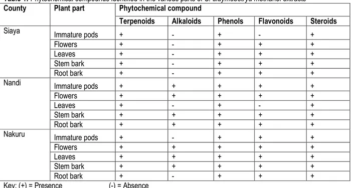
Phytochemical screening of methanol extracts of S. didymobotrya: The methanolic extracts of S. didymobotrya contained a mixture of compounds namely, steroids, alkaloids, flavonoids, phenols and terpenoids (Table 1). Terpenoids, phenols and steroids were present in all plant parts extracts. Flavonoids were present in all
plant-parts extracts except in immature pods from Siaya and Flowers in Nandi. Alkaloids were not detected in all plant parts from Siaya. Alkaloids were present in immature pods, flowers, stem and root barks (Nandi) and in flowers, leaves and stem bark (Nakuru)
Table 1: Phytochemical compounds identified in the various parts of S. didymobotrya methanol extracts
County Plant part Phytochemical compound
Terpenoids Alkaloids Phenols Flavonoids Steroids
Siaya Immature pods + - + - +
Flowers + - + + +
Leaves + - + + +
Stem bark + - + + +
Root bark + - + + +
Nandi Immature pods + + + + +
Flowers + + + + +
Leaves + - + - +
Stem bark + + + + +
Root bark + + + + +
Nakuru Immature pods + - + + +
Flowers + + + + +
Leaves + + + + +
Stem bark + + + + +
Root bark + - + + +
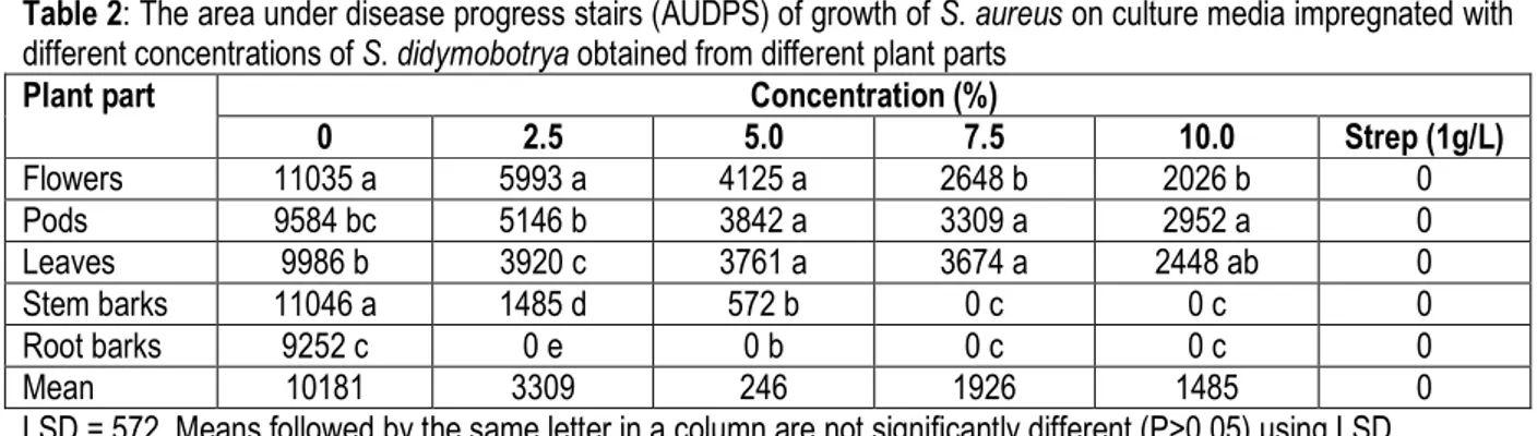
Table 2: The area under disease progress stairs (AUDPS) of growth of S. aureus on culture media impregnated with different concentrations of S. didymobotrya obtained from different plant parts
Plant part Concentration (%)
0 2.5 5.0 7.5 10.0 Strep (1g/L) Flowers 11035 a 5993 a 4125 a 2648 b 2026 b 0 Pods 9584 bc 5146 b 3842 a 3309 a 2952 a 0 Leaves 9986 b 3920 c 3761 a 3674 a 2448 ab 0 Stem barks 11046 a 1485 d 572 b 0 c 0 c 0 Root barks 9252 c 0 e 0 b 0 c 0 c 0 Mean 10181 3309 246 1926 1485 0
LSD = 572, Means followed by the same letter in a column are not significantly different (P>0.05) using LSD. Results in Table 2 indicated that the negative control (0%)
had a mean range colony growth AUDPS of 9252 – 11035. There were no colony growth of S. aureus (Table 2) observed on plates inoculated with streptomycin sulphate. When media were impregnated with 2.5% concentration of the extract, there was no culture growth observed on the medium containing root bark extract. This was also observed on medium impregnated with 5%, 7.5% and 10% of the root bark extracts. Media
impregnated with 2.5% stem bark extract had mean colony AUDPS of 1485. Media treated with immature pods, leaves and flowers had lower colony growths as compared to the negative control. The same trend was observed at 5% where root bark extract had no culture growth on medium whilst stem bark had mean culture growth AUDPS of 572. When media was impregnated with 7.5% and 10% of stem bark and root bark extracts there was no growth of bacteria observed.
Table 3: The area under disease progress stairs (AUDPS) of growth of E. coli on culture media impregnated with different concentrations of S. didymobotrya obtained from different plant parts.
Plant part Concentration (%)
0 2.5 5.0 7.5 10.0 Strep (1g/L) Flowers 7478 a 6658 a 6218 a 4001 a 3421 a 0 Pods 6422 b 5927 a 4880 b 4769 a 3004 a 0 Leaves 5785 b 4208 b 4141 b 3817 b 3541 a 0 Stem barks 5824 b 1309 c 173 c 0 c 0 b 0 Root barks 5620 b 929 c 0 c 0 c 0 b 0 Mean 6226 3806 3082 2517 1993 0
LSD = 871, Results are means of three replicate values. Means followed by the same letter in a column are not significantly different (P > 0.05) using LSD
Table 3 shows the negative control treatments had AUDPS range of 5619.7-7478.2. When medium was impregnated with 2.5% extract, root bark and stem bark had AUDPS of 929.1 and 1308.8 respectively. These were not significantly different (p > 0.05). Flowers, immature pods and leaf extracts had AUDPS range of 4208.3-6658.3. At concentration of 5%, the AUDPS for root bark was 0 whilst that of stem bark was 172.7. They
were significantly different (P < 0.05). When medium was impregnated with 7.5 and 10% of stem bark and root bark, there was no culture growth. At 7.5% and 10%, the flowers, immature pods and leaves had lower culture growth compared to the negative control. There were no colony growth of E. coli (Table 3) observed on plates inoculated with streptomycin sulphate
DISCUSSION
Phytochemical screening of S. didymobotrya: The presence of a good number of phytochemicals in the plant extracts screened can be a good source of bioactive components with antimicrobial potency, as they can be responsible together with the unscreened ones for the
antimicrobial activity of the extracts. This study showed that all plant parts of S. didymobotrya extracts contained terpenoids, phenols and steroids. The presence of alkaloids varied greatly depending on the location and plant part samples (Table 1). This may be attributed to the
great structural diversity of alkaloids (Fattorusso and Taglialatela- Scafati, 2008). Terpenoids have medicinal value such as antimicrobial, anti-carcinogenic, antimalarial and antidiuretics activity (Pichersky and Gershezon, 2002; Deganhart, 2003). They have shown great potentials in treatment against disease causing microorganisms. In addition, terpenoids have exhibited antibacterial activity against E. coli, S. aureus (Santos et al., 2008; Piera et al., 2011). The presence of alkaloids in the plant justifies its medicinal value since they have been identified for their functions, which include analgesic, antispasmodic, hypertensive effects, cytotoxic, anti-inflammatory and antimalarial. Kokwaro (2009) indicated that most of the savannah and semi - arid plant contains large percentages of alkaloids. Phenolic compounds have excellent antioxidant properties (Narayana et al., 2001). They, have biological and pharmacological properties especially their antimicrobial activity (Anpin Raja et al., 2011), antiviral, anti-inflammatory and cytotoxic activity (Mungole et al., 2010). Flavonoids are known to be antioxidant compounds, which protect human, animal and plant cells against the damaging effects of free radicals. Flavonoids and its derivatives have long been known to function as antimicrobial defense compounds (Kazmi et al., 1994). Other authors have shown that flavonoids have effective antimicrobial activity against a wide range of microorganisms (Schewe and Sies 2003; Yadav and Agarwala, 2011). Plant steroids are known to be important for their cardiotonic activities, possess insecticidal and antimicrobial properties (Ngbede et al., 2008; Anpin Raja et al., 2011). These secondary metabolites play a defensive and attractive role in the interactions between plants and their environment with other plants, herbivores, pathogens and pollinators (Figuiredo et al., 2008). Secondary metabolites have been found to have a distribution, which is sometimes confined to a genus. Species frequently have different production and accumulation sites (Cowen, 2008). The presence of phytochemicals in different plant parts of S. didymobotrya have been observed in previous studies (Nyaberi et al., 2013; Ngule et al ., 2013; Nyamwamu et al 2015). Presence of phytochemicals has also been reported in different plant parts of Bombax buonopozense and Monodora myristica (Firempong et al ., 2016) and Moringa oleifera (Abubakar and Usman, 2016). The phytochemicals in the present study differed in their diversity with geographical areas or ecozones and the plant parts (Table 1). These results forms the scientific basis for the use of these plants by the herbal practitioners in the Nandi and Kipsigis communities community, since these secondary metabolites in plants
have been shown to possess biological activity (Jeruto et al ., 2008; Korir et al ., 2012). Generally, the composition of phytochemical compounds within the same plant species differed from one part to another. These observations clearly demonstrated the reason as to why traditional healers use more than one plant parts to make a concoction for the treatment of a given disease since they have some additive or synergistic activity among various phytochemicals against the pathogens of interest (Addo-Fordjour et al ., 2008; Ruttoh et al ., 2009). Bacterial bioassays: Staphylococcus aureus is a gram - positive bacterium that produces staphylococcal enterotoxin (SE) A which is responsible for almost all staphylococcal food poisoning, a form of gastroenteritis (Argudin et al ., 2010). It may cause non - food related health issues such as skin inflammations, mastitis, respiratory infections, wound sepsis and toxic shock syndrome (Stewart, 2003; Montville and Mathews, 2008; Musa et al ., 2011). Over time, staphylococcus bacteria have become difficult to treat with antibiotics related to penicillin. These resistant forms of staphylococcus are referred to as Methicillin - Resistant S. aureus, or MRSA. Other pharmaceuticals used include ampicillin and vancomycin, which has been the most reliable antibiotic against MRSA, though currently resistance strains have been, cited (Harris et al., 2002). Attempts have been carried out by several authors in the world in managing S. aureus using alternative and herbal medicine. Results presented in Table 2 indicated that there was no growth of S. aureus on cultures impregnated with 2.5% root bark extract and 7.5% stem bark extracts. These were the only extracts that suppressed the growth of S. aureus within the range of concentrations studied. Even though the extracts from leaves, immature pods and flowers did not completely inhibit colony growth it was observed that there was significant suppression of the colony growth as the concentrations were increased. Thus, it was possible that the extracts from the other plant parts would completely suppress the growth of the organism at higher concentrations as the trend indicated (Table 2). In other studies, Anthoney et al. (2013b) reported that hot water extract of S. didymobotrya leaves inhibited the growth of all the gram positive and gram-negative bacteria that was tested against it. The extract inhibited the growth of S. aureus with a zone of inhibition of 11.00±0.577. Ngule et al. (2013) reported that the antibacterial activity of S. didymobotrya leaves was associated with the presence of phytochemicals, which included tannins and alkaloids. Observation that extracts from leaves, immature pods and flowers were not effective (P ≥ 0.05) may be because they yielded less phytochemicals than in stem and root
barks. Other researchers have demonstrated the antimicrobial activities of some plants against S. aureus. The leaf aqueous extracts of Eucalyptus camaldulensis Dehnh. using agar well diffusion technique was active against S. aureus (gram positive bacteria) even from a low concentration (12 mg / ml), the zones of inhibition increased in a dose dependent manner and it was bactericidal at 50 mg/ml (Musa et al ., 2011). The crude extracts showed activity that surpassed those of the standard antibiotics suggesting that they could be used in treating diseases caused by S. aureus. In their investigations on antibacterial investigations of moringa (Moringa oleifera) leaf extract on S. aureus, Abubakar and Usman (2016) reported that methanol extract had more antibacterial activity than the water extract, more so the extracts were discovered to be more active at higher concentration. They concluded that methanol extract of M. oleifera leaves have a potential antibacterial activity and to cure infections caused by bacteria such as E. coli, S. aureus and S. pneumonia. Escherichia coli is a gram-negative bacterium that lives in the digestive tracts of humans and warm-blooded animals. E. coli is a food borne pathogen that causes diarrhoea, hemorrhagic colitis and haemolytic uremic syndrome, which is a killer disease worldwide (Omori et al ., 2012). Young children, the elderly and immune-compromised individuals are particularly at risk (Zhao et al ., 2013). E. coli infection usually goes away on its own however the main treatment / prevention is to drink plenty of water since diarrhoea causes dehydration, which is especially dangerous for babies and older adults. With bloody diarrhoea, medicine or antibiotics can be used. Stem bark and root bark extracts completely inhibited colony growth of E. coli at concentrations of 5.0% and 7.5% respectively (Table 3). The other plant parts did not completely inhibit the colony growth at the highest concentration studied (10.0). However, a decrease in colony size was observed in
these extracts as the concentrations increased. The results obtained in this study are in conformity with those obtained by Anthoney et al . (2013 a), in which the plant roots were found to inhibit the growth of E. coli with significant zone of inhibition of 13.33± 0.667. In other studies, Chandrabhan et al . (2011) found that with increased concentration of up to 15% methanol and aqueous extracts of Terminalia chebula inhibited growth of both E. coli and S. aureus. Using agar well diffusion method, methanolic extracts showed a greater antibacterial activity compared to aqueous extracts (Lakshmidevi et al ., 2011) with Emblica officinalis leaf extract giving a zone of inhibition of 20 mm on E. coli. S. didymobotrya stem bark and root barks have shown effective antimicrobial activities against S. aureus and E. coli at low dosages/ concentrations. There is need to carry our research on these plant part extracts to identify the quantity of active phytochemicals that contribute to their high efficacies as compared to leaf, flower and immature pods extracts. On the conservation front, harvesting of these plant parts may lead to depletion of S. didymobotrya because they are potentially destructive unlike harvesting of the leaves, flowers and immature pods that regenerate every season hence reforestation of the plant is recommended for sustainability. S. didymobotrya extracts possessed antibacterial activity against bacterial pathogens thus supporting their folkloric usage, promising a future scope for its use against microbial populations. Methanolic extracts possessing high antibacterial effects should be further investigated for their therapeutic utility. This would be related to the presence of bioactive metabolites, which are soluble in methanol. There is need to explore further the quantities of phytochemicals in the root and stem barks that make them more potent than the other plant parts. The structures of the bioactive metabolites should be examined in future.
ACKNOWLEDGEMENTS
We acknowledge financial support by Deutscher Akademischer Austauschdienst (DAAD) through in-country scholarship and the National Commission for Science, Technology and Innovation (NACOSTI) through the Women Scientist Program Call II. The Kenya Medical Research Institute - Kisumu enabled us carry out this research by making available the test microorganism. We highly appreciate the assistance of Prof. Netondo and Prof. Mang’uro of Maseno University for allowing us to use the laboratory facilities at the Department of Botany
and Department of Chemistry respectively. We wish to thank the technical staff of both laboratories for their technical support and our colleagues at Jaramogi Oginga Odinga University of Science and Technology collaborators who accorded us moral support and assistance during the study. We thank all local communities in Nandi, Nakuru and Siaya counties. Disclosure of conflict of interest: We the authors declare that we do not have conflicts of interest.
REFERENCES
Abubakar, I., and Usman, A. 2016. Phytochemical and antibacterial investigations of moringa (Moringa oleifera) leaf extract on selected bacterial pathogens. Journal of Microbiology and Antimicrobials; 8 (5): 28-33
Addo-Fordjour P., Anning A. K., Belford E.J.D, Akonnor D. 2008. Diversity and conservation of medicinal plants in the Bomaa community of the Brong Ahafo Region, Ghana. J. Med. Plants Res.; 2(9):226-233.
Akanwariwiak, W. G., Addo - Fordjour, P. and Musah, A. A. 2012. Effects of combining crude ethanolic extract of Jatropha curcus L. leaf and some antibiotics against some selected microorganisms. Global Journal of Research on Medicinal Plants and Indigenous Medicine, 1 (5): 140 - 148.
Alo, M. N., Anyim, C., Igwe, J. C., Elom, M. and Uchenna, D. S. 2012. Antibacterial activity of water, ethanol and methanol extracts of Ocimum gratissimum, Vernonia amygdalina and Aframomum melegueta. Advances in Applied Science Research, 3 (2): 844 - 848.
Anpin Raja, R.D., Jeeva S., Prakash, J.W., Johnson, M. and Irudayaraj, V. 2011. Antibacterial activity of selected ethnomedicinal plants from South India. Asian Pac J Trop Med; 4: 375-378. Anthoney, S. T., Ngule, M. C. and Obey, J. K. 2013.
Phytopharmacological analysis of methanolic- aqua extract (Fractions) of Senna didymobotrya roots. International Journal of Bioassays, 2 (11):1473 - 1479.
Anthoney, S. T., Ngule, M. C. and Obey, J. K. 2013b. Evaluation of in vitro antibacterial activity in Senna didymobotrya roots methanolic-aqua extract and the selected fractions against selected pathogenic microorganisms. International Journal of Current Microbiology and Applied Sciences, 3 (5): 362 - 376. Argudin M. A, Mendoza, M. C. and Rodicio, M. R. 2010.
Food poisoning and Staphylococcus aureus enterotoxins. Toxins, 2 (7): 1751 - 1773. Bett, S. (2010). Mursik: Indigenous milk preservation
technology among the Kalenjins of Kenya. Ng’arua Maarifa Center. Article released on 3rd
November 2010. Internet: http:// ngaruamain.blogspot.Com / 2010 / 11 / mursik – indigenous – milk - preservation. Html downloaded on 27th September 2013.
Chad, B. 2013. Antibacterial effect of garlic (Allium sativum) and ginger (Zingiber officinale) against Staphylococcus aureus, Salmonella typhi, Escherichia coli and Bacillus aureus. Journal of Microbiology, Biotechnology and Food Sciences, 2 (4): 2481 - 2491.
Chandrabhan, S., Sachin, K. and Hotam, S. C. 2011. In vitro antibacterial study of aqueous and methanolic extracts of some selected medicinal plants. Journal of Chemical and Pharmaceutical Research, 3 (4): 854 - 860.
Cowen, L. E. 2008. The evolution of fungal drug resistance: Modulating the trajectory from genotype to phenotype. Natural Reviews Microbiology, 6: 187 - 198.
Deganhardt, J. 2003. Attracting friends to feast on foes: Engineering terpene emission to make crop more attractive to herbivores enemies. Current Opinion in Biotechnology, 14: 169 - 176. Edeoga, H. O. and Okwu, D. E. 2005. Phytochemical
constituents of some Nigerian medicinal plants: African Journal of Biotechnology, 4 (7): 685 - 688.
Fattorusso, E. and Taglialatela - Scafati, O. 2008. Modern alkaloids: Structure, isolation, synthesis and biology, Wiley – VCH.
Figueiredo, A. C., Barroso, J. G., Pedro, L. G. and Scheffer, J. J. C. 2008. Factors affecting secondary metabolites production in plants: volatile components and essential oils. Flavour and Fragrance Journal, 23: 213 - 226.
Firempong, C.K., Andoh, L.A. Akanwariwiak W.G., Addo-Fordjour P. and Adjakofi, P. 2016. Phytochemical screening and antifungal activities of crude ethanol extracts of red-flowered silk cotton tree (Bombax buonopozense) and Calabash nutmeg (Monodora myristica) on Candida albicans Journal of Microbiology and Antimicrobials, 8(4):22-27
Gachathi, F. 1989. Kikuyu botanical dictionary of plant names and uses. Nairobi; Kenya: AMREF. Harborne JB. 1984. Phytochemical methods. A guide to
modern techniques of plant analysis. London, New York: Chapman and Hall Ltd; p. 49–188. Harborne, J. B. 1998. Phytochemical methods: A guide to
modern techniques of plant analysis, Chapman and Hall, London. 3rd Edition: pp. 182 - 190.
Harris, L. G., Foster, S. J. and Richards, R. G. 2002. An introduction to Staphylococcus aureus, and
techniques for identifying and quantifying S. aureus adhesives in relation to adhesion to biomaterials: Review. European Cells and Materials, 4: 39 - 60.
Jeruto, P., Lukhoba, C., Ouma, G., Mutai, C. and Otieno, D. 2008. An Ethnobotanical study of medicinal plants used by the Nandi people in Kenya. Journal of Ethnopharmacology, 116: 370 - 376. Kareru, P. G., Kenji, G. M., Gachanja, A. N., Keriko, J. M. and Mungai, G. 2007. Traditional medicines and healing methods among Embu and Mbeere peoples of Kenya: African Journal of Traditional, Complementary and Alternative Medicine, 4 (1): 75 - 86.
Kazmi, M. H., Malik, S., Hameed, S., Akhtar, N. and Ali, S. N. 1994. An anthraquinone derivative from Cassia italica. Phytochemistry, 36: 761 - 763 Kigondu, E. V. M., Rukunga, G. M., Keriko, J. M., Tonui,
W. K. and Gathirwa, J. W. 2009. Anti - parasitic activity and cytotocity of selected medicinal plants from Kenya. Journal of Ethnopharmacology, 123: 504 - 509.
Kokwaro, J. O. 2009. Medicinal plants of East Africa. 3rd
Edition. Nairobi, Kenya: University of Nairobi Press; ISBN 9966 – 846 – 84 – 0.
Korir, R. K., Mutai, C., Kiiyukia, C. and Bii, C. 2012. Antimicrobial activity and safety of two medicinal plants traditionally used in Bomet district of Kenya. Research Journal of Medicinal Plant, 1 - 13. ISSBN 1819 – 3455 / DOI: 10.3923 / rjmp.2012.
Lakshmidevi, N., Kavishankar, G. B., and Murthy, M. S. 2011. Phytochemical analysis and antimicrobial properties of selected medicinal plants against bacteria associated with diabetic patients. International Journal of Pharmacology and Biosciences, 2 (4): 509 - 518.
Madureira, A. M., Ratmalhete, C., Mulhovo, S., Duarte, A. and Ferreira, M. 2012. Antibacterial activity of some African medicinal plants used traditionally against infectious diseases. Pharmaceutical Biology, 50 (4): 484 - 489. ISSN 1388 - 0209 print / ISSN 1744 - 5116 0nline. DOI:10.3109/13880209.2011.615841.
Mazumnder, P. M., Percha, V., Farswan, M. and Upaganlawer, A. 2008. Senna: A wonderful gift of medical sciences. International Journal of Community Pharmacy, 1 (2): 17 - 38.
Montville, T. J. and Matthews, K. R. 2008. Food microbiolgy: An introduction. 2 Ed, ASM Press,
Washington D. C.
Mungole, A.J., Awati, R., Chaturvedi, A. and Zanwar, P. 2010. Preliminary Phytochemical screening of Ipomoea obscura (L) -A hepatoprotective medicinal plant. Intern J Pharm Tech Res; CODEN (USA), 2(4): 2307-2312.
Muregi, F. W., Chhabra, S. C., Njagi, E. N. M., Langat - Thoruwa, C. C., Njue, W. M., Orago, A. S. S., Omar, S. A. and Ndiege, I. O. 2004. Anti - plasmodial activity of some Kenyan medicinal plant extracts singly and in combination with chloroquine. Phytotherapy Research, 18: 379 - 384.
Musa, D. A., Nwodo, F. O. C. and Ojogbane, E. 2011. Phytochemical, antibacterial and toxicity studies of the aqueous extract of Eucalyptus camaldulensis Dehnh. Asian Journal of Plant Science Research, 1 (3): 1 - 10.
Narayana, K. R., Reddy, M. S., Chaluvadi, M. R. and Krishna, D. R. 2001. Bioflavonoids classification, pharmacology, biochemical effects and therapeutic potential. Ind J. Pharmacol., 33:2-16.
Ngbede, J., Yakubu, R. A. and Nyam, D. A. 2008. Phytochemical Screening for Active Compounds in Canarium schweinfurthii (Atile) Leaves from Jos North, Plateau States, Nigeria. Res J Biol Sci.; 3:1076-1078.
Njoroge, G. N., Bussmann, R. W., Gemmill, B., Newton, E. L. and Ngumi, W. (2004). Utilization of weed species as sources of traditional medicines in Central Kenya. Lyonia a Journal of Ecology and Application, 7 (2): 71 - 87.
Njoroge, G. N. and Bussmann, R. W. 2007. Ethnotherapeutic management of skin diseases among the Kikuyus of Central Province of Kenya. Journal of Ethnopharmacology, 11: 303 - 307.
Njoroge, G. N. and Kibunga J. W. 2007. Herbal medicine acceptance, sources and utilization for diarrhoea management in a cosmopolitan urban area (Thika, Kenya). African Journal of Ecology, 45 (suppl.1): 65 - 70.
Ngule, C. M., Swamy, T. A. and Obey, J. K. 2013. Phytochemical and bioactivity evaluation of Senna didymobotrya Fresen Irwin used by the Nandi community in Kenya. International Journal of Bioassays, 2 (7): 1037 - 1043.
Nyaberi, M. O., Onyango, C. A., Mathoko, F. M. Maina, J. M. Makobe, M. and Mwaura, F. 2013. Bioactive fractions in the stem charcoal of Senna didymobotrya Freasen Irwin and Barney used by
pastoral communities in West Pokot to preserve milk. Natural Resource Management, 16: 980 - 985.
Nyamwamu, L.B., Ngeiywa, M., Mulaa, M. Lelo, A.E., Ingonga, J. and Kimutai, A. 2015. Phytochemical constituents of Senna didymobotrya Fresen Irwin roots used as a traditional medicinal plant in Kenya International Journal of Education and Research, 3(6):431-442
Omori, E. O., Calistus, O., Mbugua, P. K. and Okemo, P. O. (2012). Ethnobotanical identification and anti - microbial evaluation of some anti - diarrhoreal plants used by the Samburu community, Kenya. Malaysian Journal of Microbiology, 8 (2): 68 - 74.
Parekh, J. and Chanda, V. (2007). In vitro antimicrobial activity and phytochemical analysis of some Indian medicinal plants. Turkey Journal of Biology, 31: 53 - 58.
Pichersky, E. and Gershezon, J. (2002). The formation and function of plant volatiles, perfumes for pollinator attraction and defence. Current Opinion in Plant Biology, 5: 237-243.
Piera, F. A., Souza, C. F., Costa, J. C M., Barreto, M. A. O., Espescheit, I. F., Silva, V. O. and Moreira, M. A. S. (2011). Inhibition of E. coli from mastitic milk by Agrarias. Londrina, 32: 1929 - 1934 Rajakaruna, N., Harris, S. C. and Towers, G. H. N.
(2002). Antimicrobial activity of plants collected from Serpentine outcrops in Sri Lanka. Pharmacological Biology, 40: 235 - 244. Ruttoh, E. K., Tarus, P. K., Bii, C. C., Machocho, A. K.,
Karimi, K. L. and Okemo, P. O. (2009). Antibacterial activity of Tabernaemontana stapfiana Britten (Apocynaceae) extracts. African Journal of Traditional, Complementary and Alternative Medicine, 6 (2): 186 - 194. Santos, A. O., Ueda-Nakumura, T., Dias Filo, B. P.,
Veiga, V. F. J., Pinto, A. C. and Nakamwa, C. V. (2008). Antimicrobial activity of Brazilian Copapipa oils obtained from different species of the Copalifera genes. Memorias do Instituto Oswaldo Cruz, 103: 277-281.
Schewe, T. and Sies, H. (2003). Flavonoids as protectants against prooxidant enzymes, Research monographs, Institut fur Physiologische Chemie 1; Heinrich- Heine- Universitat Dusseldorf- Date of access: 7.07.2009. Available from:
http://www.uni-duesseldorf.de/WWW/MedFak/PhysiolChem/ind ex.html.
Sharma, H. K and Alagarsamy, V. (2012). In vitro antimicrobial activity of various extracts of Dyschoriste littoralis Nees. International Journal of Phytopharmacology, 4 (3): 212 - 216. Sikuku, P. A., Netondo, G. W., Onyango, J. C. and
Musyimi, D. M. (2010). Chlorophyll fluorescene, protein and chlorophyll content of three Nerica rainfed rice varieties under varying irrigation regimes. Journal of Agricultural and Biological Sciences, 5 (2): 19 - 25.
Simko, L. and Piepho, H. P. (2012). The area under the disease progress stairs: Calculation, advantage and application. Phytopathology, 102: 381 - 389. Stewart, C. M. (2003). Staphylococcus aureus and
staphylococcal enterotoxins. Ch 12 In: Hocking A.D (ed). Foodborne microorganisms of public health significance. 6th Ed, Australian Institute of Food Science and Technology (NSW Branch), Sydney: 359 - 380.
Sunarno, B. (1997). Senna didymobotrya (Fresenianus) Irwin & Barneby. In Faridah Hanum, I. and van derMaesen, L. J. G. (Eds.): Plant Resources of South - East Asia. No. 11. Auxillary Plants. Prosea Foundation, Bogor, Indonesia: 229 – 231.
Wong, E. and Cuervo, A. M. (2010). Autophagy gone awry in neurodegenerative diseases. Nature Neuroscience, 13 (7): 805 - 811.
World Health Organization (WHO). (2002). WHO Traditional Medicine Strategy 2002 - 2005.World Health Organization, Geneva, WHO/EDM/TRM/2002.1
World Health Organization (WHO) 2003. WHO guidelines on good agricultural and collection practices (GACP) for medicinal plants. World Health Organization, Geneva, Switzerland, 7 − 9 July 2003 Geneva: 1 - 80.
Yadav, R. N. S. and Agarwala, M. (2011). Phytochemical analysis of some medicinal plants. Journal of Phytology, 3 (12): 10 - 14.
Zhao, L, Tyler, P. J., Starnes, J., Rankins, D. McCaskey, T. A. and Wang, L. (2013). Evaluation of the effects of weaning diets on Escherichia coli O157 shedding, body weight, and faecal bacteria communities in beef calves. Foodborne Pathogens and Disease 0 (0): 1 - 6.