HAL Id: tel-01124241
https://tel.archives-ouvertes.fr/tel-01124241
Submitted on 6 Mar 2015HAL is a multi-disciplinary open access archive for the deposit and dissemination of sci-entific research documents, whether they are pub-lished or not. The documents may come from teaching and research institutions in France or abroad, or from public or private research centers.
L’archive ouverte pluridisciplinaire HAL, est destinée au dépôt et à la diffusion de documents scientifiques de niveau recherche, publiés ou non, émanant des établissements d’enseignement et de recherche français ou étrangers, des laboratoires publics ou privés.
Differential abnormalities of two spinal motoneuron
populations in the SOD1 G93A neonatal mouse (model
of the amyotrophic lateral sclerosis)
Félix Leroy
To cite this version:
Félix Leroy. Differential abnormalities of two spinal motoneuron populations in the SOD1 G93A neonatal mouse (model of the amyotrophic lateral sclerosis). Human health and pathology. Université René Descartes - Paris V, 2013. English. �NNT : 2013PA05T063�. �tel-01124241�
Thèse de doctorat de l’université Paris Descartes
Spécialité Neurosciences Laboratoire de Neurophysique et Physiologie UMR 8119 Ecole doctoraleCerveau – Cognition – Comportement
Présentée par M. Félix Leroy Pour obtenir le grade de
Docteur de L’Université Paris Descartes
Atteinte différentielle de deux populations de
motoneurones spinaux chez le souriceau SOD1
G93A (modèle de la maladie de Charcot)
Soutenue publiquement le 6 décembre 2013
Devant le jury composé de:
M. Pascal Branchereau M. George Mentis M. Frédéric Charbonnier M. Stéphane Charpier M. Boris Lamotte d’Incamps M. Daniel Zytnicki
Université Bordeaux 1 Columbia University Université Paris Descartes Université Pierre et Marie Curie Université Paris Descartes Université Paris Descartes
Rapporteur Rapporteur Examinateur Examinateur Examinateur Directeur de thèse
Differential abnormalities of two spinal motoneuron
populations in the SOD1 G93A neonatal mouse
(model of the amyotrophic lateral sclerosis)
Doctorate PhD thesis defended by Mr Félix Leroy
On the 6
thof december 2013
Remerciements
En premier lieu, je tiens à remercier George Mentis, Pascal Branchereau, Stéphane Charpier et Frédérique Charbonnier d’avoir accepté de faire partie de mon jury thèse.
Je remercie ensuite chaleureusement Daniel Zytnicki pour m’avoir encadré durant la thèse. Merci pour la patience et la confiance dont il a fait preuve à mon égard. Je remercie également Boris Lamotte d’Incamps de m’avoir encadré et appris d’innombrables choses en particulier en ce qui concerne la technique de patch-clamp et la préparation que nous utilisons.
Un grand merci à tous les membres de mon équipe, Marin Manuel, Rebecca Manuel, Nicolas Deslestrée et Clémence Martinot aux côtés desquels j’ai pris plaisir à travailler durant ces 3 années.
Je remercie de même les personnes de l’UMR 8119, en premier lieu Claude Meunier qui m’a accueilli au laboratoire, mais aussi Visou Ady, Arthur Leblois, Bill Wood, Hervé Suaudeau, Carole Sens ainsi que tous les autres…
Merci aussi à tous les membres de l’UMR 8118, où j’ai effectué les dissections et avec lesquels j’ai eu de nombreuses discussions stimulantes sur nos projets respectifs.
Merci aussi à Patrice Jégouzo pour son aide précieuse. Merci aussi à l’équipe de Frédéric Charbonnier qui m’a accueilli à bras ouvert dès que je passais leur porte. Merci à Jamilé Hazan pour ses conseils concernant la technique de RT-PCR. Merci aussi à Ali Jalil qui m’a enseigné les subtilités des anticorps.
Je souhaite remercier particuliérement ceux qui, en dehors de mes encadrants ont bien souvent relus mes bafouilles: CJ Heckman, Kathy Quinlan et Marin Manuel. Un grand merci spécialement à Philippe Asher pour son oreille attentive et ses relectures assidues.
Enfin, je remercie vivement mes parents, Patrick et Patricia Leroy, ma famille, mes amis et surtout mon épouse Laëtitia Herbaut qui me soutiennent depuis des années et sans qui rien n’aurait été possible.
Table of content
Introduction (p6)
1/ Different motoneuron sub-types in the adult mouse 2/ Motoneurons are uneven when facing the ALS 3/ Presentation of the work done
4/ Thesis content
I - Differentiation of the heterogenous motoneurons populations (p15)
1/ Mature motoneurons segregate
2/ Henneman’s “size principle” for mature motoneurons 3/ Motor units differentiation
4/ Motoneuron maturation and differentiation
II - Two subpopulations of alpha spinal motoneurons displaying different discharge profiles caused by different potassium currents (p35)
1/ Introduction
2/ A-like and slow inactivating potassium currents determine recruitment and firing properties in a subpopulation of spinal motoneurons in neonatal mice 3/ Correlation of the discharge patterns with fast and slow types
4/ Conclusion
III - Heterogenous vulnerability of the motoneurons during the ALS (p70)
1/ Generality on the ALS, molecular substrate and order of degenerescence 2/ Evolution of the disease
3/ Excitotoxic hypothesis and other theories 4/ Differential vulnerability to the disease
IV - Electrical and morphological properties of spinal motoneurons are differentially affected depending upon their firing pattern in neonatal G93A
SOD1 mice (p83)
1/ Introduction 2/ Results 3/ Conclusion
Discussion (p112)
1/ Lack of tonic synaptic inputs in my preparation 2/ Motor units maturation
3/ Differential effect of the SOD1 G93A mutation on the two populations 4/ Possible change in excitation leading to morphological alterations
References (p123)
Supplementary material (p139)
1/ Synaptic inputs have no tonic effect on the motoneurons intrinsic properties 2/ Other conductances expressed in spinal neonatal mouse motoneurons 3/ Protocols
Table of figures
Figure 1: Schematic representation of the different types of motoneurons. (p8)
Figure 2: Motoneurons subtypes segregate according to their intrinsic properties. (p15)
Figure 3: Summary of important events in the development of neuromuscular connectivity. (p19) Figure 4: Polyneuronal innervation of muscle fibers. (p22)
Figure 5: Neonatal motor unit are already largely homogenous in their fibers composition. (p23) Figure 6: Ontogeny of the A-, T- and L-currents in motoneurons. (p27)
Figure 7: Chodl+ cells have electrophysiological properties of fast motor neurons. (p33)
Figure 8: Electrical properties of delayed firing and immediate firing motoneurons strongly suggest that they are fast and slow type motoneurons. (p38)
Figure 9: Delayed firing motoneurons display more complex dendritic arborization. (p41) Figure 10: Setting of the single cell RT-PCR. (p42)
Figure 11: Immediate and delayed firing motoneneurons co-express NeuN and Errγ. (p43) Figure 12: Chodl is only expressed in a sub-population of the delayed firing motoneurons. (p44) Figure 13: Contribution of mutant SOD1 within different cell types in ALS. (p71)
Figure 14: Time course of neurodegeneration in the SOD1G93A mouse model of (ALS). (p73) Figure 15: Proposed mechanisms of toxicity in SOD1-mediated ALS. (p78)
Figure 16: Neuromuscular junction phenotypes in fast-fatigable (FF) and slow (S) motor units in mutant SOD1 mice. (p81)
Figure 17: Some motoneurons express the H-current. (p141) Figure 18: activation of Ican in delayed firing motoneuron. (p142)
Introduction
Motoneurons are the link between the nervous system and the muscles. They are the final common pathways of every neural circuits involved in motricity. Since Charles Sherrington’s work we know that all motoneurons share the ability to excite muscle fibers. In the adult mammalian, one motoneuron innervates many muscle fibers but each muscle fiber is innervated by only one motoneuron (Liddel & Sherrington, 1925; Redfern, 1970; Jansen & Flatby, 1990; Van Essen et al., 1990). A motor unit comprises the motoneuron and all its target muscle fibers that usually share similar contractile properties.
Motoneurons receive tens of thousand of peripheral as well as central inputs (Henneman & Mendell, 1981; Kernell, 2006). Descending systems (corticospinal, vestibulospinal, rubrospinal and reticulospinal) project either directly or indirectly on the motoneurons modifying phasically (voluntary action) or tonically (serotonin liberation during locomotion) the motoneurons voltage. Peripheral inputs project directly, or indirectly, on motoneurons. The muscular mechanoreceptors (neuromuscular spindles, Golgi tendon organs) but also the joint and cutaneous receptors allow proprioceptive information to drive the motoneurons activity as well. Ultimately all these inputs are summed in the motoneurons soma and generate trains of action potentials eliciting the recruitment of motor units and the muscle contraction. Far from being a linear integrator (Kernell, 2006), motoneurons exhibit numerous voltage-gated conductances allowing non-linear transformation of the inputs (membrane bistability, hysteresis of the response to triangular ramps). The most extendedly studied is the calcium persistent inward current that amplifies distal tonic inputs (Lee & Heckman, 1998a). Another kind of persistent inward current arises from a sodium component. Its faster time constant suggests a role in amplifying faster inputs (Li et al., 2004; Harvey et al., 2006; Theiss et al., 2007). Other conductances active at membrane resting potential filter the cell membrane potential below firing threshold. This is the case of the H-current that creates, in conjunction with the membrane passive properties, a peak of resonance amplifying inputs of specific frequency (Manuel et al., 2007).
1/ Different motoneuron sub-types in the adult mouse
The motoneuron population can be divided in many sub-populations in adults (Figure 1). γ -motoneurons innervate intrafusal fibers whereas α -motoneurons innervate extrafusal fibers and beta-motoneurons innervate both. γ- and β-motoneurons further divide between static and dynamic motoneurons according to the way they modulate the spindle activity. When investigating the properties of the motor units, Burke et al. (1971; 1973) defined three physiological types: fast and slow motor units according to the contraction time of the muscle fibers and within the fast motor units fatigue-resistant and fatigable motor units according to the way the contractile material of their muscle fibers is able or not to sustain repeated stimulations. Since each motor unit comprises a single motoneuron we may therefore talk about slow motoneurons (S motoneurons), fast fatigue-resistant motoneurons (FR motoneurons) and fast fatigue-fatigable motoneurons (FF motoneurons). The investigation of the motor unit properties was initially carried out in the cat but similar results were obtained in many other mammals (rat, rabbit, human, guinea-pig, (Kernell, 2006)). However some authors argued that, in the mouse, FR and FF motor units form a continuum rather than discrete populations, especially in rodents in which motor neuron size differences are less marked (Bakels & Kernell, 1993a, b) (Gardiner, 1993). In mammals every muscle embodies the three types of muscle units although in highly variable proportions. Thanks to the glycogen depletion method, Burke et al. (1971, 1973) further demonstrated that all the muscle fibers of a given motor unit exhibited the same histochemical profile. Furthermore, they found a correlation between the physiological types and the histochemical profiles: type S motor units have type I muscle fibers, type FR motor units have type IIA muscle fibers, and type FF motor units have type IIB muscle fibers (Figure 1). Therefore Burke et al. (1971, 1973) showed an exquisite correlation between the muscle unit contractile properties (contraction velocity and fatigue resistance) and the molecular identity of the fibers (difference in the amount of oxidative enzymes such as SDH or in the amount of myofibrillar ATPases, (Edstrom & Kugelberg, 1968), reviewed in (Schiaffino & Reggiani, 2011). Later on, staining of the different isotypes of the myosin heavy chain proteins was established (Schiaffino et al., 1988) allowing to tighten the link between the contractile proteins and the muscle unit properties.
The fact that different motoneurons impinge on different muscle units and that a given motoneuron connects exclusively to a homogenous muscle fiber population calls for a matching between a motoneuron and its efferent fibers characteristics (i.e the motoneurons electrophysiological properties need to be tuned to their output). Indeed, motoneurons properties are widely different. Among all characteristics, motoneurons of a given kind segregate according to their electrotonic size (evaluated by the measure of the input conductance, (Zengel et al., 1985). In addition, Friese et
al. (2009) showed that the average size of the soma already allowed to discriminate
between γ - and α -motoneurons as early as P14 in the mice. Motoneuron size therefore increases from γ-motoneuron to α-FF motoneuron (γ < α-S < α-FR < α-FF).
Given the evidence indicating that α-motoneurons receive a common input (Mendell & Henneman, 1968; Henneman, 1985), Henneman et al. (1957) posited an orderly recruitment of the motor units based on the size principle. That is to say that considering a fixed input, the most electrotonically compact motoneurons will see their voltage rise more and will reach their firing threshold prior the less compact ones.
In the neonatal rodent, heterogeneity between motor units is already established (Close, 1967) and motor units present a biased composition in fiber types as early as E16 (Condon et al., 1990). But what about putative differences between neonatal motoneurons electrical intrinsic properties? So far, we do not know how the functional properties of the different sub-types of spinal motoneurons maturate. It is well known that, in adult mammals, spinal motoneurons start to fire without any delay at the onset of a liminal current pulse (Kernell, 2006; Manuel et al., 2009). In sharp contrast, at least two different firing behaviors were reported during the second postnatal week for the rat abducens motoneurons (Russier et al., 2003) or the mouse spinal motoneurons (Pambo-Pambo et al., 2009). Upon injection of liminal current pulses, the discharge starts at the current onset in some motoneurons but it is delayed in others.
The first part of my thesis aims at answering the following questions: What are the currents underlying the different patterns of discharge in neonatal mice? Are the differences in firing patterns linked to diverse motoneurons functional sub-types? 2/ Motoneurons are uneven when facing the ALS
The amyotrophic lateral sclerosis also called Charcot’s disease or Lou Gehrig’s disease is a neurological disease relatively frequent of cause unknown and somber prognosis. As Charcot stated it 140 years ago “the prognosis up to present, is of the gloomiest. There does not exist, so far as I am aware, a single example of a case where, the group of symptoms just described having existed, recovery followed”. It is the most frequent motoneuron disease with a prevalence of 4/100000. Both sexes are concerned with a slight prevalence toward the males. Onset generally occurs around 50 years. The disease is characterized by a progressive degenerescence of
the spinal cord motoneurons as well as the motor cortex neurons and a destruction of the pyramidal tract (Goetz, 2000). So far only one drug has been approved by the FDA: the riluzole, an inhibitor of presynaptic glutamate release, only prolong the patient life by a few months. Superoxide Dismutase 1 (SOD1) is the most commonly mutated protein in cases of familial ALS (20%). However numerous different proteins and mutations have also been involved in ALS (Ling et al., 2013). They will be reviewed in the chapter III.
The ALS symptoms usually begin with an obvious weakness and/or muscle atrophy. The majority of the cases experience "spinal onset" ALS, i.e., symptoms starts in the arms or legs. Patients with the leg-onset form may suffer difficulties when walking or running or notice a tendency to stumble. Arm-onset patients may encounter increased difficulty with fine manual tasks such as buttoning their clothes, writing or play an instruments. In rare cases the affection remain confined to a single limb for a long time, this is the monomelic atrophy. Most of the other cases are "bulbar onset" ALS. These patients first notice difficulty of speaking clearly or swallowing. Speech may become nasal in character, slurred or softer. Other symptoms include difficulty of swallowing accompanied by a decrease in the tongue mobility. A very minor percentage of the patients suffer from "respiratory onset" ALS, where the intercostal muscles that support breathing degenerate primarily. Noteworthy, a few patients present symptoms similar to frontotemporal dementia and later include more typical ALS signs. Regardless of the part of the body first affected by the disease, muscle weakness and atrophy spread to other regions of the body as the disease progresses. In bulbar-onset ALS symptoms typically spread to the arms before the legs, whereas in limb-onset ALS, symptoms usually spread from the affected limb to the opposite limb before affecting a new body part. Furthermore some motor nuclei proved to be insensitive to the disease progression even during the latest stages. Clinical studies show that in most patients, ocular movement and voluntary control of eliminative functions remain unimpaired until terminal stages (Mitsumoto et al., 2006). These functions are controlled by motor neurons of the oculomotor, trochlear, and abducens nuclei in the midbrain/hindbrain, and by Onuf’s nucleus in the lumbosacral spinal cord, respectively. Post-mortem studies confirmed motoneurons from these motor pools remained intact (Schroder & Reske-Nielsen, 1984; Gizzi et
models also show almost complete resistance of oculomotor, trochlear, abducens (Ferrucci et al., 2010) and Onuf’s (Hamson et al., 2002) nuclei.
To study the disease, a mutated version of the human SOD1 gene was introduced by transgenesis into the mouse genome (Gurney et al., 1994). I used the G93A high expressor line SOD1 model to study the effect of ALS in neonatal mouse motoneurons. In this model, degeneration of the motoneurons starts at about 50 days (failing of the neuro-muscular junctions) and follows an order that depends on the motor unit type (Pun et al., 2006; Hegedus et al., 2008). Motoneurons innervating the fast-contracting and fatigable muscle fibers degenerate before those innervating the fast-contracting and fatigue-resistant fibers whereas those innervating the slow-contracting fibers are affected only during the final stages of the disease (Hegedus et
al., 2008).
Excitotoxicity, due to an excessive entry of calcium in the cell, is one of the main hypotheses to explain the motoneurons degenerescence. Excitotoxicity may come from intrinsic and extrinsic reasons. From the outside, a shift of the excitatory / inhibitory balance toward more excitation or a less efficient glutamate recapture by the astrocytes may induce an increase in the calcium entering the cell. From the inside, every time an action potential is emitted and travels along the axon, voltage-gated calcium conductances open and allow calcium to enter the cell. Therefore any modifications leading to broader or more numerous spikes will increase the total amount of calcium entering and might induce the death of the motoneuron. By definition the neuron is said hyper-excitable. Besides broadening of the spikes, critical changes in the electrophysiological properties include lowering of the recruitment threshold and/or increase of the spiking frequency for a given current density. Altogether any modifications of the intrinsic properties that lead to increase the number of spikes may induce hyperexcitability. In addition, evidence evidence indicates that motoneurons display low levels of calcium buffers (Lips & Keller, 1998). Several studies pointed out early alterations in the electrical and geometrical properties of cultured, spinal or hypoglossal motoneurons of embryonic and neonatal SOD1 mice. However, the results were somehow contradictory. In embryos, the excitability of the mutated motoneurons was higher than the WT motoneurons (Pieri
(Martin et al., 2013). In neonates, motoneurons were described as hypo-excitable by Bories et al. (2007) whereas for Pambo-Pambo et al. (2009) they were hyperexcitable. However, Quinlan et al. (2011) found that the excitability of motoneurons is homeostatically maintained despite an increase of their input conductance. This increase of input conductance was partly accounted for an over-branching of the dendritic tree (Amendola & Durand, 2008; Elbasiouny et al., 2010). These discrepancies might be due to the fact that the spinal α -motoneurons are a heterogeneous population even at these early stages.
The heterogeneous vulnerability of SOD1 mutated motoneurons raises the possibility that, similarly to what happens in the adult, the sub-types found among the spinal motoneurons population in neonates are not affected in the same way by the SOD1 G93A mutation.
The second part of my thesis thereby aims at answering the following question: Is it possible to identify in the neonatal mouse subsets of motoneurons specifically affected by the mutation?
3/ Presentation of the work done
The common hypothesis underlying the questions defined above is that motoneurons electrical properties already clusterize in the neonatal mouse allowing the discrimination of different functional populations and that these populations are differentially affected by the disease.
To test this hypothesis I conducted patch-clamp recordings of P6-P10 L5 spinal motoneurons in an in vitro slice preparation (Lamotte d'Incamps et al., 2012). The direct stimulation of the ventral rootlet allowed for identification of the motoneurons by the mean of the antidromic action potential. Experiments were conducted during the second postnatal week, a few days prior the mice begin walking. We thus hope to be able to disambiguate early causes of the ALS from downstream adaptations and coping mechanisms that might compensate early modifications. Furthermore, in the wild-type mouse we know very little regarding the motoneurons properties maturation.
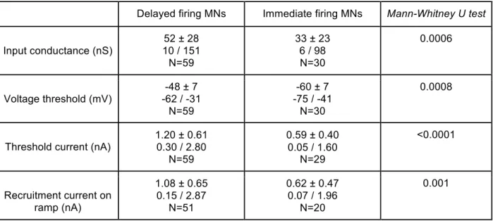
In the first part of my work, I recorded wild-type (WT) motoneurons and found the two discharge patterns described previously (Pambo-Pambo et al., 2009). In order to characterize the underlying currents, I conducted a series of voltage-clamp experiments coupled with pharmacology in order to decipher the currents underlying both discharge patterns. I found that the delayed firing motoneurons express two potassium currents not found in the other immediate firing motoneurons. These currents prevent the discharge before they inactivate, allowing the membrane potential to rise to the voltage threshold and the cell to fire.
This work gave birth to a manuscript entitled “A subpopulation of neonatal mouse
spinal motoneurones displays a delayed firing profile due to an A-like and a slow-inactivating potassium currents” that has been recently submitted for
publication.
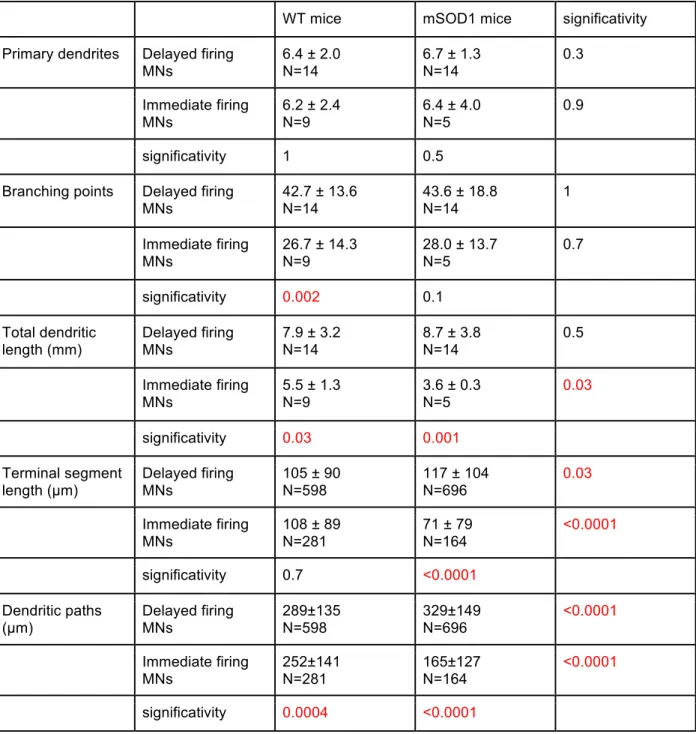
In addition to exhibiting different discharge patterns, the delayed and immediate firing motoneurons display differences in their electrophysiological and morphological properties. Following the presentation of the first article I will detail three kinds of evidence (morphological, electrical and molecular) suggesting that the motoneurons exhibiting a delayed discharge in response to a liminal pulse of current innervate fast-contracting fibers whereas those with an immediate discharge innervate slow-contracting fibers.
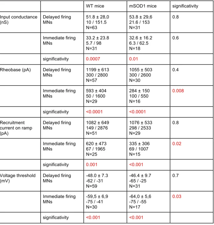
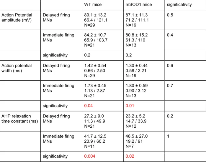
In the second part of my work, I recorded motoneurons from SOD1 G93A mice as well as their non-mutant littermates. Mutant and WT motoneurons presented delayed and immediate firing patterns in the same proportions. Based on the known differential vulnerability to the disease between motoneurons of different sub-types I conducted the type-by-type comparison of the SOD1 mutation effect on the delayed firing motoneurons parameters on one side and immediate firing motoneurons on the other. I found that only the immediate firing motoneurons are hyperexcitable in the mSOD1 mice. The excitability of the delayed firing motoneurons is unchanged. A second manuscript entitled “Only spinal motoneurons that display the
“immediate firing” profile are hyperexcitable in SOD1 G93A neonatal mice” was
4/ Thesis content
In the first chapter I will review what we know about the differentiation of the motor units and motoneurons leading to their extensive heterogeneity. In the second chapter I will summarize my results showing how the expression of specific potassium conductances in 66% of the wild-type motoneurons allows for the delayed firing pattern (in opposition to the left 33% of the motoneurons that fire at the pulse onset). The corresponding submitted article, that fully describes the results, is appended following the chapter 2. The second chapter continues presenting additional results to the first article that attempt to correlate the discharge patterns with known the motoneurons fast and slow subtypes. The third chapter will review the data regarding the impact of the SOD1 G93A mutation on the motoneurons properties. In the fourth chapter I will summarize my type-by-type analysis of the delayed and immediate motoneurons population. It will enlighten interesting opposite effects of the SOD1 mutation for each type of neonatal motoneurons. The corresponding article (to be submitted), that fully decribes the results, is appended following the chapter 4. Material and methods of the experiments are detailed in the two articles or in the protocol section at the end of the thesis. A global discussion on the work performed and its future developments completes this manuscript. After discussing the results concerning the maturation of the motor units and the differential effects of the SOD1 mutation, the discussion will open to consider whether a change in the cell excitation (i.e its synaptic inputs) could take place during the ALS and how this could be experimentally tested.
Chapter I: Differentiation of the heterogenous
motoneuron populations
1/ Mature motoneurons intrinsic properties segregate
a/ According to their morphological parametersMany of the motoneurons properties vary according to the motor unit type they belong to. Horseradish peroxidase stainings of cat motoneurons. Burke et al. (1981) have shown that the somas of motoneurons innervating FF motor unit tend to be are larger (50 - 80 µm) than those of motoneurons innervating FR motor units (50 - 70 µm), which themselves are larger than those of motoneurons innervating the S motor unit (45 - 70 µm). There is however an extensive overlap between the values. We know that γ-motoneurons are even smaller than the α slow motoneuron (Horcholle-Bossavit et al., 1990). The difference seen in the average sizes can be expected to influence on many electrophysiological properties such as the axonal conduction velocity, input conductance and rheobase.
b/ According to their electrophysiological parameters
Zengel et al. (1985) realized a meta-analysis of 73 motoneurons recorded in vivo in the adult cat medial gastrocnemius that were type-identified based on the mechanical properties of their motor unit. They found that FF and FR motoneurons presented similar axonal conduction velocities (99 ± 1 m/s and 100 ± 1 m/s respectively) which was higher than that of S motoneurons (86 ± 1 m/s). Input resistances were comprised between 1.0 MΩ and 2.8 MΩ (1.6 ± 0.1 MΩ) for S motoneurons, between 0.4 MΩ and 1.6 MΩ (0.9 ± 0.1 MΩ) for FR motoneurons and between 0.1 and 1 (0.6 ± 0.1 MΩ) for FF motoneurons. Rheobase, defined as the minimum amount of current to inject during a 50 ms pulse to elicit firing, also varies between types being highest in FF motoneurons (21.3 ± 0.5 nA), intermediate in FR motoneurons (12.0 ± 0.4 nA) and lower in S motoneurons (5.0 ± 0.3 nA).
The overlap values of each individual electrophysiological parameters require using several parameters to properly classify a motoneuron. A single parameter could never reach a classification rate higher than 80% (Zengel et al., 1985). Using the principal component analysis method Zengel et al. (1985) showed that a
classification criterion combining the input resistance and the AHP half-relaxation time they could correctly classify of 94% of their sample. Zengel et al. (1985) therefore chose boundaries in the input resistance and half-AHP duration spaces that allow for reliable motoneuron classification. A similar work on mouse motor units is currently underway in the lab as an in vivo mouse preparation was developed (Marin Manuel personnal communication). Preliminary results show that the electrophysiological properties of adult mouse motoneurons allow the characterization of three different groups of motoneurons. As observed in the cat, the motoneurons innervating the same type of muscle fibers share similar electrophysiological properties. Input conductance and AHP duration allow for a good discrimination of the motor unit type (Figure 2, Marin Manuel personal communication).
2/ Henneman’s “size principle” for mature motoneurons
The clusterization of the motoneuron electrophysiological properties allowed Henneman (1957) to forge the “size principle” to explain the orderly recruitment of motoneurons from the same motor pool. During the stretch reflex of a decerebrated cat sural triceps, the smaller motor units (S motor units) are recruited first. Henneman (1957) hypothesized that during a voluntary movement the smaller motor units exhibiting less force and longer contraction time (S motor units) are mobilized first followed by those producing more force and contracting more rapidly (FR and FF motor units). This was supported by many other experiments (Burke, 1981; Henneman & Mendell, 1981). If we assume that a common synaptic current drive for all α-motoneurons belonging to a common motor pool then because of their smaller input resistance the S motoneuron will reach its spiking threshold first. Then when the input increases the FR motoneurons will be recruited and ultimately the FF motoneurons. The longer time constant of S motoneurons also allows for an easier summation of the slow frequency synaptic inputs and favors similarly their selective recruitment at low frequencies. In this view, recruitment of different motor units starts by the smaller motor units of lesser force and longer contraction time allowing for a smooth gradation of the global force developed at the level of the entire muscle. The “size principle” was posited at a time when motoneurons were thought to be passive integrators. We now know that the motoneuron membrane bears various voltage-dependant conductances allowing them to act as a non-linear integrators (Manuel et al., 2007). However channels carrying the persistent inward currents are more strongly expressed on the slow type motoneurons which are the first recruited (Lee & Heckman, 1998a, b). Therefore this voltage-dependant conductance strengthens the orderly recruitment. Indeed, persistent inward currents were likely present during the experimental protocol Henneman used (Henneman, 1957). Some studies argued that motoneurons receive different inputs (Kanda et al., 1977; Heckman & Enoka, 2012) and that the Henneman’s principle should be supplemented by other principles such as an orderly recruitment among motoneurons supplying different muscles to better explain fast ballistic movements and smooth trajectories (Cope & Sokoloff, 1999) or “neuromechanical” principle to explain respiratory movements (Butler & Gandevia, 2008). Nevertheless, a multitude of studies in humans have shown the size principle to apply in multiple tasks,
muscles and movement speeds (Desmedt & Godaux, 1977; Jones et al., 1994; Feiereisen et al., 1997; Scutter & Turker, 1998), which fully validate it as a genuine physiological principle.
3/ Motor units differentiation
This section will briefly summarize the muscles and motoneurons development leading to the establishment of a functional adult motor unit in the mouse.
a/ Myogenesis
Myogenesis begins when stem cells originating from the somites migrate to the limb buds. They begin to synthesize myosin as early as E10 (Figure 3) in the mouse embryo (Jansen & Flatby, 1990). Myoblasts divide into primary myotubes. After a time lag myotubes are reached by motor axon nerves around E12. Myogenesis resume and a second wave of myogenesis build upon the first one adding the secondary myotubes (Figure 3). This second myogenesis ends around birth: before birth for respiratory muscles that will be used later on (the mice begin to stand around P10. The assembly of the secondary generation of myotubes requires innervation of the muscle and cholinergic activation of the primary fibers (Harris, 1981; McLennan, 1983). In rodents the early myotubes express both embryonic myosin heavy chain (MHC) and adult slow-type MHC. Many also express a distinct neonatal MHC (Weydert et al., 1987). Primary generation myotubes remain mostly slow, while some eventually suppress the expression of slow-type MHC, to produce adult fast-type MHC instead. Such a transition is thought to be highly activity dependent. The second generation myotubes initially express embryonic and neonatal MHC. The majority develop into fast fibers. In slow muscles such as the soleus a more substantial fraction of the secondary myotubes develops into slow fibers (Narusawa
et al., 1987). The final numbers of fast and slow fibers and their distribution are
characteristically different for each muscle and far from being fixed, reorganization occurs during the life upon injury or aging.
b/ Motor Columns
During development, newly postmitotic motoneurons are grouped into motor columns stretching along the rostrocaudal extent of the neural tube. This is due to gradients of diffusible molecules emitted from the neural tube which interact with the motoneuron transcription factors (reviewed in (Dasen & Jessell, 2009). For all mammals the motor columns are the following: the medial motor column (MMC), which projects to epaxial muscles of the dorsal body region, the hypaxial motor column (HMC), which projects to hypaxial muscles of the ventral body wall, and the lateral motor columns (LMC), which project to the limbs. The preganglionic column (PGC) innervates sympathetic ganglia and the smooth muscles. Within each column the ensemble of motoneurons innervating a single muscle is called a motor pool. It is interesting to note that in parallel to the distribution of motoneurons each motor pool comprises all the different functional sub-types described before. The presence of these two levels of diversity renders comparison between motoneurons of the same sub-type fully relevant only when operated within one motor pool. Once the motoneurons somas have reached their location in the spinal cord they begin sending their axons to innervate their target muscle. The motor axons exit the spinal cord through their segmental ventral roots and join their fellow motor axons belonging to the same motor pool but exiting from another segment in the lumbar plexus. The axons of the same motor pool then
fasciculate and travel along peripheral nerve trunks to leave these at specific branch points in order to reach the appropriate muscle.
c/ Formation of the neuromuscular junctions
The axon terminals reach the embryonic muscle prior to the fusion of the first myoblasts. Functional neuro-muscular junctions (NMJs) are formed shortly. In mammalian muscles, all NMJs are found located in a tightly regulated mid-part of the muscle fibers. Indeed, the medial part remain more or less fixed during the fiber contraction and the muscular action potential can propagate symmetrically toward both ends. Furthermore the close proximity between the different NMJs favors competition between them during the elimination of poly-innervation. The synapse formation and maturation requires intensive interactions between pre- and post-partners for correct alignment but also for motoneurons and fibers matching (Hall & Sanes, 1993). Evidence points out that future γ -motoneurons already connect to future intra-spindle fibers (Thompson et al., 1990). Similarly, a few studies conducted on immature motor units (Thompson et al., 1984; Flatby & Jansen, 1990) seem to indicate that most fibers are correctly grouped into muscle units prior the late events of axonal pruning and diminution of the muscle fiber poly-innervation.
d/ Motoneurons death
A massive developmental death begins when the axons reach the muscle embryos. From E12 in the mouse(Lance-Jones, 1982) and over the following week about half of the motoneurons will enter apoptosis and die (Figure 3A). Such a large loss is intriguing since all axons from a motor pool contact its appropriate muscle. One hypothesis is that the programmed death results from the wrong wiring within the muscle between inappropriate functional sub-types (mismatch between the motoneuron and fiber sub-types (Jansen & Flatby, 1990).
e/ Loss of the poly-innervation
In the adult each fiber is contacted by a single motoneuron. However around birth all muscle fibers are poly-innervated. All muscles therefore undergo a process of elimination of the poly-innervation. As a result the number of fibers in each motor unit decreases strongly. The size of motor units decreases accordingly (Figure 3B, (Jansen & Flatby, 1990). Dennis et al. (1981) measured in the rat intercostal muscle
the average number of NMJs present on each muscle fibers (Figure 4A). They observed an increase in the degree of poly-innervation followed by a plateau and then a decrease. However the degree of poly-innervation as well as the moment at which it begins to decrease vary extensively between muscles. In the rat diaphragm and intercostal muscles the degree of poly-innervation has peaked already a day before birth and is distinctly reduced at birth (Figure 4A, Bennett & Pettigrew, 1974; Dennis et al., 1981). In the soleus, on the other hand, the expanded motor units appear to remain at a plateau until P10 with about 5 terminals converging on each fiber before stooping down quickly to 1 terminal by P15 (Figure 4B, (Brown et al., 1976). Although different in time and intensity there are two process concomitantly at stake in the maturation of muscles around birth. On one hand new fibers (secondary myogenesis) are added and rapidly innervated and on the other hand supernumerary synapses are deleted. This situation can give rise to several scenarios depending on whether the events are disjoint or overlap. Due to the difficulty to properly follow the degree of poly-innervation around birth researchers tried to compare the rate of decrease in the poly-innervation. However the rate of decrease they measured only reflects the ratio of new contacts over the one being eliminated when only the number of axon terminals retracting truly matters. Additionally an important caveat was the absence of fiber type identification when assessing the poly-innervation. Some studies suggested that most NMJ were already type-matched prior the reduction of the poly-innervation. Callaway et al. (1989) measured the fractional decline in motor unit tension in the rabbit soleus and demonstrated a substantially higher initial degree of poly-innervation and rate of elimination for slow than for fast motor units.
Flatby and Jansen (1990) studied the composition in muscle fibers of the motor units of the mouse neonatal soleus at P2, P5 and P14. At P2 and P5 the muscle is still at its neonatal peak of polyneuronal innervation (on average 6 NMJ/fiber) while at P14 poly-innervation is virtually over. Stimulating one axon (i.e one motor unit) per experiment they were able to sample the muscle fibers connected to it. For recordings at P14 they used the glycogen depletion method. The group of Kugelberg (Edstrom & Kugelberg, 1968) first showed that in the adult individual motor axons innervate a homogenous group of muscle fibers. This was achieved by stimulating individual motor axons under anoxic conditions until the glycogen stores of their muscle fibers were depleted (Edstrom & Kugelberg, 1968).They could then identify the fibers histochemically on cross sections of the muscle using the periodic acid Schiff (PAS) reaction. However this method could not be used in immature muscle fibers at P2 and P5. The authors then randomly recorded muscle fibers and filled them with Lucifer Yellow when a plaque potential was recorded following the motor unit stimulation (Figure 5a,b and c). They plotted the histogram distribution of the fraction of fast fibers found in each recorded motor unit (Figure 5d). At P2 the histogram distribution is already slightly bimodal (Figure 5e) when an unimodal distribution would have been expected if the wiring were random (Figure 5d). Therefore already at P2, the motor units composition is already biased toward either
a majority of fast or a majority of slow fibers. The bimodal distribution refines at P5 and P14 (Figure 5f and 5g). Given to this biased composition motor units already displayed an emerging fast/slow cleavage in their contractile velocity as soon as P5. The authors concluded that most of the fast/slow separation of the muscle fibers takes place prior to the reduction in poly-innervation begins.
The following elimination of supernumerary NMJ would not be a major factor of the fast / slow fibers segregation but only a competition between motoneurons of the same sub-type to keep innervating the largest subset of fibers. Unfortunately, technical limitations precluded Flatby and Jansen from recording the properties of the motoneurons they stimulated. Although the total number of NMJs remains constant between P2 and P5 the histogram distribution shows an extensive refining in the motor unit composition. Presumably slow motoneurons loose synapses impinging on slow primary myotubes and slow secondary myotubes while gaining synapses on the neonatal secondary myotubes. Given that all primary myotubes are slow (Weydert et
al., 1987) and that we know that some slow motoneurons contact slow fibers at birth
it is safe to assume that slow motoneurons innervate slow primary myotubes prior to the second myogenesis. We should keep in mind that activity-dependent transformation of the fibers is possible (reviewed in Schiaffino & Reggiani, 2011) and
cannot be entirely ruled out, although it probably only takes place during non-developmental remodeling following an injury.
To conclude, the formation and maturation of the motor unit is a highly controlled process leading to the correct wiring between matching motoneurons and muscle fibers. Motoneurons from a motor pool connect all to its muscle and within the motor pool the different motoneurons sub-types connect appropriately to their matching fibers. Assessing the properties of the motoneuron activating the recorded muscle units is the only way to properly correlate the two elements forming the motor unit. However technical limitation in the neonatal mouse have so far precluded performing simultaneous recordings from the muscle and the spinal cord. Such preparation remains to be developed. Thus no correlation has been made at an early age between the motor units and their motoneurons. Are the motoneurons properties already differentiated or do they differentiate later once the motoneurons are appropriately connected to their subset of homogenous muscle fibers? In the next section we will review the available data regarding the evolution of conductances, morphology and electrophysiological properties in the maturing motoneurons.
4/ Motoneuron maturation and differentiation
a/ MorphologyInitially, motoneurons precursors all begin growing dendritic tree once their somas finished migrating to the future place of the motor pool. The distribution of the somas is unimodal around birth and then becomes quickly bimodal reflecting the differentiation of gamma and alpha motoneurons (Horcholle-Bossavit et al., 1990, for kitten; Friese et al., 2009 for mice) Dendrite growth appeared to be proportional throughout the tree. Furthermore, the elongation was proportional to enlargement of overall spinal cord dimensions. The topology of cat and rat motoneuron dendritic tree is submitted to remodelling and pruning shortly after birth (Nunez-Abades et al., 1994; Nunez-Abades & Cameron, 1995). The number of axon collateral changes as well for in the cat (Horcholle-Bossavit et al., 1990). However, for mouse motoneurons dendritic trees, the topology remains similar from E17.5 to P11 (Li et al., 2005; Martin
et al., 2013; personal observations). During the first post-natal weeks maturation
arborization, which appear to follow the significant overall growth of the surrounding tissue during this period. A small increase in number of nodes and terminals can be seen but overall mouse motoneurons dendritic tree mostly grow homogeneously in size (Li et al., 2005; Martin et al., 2013; personal observations).
Motoneuron morphology in adults is highly dependant of their motor pool as well as their functional sub-type. Dendrites initially projects in all directions to invade the surrounding space, but for motor pool close from the ventral horn boundary the most extensive branching takes place within the gray matter dorsal and medial to the somatic location (Li et al., 2005; personal observations) when much less dendrites project rostro-caudally. Motor axons arise directly from the soma or from a thick proximal dendrite and then extend into the nearest ventral root (Li et al., 2005; personal observations). Motor axons have one or two recurrent collaterals that branched profusely within the territory occupied by the dendrites of the same neuron, projecting notably to the Renshaw cells impinging back on the motoneurons (Li et al., 2005; personal observations).
Cullheim et al., 1987a, b) were among the first to study the size and topology of the type-identified motoneurons in adult mammals. Staining of the triceps surae alpha-motoneuron by HRP allowed reconstruction of the alpha-motoneuron and measurement of the membrane area. They showed that S-type motoneurons dendritic area was on average 22% smaller than F-type motoneurons although the populations overlap. The branching structures differed as well. S-type motoneurons were less prone to branching and expand more radially than F-type motoneurons. Overall the structural differences suggested that the F and S groups of α-motoneurons could be viewed as intrinsically distinct cell types and not as members of the same population with different sizes (Cullheim et al., 1987a). In a companion paper the same authors tried to assess the ways in which dendrites occupy three-dimensional space. They concluded that despite considerable scatter, the results showed that the space occupied by the branches tends to be approximately the same in large and small dendrites, and in F and S cell groups (Cullheim et al., 1987b). Further studies of adults motoneurons confirmed that the different motoneuron populations segregate relatively to their size in the following order (γ-motoneurons < S motoneurons < α-FR motoneurons < α -FF motoneurons). Extensive overlap between populations persists however all life long.
b/ Conductances
Motoneurons are endowed with a set of common voltage-dependent channels which include the fast sodium channels and the delayed rectifier potassium channels allowing the emission of action potentials (Barrett & Barret, 1976; Schwindt & Crill, 1984; Delgado-Lezama et al., 1999). It also includes a calcium-dependent potassium channel responsible for the mAHP as well as a Ca2+ channel allowing calcium influx during the spikes. Drug sensitivity supports the idea that these channels are respectively the SK channel (Barrett & Barret, 1976; Schwindt & Crill, 1984; Delgado-Lezama et al., 1999) and the N-type calcium channel (Perrier & Hounsgaard, 2000). Additionally motoneurons also express a non-specific cation channel generating the H-current (Ito & Oshima, 1965; Schwindt & Crill, 1984; Manuel et al., 2009). Some motoneurons also exhibit additional currents such as the Ca2+ persistent inward current (Hounsgaard & Mintz, 1988), the Ca2+ transitory T-type current, the Na+ persistent inward current, the potassium A-current or the Ca2+ activated non-selective cationic current (Perrier & Hounsgaard, 1999). Among all currents identified in the motoneurons we will focus on three of them which expression has been shown to be modulated during development: the T-type calcium current, the transient potassium A-current, and the L-type Ca2+ current.
i/ T-type Ca2+ current
The T-type Ca2+ channel is a low-voltage activated Ca2+ channels (Figure 6C). It may induce a rebound of the membrane potential following prolonged hyperpolarization since it is inactivated at rest and de-inactivated upon hyperpolarization. Evidence for transient expression of T-type calcium channels in spinal motoneurons during development has been obtained from chick (McCobb et al., 1989), mouse and rat (Figure 6A). In the mouse it has been detected in spinal motoneurons from E14 to P8 (Mynlieff & Beam, 1992a, b). In the rat, a T-type calcium current facilitated by serotonin was demonstrated in spinal motoneurons at P3-P4 (Berger & Takahashi, 1990) but it was never documented at later stages. It appears that its decline in spinal motoneurons matches the time of motoneuronal developmental death (McCobb et al., 1989; Mynlieff & Beam, 1992a, b). However its expression persisted in the rat hypoglossal motoneurons (Berger et al., 1995) and abducens (Russier et
al., 2003) much later after birth, suggesting a different role for this current in these
early expression of T-channels in motoneurons could be responsible for the burst discharge pattern that characterizes intra-uterine motor activity. So far, this burst mechanism has only been confirmed for neonatal hypoglossal motoneurons (Berger
et al., 1995).
ii/ Potassium A-current
The fast transient outward A-current was originally described in the mollusc Doridiae
Anisodoris by Connor and Stevens (1971). In vertebrates, it is mediated by numerous
Kv isoforms including the Shaker-related Kv 1.4, the Shaw-related Kv 3.3 and Kv 3.4 as well as the Shal-related Kv 4.1, Kv 4.2 and Kv 4.3 (Pongs et al., 1999). The addition of a β subunit to the α pore-forming subunits might also confers A-like properties to any α pore-forming subunit (Yao & Wu, 1999). Its activation requires a
period of hyperpolarization followed by a depolarization. Both the activation and the inactivation occur relatively quickly: a few milliseconds for the activation and a few tens of milliseconds for the inactivation. The A-current is suppressed by millimolar concentrations of 4-aminopyridine (4-AP, Figure 6B). The A-current is expressed in spinal motoneurons in the chick embryo as early as embryonic day 4 (E4) and the current density strongly increases at least until E11 (McCobb et al., 1990). The A-current is also transiently expressed in motoneurons in the rat spinal cord from E14 until postnatal period P2–P8 (Safronov & Vogel, 1995; Gao & Ziskind-Conhaim, 1998; Svirskis & Hounsgaard, 1998; Alessandri-Haber et al., 1999). No expression of the A-current was ever observed in adult spinal motoneurons of mouse, chick, rat, cat, frogs or turtle (Figure 6A). On the contrary, trigeminal motoneurons of adult guinea-pig also express the A-current (Conway et al., 1988). Lappe and Nistri (1999) identified the A-current in hypoglossal motoneurons until P9. Russier et al. (2003) found the A-currrent to be expressed in a subset of the neonatal abducens motoneurons from P2 to P9. The rapid hyperpolarizing effect of the A-current makes it fit to shorten the spike duration (McCobb et al., 1990; Svirskis & Hounsgaard, 1998), reduce the amount of calcium entering the neurons and perhaps spare them from apoptosis. In this view the A-current transient expression before birth would counterbalance the apoptotic effect of the T-type current. If a significant amount of A-current is de-inactivated prior to a pulse, the A-A-current may delay the discharge over a relatively short time (order of magnitude 30 ms). In neonatals, such an effect has been observed in spinal motoneurons (Gao & Ziskind-Conhaim, 1998; Perrier & Hounsgaard, 2000), as well as in brainstem motoneurons (Lape & Nistri, 1999; Russier et al., 2003).
iii/ L-type Ca2+ persistent inward current
The Ca2+ persistent inward current carried by the L-type channels does not have a transient expression around birth but gradually appears after birth (Figure 6A). The L-type channels open during depolarizations and inactivate very little. They may therefore induce plateau potentials and bistability in the motoneurons, requiring a hyperpolarizing event to end the plateau (Figure 6D). In mouse and rat motoneurons L-type Ca2+ current is present at E14–15 (Mynlieff & Beam, 1992a; Hivert et al., 1995)). In the mouse, the expression of L-type Ca2+ channels as measured by membrane current density continues to increase after birth (Mynlieff & Beam, 1992b)
and L-type channels contribute detectably to motor behavior after P7 (Jiang et al., 1999). Indeed, plateau potentials resulting from the current activation have been found in adult cat and mouse spinal motoneurons (Schwindt & Crill, 1980; Bennett et
al., 1998; Kiehn & Eken, 1998; Lee & Heckman, 1998b; Jiang et al., 1999). Similarly,
the adult rats and humans motoneurons exhibit correlates of plateau potentials (Eken & Kiehn, 1989; Gorassini et al., 1998; Kiehn & Eken, 1998). Indirect evidence suggests that plateau potentials contribute to motor behaviour by shaping motor output patterns and provide mechanisms for self-sustained activation of motor units (Kiehn & Eken, 1997, 1998; Delgado-Lezama et al., 1999; Russo & Hounsgaard, 1999).
To conclude, in spinal motoneurons, the expression of the A-current and the T-type current starts during the early embryonic development and peaks during the motoneuronal death suggesting a regulatory role. Its role at later stages in trigeminal, hypoglossal or abducens motoneurons remains however elusive. In parallel the L-type channels expression increases slowly after birth and becomes stable around the time the animal engages in locomotor postural activity. The distinct set of conductances expressed at embryonic or adult stage may reflect distinct role for the motoneurons intrinsic properties. At a young age, these properties may be part of the signaling system that regulates growth, pathfinding, cell death, and formation of pre- and post-synaptic contacts (Perrier & Hounsgaard, 2000). In the adult, motor system is mature and the intrinsic properties need to be tuned to support the motoneurons functions. This led us to question whether all types of motoneurons belonging to a same pool express the same conductances in similar densities. Since they have to perform various functions it seems crucial their currents match their needs.
c/ Delayed firing in a subpopulation of neonatal motoneurons
So far, we do not know whether and how the functional properties of spinal motoneurons maturate at the same time as those of their muscle fibers. It is well known that, in adult animals, spinal motoneurons start to fire almost without any delay at the onset of a liminal current pulse (Kernell, 2006; Manuel et al., 2009) for mice motoneurons). In sharp contrast, the firing behavior is heterogeneous during the second postnatal week (Russier et al., 2003; Pambo-Pambo et al., 2009; Zhu et al., 2012). For liminal current pulses, the discharge starts at the current onset in some
motoneurons but it is delayed in others. Pambo-Pambo et al. (2009) worked on the mouse spinal cord slice preparation between p6 and p10. They reported that some neonatal spinal motoneurons fired at threshold with around 100 ms of delay and that this delay disappeared when 10 mM TEA and 4 mM 4-AP were applied. Zhu et al. (2012) reported that two weeks following birth, the spinal rat motoneurons exhibited several firing patterns. While 42% discharged at pulse onset, 30% presented delayed onset discharge. The remaining motoneurons displayed irregular firing patterns. Russier et al. (2003) analyzed the two populations of rat abducens neonatal motoneurons in a preparation of brainstem slices from P1 to P13. They observed two types of motoneurons according to their firing profile during prolonged depolarizations. The first type, accounting for 70% of the total, exhibited a burst of action potentials followed by an adaptation of the discharge (bursting discharge). Their firing pattern was similar to that of adult abducens motoneurons. They exhibited T-type as well as H-currents. The second population of abducens motoneurons displayed a delayed discharge, the was due to the potassium A-current and was observed only between P4 and P9. According to the authors, these motoneurons exhibited immature characteristics: simple AHP and no separated fast and medium AHPs, as well as a linear current–voltage relationship and a small input conductance. Given that the total number of abducens motoneurons remained unmodified and that only motoneurons presenting a bursting discharge were encountered after P9 the authors concluded that the expression of the A-current was transient and that motoneurons of the second type (delayed discharge) turn into the other type (bursting discharge) right before the rat begin using the muscles controlling the eye (P11-P12). Thus, the transient delayed firing pattern due to the A-current observed in a sub-set of neonatal motoneurons appears to be a common property of motoneurons. Noteworthy Lape and Nistri (1999) also identified a second slow inactivating potassium current that may also delay the discharge on a longer time-scale. During the second postnatal week, rat hypoglossal motoneurons exhibit either a decrementing or an incrementing firing pattern in response to square pulses (Viana
et al., 1995), while in adult spinal motoneurons, the firing frequency is always
decreasing during a pulse (Brownstone, 2006). Similarly, facial motoneurons (Magarinos-Ascone et al., 1999) of neonates also presented heterogenous firing behaviors and frequency adaptations. In general it seems that a sub-population of developing motoneurons express additional potassium currents modulating the
discharge onset and its adaptation. Since we know that muscle fibers are already differentiated and that the sub-types are grouped in homogeneous motor units during the second post-natal week we may wonder whether motoneurons from different types have different discharges and if this difference is required for the proper maturation of the system.
Besides these two patterns of discharge very little is known about the maturation of conductances within the same motor pool. Ramp-response analysis suggests that in the adult S-type motoneurons have more Ca2+ persistent inward currents than the F-type ones (Lee & Heckman, 1998a, b). This fits with the idea that slow motoneurons are engaged more often in postural activity (Burke, 1981) and need to sustain prolonged activation of their muscle fibers.
d/ Labelling of motoneuron molecular markers: dissecting the sub-populations
Research for molecular markers of different motoneuron types is of the utmost importance. Indeed when working on a slice preparation, the efferent muscle fibers are missing and an easy molecular tool would allow to establish a correlation between recorded cell and motor unit type. However, the detection of markers requires comprehensive analysis of the genes expressed by distinct cell populations and the difficulty to harvest sufficient amounts of mRNA from identified motoneurons have hindered this analysis for a long time.
Specific markers of γ - and α -motoneurons were recently discovered recently. The transcription factor Estrogen-related receptor 3 (Err3), an orphan receptor of the nuclear hormone, can separate γ- from α -motoneurons in the spinal cord. Its expression first localizes to the cell bodies and becomes restricted to γ-motoneurons during the first two postnatal weeks (Friese et al., 2009). Adult γ-motoneurons also express higher levels than α-motoneurons of the glial cell line–derived neurotrophic factor receptor subunit GFRα1 as detected using reporter mice (Shneider et al., 2009). Conversely, the neuron-specific nuclear protein named nuclear nuclei (NeuN, Mullen et al., 1992) antigen is strongly expressed by α motoneurons but not by γ -motoneurons (Friese et al., 2009; Shneider et al., 2009). More recently, the serotonin receptor 1d (5HT1d) was found to be expressed specifically by γ-motoneurons after P11 (Enjin et al., 2012).
Concerning the fast and slow motoneurons, the first identified marker was the calcitonin gene (Calca) and its gene product calcitonin gene-related peptide (CGRP). Several studies showed a correlation between its expression and the amount of fast motoneurons within a motor pool (Forsgren et al., 1993; Piehl et al., 1993). γ -motoneurons appear to lack Calca expression as well. Building on this results Enjin
et al. (2010) proved that the expression of another protein, the chondrolectin,
colocalized with Calca (Figure 7A). Then Enjin et al. (2010) correlated Chodl expression with one of two distinct motoneurons populations recorded at the second post-natal week. Motoneurons from the two populations exhibited different half-AHP durations (Figure 7B and D), different rheobases (Figure 6E) and different input resistances (Figure 7F). As a result the two populations segregated along the AHP duration and rheobase axis (figure 7G) similarly to what has been demonstrated in the adult cat fast and slow motoneurons (Zengel et al., 1985) and probably in the adult mouse motoneurons as well (figure 2). Finally Enjin et al. (2010) showed that motoneurons non-expressing Chodl expressed the estrogen-related receptor β (ERRβ) making it a putative slow motoneurons marker. In parallel in situ hybridization also showed that synaptic vesicle protein 2A (SV2A) was selectively localized in motor nerve terminals on slow (type I and small type IIA) muscle fibers. SV2A is broadly expressed at birth, then fast motoneurons downregulate its expression during the first postnatal week (Chakkalakal et al., 2010). Lately the expression of the Ca2+ -activated K+ channels 3 (SK3) was found to be restricted to putative slow motoneurons in the rat motoneurons (Deardorff et al., 2013). However SK3 expression was weak during the first two weeks following birth. This new marker is particularly promising since SK channels carry the mAHP current and the slow motoneurons AHP is significantly longer than the fast motoneurons AHP in the adult cat, rat and mouse (Kernell, 2006; Manuel et al., 2009). This new marker may therefore be the first to bear a causal relationship with a major electrophysiological property used as a discriminative criterion between populations.
Conclusion
Motoneurons are subject to a profound reorganization of their intrinsic excitability during postnatal development. Evidence points out that the motoneuron sub-type is determined early on, probably before birth. The motoneurons grow in size during the entire animal life but in a hierarchical fashion. Size differences are noticeable in the weeks following birth.
The time window (P6-P10) that I have investigated during my thesis work is a period of intense shaping of the connectivity pattern within the muscle (reduction of poly-innervation and refinement of the motor unit homogeneity). During the second post-natal week the spinal cord is still immature (descending tracts are not functional) and the mouse cannot walk before P10. It is likely that the properties of the different motoneuron populations are still maturing leading to further differentiation between sub-types. Similarly to what had been done for the muscle fibers, I investigated how and when the differentiation between motoneuron electrophysiological properties arises. Heterogeneous firing is a common feature of neonatal motoneurons. Can we correlate the firing patterns with a subtype? What are the effects of the different activities on the respective maturation?
Chapter
II:
Two
subpopulations
of
spinal
motoneurons displaying different discharge profiles
1/ Introduction
The second postnatal week is crucial in the development of the mouse neuromuscular system. During this week, postural activity is acquired, allowing the locomotor behavior to switch from crawling to walking (Jiang et al., 1999). This transformation calls for a remodeling of the sensory and the descending neural controls (Clarac et al., 2004).The motor units also undergo extensive maturational processes in order to ultimately differentiate into three mature subtypes: the slow contracting motor units (S type), the fast contracting but fatigue resistant motor units (FR type) and the fast contracting and fatigable motor units (FF type; Burke, 1981). During the second post-natal week the distal part of motor unit (the muscle unit composed of all muscle fibers innervated by a single motoneuron) is already differentiated. However, so far we do not know when and how the functional properties of spinal motoneurons maturate, particularly when and how the properties of slow motoneurons differentiate from those of fast motoneurons. It is well known that, in adults, spinal motoneurons start to fire without any delay at the onset of a liminal current pulse (Kernell, 2006; Manuel et al., 2009), 2009 for mice motoneurons). In sharp contrast, the firing behavior is heterogeneous during the second postnatal week (Russier et al., 2003; Pambo-Pambo et al., 2009). For liminal current pulses, the discharge starts at the current onset in some motoneurons but it is delayed in others.
In a first time I characterized the currents underlying the delayed discharge exhibited by some of the spinal motoneurons and, I characterized their impact on the recruitment threshold and on the F-I relationship (article 1). The second part of this chapter presents arguments of electrophysiological, morphological and molecular nature to support the view that motoneurons with delayed firing might be fast motoneurons and motoneurons with immediate firing might be slow motoneurons.