HAL Id: hal-01849228
https://hal.archives-ouvertes.fr/hal-01849228
Submitted on 5 Jun 2019
HAL is a multi-disciplinary open access archive for the deposit and dissemination of sci-entific research documents, whether they are pub-lished or not. The documents may come from teaching and research institutions in France or abroad, or from public or private research centers.
L’archive ouverte pluridisciplinaire HAL, est destinée au dépôt et à la diffusion de documents scientifiques de niveau recherche, publiés ou non, émanant des établissements d’enseignement et de recherche français ou étrangers, des laboratoires publics ou privés.
Plasma membrane coating with cationic silica particles
and osmotic shock alters the morphology of bovine
aortic endothelial cells
Claire Millot, Véronique Le Berre, Jean-François Tocanne, Jean-François
Tournier
To cite this version:
Claire Millot, Véronique Le Berre, Jean-François Tocanne, Jean-François Tournier. Plasma mem-brane coating with cationic silica particles and osmotic shock alters the morphology of bovine aortic endothelial cells. Biochimica et Biophysica Acta:Biomembranes, Elsevier, 2000, 1467 (1), pp.85 - 90. �10.1016/S0005-2736(00)00207-8�. �hal-01849228�
Plasma membrane coating with cationic silica particles and osmotic
shock alters the morphology of bovine aortic endothelial cells
Claire Millot *, Ve¨ronique Le Berre-Anton, Jean-Franc°ois Tocanne,
Jean-Franc°ois Tournier
Institut de Pharmacologie et de Biologie Structurale du CNRS, 205, route de Narbonne, F-31077 Toulouse, Cedex, France Received 2 March 1999; received in revised form 29 February 2000; accepted 21 March 2000
Abstract
We have used a published method of membrane preparation based on the precoating of the apical membrane of aortic endothelial cells with cationic silica microbeads (with or without polyacrylic acid) in combination with an osmotic shock and mechanical shearing to isolate the apical from the basal plasma membranes of these cells, in vitro. After labeling of the plasma membrane of adherent endothelial cells with a fluorescent derivative of phosphatidylcholine and by using laser confocal fluorescence scanning microscopy, we found that this method of membrane isolation rapidly induced invaginations of the basal plasma membrane to an extent which makes this method unsuitable for further membrane lipid analysis. Morphological analysis of the cells and fluorescence recovery after photobleaching experiments on the plasma membranes were performed at each step of the purification procedure and showed that only hypotonic shock and mechanical shearing of the cells enabled the basal plasma membranes to be purified without significant morphological changes. ß 2000 Elsevier Science B.V. All rights reserved.
1. Introduction
Aortic endothelial cells resting on their extracellu-lar matrix occupy a biological interface, in vivo, be-tween the blood and the smooth muscle cells of the arterial wall. The extreme environmental di¡erence between the apical and the basal plasma membrane of this cell monolayer has led to numerous studies related to their respective compositions and functions and then to the recurrent need for e¤cient methods
of plasma membrane isolation. Various approaches can be used for this purpose, among which, the one described by Stolz and Jacobson [1] is particularly attractive. This method is based on a combination of coating of the apical membranes of aortic endo-thelial cells with cationic colloidal silica microbeads and then polyacrylic acid followed by exposure of the cells to an osmotic shock and mechanical shear-ing. When using this approach, the basal membrane remains attached to the solid support while the coated apical membranes in suspension are easy to separate from other organelles by centrifugation. However, morphological examination of the prepa-rations at each step of any cell fractionation proce-dure must be considered as very important. In the case of plasma membranes, this can be done on liv-ing cells by the use of laser confocal scannliv-ing
micros-* Corresponding author. Fax: +33-5-6117-5993; E-mail: cmillot@ipbs.fr
copy. For this purpose, the outer lea£et of the plas-ma membrane of the endothelial cells was labeled with a £uorescent derivative of phosphatidylcholine [2^4], and the morphology of these non-refringent organelles was checked at each step of their puri¢ca-tion. This approach showed that the association of precoating of the cells with cationic silica and expo-sure to osmotic shock induced dramatic morpholog-ical changes in the basal plasma membrane, which render the method unsuitable for further lipid and membrane structure analysis. We show that osmotic shock without precoating of the cells followed by mechanical shearing is preferable to ensure basal plasma membrane isolation. In addition to laser con-focal microscopy, the quality of the preparations of basal plasma membranes was assessed by £uores-cence recovery after photobleaching (FRAP) experi-ments.
2. Materials and methods 2.1. Chemicals
1-Acyl-2-[6-[N-(7-nitrobenz-2-oxa-1,3-diazol-4-yl)-amino]caproyl]-phosphatidylcholine (C6-NBD-PC) was obtained from Avanti Polar Lipids (Pelham, AL, USA). Dioleoylphosphatidylcholine (DOPC) was purchased from Sigma (Saint Louis, MO, USA). Recombinant basic FGF was kindly provided by Dr. Bouche (Institut de Biologie Cellulaire et de Ge¨ne¨tique in Toulouse (France)). Cationic colloidal silica microbeads were a generous gift from Drs. B.S. Jacobson and B. Larijani (Lederle Graduate Re-search Center, Massachussets, USA).
2.2. Cells and cell culture
Vascular endothelial cell cultures were established and cloned twice from ox aortic arch and character-ized as previously described [5^7]. In most experi-ments, cells resting on 22U22 mm coverslips were used near con£uence because membrane isolation procedure occasionally resulted in whole detachment of postcon£uent endothelial cell monolayers as an intact sheet or in poor basal plasma membrane re-covery as previously reported by Stolz and Jacobson [1].
2.3. Isolation of plasma membrane domains from tissue culture coverslips
The procedure used for the coating of the cells was that described by Stolz and Jacobson [1]. Endothelial cell monolayers were washed twice with cold PBS and then twice with the coating bu¡er, CB (20 mM 2[N-morpholino]ethanesulfonic acid, 130 mM NaCl, 1 mM MgCl2, 0.1 mM CaCl2, pH 6.5). Cells were
coated for 10 s with a 5% (w/v) cationic colloidal silica suspension in CB which was subsequently di-luted 10-fold by the addition of CB in the Petri dishes. The monolayers were rinsed four times with CB and exposed (or not) for 10 s to a solution of polyacrylic acid (1 mg/ml in CB) which was then diluted 10-fold with CB. After one wash with lysis bu¡er, LB (2.5 mM Tris, 1 mM MgCl2, 0.5 mM
CaCl2, pH 7.4), the cells were allowed to stand on
ice for 3 min in LB. To isolate their basal plasma membranes, mechanical shearing was applied to the cell monolayer by squirting the cells with cold LB at about a 45³ angle using a 5-ml syringe ¢tted with a £attened 18-gauge needle. The progress of cell lysis was continuously monitored under a phase-contrast microscope. Isolated basal plasma membranes anch-ored on glass coverslips were ¢nally rinsed three times with PBS.
2.4. Plasma membrane labeling and £uorescence intensity measurements on endothelial cells All these procedures were performed as described previously [2^4]. Mixed vesicles of DOPC and the £uorescent probe C6-NBD-PC, in the 1/1 (w/w) ratio were prepared by injection of an ethanol solution of the lipids in PBS at room temperature [8]. The ¢nal lipid concentration was 40 Wg/ml (20 Wg/ml of each lipid) and the vesicles were used within 1 day. Endo-thelial cell monolayers were washed twice with cold PBS and chilled on an ice-water bath for 10 min. The medium was removed and cells were incubated for 10 min (still on an ice-water bath) with a cold lipid vesicle suspension containing C6-NBD-PC. Fluores-cence originating from the membrane surface was measured with the FRAP apparatus, by illuminating small basal plasma membrane areas of 1.95 Wm ra-dius either on entire cells or on isolated sheets. For each measurement, 120^cells were examined.
C. Millot et al. / Biochimica et Biophysica Acta 1467 (2000) 85^90 86
2.5. Fluorescence recovery after photobleaching experiments
FRAP experiments were carried out under condi-tions of constant incident light intensity and of `uni-form disk illumination' using apparatus and experi-mental conditions previously described [9]. The di¡usion coe¤cient D and the mobile fraction M were obtained by double ¢tting of the experimental recovery data, using a ¢nite di¡erentiation method and statistical analysis [9]. D and M values are given at 95% con¢dence level. FRAP experiments were car-ried out at a temperature of 20³C and at least 60^80 cells were tested for each kind of experiment. 2.6. Confocal £uorescence microscopy
Cells were observed with the LSM 410 inverted confocal microscope from Zeiss (Germany) equipped with a U63 oil-immersion objective. Excitation and emission wavelength were 488 and 530 nm, respec-tively.
3. Results
3.1. Morphological analysis
When compared to control cells (Fig. 1A), coating of endothelial cells with colloidal silica and poly-acrylic acid did not produce obvious morphological change (Fig. 1B) and a homogeneous labeling pat-tern of the plasma membrane could be seen in both cases. A subsequent brief exposure (less than 3 min at 4³C) to hypotonic bu¡er (Tris 2.5 mM, 0.5 mM Ca2, 1 mM Mg2, pH 7.2) brought about a large
swelling of the cells with the appearance of intracel-lular £uorescent labeled vesicles located at the cell junctions or originating from the basolateral domain (Fig. 1C). After 5^10 min of this treatment, £uores-cence appeared mainly in grain-like forms inside the cells (data not shown). In contrast, exposure of the cells to the hypotonic bu¡er only led to an instanta-neous and large expansion of the apical plasma membrane, but without appearance of the vesicles (Fig. 1D). Because the folding of the plasma mem-brane at junctional sites could provide su¤cient ma-terial, when stretched, to explain the origin of the
large apical expansion, only osmotic shock was per-formed on sparse cells. Homogeneous membrane la-beling was observed in swollen sparse cells and an instantaneous and large apical membrane expansion could be observed (Fig. 1E) while their basal area remained nearly unchanged when compared to con-trol cells (data not shown). In the swollen near-con-£uent and sparse cells, £uorescence appeared to be less intense in the apical than in the basal pole (see Z sectioning in Fig. 1D,E). This could be due to the fact that the probe was relatively more diluted in the apical than in the basal pole and/or to attenuation of the recorded £uorescence intensities with increasing penetration of the laser beam in the watery medium. For that reason and to determine more precisely the origin of this membrane expansion, the £uorescence intensities originating from the basal membrane were compared after each of the following conditions: (1) con£uent cells labeled with C6-NBD-PC; (2) cells labeled and then exposed 3 min at 4³C to the tonic bu¡er; and (3) cells ¢rst exposed to the hypo-tonic bu¡er and then labeled. The means of the £uo-rescence intensities per unit of membrane area were found to be equal when the labeling was performed on native plasma membranes or when the labeling was performed after the osmotic shock on the swol-len cells. However, we have observed that when the cells were ¢rst labeled and then exposed to the hypo-tonic medium, the swelling of the labeled cells is accompanied by a decrease of about 40% of the £uo-rescence intensities per unit of membrane area. This suggests that the membrane surface area which could be labeled with the £uorescent probe after the os-motic shock originated probably from a pre-existing intracellular compartment. However, when the swol-len cells were disrupted by squirting the cell mono-layer with the hypotonic bu¡er, the resulting basal plasma membrane displayed homogeneous labeling whenever the labeling step was performed, i.e. on the native cells before the isolation procedure (Fig. 1F), after the osmotic shock on unlabeled cells, or directly on the puri¢ed basal plasma membrane (data not shown).
3.2. Fluorescence recovery after photobleaching experiments
mem-Fig. 1. Laser confocal £uorescence scanning microscopy of vascular endothelial cells labeled with C6-NBD-PC. (A) Micrograph of con£uent cultures before and (B) after coating with cationic silica or (C) after coating with cationic silica and exposed to hypotonic shock or (D) exposed only to the lysis bu¡er. In E, a micrograph of labeled sparse cells incubated in the hypotonic medium is shown; and in F, basal plasma membranes isolated from con£uent cells after osmotic shock and mechanical shearing. The upper part of each micrograph was obtained in the xy plane while the lower part is an optical Z sectioning of the corresponding sample following the white line when indicated. The entire width of micrographs was 200 Wm in A, B, D and F, and 80 Wm in C and E.
C. Millot et al. / Biochimica et Biophysica Acta 1467 (2000) 85^90 88
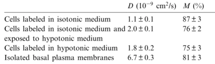
brane obtained from swollen cells, FRAP experi-ments were carried out on the basal pole of either whole cells before or after swelling and on isolated basal membrane domains. The lateral di¡usion coef-¢cient of C6-NBD-PC was found to slightly increase in the basal pole of entire cells labeled before or after the osmotic shock (D = 1.8 þ 0.2U1039 cm2/s) when
compared to control cells (D = 1.1 þ 0.1U1039cm2/s,
Table 1). Concomitantly, the fraction of probes which were free to di¡use decreased from 87 þ 3% in control cells to 75 þ 3% in swollen cells (Table 1). Lateral di¡usion parameters obtained in basal membranes after their isolation (D = 6.7 þ 0.3U1039
cm2/s and M = 81 þ 3%, Table 1) indicated either a
further modi¢cation of membrane properties after disruption of the cells or a possible redistribution of the £uorescent probe between the two lea£ets of the plasma membrane or both. However, it should be noted that the fraction of the probes which were free to di¡use in isolated basal plasma membranes (M = 81%) was similar to that measured on whole cells (M = 87%). Large M value is a good indication of the absence of contamination by intracellular or-ganelles. In fact, they would contribute to the photo-bleaching process, but their small sizes could not constitute surfaces su¤ciently large to allow the re-covery process in the area of the measurements. In this respect, FRAP experiments performed on baso-lateral domains obtained after mechanical shearing of silica-coated and swollen cells produced non-measurable D and M values. Very low M values were also recorded on unsuccessful preparations which, in this case, were due to intracellular mem-branes. This indicates a disruption of the lateral dif-fusional continuum of the lipids which may be
cor-related to the appearance of the intracellular vesicles described above (see Fig. 1C) when the cells are pre-coated with colloidal silica beads.
4. Discussion
Membrane isolation procedure using precoating of numerous cell types with cationic silica particles [1,10^14] proved to be a very useful tool in the course of the identi¢cation of the proteins associated with the plasma membrane compartment, as well as in the puri¢cation of associated structures like cav-eolae [15,16]. However, little attention has been fo-cused on the behavior of their lipid molecules in the course of the puri¢cation process, probably owing to severe technical di¤culties locating these ubiquitous molecules precisely. In the present study, we wanted to use this e¤cient method to further determine the lipid composition of the apical vs. the basal plasma membrane of aortic endothelial cells and a major aim was, of course, to avoid the contamination of the isolated fractions with intracellular lipids. The data presented above clearly indicate dramatic plasma membrane remodeling when using the combination of coating the apical membrane with cationic silica (with or without polyacrylic acid) and osmotic shock. For that reason, only osmotic shock and mechanical shearing were further used. However, with the excep-tion of bovine corneal endothelial cells which disrupt instantaneously in distilled water ([17] and unpub-lished observations), the swelling of mammalian cells under hypotonic conditions is a well known phenom-enon but little attention has been focused on the origin of the new emerging plasma membrane mate-rial. Here we show that this apical plasma membrane surface could not be accounted for by the folding of the plasma membrane at the cell junctions nor by the stretching of microvilli which are not present in the aortic endothelial cells [7]. However, as observed on giant unilamellar vesicles, contribution of stress in-duced apparent area changes under hypotonic con-ditions cannot be excluded [18]. The origin of the intracellular lipids mobilized in the plasma mem-brane during the osmotic shock remained to be de-termined. Lastly, the phosphatidylcholine £uorescent analog inserted in the puri¢ed sheets of basal plasma membrane domains exhibited a higher M value than
Table 1
Di¡usion coe¤cient D and mobile fraction M of the £uorescent derivative of phosphatidylcholine inserted in the plasma mem-brane of bovine aortic endothelial cells exposed or not to hypo-tonic medium, or after basal plasma membrane isolation with-out cationic silica precoating.
D (1039 cm2/s) M (%)
Cells labeled in isotonic medium 1.1 þ 0.1 87 þ 3 Cells labeled in isotonic medium and
exposed to hypotonic medium 2.0 þ 0.1 76 þ 2 Cells labeled in hypotonic medium 1.8 þ 0.2 75 þ 3 Isolated basal plasma membranes 6.7 þ 0.3 81 þ 3
in entire cells. This could be related to a global change in membrane structure, but also to random-ization of lipids between the two lea£ets of the plas-ma membrane. However, such complete randomiza-tion was already observed for a series of partially water soluble phospholipid probes during hypotonic hemolysis of human erythrocytes in the absence of Mg-ATP, i.e. in the absence of aminophospholipid translocase activity [19]. In view of the previous work of Stolz et al. [1] and of the present study, it seems reasonable to propose that in association with me-chanical shearing, the use of the precoating of cells with cationic silica and an osmotic shock is suitable only for the isolation of the apical membrane of the aortic endothelial cells, but an osmotic shock without coating is preferable for isolation of the basal ones. Acknowledgements
The authors are very grateful to Ce¨line Abadie for her skilful technical assistance in FRAP and confocal £uorescent microscopy experiments.
References
[1] D.B. Stolz, B. Jacobson, Examination of transcellular mem-brane protein polarity of bovine aortic endothelial cells in vitro using the cationic colloidal silica microbead membrane-isolation procedure, J. Cell Sci. 103 (1992) 293^302. [2] M. Julien, J.F. Tournier, J.F. Tocanne, Di¡erences in the
transbilayer and lateral motions of £uorescent analogs of phosphatidylcholine and phosphatidylethanolamine in the apical plasma membrane of bovine aortic endothelial cells, Exp. Cell Res. 208 (1993) 387^397.
[3] M. Julien, J.F. Tournier, J.F. Tocanne, Basic ¢broblast growth factor modulates the aminophospholipid translocase activity present in the plasma membrane of bovine aortic endothelial cells, Eur. J. Biochem. 230 (1995) 287^297. [4] M. Julien, C. Millot, J.F. Tocanne, J.F. Tournier,
12-O-Tet-radecanoylphorbol-13-acetate inhibits aminophospholipid translocase activity and modi¢es the lateral motions of £uo-rescent phospholipid analogs in the plasma membrane of bovine aortic endothelial cells, Exp. Cell Res. 234 (1997) 125^131.
[5] D. Gospodarowicz, J. Moran, D. Braun, C. Birdwell, Clonal growth of bovine vascular endothelial cells: ¢broblast growth factor as a survival agent, Proc. Natl. Acad. Sci. USA 73 (1976) 4120^4124.
[6] J.F. Tournier, A. Lopez, J.F. Tocanne, E¡ect of cell sub-stratum on lateral mobility of lipids in the plasma membrane of vascular endothelial cells, Exp. Cell Res. 181 (1989) 105^ 115.
[7] J.F. Tournier, A. Lopez, N. Gas, J.F. Tocanne, The lateral motion of lipid molecules in the apical plasma membrane of endothelial cells is reversibly a¡ected by the presence of cell junctions, Exp. Cell Res. 181 (1989) 375^384.
[8] J.M.H. Kremer, M.W.J. van der Esker, C. Pathmamanohar-an, P.H. Wiersema, Vesicles of variable diameter prepared by a modi¢ed injection method, Biochemistry 16 (1977) 3932^3935.
[9] A. Lopez, L. Dupou, A. Altibelli, J. Trotard, J.F. Tocanne, Fluorescence recovery after photobleaching (FRAP) experi-ments under conditions of uniform disk illumination, Bio-phys. J. 53 (1988) 963^970.
[10] D.B. Stolz, M.G. Mahoney, B. Jacobson, The impenetrabil-ity of 5-(N-hexadecanoyl)amino£uorescein through endothe-lial cell monolayers is dependent upon its solution proper-ties, not the of presence of tight junctions, Biochem. Biophys. Res. Commun. 184 (1992) 160^166.
[11] P.W. Mason, B.S. Jacobson, Isolation of the dorsal, ventral and intracellular domains of HeLa cell plasma membranes following adhesion to a gelatin substrate, Biochim. Biophys. Acta 821 (1985) 264^276.
[12] R. Schmit, G. Pautrat, S. Michel, M.T. Cavey, J. Gazith, C. Dalbiez, U. Reichert, High-yield puri¢cation of plasma membranes from transformed human keratinocytes in cul-ture, J. Invest. Dermatol. 85 (1985) 50^53.
[13] Y. Sambuy, E. Rodriguez-Boulan, Isolation and character-ization of the apical surface of polarized Madin^Darby ca-nine kidney epithelial cells, Proc. Natl. Acad. Sci. USA 85 (1988) 1529^1533.
[14] L. Cezanne, L. Navarro, J.F. Tocanne, Isolation of the plas-ma membrane and organelles from Chinese hamster ovary cells, Biochim. Biophys. Acta 1112 (1992) 205^214. [15] J.E. Schnitzer, H. Oh, B.S. Jacobson, A.M. Dvorak,
Caveo-lae from luminal plasmalemma of rat lung endothelium: microdomains enriched in caveolin, Ca2-ATPase, and
ino-sitol triphosphate receptor, Proc. Natl. Acad. Sci. USA 92 (1995) 1759^1763.
[16] J.E. Schnitzer, D.P. McIntosh, A.M. Dvorak, J. Liu, P. Oh, Separation of caveolae from associated microdomains of GPI-anchored proteins, Science 269 (1995) 1435^1439. [17] J.B. Soltau, L.X. Zhou, B.J. McLaughlin, Isolation of
plas-ma membrane doplas-mains from bovine corneal endothelial cells, Exp. Eye Res. 56 (1993) 115^120.
[18] B.L. Mui, P.R. Cullis, E.A. Evans, T.D. Madden, Osmotic properties of large unilamellar vesicles prepared by extru-sion, Biophys. J. 64 (1993) 443^453.
[19] S.L. Schrier, A. Zachowski, P. Herve¨, J.C. Kader, P.F. De-veaux, Transmembrane redistribution of phospholipids of the human red cell membrane during hypotonic hemolysis, Biochim. Biophys. Acta 1105 (1992) 170^176.
C. Millot et al. / Biochimica et Biophysica Acta 1467 (2000) 85^90 90