Production, by co-grinding in a media mill, of porous biodegradable polylactic
acid
–apatite composite materials for bone tissue engineering
Nadine Le Bolay
a,⁎
, Véronique Santran
b, Gérard Dechambre
b, Christèle Combes
b, Christophe Drouet
b,
Alain Lamure
b, Christian Rey
ba
Laboratoire de Génie Chimique– UMR UPS-INPT CNRS 5503 – ENSIACET – 5 rue Paulin Talabot, BP 1301, 31106 Toulouse cedex 1, France
b
Institut Carnot CIRIMAT– UMR UPS-INPT CNRS 5085 – ENSIACET – 118 route de Narbonne – 31077 Toulouse cedex 4, France
A B S T R A C T Keywords: Composite biomaterial Biodegradable Polylactic acid Apatite Co-grinding Porosity
This paper presents the results of a study of the production of porous biodegradable composite materials by co-grinding, followed by scaffolding. Dry powders of polylactic acid and nanocrystalline carbonated apatite, analogous to bone mineral were co-ground in a tumbling ball mill in order to disperse the mineralfiller within the polymer. Porous scaffolds were then made by hot moulding the mixture of the two components along with a pore-forming agent which was subsequently eliminated by washing. The mechanical resistance of the scaffolds was evaluated in order to determine the best operating conditions to produce implants offering optimised properties for use as bone substitutes. It was shown that 30 wt.% offiller and 70 wt.% of pore-forming agent produce scaffolds which are sufficiently porous and resistant.
1. Introduction
Polymer nanocomposites werefirst developed in the late 1980s and the recent scientific discoveries and technical breakthroughs have allowed simple commodity plastics to be turned into ‘hi-tech’ materials with much improved properties.
The improvement of the biological activity and performance of bone substitute materials is one of the main concerns in orthopaedic and dental surgery specialists. Among a large variety of biomaterials, an increasing number of bioceramics are specifically prepared to give a biological activity promoting the integration of the implants into biological tissues and favouring their repair. The association of calcium phosphates (CaP) with organic molecules (polymer or protein), as mineral-organic composites, can impart bioactive properties and mechanical properties (i.e. better structural integrity andflexibility) closer to those of bone as compared to pure calcium phosphate ceramics. Recently, such composites have gained increasing interest in thefield of tissue engineering which appears as a promising concept for bone reconstruction. This concept requires a biodegradable host matrix (composite for example) acting temporarily as a mechanical support and directing osteoblast cell growth and tissue neoformation once implanted. High porosity is necessary to promote the osteointegration of the implant and especially neoformed bone invasion. Furthermore the shape, porous architecture and pore morphology are important factors
for the performance of tissue engineering supports. Numerous studies related to porous scaffolds based on composites CaP-protein or polymer (e.g. HAP-collagen, -cellulose, -polyethylene, -polysaccharides, -polyesters) can be found in the literature[1–3].
Generally, composite materials (polymer-ceramic) are obtained by melt blending or dispersion of the strengthening agent in the polymer in solution. Especially in the case of composites for bone tissue engineering, other techniques inspired by in vivo hard tissue cal-cification processes have emerged. For example, organised organic matrices with numerous sites favourable for nucleation of calcium phosphates after phosphatation or silanation treatments were used [4]. Many studies can be found in the literature where the aim is to produce biomimetic artificial bone-like tissue involving HAP and collagen asfiber, gel or gelatin (denatured collagen)[5–7].
Macroporosity can be created within the composite using different methods: solvent casting/particulate leaching, emulsion freeze drying or thermally induced phase separation.
Biodegradable porous composites including resorbable polymer such as polylactic acid (PLA) and/or polyglycolic acid (PGA) and a resorbable apatite can be prepared at ambient temperature[8,9]. This kind of association has the advantage of a controlled pH in the surrounding medium during the composite degradation, since apatite (whose degradation involves the release of alkaline components) can moderate the pH drop resulting from polyester (PLA or PGA) bio-degradation (hydrolysis of ester bond). Other good reasons for using such synthetic or natural polymers are: a) the ease of processing, b) the possibility of controlling the polymer degradation rate depending on its composition (polymer or copolymer), molecular weight and
⁎ Corresponding author. Tel.: +33 5 34 61 52 67; fax: +33 5 34 61 52 53. E-mail address:[email protected](N. Le Bolay).
crystallinity. Moreover, in vivo degradation of PLA releases lactic acid, which is a natural metabolite that the organism can eliminate by natural ways. The biomaterial 3D architecture (dense or porous composite) also has an effect on the rate of biodegradation which is lower in the case of porous composites where the acid released from PLA degradation can be leached with biologicalfluids whereas this acid concentrated in the bulk of dense composite should autocatalyse composite biodegradation. However to date such composites have found only limited industrial development.
The formation of a polymer-apatite composite can also correspond to afirst step in the preparation of nanocrystalline apatite porous ceramic. For example, Tadic et al.[10] reported the preparation of nanocrystalline apatite-based porous bioceramics using both sodium chloride salt and polyvinyl alcohol fibers as water-soluble pore-forming agents and cold isostatic pressing without the need to sinter. Properties of composites are strongly dependent, not only of the interfaces between the matrix and the filler (i.e. affinity between the two components), but also of the size distribution offillers. A way that has not yet been explored is to prepare such composite biomaterials by the use of co-grinding polymer and mineralfiller. This process has been used for polymer matrix composites including calcite[11,12]. It is based on the principle of alternated fragmentation and agglomeration of particles in a mill. The materials are introduced into a mill as powders, and are first fragmented by the grinding medium. However, the materials involved do not have the same hardness, leading to different grinding rates. The fragmentation of a mineral is faster than that of a polymer. Thus, the mineral fragments adhere to the polymer particles and undergo in their turn a fragmentation. Consequently, the as-produced mineralfines agglom-erate on the free faces of the polymer particles, thus slowing down the agglomeration of the latter and supporting even their fragmenta-tion. Then, these successive stages of fragmentation and agglomera-tion of the mineral continue, generate a progressive covering of the polymer particles by mineral particles. A coating phenomenon thus operates. If the operation is continued, agglomeration can occur between the coated polymer particles. Each agglomerate thus obtained is a composite in which the filler, of very small size, is well distributed in the matrix. It has been shown [12] that co-grinding greatly improves the composite mechanical properties compared to that obtained by a simple mixing process, since the dispersion of thefiller in the matrix and its adhesion on the polymeric surface are better. Moreover, the limit minimum size that can be reached by the filler fragments in the case of co-grinding is divided by 10 compared to the size that can be obtained when grinding the mineral alone, improving the homogeneity of the composite.
Nanocrystalline apatites are the major inorganic constituent of mineralised tissues in vertebrates. Synthetic biomimetic nanocrystal-line apatites exhibit enhanced and tunable reactivity as well as original surface properties related to their composition and mode of formation. These apatites offer extensive possibilities for the design of biomaterials with improved bioactivity using unconventional proces-sing at low temperature, preserving their surface reactivity and biological properties and enabling their association with active molecules and/or ions[13,14]. All these interesting properties justified the choice of nanocrystalline carbonated apatite analogous to bone mineral asfiller for the production of the composite presented in this study.
In the present study we investigated in the possibility of producing porous biodegradable composites for bone tissue engineering by co-grinding in a tumbling ball mill and shaping at low temperature. This leads to several specific characteristics: control of porosity and of pore distribution, good mechanical resistance. The choice of PLA-nano-crystalline apatite association presents several advantages: polymer matrix biodegradability, biological and mechanical properties of the mineralfiller. Sodium chloride particles were used in this study as pore-forming agent, as it is easily dissolved by leaching.
2. Experimental procedure
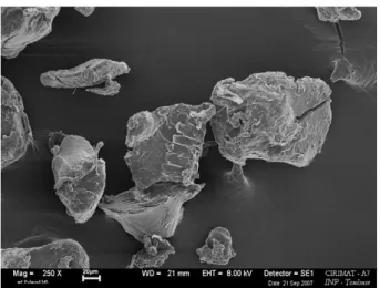
The polymeric matrix used in this study is poly-L,D-lactic acid (PLA, Galactic) with a molecular weight of 90,000 g/mol, initially available in the form of spherical beads having a diameter of about 3 mm. The beads were pre-ground in a laboratory blade mill (Janke and Kunkel). A pre-grinding temperature of 18 °C wasfixed by water circulating in a double jacket surrounding the mill chamber to avoid heating and even melting of the particles in the contact with the blades. Pre-grinding was performed for 10 min and the particles were then sieved. Only those having a size less than 250 µm were selected for this study. The initial mean size of the preground product is 157 µm. A scanning electron microscopy (SEM) micrograph of preground particles is shown inFig. 1.
The homopolymer of L-lactide (LPLA) is a semicrystalline polymer: Poly-L-lactide is about 37% crystalline, with a melting point of 173– 178 °C and a glass-transition temperature of 60–65 °C. PLA based materials exhibit high tensile strength and low elongation and consequently have a high modulus making them more suitable for load-bearing applications such as in orthopaedicfixation and sutures [15]. DL-PLA is an amorphous polymer exhibiting a random distribution of both isomeric forms of lactic acid, and is unable to arrange into an organised crystalline structure. This material has lower tensile strength, higher elongation, and a much more rapid degradation time, making it more attractive for drug delivery applications: the degradation time of LPLA is requiring more than 2 years to be completely absorbed.
The mineral charge is nanocrystalline carbonated apatite which has been prepared at ambient temperature and physiological pH by double decomposition of a diammonium hydrogenphosphate and sodium hydrogencarbonate (90 g of (NH4)2HPO4and 90 g of NaHCO3
in 1500 mL of deionised water) and a calcium nitrate solution (52.2 g of Ca(NO3)24H2O in 750 mL of deionised water). The calcium solution
is rapidly poured into the phosphate and carbonate solution at room temperature (20 °C) and stirred only for a few minutes. The apatite was left for 15 days to mature after precipitation at room temperature in the mother solution without stirring and in a stoppered vial to minimise the release and uptake of CO2at physiological pH. At the end
of maturation period, the precipitate is thenfiltered under vacuum and washed with deionised water (2 L). Then the gel is freeze-dried andfinally stored in a freezer to prevent further maturation of the apatite before its use.
The carbonate content of the as-prepared apatite (4.5% w/w of CO3), was determined using a CO2coulometer (UIC Inc., USA). Calcium
concentration was determined by complexometry with EDTA and the phosphorus concentration by visible spectrophotometry of the phospho-vanado-molybdenum complex. The Ca/P atomic ratio of
Fig. 1. SEM micrograph of preground polymer particles.
the prepared apatite (Ca/P = 1.46) was calculated from the result of these two analyses.
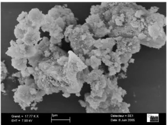
The evaluation of the average crystal dimensions of the synthe-sised apatite is based on X-ray diffraction line broadening analysis applying Scherrer's formula. Platelet-like apatite nanocrystals have an average length of 160 Å and an average width/thickness of 54 Å. These platelets are highly agglomerated in the initial powder and the initial mean particle size (37.4μm) was determined using laser diffraction granulometer (Malvern Mastersizer 2000). Fig. 2 presents a SEM micrograph of such apatite particles.
Dry batch grinding and co-grinding experiments were performed using a laboratory tumbling ball mill. It consists in a 1 L cylindrical ceramic chamber, rotating around its horizontal axis, and containing ceramic balls with a size of 10 mm. The rotating speed of the chamber wasfixed at 100 rpm, i.e. at 75% of the critical speed, while the ball loading volume represented 20% of the whole volume of the chamber. The powderfilling rate was 10 vol.% of the total void space of the balls. The balls and powder filling rates may be considered as small compared to usual rates used in industrial tumbling ball mills. However, these values were chosen to minimise the consumption of high-cost components. The polymer and the mineral were ground separately, but also together (co-grinding), in the case of mixtures containing between 10 and 40 wt.% of apatite. The co-grinding time was varied from 10 min to 10 h.
Powder samples were taken from different regions of the chamber at various times to be analysed. The sample mass was small enough, so as not to modify significantly the powder proportion in the mill.
Dry measurement of particle size distributions of PLA and apatite powders before and after grinding were made with a laser diffraction granulometer (Malvern Mastersizer 2000). The data was treated by Mie theory which permits to limit artefacts at small sizes of the distribution. The mean diameter, d50, corresponding to cumulated
volume fractions of 50%, was calculated from the size distribution. Selected samples were analysed by scanning electron microscopy (LEO 435 VP) after gold-plating. The glass transition temperature of PLA and apatite mixtures was registered in a nitrogen atmosphere using a thermobalance SDT Q 600 TA Instruments.
The scaffolding feasibility was tested by producing porous tablets. Thus, a pore-forming agent (sodium chloride particles with a mean size of 300 µm) was added to the powders produced by co-grinding, the salt representing between 50 and 90 wt.% of the whole. The samples were then mixed to obtain homogeneous systems, and then placed in a Teflon mould to form discs with a diameter of 1 cm and a thickness of 3 mm. They were heated at 190 °C for 45 min and cooled to room temperature. The discs were then placed in distilled water to dissolve the sodium chloride crystals and generate porosity, and
finally dried at ambient air. Their porosity was controlled by SEM observation and measured with a multi-gas porosimeter (ASAP 2010 M Micromeritics).
Due to the size and shape of the mould used, the composite materials were disc-shaped. Therefore, usual compression tests (requiring a height to diameter ratio greater than 1.5) could not be performed; mechanical testing was carried out by diametral compres-sion tests, also referred to as Brazilian disc tests [16]. In such experiments, an increasing force (F) is applied on the discs placed vertically until rupture. This leads to a tensile strain perpendicular to F, and the related tensile stress (σ), which is characteristic of the mechanical strength of the disc, is related to F by the equation:
r ¼ 2F=p:D:H ð1Þ
where D and H are respectively the diameter and height of the disc. As this test is highly sensitive to the disc quality, the results obtained will be used only to compare with each other the samples tested in the present study and not with results obtained for composites prepared in other studies found in the literature. 3. Characterization of the powders after co-grinding
Since each particulate system has a specific behaviour during grinding or co-grinding, it isfirst of all appropriate to separately grind the two components and follow the evolution of their size with time. With regard to the polylactic acid, whose initial average size is of 158 µm, grinding does not decrease the particle size. On the contrary, the size increases gradually during grinding because of agglomeration. Thus, it reaches 190 µm after 1 h of treatment and 415 µm after 3 h. Beyond this time, one observes the formation of chips resultingfirst from the agglomeration of the particles, and then from the compac-tion of the agglomerates under the effect of the balls (Fig. 3).
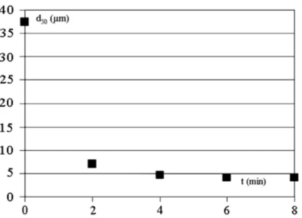
The size of apatite particles is quickly reduced as shown inFig. 4. However a size limit of 4 µm is reached after 6 min processing. This fast fragmentation is due to the fact that the particles consist of nanocrystals agglomerates, which are partially dissociated during grinding. This suggests that the mineral will be able to be distributed easily within the matrix by co-grinding.
With regard to the mixtures, their behaviour differs according to the mineral proportion. Indeed, with 10 wt.% apatite, the mixture tends to agglomerate like polymer alone. On the other hand, for mixtures including more than 30% of apatite, one notes a progressive reduction of the average size of the particles. After 10 h of co-grinding, the particle size reaches 91 µm for mixtures with 30% of apatite, and 63 µm for those including 40% of apatite. The apatite particles are
Fig. 3. SEM micrograph of polymer agglomerates. Fig. 2. SEM micrograph of an apatite particle.
broken during grinding. When the fragments reach a few microns, they adhere to the surface of the polymer particles, preventing agglomeration (seeFig. 5). A prolonged co-grinding treatment even makes it possible to erode the mineral fragments stuck on the polymer surfaces and to reduce their size more than when the mineral is ground alone, as already observed in a previous study with other materials[11]. Thus, one can see on the SEM micrograph presented in Fig. 5that apatite fragments with a size lower than a micron are stuck on polymer. EDX analysis shows that the small fragments were the mineralfiller. This one is thus perfectly dispersed within the matrix. This dispersion is supported by the reduction of the size of the polymer particles. Moreover, the adhesion of thefiller on polymer makes it possible to avoid a later segregation of the components during handling, thus preserving the homogeneity of the mixture. From the analysis of the size and morphology of the particles, it seems that a filler percentage of 30 or 40 wt.% should be used for the continuation of the study. These compositions have a sufficiently high proportion of mineral to take part in tissue repair (osteoconduction) by supporting the cellular colonization of the implant and the minerali-zation of the extracellular matrix.
The glass transition temperature of pre-ground polymer samples, without apatite, and of mixtures containing 20 to 50 wt.% offiller co-ground 10 min and 10 h was measured. An example of the results is shown inFig. 6. The same kind of data was found for all the operating conditions. The glass transition temperature is equal to 54 °C for the pre-ground polymer (in agreement with the range 55–60 °C given in literature[15]and the specifications of the supplier), as for the ground mixtures obtained after 10 min. For the mixtures which were co-ground for 10 h, the glass temperature increases to 59 °C and thefiller
percentage does not influence this value. This difference in temperature is significant because the analysis was carried out several times and the difference between the glass transition temperatures determined for the same sample did not exceed 2%. These results show that thefiller rigidifies the supramolecular structure of the polymer.
4. Feasibility of processing scaffolds
Considering bone reconstruction, composite scaffolds must allow the migration of cells and vascularisation. Thus, the objective is to produce sufficiently porous implants, in terms of porosity and pore size, but being sufficiently resistant.
In this way, we manufactured scaffolds made up on the one hand of mixtures co-ground during various times, containing 30 or 40 wt.% of apatite, and on the other hand of sodium chloride crystals at 50 to 90 wt.% in the whole samples. Salt was then eliminated by washing the scaffolds in distilled water during 48 h. The complete leaching of the scaffold was checked by SEM. Moreover, the scaffolds were qualitatively observed to control that they did not crumble during this washing step. Finally, the porous scaffolds were dried. Their ability to crumble was also controlled at this stage. They were then observed by SEM and their porosity was measured.
The scaffolds containing 90 wt.% of sodium chloride were transformed into powder during washing because the polymer was not present in sufficient quantity to constitute a continuous network around the salt crystals. The scaffolds containing up to 80 wt.% of salt satisfied this processing step. The scaffolds containing the composite with 40 wt.% of apatite were very friable after the stage of drying and they were transformed into powder during their handling. Thefiller proportion in these mixtures was probably too high, not making it possible the matrix to correctly play its role of binder. Only the scaffolds containing the mixtures with 30 wt.% of apatite and 80 wt.% of sodium chloride or less were selected for the later analyses.
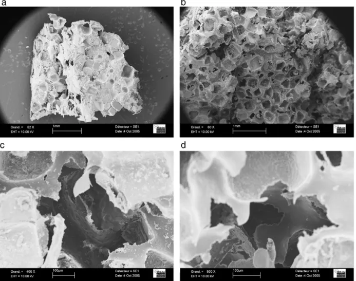
The micrographs presented inFig. 7.a and .b show scaffolds before and after washing. On thefirst photo, the salt crystals are clearly visible and dispersed in the composite. After washing, pores are created. As expected these are cubic shaped with a size of about 200– 300 µm. Observation at higher magnification (see onFig. 7.c) shows that very small apatite crystals are stuck on the pores walls, and they will be accessible when used in vivo. Moreover, there are inter-connections between pores (Fig. 7.c and .d), of a diameter varying between 100 and 200 µm, resulting from the contacts between sodium chloride salt crystals during the processing of the scaffolds.
The distribution of the pores within the scaffolds was determined by porosimetry. An example is presented onFig. 8corresponding to a scaffold initially made of 80 wt.% of sodium chloride (that is to say 74 vol.%) and 20 wt.% of a mixture containing 30 wt.% of apatite. The average size of the pores is equal to 133 µm and the porosity 60 vol.% for the as-prepared scaffold. This percentage is a little lower than the
Fig. 5. SEM micrograph of the surface of a composite particle after 10 h of co-grinding.
Fig. 6. Thermograms of the preground polymer and the PLA–apatite mixtures. Fig. 4. Variation of apatite particles mean size during grinding.
initial salt rate. Indeed, during the scaffold processing, the sodium chloride crystals being denser than the composite mixture, leads to slight segregation, so that the real proportion of salt in the scaffolds is a little lower than the initial value.
Similar results were obtained with all the scaffolds containing the composite with 30 wt.% offiller, whatever the time of co-grinding. Nevertheless, the difference between the percentage of porosity of the implants and the percentage of pore-forming agent initially intro-duced are reintro-duced as co-grinding time increases because of a reduc-tion in the segregareduc-tion phenomena between the components. The
pore size and the proportion of air voids are satisfactory to allow the accessibility of the whole implant to the cells.
5. Mechanical resistance of the scaffolds
Another important characteristic for application of such scaffolds as bone tissue engineering scaffolds is their mechanical resistance. Indeed, they must be easily handled by the surgeon, and preserve their shape after implantation in vivo. Generally, the mineralfiller controls this property. Nevertheless, in the case of porous composites having a high porosity, the presence of the pores can limit the effect of thefiller. This is why tests on mechanical resistance were performed. Two series of experiments were carried out. In the first series, Brazilian tests were carried out on scaffolds obtained under conditions as described above: weight percentage of apatite = 30%; time of co-grinding= 10 min; weight percentage of salt = 65, 70, 75 and 80%; heating temperature = 190 °C; processing at environmental pressure.Table 1 presents the results of the mechanical resistance measured for samples including various percentages of pore-forming agent. One can note that up to 70% of pore-forming agent, the mechanical resistance is
Fig. 7. SEM micrographs of scaffolds: a- before washing; b- after washing; c- mineral crystals on the pores surface; d- interconnections between the pores.
Fig. 8. Volume distribution of the pores.
Table 1
Influence of the salt (porogenous agent) percentage on the resistance of the porous scaffolds
% of salt 65 70 75 80 Resistance (MPa) 2.2 2.2 1.0 –
satisfactory. It decreases significantly for higher percentages because the proportion of polymer is too small to maintain sufficiently a rigid structure.
A second series of experiments was carried out by decreasing the processing temperature and by applying a pressure on the surface of the scaffolds during processing. A temperature of 130 °C wasfixed with the aim of limiting the important loss in the apatite surface reactivity occurring when processing at higher temperatures. Several pressures were applied: 20, 50 and 75 MPa. The results are given inTable 2. As previously, one can note that the results are more satisfactory with the smallest percentages of salt. Moreover, the resistance of the scaffolds is improved by an increase of the pressure applied to their surface during processing. However, whatever the conditions, the mechanical resistances remain lower than those obtained for thefirst series of experiments, showing the important role of the processing tempera-ture in scaffoldfinal mechanical properties.
6. Conclusions
Dry co-grinding was used to produce biodegradable porous composite materials. For this purpose, poly-L,D-lactic acid was associated with a nanocrystalline carbonated apatite analogous to bone mineral and used as mineralfiller. It was shown that the addition of 30 to 40 wt.% offiller aids the fragmentation of polymer particles and allows a satisfactory dispersion of the mineral within the matrix. A high percentage offiller is unfavourable for a processing of porous scaffolds, as it prevents the matrix from playing its role of binder. The processing of scaffolds was carried out by adding a pore-forming agent which then can be eliminated by simple leaching, thus generating the porosity. The percentage of pore-forming agent introduced into the mixture controls the proportion of air voids of the scaffolds. To avoid a disintegration of the scaffolds after leaching, one should not add more than 80 wt.% of pore-forming agent. Another important criterion is the mechanical resistance of the scaffolds. It is satisfactory if the percentage of pore-forming agent does not exceed 70 wt.%.
In order to be regarded as satisfactory, a scaffold needs to contain a sufficient amount of apatite supporting mineralisation and cellular adhesion. Also, the pores should have a sufficient size and should be numerous enough to facilitate cell invasion and proliferation and the vascularisation of the scaffold. Finally, it needs to exhibit sufficient mechanical resistance once implanted in vivo. Afiller percentage of 30 wt.%, a co-grinding time of 10 min, a percentage and a size of the
pore-forming agent respectively equal to 70 wt.% and 300 µm and a processing temperature of 190 °C were shown in this work to make it possible to achieve these goals.
Nomenclature
d50 mean diameter of the particles corresponding to cumulated
volume fraction of 50% m D disc diameter in Brazilian tests m F applied force in Brazilian tests N H disc height in Brazilian tests m
t time min
T temperature °C
σ tensile stress Pa
References
[1] H. Petite, V. Viatteau, W. Bensaïd, A. Meunier, C. De Pollak, M. Bourguignon, K. Oudina, L. Sedel, G. Guillemin, Tissue-engineered bone regeneration, Nat. Tech. 18 (9) (2000) 569–579.
[2] L. Kong, Y. Gao, W. Cao, Y. Gong, N. Zhao, X. Zhang, Preparation and characterization of nano-hydroxyapatite/chitosan composite scaffolds, J. Biomed. Mater. Res. 75 (2005) 275–282.
[3] V.M. Rusu, C.H. Ng, M. Wilke, B. Tiersch, P. Fratzl, M.G. Peter, Size-controlled hydroxyapatite nanoparticles as self-organized organic–inorganic composite materials, Biomaterials 26 (2005) 5414–5426.
[4] F. Miyaji, H.M. Kim, S. Handa, T. Kokubo, T. Nakamura, Bonelike apatite coating on organic polymers: novel nucleation process using sodium silicate solution, Biomaterials 20 (1999) 913–919.
[5] A. Tampieri, G. Celotti, E. Landi, M. Sandri, N. Roveri, G. Falini, Biologically inspired synthesis of bone-like composite: self-assembled collagenfibers/hydroxyapatite nanocrystals, J. Biomed. Mater. Res. 67 (2003) 618–625.
[6] M. Kikuchi, T. Ikoma, S. Itoh, H.N. Matsumoto, Y. Koyama, K. Takakuda, K. Shinomiya, J. Tanaka, Biomimetic synthesis of bone-like nanocomposites using the self-organization mechanism of hydroxyapatite and collagen, Compos. Sci. Technol. 64 (2004) 819–825.
[7] H.W. Kim, J.C. Knowles, H.E. Kim, Porous scaffolds of gelatin-hydroxyapatite nanocomposites obtained by biomimetic approach: characterization and anti-biotic drug release, J. Biomed. Mater. Res. 74B (2005) 686–698.
[8] W. Linhart, F. Peters, W. Lehmann, K. Schwarz, A.F. Schilling, M. Amling, J.M. Rueger, M. Epple, Biologically and chemically optimized composites of carbonated apatite and polyglycolide as bone substitution materials, J. Biomed. Mater. Res. 54 (2001) 162–171.
[9] G. Chen, T. Ushida, T. Tateishi, Poly(DL-lactic-co-glycolic acid) sponge hybridized with collagen microsponges and deposited apatite particulates, J. Biomed. Mater. Res. 57 (2001) 8–14.
[10] D. Tadic, F. Beckmann, K. Schwarz, M. Epple, A novel method to produce hydroxyapatite objects with interconnecting porosity that avoids sintering, Biomaterials 25 (2004) 3335–3340.
[11] C. Zapata-Massot, C. Frances, N. Le Bolay, On the use of scanning electron microscopy for the modelling of co-grinding kinetics in a tumbling ball mill, Powder Technol. 143-144 (2004) 215–229.
[12] C. Zapata, C. Frances, N. Le Bolay, S. Molina-Boisseau, Production of small composite particles by co-grinding in a media mill— characterization of the granulometric and the mechanical properties, Trans. IChemE, Part A, Chem. Eng. Res. Des. 82 (A5) (2004) 631–636.
[13] S. Cazalbou, C. Combes, D. Eicher, C. Rey, Adaptative physico-chemistry of bio-related calcium phosphates, J. Mater. Chem. 14 (2004) 2148–2153.
[14] C. Rey, C. Combes, C. Drouet, H. Sfihi, A. Barroug, Physico-chemical properties of nanocrystalline apatites: Implications for biominerals and biomaterials, Mater. Sci. Eng. 27 (2007) 198–205.
[15] K. Van de Velde, P. Kiekens, Biopolymers: overview of several properties and consequences on their applications, Polym. Test. 21 (2002) 433–442.
[16] A. Brückner-Foit, T. Fett, D. Munz, K. Schirmer, Discrimination of multiaxiality criteria with the Brazilian disc test, J. Eur. Ceram. Soc. 17 (5) (1997) 689–696. Table 2
Influence of pressure and salt percentage on the resistance of the porous scaffolds Pressure (MPa) % of salt Resistance (MPa)
75 65 2.0 70 0.9 75 0.1 80 0.1 50 65 0.6 80 0.1 20 65 0.6 80 –