Crystal structure of the C47S mutant of human peroxiredoxin 5
Christine Evrard,(1) Aude Smeets,(1) Bernard Knoops,(2) and Jean-Paul Declercq(1)
(1) Unit of Structural Chemistry (CSTR), Université catholique de Louvain, 1 place Louis Pasteur, B-1348 Louvain-la-Neuve, Belgium. (2) Laboratory of Cell Biology, Institut des Sciences de la Vie, Université catholique de Louvain, 5 place Croix du Sud, B-1348 Louvain-la-Neuve, Belgium.
Abstract
In the crystal structure of the reduced form of the wild-type human peroxiredoxin 5, the presence of a benzoate ion in direct interaction with the peroxidatic cysteine (Cys 47) appeared as a rather intriguing feature since it is known that the benzoate ion can play the role of a specific hydroxyl radical scavenger. The crystal structure of the C47S mutant of the same enzyme has been crystallized in the tetragonal system, space group P41212, with a = 65.65 Å, c = 122.04 Å. It confirms the presence of this benzoate ion in spite of the mutation into a serine of the Cys 47 residue to which the benzoate ion was directly linked in the wild-type structure. The benzoate ion seems to be stabilized by hydrophobic contacts on both sides of the aromatic ring. In this matter, the α5 helix, which is specific to peroxiredoxin 5 among mammalian peroxiredoxins, plays an important role. These hydrophobic contacts also allow to suggest why the benzoate ion disappears when the molecule is oxidized.
Keywords : Antioxidant enzyme ; peroxiredoxin ; thioredoxin fold ; thioredoxin peroxidase.
INTRODUCTION
The peroxiredoxins (PRDXs) define a large family of peroxidases able to reduce hydrogen peroxide and alkyl hydroperoxides from prokaryotes to eukaryotes.1-3 Functionally, PRDXs have been implicated in antioxidant protection and regulation of redox-dependent signaling in cells. Structurally, all PRDXs contain a conserved peroxidatic Cys residue in the N-terminal portion of the protein, that attacks the peroxide and is oxidized to Cys sulfenic acid. PRDXs are classified into three subgroups depending on the number of Cys residues directly involved in catalysis and on the resolution mechanism of the Cys sulfenic acid. PRDX1-PRDX4 represent the typical 2-Cys mammalian subgroup and are obligate homodimers. In this subgroup, the N-terminal Cys residue (Cys51 in human PRDX2) oxidized to sulfenic acid is attacked by the resolving Cys (Cys 172 in human PRDX2) located in the C-terminus of the other subunit resulting in the formation of an intermolecular disulfide bond. PRDX5 is the only identified mammalian member of the atypical 2-Cys subgroup. In atypical 2-Cys PRDXs, the C-terminal resolving Cys is contained in the same protein and the reaction with the peroxidatic Cys results in the formation of an intramolecular disulfide bond. Finally, PRDX6 is classified in the mammalian 1-Cys subgroup in which the N-terminal peroxidatic Cys (Cys47) is conserved and the resolving C-terminal Cys is missing.
Crystal structures are known for members of the typical 2-Cys subgroup (PRDX1,4 PRDX22,5,6). PRDX6 represents the mammalian 1-Cys subgroup for which a crystal structure is also available.7 In typical 2-Cys and 1-Cys subgroups, the crystal structures are characterized by the formation of tightly associated dimers resulting from the association of the β sheets of two monomers to form a 10- or 14-stranded β-sheet. PRDX5, the
mammalian atypical 2-Cys PRDX, was characterized8,9 and the structure of its reduced state was recently solved by our group in two different crystal forms.10,11 Another crystal form simultaneously containing the reduced state and an unexpected dimeric oxidized state was also described.12 All the reduced molecules show that the sulfur atoms of Cys47 and Cys151 are positioned too far apart (13.8 Å) to interact in the expected intramolecular way without large conformational changes. The dimer present in all other known PRDXs structures and resulting from the association of β sheets is not observed in PRDX5. Also, the crystal structure of the reduced form of PRDX5 allowed to discover the presence of a benzoate ion, a specific scavenger of hydroxyl radical,13 covering the active site pocket and interacting with the sulfur atom of the conserved peroxidatic cysteine residue (Cys47). Since the mutation of the peroxidatic Cys47 into a Ser residue (C47S) in human PRDX5 inhibits its peroxidase activity9 and its antioxidant protection in vitro and in vivo,14 and since benzoate ion known as a scavenger of
investigations were necessary to determine whether a loss of antioxidant activity of human PRDX5 C47S mutant could only be due to the replacement of the catalytic Cys by a Ser or also to conformational changes leading to the release of the benzoate ion.
Here we present the crystal structure analysis of human PRDX5 C47S mutant. The crystal structure refined at 1.7 Å shows the presence of the benzoate ion in spite of the mutation of Cys47 into a Ser. The benzoate ion appears to be stabilized by hydrophobic contacts on both sides of the aromatic ring.
EXPERIMENTAL SECTION
The expression and purification of wild-type human PRDX5 and mutant C47S were previously described.10,14 The crystals of the 6xHis-tagged molecule were grown in reducing conditions by hanging or sitting drop vapour diffusion at 18°C with the well solution (500 µL) consisting of 1.6 M ammonium sulfate and 0.2 M potassium sodium tartrate as precipitant, 0.1 M sodium citrate buffer pH 5.3, 10-3 M 1,4-dithio-dl-threitol as antioxidant and 0.02% (w/v) sodium azide. The crystallization drop was formed by mixing equal amounts (2-10 µL) of the protein solution (10 mg mL-1) and of the well solution.
Before data collection, the crystal was cryo-soaked for a few seconds in paratone and flash cooled at 100 K. Data were collected at 1.7 Å resolution on the synchrotron BW7A beam line at EMBL c/o DESY using a MAR-ccd detector. The images were processed with the program DENZO15 and merged with the program SCALEPACK.15 The crystals are tetragonal, space group P41212, with a = 65.65 Å, c = 122.04 Å, and one protein molecule in the asymmetric unit. Five percent of the reflections were flagged for use in Rfree calculations.16 Some statistics of data collection and processing are presented in Table 1.
Since the space group and the cell dimensions of the C47S mutant of PRDX5 allowed to suppose a probable isomorphism with the tetragonal structure of the wild-type enzyme10 (pdb entry 1hd2), these coordinates were used for starting the refinement after applying the appropriate mutation. A rigid body refinement using
REFMAC517 belonging to the CCP4 suite18 yielded values of 0.343 and 0.348 for R and Rfree, respectively. Some minor adjustments were performed with O19 and were followed by cycles of refinement with REFMAC5. The N-terminal His tag and the short linker were not observed except for the last residue (Ser 0). Electron density for a benzoate ion was apparent in the initial electron density map despite its absence in the initial model used for phasing. Solvent molecules were progressively introduced by ARP/wARP.20 A significant improvement was reached by refining the mean square displacements of the unique molecule considered as a rigid body and defined as a TLS group. It was necessary to introduce alternate conformations for a number of side chains, mainly located at the surface of the molecule: Glu13, Glu18, Asn21, Glu27, Glu57, Gln68, Ser74, Glu83, Lys89, Glu91, Asp114, Ser159. The final R value is 0.146 (Rfree = 0.169) for a model containing 3 chloride ions and 285 solvent molecules.
Table 1. Statistics of Data Collection and Refinement
Wavelength (Å) 0.9911
Resolution (Å) Overall (ov) 37-1.70
Highest shell (hs) 1.74-1.70 Measured reflections 184,830 Unique reflections 30,149 Completeness (%) ov/hs 99.8/100 Rsym ov/hs 0.053/0.292 I/σ(I) ov/hs 32/5.5 Refinement
R factor (Rfree) Overall 0.146 (0.169)
Highest shell 0.179 (0.171)
Estimated overall coordinate error (Å) Based on Rfree 0.068
Based on maximum likelihood 0.040
r.m.s. deviations from ideality Bonds (Å) 0.019
Angles (°) 1.63
r.m.s. deviations in B values (bonded atoms) (Å2) Main chain 0.82
Side chain 2.44
Side-chain atoms 23.2
Solvent molecules 40.5
Chloride ions 25.4
Benzoate ion 19.9
Ramchandran plot: non-glycine residues located in The most favored regions 89.4%
Disallowed regions None
RESULTS AND DISCUSSION
All the 161 residues of the protein are well defined in the electron density. As shown in Fig. 1, the overall structure is characterized by the presence of a thioredoxin fold consisting of a four-stranded β sheet and three flanking α helices23 and the benzoate ion remains present at the surface of the active site pocket. Each molecule comprises three additional α helices and three β strands, one of which is associated with the thioredoxin β sheet to form a fifth strand while the two remaining ones form an additional two stranded β sheet in the N-terminal part of the molecule. Since the overall folding is very similar to that described for the two crystal forms of the wild PRDX5 structures,10,11 it will not be further discussed.
In the tetragonal (1hd2) structure of the reduced wild-type PRDX5 enzyme, the presence of a benzoate ion located at the surface of the active site pocket10 appears as a completely unexpected structural feature. This ion was also found in the monoclinic structure (1h4o)11 for each of the eight independent molecules of the
asymmetric unit. As benzoate is known to be a specific hydroxyl radical scavenger, its presence close to the active site might not be fortuitous. The present study confirms the presence of the benzoate ion (Fig. 1) in the C47S mutant. In lhd2, the distance between the oxygen atom O1 of the benzoate ion and the sulfur atom of Cys47 is 3.4 Å and we now observe a distance of 2.92 Å with the Oγ atom of Ser47, confirming the presence of a
hydrogen bond between the two oxygen atoms. Other distances involving the carboxylate oxygen atoms of the benzoate ion are compared in Table 2a and show that in the C47S mutant this part of the benzoate is even better anchored to the active site pocket than in the wild-type enzyme.
Fig. 1. The structure of the C47S mutant of human peroxire-doxin 5. The structural elements representative of
the thiore-doxin fold are colored green (helices) and red (sheets). The benzoate ion and the side chains of some important residues discussed in the text are represented. Figure prepared using MOLSCRIPT21 and
Table 2. Distances (Å) Involving Atoms of the Benzoate Ions
C47S mutant Wild type
(a) Distances involving the carboxylate oxygen atoms
Benzoate O1 Thr44 Oγ1 3.07 3.38 Gly46 N 3.26 3.01 Cys/Ser47 N 2.98 3.19 Cys Sγ/Ser47 Oγ 2.92 3.40 Arg127Nε2 2.79 2.99 Benzoate O2 Gly46 N 2.67 2.68
(b) Hydrophobic contacts involving the aromatic ring
Benzoate C3 Ile119Cδ1 4.70 4.74
Benzoate C4 Leu116Cδ1 3.99 3.87
Phe120 Cδ 3.57 3.73
Benzoate C5 Thr147 Cγ2 3.62 3.66
Note. Comparison of the distances observed in the C47S mutant and in the wild-type enzyme (lhd2).
The presence of a benzoate ion in contact with the active site residues has already been described in the crystal structure of chloroper-oxidase from S. aureofaciens Tü24 (pdb entry 1a8u).24 An organic acid is required in this enzyme for the haloperoxidase activity. The complex with benzoate was obtained after soaking the crystals in a solution containing sodium benzoate. However, the benzoate is not specific since complexes were also obtained with propionate and acetate. In these complexes, both carboxyl oxygen atoms of the organic acids are within hydrogen bonding distance from Ser98 Oγ. Interestingly, these carboxyl oxygen atoms are also hydrogen bound
to backbone nitrogen atoms, to His257 Nε2 belonging to the active site and to a Thr Oγ. Table 2a shows that this
situation is very similar to our observations in PRDX5 (wild-type and C47S mutant) with the replacement of His257 Nε2 by Arg127 Nε2 whose Nε1 interacts with the Sγ atom of Cys47 (wild-type) or the Oγ atom of Ser47
(mutant). In PRDX5, it is however worth remembering that the benzoate ion was not added at any stage of the production, of the purification, or of the crystallization. However, we cannot completely exclude that the benzoate ion was present in bacterial culture media (LB Broth base including peptone 140 and yeast extract) used in the production of E. coli recombinant human PRDX5. The remaining contacts of the benzoate ion in PRDX5 involve its aromatic part and are thus mainly hydrophobic. The buried surfaces of the protein by the presence of this aromatic ring are very similar in the cases of the C47S mutant and of the wild-type enzyme, respectively 47 Å2 in the C47S mutant compared to 52 Å2 in the wild-type enzyme. These hydrophobic contacts between the protein and the benzoate ion concern four residues at the level of the protein and the shortest distances between these residues and the aromatic ring are given in Table 2b. As shown in Fig. 1, one residue, Thr147, appears on one face of the aromatic ring.
The three remaining residues (Leu116, Ile119, and Phe120), located on the other face, belong to the α5 helix which is a characteristic of PRDX5, since it is not present in other PRDXs structures. This α5 helix which appears as a bump at the surface of the protein thus seems to be an essential feature for holding the benzoate ion in its position. It constitutes one side of a kind of pincer in which the benzoate is locked, the other side being the Thr147 residue. When PRDX5 is oxidized, it was shown12 that the region 146-147 undergoes an important reorganization in order to bring the resolving residue Cys151 in the neighborhood of the peroxidatic residue Cys47 and to allow the formation of the disulfide bond. During this movement, one side of the pincer (Thr147) is thus relaxed and the benzoate ion can escape from the trap. From the comparison of nine independent molecules of PRDX5, the region of residues 146-147 was also recognized for its important flexibility.11
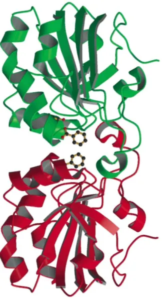
Interestingly, it was previously noticed12 that the α5 helix was involved in the formation of a non-covalent dimer between two PRDX5 molecules. This kind of dimer buries about 790 Å2 per molecule and since it is present in structures of reduced and oxidized forms of PRDX5 belonging to different space groups and systems (tetragonal for 1hd2,10 monoclinic for 1h4o,11 orthorhombic for loc312) it is certainly not an artefact of crystallization. This dimer is also observed in the structure of the C47S mutant and is formed by the association of two molecules related by a crystallographic twofold axis. This association leads to the formation of a channel at the interface between the two molecules (Fig. 2) with the two benzoate ions obstructing both extremities of this channel. The benzoate ions appear as the guardians of the two gates giving access to the channel.
What could be the exact role of the benzoate ion in the mechanism of PRDX5 remains to be elucidated. Does it react with hydroxyl radicals produced during the enzymatic reaction to be decarboxylated13 or hydroxylated?25 Does it undergo a nucleophilic attack on the carboxyl carbon, as in haloperoxidases?24 Is it involved in a
completely different reaction? Or is its presence only fortuitous? This remains an open question.
Fig. 2. The benzoate ion appears at the extremities of a kind of channel at the interface of two molecules of the
C47S mutant of PRDX5 associated in a non-covalent dimer. The side chain of Ser47 is shown. Figure prepared using MOLSCRIPT21 and RASTER3D.22
ACKNOWLEDGMENTS
This work was supported by grants from the "Fonds National de la Recherche Scientifique" (Belgium) and the "Communauté française de Belgique-Action de Recherches Concertées." We also thank the European
Community for Access to Research Infrastructure Action of the Improving Human Potential Programme to the EMBL Hamburg Outstation, contract number HPRI-1999-CT-00017. We are grateful to the members of the scientific staff at the EMBL-Hamburg outstation for their help during data collection.
Supplementary material the coordinates and the structure factors have been deposited with the Protein Data Bank under accession code 1urm. Further details of the crystal structure investigations may be obtained from
http://www.rcsb.org/pdb/ or http://rutgers. rcsb.org/pdb/. References
2. Wood, Z.A.; Poole, L.B.; Hantgan, R.R.; Karplus, P.A. Biochemistry 2002, 41, 5493. 3. Hofmann, B.; Hecht, H.-J.; Flohé, L. Biol. Chem. 2002, 383, 347.
4. Hirotsu, S.; Abe, Y.; Okada, K.; Nagahara, N.; Hori, H.; Nishino, T.S.; Hakoshima, T. Proc. Natl. Acad. Sci. U.S.A. 1999, 96, 12333. 5. Schröder, E.; Littlechild, J.A.; Lebedev, A.A.; Errington, N.; Vagin, A.A.; Isupov, M.N. Structure 2000, 8, 605.
6. Alphey, M.S.; Bond, C.S.; Tetaud, E.; Fairlamb, A.H.; Hunter, W.N. J. Mol. Biol. 2000,300, 903. 7. Choi, H.J.; Kang, S.W.; Yang, C.H.; Rhee, S.G.; Ryu, S.E. Nat. Struct. Biol. 1998, 5, 400.
8. Knoops, B.; Clippe, A.; Bogard, C.; Arsalane, K.; Wattiez, R.; Hermans, C.; Duconseille, E.; Falmagne, P.; Bernard, A. Biol. Chem. 1999, 274, 30451.
9. Seo, M.S.; Kang, S.W.; Kim, K.; Baines, I.C.; Lee, T.H.; Rhee, S.G. J. Biol. Chem. 2000, 275, 20346. 10. Declercq, J.P.; Evrard, C.; Clippe, A.; Vander Stricht, D.; Bernard, A.; Knoops, B.J. Mol. Biol. 2001, 311, 751. 11. Declercq, J.P.; Evrard, C. Acta Crystallogr. 2001, D57, 1829.
12. Evrard, C.; Capron, A.; Marchand, C.; Clippe, A.; Wattiez, R.; Soumillion, P.; Knoops, B.; Declercq, J.P. J. Mol. Biol. 2004, 337, 1079. 13. Sagone, A.L.; Decker, M.A.; Wells, R.M.; Democko, C. Biochim. Bwphys. Acta 1980, 628, 90.
14. Plaisant, F.; Clippe, A.; Vander Stricht, D.; Knoops, B.; Gressens, P. Free Rod. Biol. Med. 2003, 34, 862. 15. Otwinowski, Z.; Minor, W. Methods Enzymol. 1997, 276, 307.
16. Brünger, A.T. Nature 1992, 355, 472.
17. Murshudov, G.N.; Vagin, A.A.; Dodson, E.J. Acta Cryst. 1997, D53, 240. 18. Collaborative Computational Project, Number 4. Acta Cryst. 1994, D50, 760. 19. Jones,T.A.; Zou,J.-Y.; Cowan,S.W.; Kjeldgaard,M. Acta Cryst. 1991, A47, 110. 20. Perrakis,A.; Morris,R.M.; Lamzin, VS. Nat. Struct.Biol. 1999, 6, 458. 21. Kraulis, P.J. J. Appl. Cryst. 1991, 24, 946.
22. Merritt, E.A.; Bacon, D.J. Methods Enzymol. 1997, 277, 505. 23. Martin, J.L. Structure 1995, 3, 245.
24. Hofmann, B.; Tölzer, S.; Pelletier, I.; Altenbuchner, J.; van Pée, K.H.; Hecht, H.J. J. Mol. Biol. 1998, 279, 889. 25. Halliwell, B.; Grootveld, M.; Gutteridge, J.M.C. Meth. Biochem. Anal. 1988,33, 59.