Cytokine Expression in Squamous Intraepithelial Lesions of the Uterine Cervix: Implications for the Generation of Local Immunosuppression
7
0
0
Texte intégral
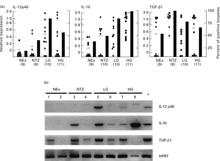
(2) 184. S. L. Giannini et al.. tumour necrosis factor-beta (TNF-b) (type I), or IL-4, IL-5, IL-6 and IL-13 (type II) [8]. In addition, several cytokines have been shown to contribute to the initiation or suppression of these immune responses, such as IL-4, IL-12, IL-10 and/or transforming growth factor-beta 1 (TGF-b1) [9–12]. These cytokines have been shown to be produced by various cell types, including macrophages, dendritic cells and keratinocytes [13–15]. It has been shown that peripheral blood mononuclear cells (PBMC) from patients with both SIL and cancer produce decreased amounts of IL-2 and IFN-g and higher levels of IL-4 and IL-10 following mitogenic stimulation, compared with the control group [16]. Recently, our group has demonstrated that basal levels of IL10 are augmented in the PBMC of patients with SIL [17]. Studies by our group and others, focused on the status of the localized immune response, have shown that SIL and/or cancer are associated with elevated levels of IL-4 and IL-6 [18,19]. Moreover, we have also shown that high-grade (HG) SIL are associated with lower densities of IL-2-producing cells compared with normal biopsies [19]. Taken together, these results argue that the development of SIL and/or cervical cancer is preferentially associated with type II (IL-4/IL-6) or immunosuppressive (IL-10) cytokines, as has been demonstrated in other types of cancers [20–23]. As an approach to understanding the factors involved in the generation and maintenance of an inefficient anti-tumour response, we have evaluated the expression of three cytokines, IL-12, IL-10 and TGF-b1, known to play an important immunomodulatory role. In this study, we used the techniques of reverse transcriptasepolymerase chain reaction (RT-PCR) and/or immunohistochemistry to analyse the expression of these cytokines in normal exocervix, transformation zone and in SIL biopsies. MATERIALS AND METHODS Biopsies Forty-three cervical biopsies (2–3 mm) were analysed in this study. Normal biopsy material was obtained from women undergoing routine examinations or hysterectomies. Biopsies from women with SIL were obtained before surgical procedures. Biopsies were immediately frozen in Tissue-Tek (Miles, Elkhart, IN) and stored at ¹808C. For each biopsy the histology was assigned to one of four categories based on histologic findings after haematoxylin–eosin staining: (i) normal exocervix from healthy women; (ii) transformation zone from healthy women; (iii) low-grade (LG) SIL including condyloma and cervical intraepithelial neoplasia (CIN) I lesions; (iv) HG SIL, including CIN II and III. Preparation of nucleic acids DNA and total RNA were prepared from all biopsies. To detect the presence of HPV, crude DNA was prepared from frozen biopsy sections (10 mm) by digesting the tissue sections with proteinase K (1 mg/ml) (Boehringer, Mannheim, Germany). RNA was extracted from 40 sections (10 mm) of frozen biopsies using the guanidinium thiocyanate method (RNAzol B; Bioprobe, Montreuil, France). PCR and RT-PCR PCR of DNA was carried out using PCR Master (Boehringer) with a standard aliquot of the DNA preparations. PCR of b-actin and L1 HPV genes was amplified for each sample using published oligonucleotide sequences [24]. The cDNA for the RT-PCR was prepared using M-MULV reverse transcriptase and 250–500 ng of total RNA from each biopsy, using standard conditions. recommended by the vendor (GIBCO/BRL, Merelbeke, Belgium). The PCR was carried out using standard aliquots of cDNA with: 1·25 U of Taq 2000 (Stratagene, La Jolla, CA), 0·2 mM dNTPs and 20 pg of each primer pair (HPRT sense GTTGGTATAAGCCAGA CTTTGTTG, antisense CAGATTTTCCAAACTCAACTTGAA, TGF-b1 sense TGAGGCCGACTACTACGCCAA, antisense GA GCCCTGGACACCAACTATT, IL-10 sense ATGCTTCGAGAT CTCCGAGA, antisense AAATCATACACGCCGTA, IL-12p40 sense ATTGAGGTCATGGTGGATGC, antisense AATGCTGG CATTTTTGCGGC and IFN-g sense GCAGAGCCAAATTGTC TCCT, antisense ATGCTCTTCGACTCGAAAC). All primer pairs spanned at least one intron. Cycling conditions were: 958C 5 min/558C or 608C 1 min/728C 1 min (35–40 cycles). A negative control without cDNA was included for each PCR reaction. The results are expressed as semiquantitative values based on the ratio of the intensity of the cytokine and HPRT PCR products analysed on ethidium bromide- or SYBR Green (Molecular Probes, Eugene, OR)-stained agarose gels (1·8%) using the BIO-PROFIL gel analyser system (Vilber Lourmat, Marne La Valle´e, France). The specificity of all PCR products was verified using the Southern blot technique with 32P-labelled internal oligonucleotides. Statistical evaluation of the PCR results was done using the Mann–Whitney test (InStat; Graph Pad Software, San Diego, CA). Immunohistochemistry for IL-10 and IL-12 Frozen sections (8 mm) were fixed in 2% paraformaldehyde and permeabilized using 0·1% saponin (UCB, Louvain, Belgium). Endogenous peroxidase was blocked with 0·2% H2O2 in PBS/ 0·1% saponin for 30 min. The slides were then incubated overnight at 48C with the cytokine-specific MoAbs (IL-12, Medgenix, Fleurus, Belgium; IL-10, Medgenix or Pharmingen, San Diego, CA) or isotype controls in PBS/0·1% saponin/2% bovine serum albumin (BSA)/0·1% NaN3. The anti-IL-12 antibody recognizes both IL-12p40 and IL-12p70. Biotin-labelled secondary antibodies (1:100 biotin goat anti-mouse IgG, Vector Labs, Burlingame, CA; 1:100 biotin-goat anti-rat, Pharmingen) were applied for 30 min at room temperature. The slides were then incubated with avidin– biotin–horseradish peroxidase (HRP) complex (Vectastain, ABCHRP kit; Vector) for 30 min. Diaminobenzidine (0·5 mg/ml; Sigma, Bornem, Belgium) was used as chromogen. Slides were counterstained with haematoxylin and mounted for light microscopy. RESULTS Expression of IL-12, IL-10 and TGF-b1 in SIL We have used the technique of RT-PCR to evaluate the expression of cytokines that could play a positive (IL-12) or negative (IL-10/ TGF-b1) role in the generation or maintenance of a cellular immune response. Included in our series of biopsies were: normal exocervix, the transformation zone, LG SIL and HG SIL. In Fig. 1 the data from the RT-PCR experiments are expressed as the relative level of cytokine expression (left ordinate) and as the percentage of biopsies expressing each cytokine (right ordinate). The relative expression level of IL-12p40 was higher in both LG and HG SIL compared with both normal exocervix (P ¼ 0·01/0·08, respectively) and transformation zone biopsies (P ¼ 0·08/0·39, respectively). The percentage of biopsies expressing IL-12p40 peaked in the LG SIL at 83% and diminished to 63% in HG SIL. In contrast, the expression of IL-10 increased continuously from a relatively low level in the normal exocervix to the highest average level and percentage in HG SIL (P ¼ 0·0004). Interestingly, the. q 1998 Blackwell Science Ltd, Clinical and Experimental Immunology, 113:183–189.
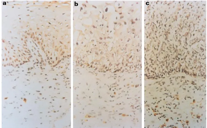
(3) (a) Relative expression. 2.0. IL-10. IL-12p40. TGF-β1 3.0. 100. 1.0 0.8. 1.0 0.8. 75. 0.6. 0.6. 0.4. 0.4. 0.2. 0.2. 0. 0. 3.0. 1.0 0.8 0.6 0.4 0.2 0 NEx (9). NTZ (8). LG (10). HG (11). 185. (16). NEx (9). NTZ (10). LG (10). HG (11). 50 25 NEx (9). NTZ (8). LG (10). Percent of positive biopsies. IL-10, IL-12 and TGF-b1 expression in SIL. HG (11). Fig. 1. Increased expression of IL-12p40 and IL-10 in squamous intraepithelial lesions (SIL). A group of 43 biopsies was evaluated for the expression of several cytokines using reverse transcriptase-polymerase chain reaction (RT-PCR). (a) The relative values of cytokine expression for IL-12, IL-10, transforming growth factor-beta 1(TGF-b1) versus HPRT. The left ordinate represents the average relative level of expression and the right ordinate represents the percentage of biopsies expressing the cytokine. Each point represents one patient and the average expression level is indicated by a bar. (b) Example of PCR products visualized on ethidium bromide (TGF-b1 and HPRT)- and SYBR Green (IL-10 and IL-12)-stained gels. NEx, Normal exocervix; NTZ, normal transformation zone; LG, low-grade SIL; HG, high-grade SIL; þ, tonsil positive control. All normal biopsies were human papillomavirus (HPV)-negative and all SIL were HPV-positive.. percentage of positive biopsies and the average expression level of IL-10 was higher in the transformation zone compared with the exocervix. Figure 1b is a representative illustration of a gel electrophoresis of RT-PCR products derived from eight of the biopsies analysed. Contrary to the differential expression of IL-10 and IL-12p40, IFN-g was expressed by a vast majority of the biopsies and the average expression levels were similar for all groups (data not shown). To complement this work we analysed several biopsies using immunohistochemistry to demonstrate the presence and the localization of the cytokine proteins. We found that the density of IL12-producing cells was higher in SIL biopsies compared with normal exocervix biopsies (Fig. 2). Compared with HG SIL, a higher percentage of LG SIL were positive for IL-12-producing cells (data not shown). IL-10-producing cells were rare (Fig. 3), but they were more frequently found in the transformation zone and SIL biopsies compared with the normal exocervix. Both IL-10- and IL-12-positive cells were found within the stroma, not within the SIL. TGF-b1 (Fig. 1) was expressed at similar levels in the exocervix biopsies and SIL. In contrast, the expression of TGF-b1 was. highest in the transformation zone, where 100% of the biopsies expressed the cytokine. Compared with the biopsies from the transformation zone, the average level of TGF-b1 expression diminished in SIL (LG P ¼ 0·05, HG P ¼ 0·03). Differential expression of IL-10 in the transformation zone The region most sensitive to SIL and cancer development is the transformation zone. Since we found that the transformation zone biopsies expressed higher average levels of IL-10 than exocervix biopsies, we wanted to evaluate the expression of this cytokine using both biopsies derived from individual patients. Figure 4 shows the results of a gel electrophoresis analysis of IL-10 expression in three individuals. We observed that in two out of the three patients (66%) analysed the transformation zone was associated with higher expression levels of IL-10. DISCUSSION In this study we have demonstrated that the progression of SIL is associated with a locally augmented expression of IL-10, an immunosuppressive cytokine. Coincident with the expression of. q 1998 Blackwell Science Ltd, Clinical and Experimental Immunology, 113:183–189.
(4) 186. S. L. Giannini et al.. Fig. 2. Detection of IL-12 protein in squamous intraepithelial lesions (SIL). Immunohistochemical staining for IL-12: (a) normal exocervix, (b) low-grade SIL, and (c) high-grade SIL. Objective: × 40.. Fig. 3. Detection of IL-10 protein in squamous intraepithelial lesions (SIL). Immunohistochemical staining for IL-10: (a) normal exocervix, (b) transformation zone, and (c) SIL. Objective: × 100 oil immersion. q 1998 Blackwell Science Ltd, Clinical and Experimental Immunology, 113:183–189.
(5) IL-10, IL-12 and TGF-b1 expression in SIL. Fig. 4. Differential expression of IL-10 in the transformation zone (TZ) and exocervix (Ex). Example of polymerase chain reaction (PCR) products visualized on ethidium (HPRT)- and SYBR Green (IL-10)-stained gels. The relative expression values are indicated below each cytokine band.. IL-10, the average levels of IL-12p40 also increased in SIL. Interestingly, we observed that the percentage of HG SIL expressing IL-12 declined compared with LG SIL, using both the technique of RT-PCR and immunohistochemistry. Thus the possibility exists that the loss of IL-12p40 expression in some HG SIL and the maintenance of IL-10 expression contributes to an efficient tumour escape mechanism. Moreover, we have observed that the transformation zone of the cervix, the region most sensitive to SIL and cancer development, is associated with higher average levels of the immunosuppressive cytokines IL-10 and TGF-b1. Since both cytokines have the ability to interfere with the efficient induction of a type I response by APC, these cytokines may contribute to the predisposition of this region to cervical carcinogenesis. Fundamental to the efficacy of IL-12 as a potent cytokine is its ability to induce IFN-g production in T and natural killer (NK) cells and the differentiation and expansion of naive T cells into Th1 cells [7,25,26]. Apart from the importance of IL-12 in the defence against intracellular pathogens such as viruses, several studies have shown that IL-12 exhibits anti-tumour activity in a variety of murine tumour models, including melanoma, bladder, colon and renal carcinoma, as well as in HPV-associated transplanted tumours ([27–29], Hallez, personal communication). IL-12 has also been shown to play a role in the inhibition of angiogenesis, a process important for tumour survival and metastasis [30]. Importantly, we observed the expression of IL-12p40 within the SIL, albeit 37% of HG SIL did not express detectable levels of the IL12p40. The lack of potential IL-12 in some HG SIL may predispose the progression of HG SIL to cancer. The relevance of the increased expression of IL-12p40 in SIL needs to be interpreted in the context of the complex regulation of bioactive IL-12, which is formed by the association of IL-12p35 and IL-12p40 subunits. Based on the reported constitutive expression of IL-12p35 [31], the potential to form bioactive IL-12p70 should exist in SIL that produce IL-12p40 protein. However, since very sensitive techniques such as bioassays and ELISAs are recommended to detect the limited levels of IL-12p70, we were unable to assay for IL12p70 in our biopsy specimens. Moreover, it is also known that the formation of IL-12p40 homodimers, which bind to the IL-12 receptor, can interfere with IL-12 bioactivity [32]. Thus, although the potential to form bioactive IL-12p70 exists in SIL, the possible formation of IL-12p40 homodimers may interfere with the activity of IL-12p70 to influence the local immune response. In contrast to IL-12, IL-10 is known to have immunosuppressive effects resulting from the down-regulation of CD80 and MHC II molecules, which are necessary for efficient antigen presentation. 187. [13,33]. Moreover, IL-10 inhibits the production of IL-12 in vitro [34,35]. Another study has shown that IL-10 can also interfere with the cytotoxic T lymphocyte (CTL) lysis of tumours [36]. In contrast to what is observed in mice, IL-10 can not be strictly associated with a type II response and has been shown to be produced by both type 1 and type 2 human T cells [37,38]. IL-10 mRNA and/or protein have been found to be augmented in several human cancers, such as renal and ovarian cancer and squamous and basal cell carcinoma of the skin [22,23,39,40]. In some cases IL-10 has been shown to be produced by the tumour cells themselves [23,41]. In addition, in vitro experiments have established that tumours can induce the production of IL-10 by PBMC [20,42]. In SIL, we observed that IL-10 was produced by stromal cells and not by preneoplastic cells within the SIL, as we previously observed for IL-4-producing cells [19]. The observed preferential expression of IL-10 in the transformation zone in some patients may contribute to the initiation of SIL by allowing HPV to subvert innate immunological surveillance mechanisms. Subsequently, the persistence of IL-10 in SIL may tolerize the immune system and permit the lesion to progress to cancer. The association of an augmented level of both IL-12 and IL-10 in some SIL may be related to the recent observation of in vitro experiments demonstrating that IL-12 induces the augmentation of IL-10 and IFN-g in human T cells [43,44]. However, in our experimental group we did not observe any correlation between the relative expression levels of IL-10 and IL-12 in SIL. Despite the expression of IL-10 in SIL, the expression pattern of IFN-g was similar in all experimental groups (data not shown). This may be relevant to the observation that IL-10 has been shown to suppress the production of IFN-g by type 1 CD4 cells and not by NK cells [45,46]. The significance of the concomitant expression of both IL12 and IL-10 in vivo within the microenviroment of a preneoplastic lesion or tumour is unknown. In cervical carcinogenesis TGF-b1 can play two contrasting roles. It may contribute to immunosuppression by down-regulating IL-2 receptor signalling in T cells and IL-12 expression by APC and by inducing the expression of IL-10 by macrophages [47–49]. In contrast, TGF-b1 can also be beneficial, in that it has been shown in vitro to inhibit the proliferation of keratinocytes and the expression of the E6 and E7 genes of HPV essential for cervical carcinogenesis [50,51]. Our results show that the overall expression of TGF-b1 is similar in the normal exocervix and SIL. In fact, a recent study has demonstrated that TGF-b1 is expressed in both normal exocervix and SIL, but that in the normal exocervix the expression is predominately in the epithelium and in SIL biopsies predominately in the stroma [52]. Surprisingly, we have observed that the transformation zone expressed the highest average levels of TGF-b1 and that 100% of the transformation zone biopsies expressed TGF-b1. Compared with the normal transformation zone, the SIL expressed diminished amounts of TGF-b1, thus being possibly more permissive to cancer progression in the absence of both the anti-proliferative effects and the transcriptional inhibition of E6 and E7 by TGF-b1. Inauspiciously, the SIL from several patients ceased to express detectable amounts of TGF-b1. Interestingly, a positive correlation was found between the expression of IL-10 and TGF-b1 in LG SIL of individual patients (r2 ¼ 0·8, data not shown). A positive correlation between IL-10 and TGF-b1 has also been demonstrated in a mouse tumour model system [53]. Despite the local expression of IL-12, cytokines associated with a type II response (IL-4/IL-6) and immunosuppression (IL-10). q 1998 Blackwell Science Ltd, Clinical and Experimental Immunology, 113:183–189.
(6) 188. S. L. Giannini et al.. appear to be associated with the progression of SIL [18,19]. An understanding of the interactions among multiple cytokines and other locally produced factors that contribute to the regulation of anti-tumour immunity will aid in the development of new strategies to treat SIL and cervical cancer. One potential candidate is IFN-a, a cytokine that has been shown to be effective in the treatment of HPV-associated genital warts and to have the ability to reduce the expression of IL-10 locally in basal cell carcinoma of the skin [23,54].. 17. 18. 19. ACKNOWLEDGMENTS This work was supported by the Belgian Fund for Medical Scientific Research, the Centre de Recherche Interuniversitaire en Vaccinologie, with a grant from the Walloon Region and SmithKline Beecham Biologicals, the Oeuvre Belge du Cancer and the Centre anticance´reux pre`s l’Universite´ de Lie`ge. P.D. is a research associate of the Belgian National Fund for Scientific Research. Antibodies for the immunohistochemistry were kindly provided by Medgenix.. 20 21. 22. 23. REFERENCES 1 Zur Hausen H. Roots and perspectives of contemporary papillomavirus research. J Cancer Res Clin Oncol 1996; 122:3–13. 2 Munger K, Phelps WC. The human papillomavirus E7 protein as a transforming and transactivating factor. Biochem Biophys Acta 1993; 1155:111–23. 3 Benton C, Shahidullah H, Hunter JAA. Human papillomavirus in the immunosuppressed. Papillomavirus Rep 1992; 3:23–26. 4 Jochmus-Kudielka I, Scheinder A, Braun R et al. Antibodies against the human papillomavirus type 16 early proteins in human sera: correlation of anti-E7 reactivity with cervical cancer. J Natl Cancer Inst 1989; 81:1698–704. 5 Viscidi RP, Sun Y, Tsuzaki B, Bosch FX, Munos N, Shah KV. Serologic response in human papillomavirus-associated invasive cervical cancer. Int J Cancer 1993; 55:780–4. 6 Frazer I, Tindle R. Cell-mediated immunity to papillomaviruses. Papillomavirus Rep 1992; 3:53–58. 7 Seder RA, Paul WE. Acquisition of lymphokine-producing phenotype by CD4þ T cells. Annu Rev Immunol 1994; 12:635–73. 8 Mosmann TR, Sad S. The expanding universe of T-cell subsets: Thl, Th2 and more. Immunol Today 1996; 17:138–46. 9 Le Gros G, Ben-Sasson SZ, Seder R, Finkelman FD, Paul WE. Generation of interleukin 4 (IL-4)-producing cells in vivo and in vitro: IL-2 and IL-4 are required for in vitro generation of IL-4producing cells. J Exp Med 1990; 172:921–9. 10 Manetti R, Parronchi P, Giudizi MG et al. Natural killer cell stimulatory factor (NKSF/IL-12) induces Th1-type specific immune responses and inhibits the development of IL-4 producing Th cells. J Exp Med 1993; 177:1199–204. 11 Moore KW, O’Garra A, de Waal Malefyt R, Vieira P, Mosmann TR. Interleukin-10. Annu Rev Immunol 1993; 11:165–90. 12 Shalaby MR, Ammann AL. Suppression of immune cell function in vitro by recombinant human transforming growth factor-beta. Cell Immunol 1988; 112:343–50. 13 de Waal Malefyt R, Abrams J, Bennett B, Figdor C, de Vries JE. Interleukin 10 (IL-10) inhibits cytokine synthesis by human monocytes: an autoregulatory role of IL-10 produced by monocytes. J Exp Med 1991; 174:1209–20. 14 Kang K, Kubin M, Cooper KD, Lessin SR, Trinchieri G, Rook AH. IL12 synthesis by human Langerhans cells. J Immunol 1996; 156:1402–7. 15 Aragane Y, Riemann H, Bhardwaj RS et al. IL-12 is expressed and released by human keratinocytes and epidermoid carcinoma cell lines. J Immunol 1994; 153:5366–72. 16 Clerici M, Merola M, Ferrario E et al. Cytokine production patterns in. 24. 25. 26. 27. 28. 29. 30. 31 32. 33. 34. 35. cervical intraepithelial neoplasia: association with human papillomavirus infection. J Natl Cancer Inst 1997; 89:245–50. Jacobs N, Giannini SL, Doyen J et al. Inverse modulation of interleukin-10 and -12 in the blood of women with preneoplastic lesion of the uterine cervix. Clin Exp Immunol 1998; 111:219–24. Tartour E, Gey A, Sastre-Garau X et al. Analysis of interleukin 6 expression in cervical neoplasia using a quantative polymerase chain reaction assay: evidence for enhanced interleukin 6 gene expression in invasive carcinoma. Cancer Res 1994; 54:6243–8. Al-Saleh W, Giannini SL, Jacobs N et al. Correlation of T-helper secretory differentiation and types of antigen-presenting cells in squamous intraepithelial lesions of the uterine cervix. J Pathol 1998; 184:283–90. Huang M, Wang J, Lee P et al. Human non-small cell lung cancer cells express a type 2 cytokine pattern. Cancer Res 1995; 55:3847–53. Huettner C, Paulus W, Roggendorf W. Messenger RNA expression of the immunosuppressive cytokine IL-10 in human gliomas. Am J Pathol 1995; 146:317–22. Nakagomi H, Pisa P, Pisa EK et al. Lack of interleukin-2 (IL-2) expression and selective expression of IL-10 mRNA in human renal cell carcinoma. Int J Cancer 1995; 63:366–71. Kim J, Moldin RL, Dubinett SM, McHugh T, Nickoloff BJ, Uyemura K. IL-10 production in cutaneous basal and squamous cell carcinomas. A mechanism for evading the local T cell immune response. J Immunol 1995; 155:2240–7. Jacobs MV, De Roda Husman AM, Van den Brule AJC, Snijders PJF, Meijer CJLM, Walboomer JMM. Group-specific differentiation between high- and low-risk human papillomavirus genotypes by general primer-mediated PCR and two cocktails of oligonucleotide probes. J Clin Microbiol 1995; 33:901–5. Chan SH, Perussia B, Gupta JW et al. Induction of interferon g production by natural killer cell stimulatory factor: characterization of the responder cells and synergy with other inducers. J Exp Med 1991; 173:869–78. Gately MK, Desai BB, Wolitzky AG et al. Regulation of human lymphocyte proliferation by a heterodimeric cytokine, IL-12 (cytotoxic maturation factor). J Immunol 1991; 147:874–82. Chen L, Chen D, Block E, O’Donnell M, Kufe DW, Clinton SK. Eradication of murine bladder carcinoma by intratumor injection of a bicistronic adenoviral vector carrying cDNAs for the IL-12 heterodimer and its inhibition by the IL-12 p40 subunit homodimer. J Immunol 1997; 159:351–9. Brunda MJ, Luistro L, Warrier RR et al. Antitumor and antimetastatic activity of interleukin 12 against murine tumors. J Exp Med 1993; 178:1223–30. Nastala CL, Edington HD, McKinney TG et al. Recombinant IL-12 administration induces tumor regression in association with IFN-g production. J Immunol 1994; 153:1697–706. Voest EE, Kenyon BM, O’Reilly MS, Truitt G, D’Arnato RJ, Folkman J. Inhibition of angiogenesis in vivo by interleukin 12. J Natl Cancer Inst 1995; 87:581–6. Ma X, Aste-Amezaga M, Trincheri G. Regulation of interleukin-12 production. Ann NY Acad Sci 1996; 795:13–23. Ling P, Gately MK, Gulbler U et al. Human IL-12 p40 homodimer binds to the IL12 receptor but does not mediate biological activity. J Immunol 1995; 154:116–27. Chang CH, Furue M, Tamaki K. B7-1 expression of Langerhans cells is upregulated by pro-inflammatory cytokines, and is down-regulated by interferon gamma or by interleukin-10. Eur J Immunol 1995; 25:394–8. D’Andrea A, Aste-Amezaga M, Valiante NM, Ma X, Kubin M, Trinchieri G. Interleukin 10 (IL10) inhibits human lymphocyte interferon gamma-production by suppressing natural killer stimulatory factor/IL12 synthesis in accessory cells. J Exp Med 1993; 178:1041–8. Koch F, Stanzl U, Jennewein P et al. High level IL-12 production by murine dendritic cells: upregulation via MHC class II and CD40 molecules and downregulation by IL-4 and IL-10. J Exp Med 1996; 184:741–6.. q 1998 Blackwell Science Ltd, Clinical and Experimental Immunology, 113:183–189.
(7) IL-10, IL-12 and TGF-b1 expression in SIL 36 Matsuda M, Salazar F, Peterson M et al. Interleukin-10 pretreatmant protects target cells from tumor- and allo-specific cytotoxic T cells and downregulates HLA class I expression. J Exp Med 1994; 180: 2371–6. 37 Yssel H, de Waal Malefyt R, Roncarolo M-G et al. IL-10 is produced by subsets of human CD4þ T cell clones and peripheral blood T cells. J Immunol 1992; 149:2378–84. 38 Del Prete G, De Carli M, Almerigogna F, Giudizi MG, Biagiotti R, Romagnani S. Human IL-10 is produced by both type 1 helper (Thl) and type 2 (Th2) T cell clones and inhibits their antigen-specific proliferation and cytokine production. J Immunol 1993; 150:353–60. 39 Pisa P, Halapi E, Pisa EK et al. Selective expression of interleukin 10, interferon g, and granulocyte-macrophage colony-stimulating factor in ovarian cancer biopsies. Proc Natl Acad Sci USA 1992; 89:7708–12. 40 Yamamura M, Modlin RL, Ohmen JD, Moy RL. Local expression of antiinflammatory cytokines in cancer. J Clin Invest 1993; 91:1005–10. 41 Kru¨ger-Krasagakes S, Krasagakis K, Garbe C et al. Expression of interleukin 10 in human melanoma. Br J Cancer 1994; 70:1182–5. 42 Kucharzik T, Lu¨gering N, Winde G, Domschke W, Stoll R. Colon carcinoma cell lines stimulate monocytes and lamina propria mononuclear cells to produce IL-10. Clin Exp Immunol 1997; 110:296–302. 43 Gerosa F, Paganin C, Peritt D et al. Interleukin-12 primes human CD4 and CD8 T cell clones for high production of both interferon-g and interleukin-10. J Exp Med 1996; 183:2559–69. 44 Windhagen A, Anderson DE, Carrizosa A, Williams RE, Hafler DA. IL12 induces human T cells secreting IL-10 with IFN-g. J Immunol 1996; 157:1127–31. 45 Mosmann TR, Moore KW. The role of IL-10 in crossregulation of Thl and Th2 responses. Immunol Today 1991; 12:A49–A53.. 189. 46 Hsu DH, Moore KW, Spits H. Differential effects of IL-4 and IL-10 on IL-2-induced IFN-gamma synthesis and lymphokine-activated killer activity. Int Immunol 1992; 4:563–9. 47 Ahuja SS, Paliogianni F, Yamada H, Balow JE, Boumpas DT. Effect of transforming growth factor-beta on early and late activation events in human T cells. J Immunol 1993; 150:3109–18. 48 Pardoux C, Asselin-Paturel C, Chehimi J, Gay F, Mami-Chouaib F, Chouaib S. Functional interaction between TGF-b and IL-12 in human primary allogeneic cytotoxicity and proliferative response. J Immunol 1997; 158:136–43. 49 Maeda H, Kuwahara H, Ichimura Y, Ohtsuki M, Kurakata S, Shiraishi A. TGF-b enhances macrophages’ ability to produce IL-10 in normal and tumor bearing mice. J Immunol 1995; 155:4926–32. 50 Coffey RJ, Sipes NJ, Bascom CC et al. Growth modulation of mouse keratinocytes by transforming growth factors. Cancer Res 1988; 48:1596–602. 51 Woodworth CD, Notario V, DiPaolo JA. Transforming growth factors beta 1 and 2 transcriptionally regulate human papillomavirus (HPV) type 16 early gene expression in HPV-immortalized human genital epithelial cells. J Virol 1990; 64:4767–75. 52 Comerci JT, Runowicz CD, Flanders KC et al. Altered expression of transforming growth factor-b1 in cervical neoplasia, as an early biomarker in carcinogenesis of the uterine cervix. Cancer 1996; 77:1107–14. 53 Maeda H, Shiraishi A. TGF-b contributes to the shift toward Th2-type responses through direct and IL-10-mediated pathways in tumorbearing mice. J Immunol 1996; 156:73–78. 54 Eron U, Judson F, Tucker S et al. Interferon therapy for condylomata acuminata. N Engl J Med 1986; 315:1059–64.. q 1998 Blackwell Science Ltd, Clinical and Experimental Immunology, 113:183–189.
(8)
Figure

Documents relatifs
We observed from tongue contour tracings that the Mid Bunched configuration generally has a lower tongue tip than the Front Bunched one in speakers who present both bunched
Objective 1: Collaborate with local partners to advocate for health policies that support cancer prevention and control and improve community access to cancer-
First introduced by Faddeev and Kashaev [7, 9], the quantum dilogarithm G b (x) and its variants S b (x) and g b (x) play a crucial role in the study of positive representations
The incidence of cancer is soaring due to rapidly ageing populations in most countries of the Region
Education in cancer pain relief and palliative care must be an essential component of any cancer control programme and should be incorporated into the health care system.. In the
b) On peut remarquer que le nuage s’allonge suivant une droite, d’où un ajustement affine est
Die Resultate der Studie zeigen, dass trotz einem erhöhten Risiko zu psychischen Folgen eines Einsatzes Rettungshelfer Zufriedenheit und Sinn in ihrer Arbeit finden können und
Nous souscrivons donc aux recommandations européennes les plus récentes concernant le traitement anticoagulant et antiagrégant des patients atteints de fibrillation auriculaire (et
L’archive ouverte pluridisciplinaire HAL, est destinée au dépôt et à la diffusion de documents scientifiques de niveau recherche, publiés ou non, émanant des
