Université de Montréal
Acute pain in domestic cats: nociceptive investigation and novel therapeutics
Par
Graeme M. Doodnaught
Département de sciences cliniques
Faculté de médecine vétérinaire
Mémoire présenté à la Faculté de médecine vétérinaire en vue de l’obtention du grade de maîtrise en sciences (M. Sc.)
en sciences vétérinaires option sciences cliniques
Février 2017
ii
Résumé
La disponibilité des médicaments analgésiques est limitée en médecine vétérinaire féline. Le but de cette étude était d'investiguer les propriétés anti-nociceptiques d'une nouvelle formule de buprénorphine (Simbadol, 1.8 mg ml-1) et tapentadol chez les chats.
Six chats étaient inclus dans deux études différentes, les deux étant prospectives,
randomisées, croisées, et aveuglées. Dans la première étude, Simbadol (1.8 mg mL-1) a été administré par voies sous-cutanée (SC;0.24 mg kg-1), intraveineuse (IV; 0.12 mg kg-1) et buccale (OTM; 0.12 mg kg-1) et les seuils thermiques ont été comparés avec ceux d'un groupe contrôle contenant de la saline (SAL; saline SC). Les concentrations plasmatiques de buprénorphine et norbuprénorphine ont été mesurées jusqu'à 72 heures suivant chaque traitement de buprénorphine. Un modèle
pharmacocinétique-pharmacodynamique adapté à 2 substances et 3 voies d'administration a été utilisé. Dans la deuxième étude, les seuils thermiques ont été comparés entre les chats recevant de la buprénorphine (0.02 mg kg−1, IM), un placébo (50 mg de dextrose oral) et deux doses de tapentadol oralement (dose réduite: 25 mg; dose élevée: 50 mg)
L'administration sous-cutanée de Simbadol a provoqué une anti-nociception thermique de longue durée (≥ 24 heures). Ces effets étaient prolongés comparativement aux traitements intraveineux (8 heures) et buccal (12 heures). Le modèle conjoint de
pharmacocinétique/pharmacodynamique a démontré des concentrations plasmatiques prolongée pour la voie sous-cutanée. Les deux doses de tapentadol ont augmenté l'antinociception thermique chez les chats. La dose élevée de tapentadol a produit une durée d'antinociception similaire à celle de la buprénorphine (2 heures) et deux fois plus longue que la dose réduite. La palatabilité de la
iii
Simbadol et tapentadol produisent une antinociception thermique comparée à la saline. Des investigations cliniques supplémentaires seront nécessaires.
Mots clés :
iv
Abstract
Analgesic drug availability is limited in feline practice. The aim of these studies was to
investigate the antinociceptive properties of a novel formulation of buprenorphine (Simbadol, 1.8 mg ml-1) and tapentadol in cats.
In two separate studies, six healthy cats (each) were included in a prospective, randomised, blinded, crossover study. In study I, Simbadol (1.8 mg mL-1) was administered by various routes: subcutaneous (SC; 0.24 mg kg-1), intravenous (IV; 0.12 mg kg-1) or buccal (OTM; 0.12 mg kg-1) route of administration and thermal thresholds (TT) were compared with a saline group (SAL; saline SC). Plasma buprenorphine and norbuprenorphine concentrations were measured up to 72 hours following each buprenorphine treatment. A bespoke pharmacokinetic-pharmacodynamic model fitted data from two analytes/three routes of administration. In study II, thermal thresholds were compared among cats receiving buprenorphine (0.02 mg kg−1, IM), placebo (50 mg oral dextrose) and two doses of oral tapentadol (low-dose 25 mg; high-dose 50 mg).
Subcutaneous administration of Simbadol provided long-lasting thermal antinociception (≥ 24 hours). These effects are prolonged compared with the IV (8 hours) and OTM (12 hours) treatments. Joint pharmacokinetic-pharmacodynamic modelling demonstrated prolonged plasma concentrations for the SC route. Both doses of tapentadol increased thermal antinociception in cats. The high-dose of tapentadol produced similar duration of antinociception as intramuscular buprenorphine (2 hours) and twice as long as the low-dose. Palatability presented a significant limitation to the drug’s administration.
Simbadol and tapentadol produced thermal antinociception when compared with saline. Additional investigation is necessary to determine if this translates to the clinical setting.
v Keywords:
vi
Table of Contents
Résumé ... ii
Abstract ... iv
Table of Contents ... vi
List of Tables ... viii
List of Figures ... x
List of Abbreviations ... xii
Dedication ...xiv
Acknowledgements ... xv
Introduction ... 1
1. Acute pain in cats ... 1
1.1 Problems in recognising acute pain ... 2
1.2. Attitudes and prevalence of analgesic administration in cats... 3
1.3 Instruments for Acute Pain Assessment ... 6
2. Nociceptive investigation ... 8
2.1 Quantitative sensory testing in people ... 8
2.2 Nociception models in animals ... 9
2.3 Nociceptive tools in feline acute pain research ... 10
3. Management of Acute Pain... 12
3.1 Opioids ... 13
3.2 Nonsteroidal anti-inflammatory drugs ... 20
3.3 Local Anaesthetics ... 22 3.4 Additional therapeutics ... 23 3.5 Non-pharmaceutical management ... 25 4. Novel investigation ... 26 4.1 Objectives ... 26 Article 1... 28
Pharmacokinetic and pharmacodynamic modelling of subcutaneous, intravenous and buccal high-concentration buprenorphine in conscious cats ... 28
vii
Introduction ... 32
Results ... 37
Discussion... 47
Conclusion ... 53
Appendix 1 – Acclimatization of cats ... 55
Appendix 2 – Analytical methods for buprenorphine and norbuprenorphine ... 56
Appendix 3: Population pharmacokinetic-pharmacodynamic modelling ... 58
Article 2... 73
Thermal antinociception following oral administration of tapentadol in conscious cats ... 73
Abstract ... 75
Introduction ... 77
Materials and methods ... 78
Results ... 81
Discussion... 87
Appendix 1 Behavior scoring scale ... 91
Combined Discussion ... 92
Conclusion... 97
viii
List of Tables
Article 1
Table 1. Skin temperature (°C; mean ± SD) in cats after saline (SAL) or
buprenorphine administration via the three treatment routes: intravenous (IV), subcutaneous (SC), and buccal (OTM).
38
Table 2. Thermal Threshold (°C; mean ± SD) in cats after saline (SAL), or
buprenorphine administration via the three treatment routes: intravenous (IV), subcutaneous (SC), and buccal (OTM).
40
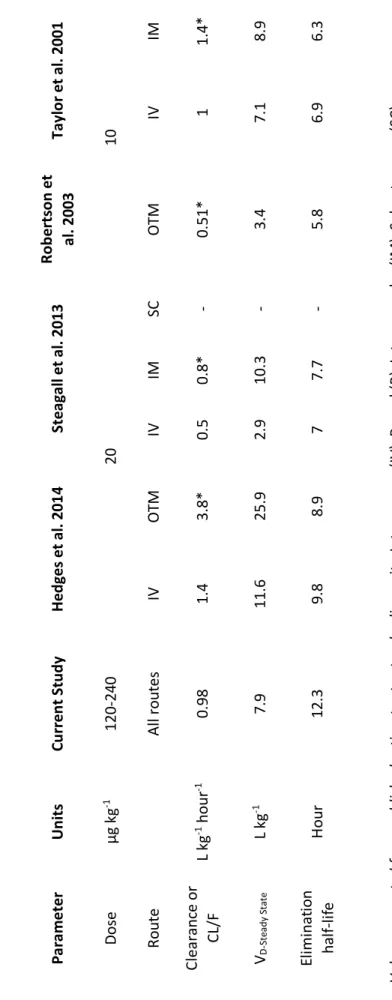
Table 3. Median pharmacokinetic estimates according to different dosage regimens and studies of buprenorphine after intravenous (IV), buccal (OTM), subcutaneous (SC), intramuscular (IM) administration in cats.
50
Table 4. Comparison of median pharmacokinetic parameters of the current study with estimates from a study using increasing doses of buprenorphine by subcutaneous (SC) administration in cats (Taylor et al., 2016).
51
Article 1 Appendix 3
Table 1: Pharmacokinetic model development showing the stepwise comparison of rival models for SC input function in joint IV, OTM and SC buprenorphine model and selection of the final, and best fit, model.
60
Table 2: Pharmacodynamic model development showing the best four models and selection of the model with the best fit.
62
Article 2
Appendix 1 Behaviour scoring scale used to evaluate cats’ behaviour at each time point. Adapted from Steagall et al., 2010.
91
Table 1 Behaviour scores [median (range)] in cats after administration of placebo (PBO, 50 mg dextrose orally), buprenorphine (BUP, 0.02 mg kg–1 IM), low-dose tapentadol (LowTAP, 25 mg orally) and high-dose tapentadol (HighTAP, 50 mg
ix
orally). Baseline (BL) values were collected before treatment administration at T0.
Table 2 Skin temperature (°C; mean ± SD) in cats after administration of placebo (PBO, 50 mg dextrose orally), buprenorphine (BUP, 0.02 mg kg–1 IM), low-dose tapentadol (LowTAP, 25 mg orally) and high-dose tapentadol (HighTAP, 50 mg orally). Baseline values (BL) were collected before treatments were administered at T0.
85
Table 3 Thermal threshold (°C; mean ± standard deviation) in cats after administration of placebo (PBO, 50 mg dextrose orally), buprenorphine (BUP, 0.02 mg kg–1 IM), low-dose tapentadol (LowTAP, 25 mg orally) and high-dose tapentadol (HighTAP, 50 mg orally). Baseline values (BL) were collected before treatment administration at T0.
x
List of Figures
Article 1
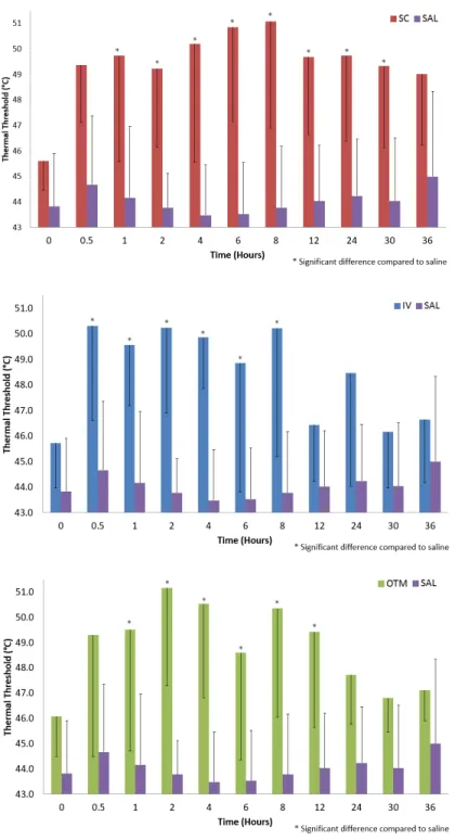
Figure 1. Thermal Threshold (mean ± SD) in cats after saline (SAL), or
buprenorphine administration via the three treatment routes: intravenous (IV), subcutaneous (SC), and buccal (OTM).
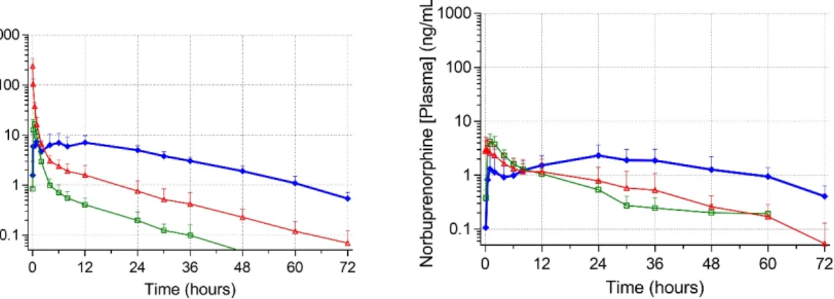
Figure 2. Mean plasma concentrations (ng ml-1) of buprenorphine and norbuprenorphine (± SD) in six conscious cats.
41
42
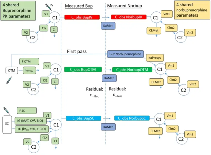
Figure 3. Pharmacokinetic-pharmacodynamic (PK-PD) model representation for buprenorphine and norbuprenorphine after subcutaneous, intravenous and buccal administration in six cats.
43
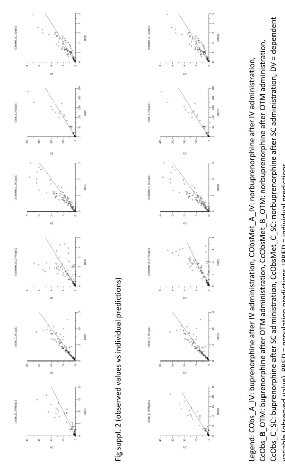
Article 1 – Appendix 3:
Results and goodness of fit plots: The goodness of fit figures for the final PK model fitting (buprenorphine and metabolite) are included thereafter
Fig suppl. 1: observed values vs population prediction Fig suppl. 2: observed values vs individual predictions
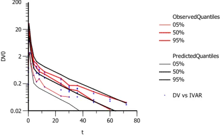
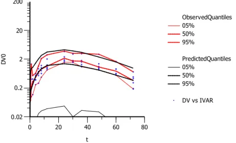
Fig suppl. 3: conditional weighted residuals vs time after dose Fig suppl. 4: conditional weighted residuals vs population prediction Fig suppl. 5 a. to f.: visual predictive check for buprenorphine IV (5.a),
norbuprenorphine IV (5.b), buprenorphine OTM (5.c), norbuprenorphine OTM (5.d), buprenorphine SC (5.e), norbuprenorphine SC (5.f)
Fig suppl. 6: observed values vs population prediction Fig suppl. 7: observed values vs individual predictions
Fig suppl. 8: conditional weighted residuals vs time after dose Fig suppl. 9: conditional weighted residuals vs population prediction Fig suppl. 10 a. to c.: pharmacodynamic visual predictive check for buprenorphine IV (10.a), OTM (5.b), buprenorphine SC (5.c)
xi Article 2
Figure 1. Thermal threshold (°C; mean ± standard error) in cats after
administration of placebo (PBO, 50 mg dextrose orally), buprenorphine (BUP, 0.02 mg kg–1 intramuscularly), low-dose tapentadol (LowTAP, 25 mg orally) and high-dose tapentadol (HighTAP, 50 mg orally). Baseline values (BL) were collected before treatment administration at T0. There were six cats in each treatment
xii
List of Abbreviations
AAFP: American Association of Feline Practitioners
ANOVA: Analysis of variance
ARRIVE: Animal research: Reporting of in vivo experiments
BL: Baseline values
BUP Buprenorphine 0.02 mg kg-1 IM
CMPS-F: Feline composite measure pain scale with grimace scale
COX: Cyclooxygenase
FDA: Food and drug administration
HighTAP High-dose tapentadol (50mg PO)
HPLC-MS High performance liquid chromatography-Mass spectrometry
IM: Intramuscular
ISFM: International Society of Feline Medicine
IV: Intravenous
LowTAP Low-dose tapentadol (25mg PO) MAC: Minimum alveolar concentration
NMDA: N-methyl-D-aspartate
NSAIDS: Non-steroidal anti-inflammatory drugs OTM: Buccal or Oral Transmucosal
PBO: Placebo
PD: Pharmacodynamics
xiii
PK-PD: Pharmacokinetics-pharmacodynamics
PO: Per-os
QST: Quantitative sensory testing
rCMPS-F: Feline composite measure pain scale
SAL: Saline treatment
SC: Subcutaneous
SD: Standard Deviation
t1/2: Half-life
TT : Thermal Threshold
UGTs: Uridine 5’-diphosphoglucuronosyl transferases
UNESP-MCPS: UNESP-Botucatu Multidimensional composite pain scale
xiv
Dedication
To my father,
I could not have achieved all that I have personally, academically, and professionally without your love, wisdom, and support.
xv
Acknowledgements
To my supervisor, Dr. Paulo Steagall, for providing me with your support and guidance throughout this program. I am truly amazed at the opportunities and experiences you provided me over the course of this masters. Multiple publications, travelled the world for conferences, and learned about the scientific method along the way. Thank you so much.
To the members of my jury, Dr. Marilyn Dunn, and chair Dr. Daniel Pang. Thank you for taking time out of your schedules to review this thesis. I greatly appreciate your contributions.
Many thanks to Zoetis for their financial support of my masters.
To everyone who helped with each of these studies. Long days (and sometimes nights) would not have been as enjoyable without your help. In particular, Dr. Beatriz Monteiro, thank you for introducing me to the cats and your tireless work throughout these studies.
To each of the cats; Hazel, Iris, Justin, Marcel, Seymour, Solomon, Emilie, Gorgeous, and Cindy. Without whom, none of this would have been possible.
To my family, no matter where in the world I am, and you be, your unconditional love, support and belief is always with me.
And finally, my dearest Julia. Your love, dedication and passion drive me to be better than I am. I could not be where I am today without you by my side.
Introduction
The intent of this literature review is to put into perspective the current knowledge regarding acute pain in cats. The beginning is intended to explain practical limitations on veterinary assessments of pain in cats, idiosyncrasies of the species and attitudes towards pain management. Next the review will explore methods of pain quantification and define and explore nociceptive research. The final topic for revision is management of feline acute pain. Comparisons are often made to the dog as a method of providing context to differences observed. Ultimately the review should provide the reader an understanding of nociceptive studies in feline medicine providing the framework for the investigation of the novel treatments in this thesis. The intent of these studies is to improve the understanding and management of acute pain in cats.
1. Acute pain in cats
Cats are the most popular household pet in Canada. There were 7 million cats living in homes across the country as reported by a survey in 2014 (1). Cats are less likely to have regular veterinary visits when compared with dogs. The same owners would pay more for life-saving procedures for dogs versus cats. Socioeconomic factors affected these perceptions, but better client communication was highlighted as essential to improving this disparity (2). When considering acute (adaptive) pain, perioperative or peri-procedural pain is more frequently recognised and treated compared to other manifestations of acute pain. Unfortunately, fewer veterinary visits result in reduced preventive medicine for cats increasing the likelihood of diseases which may produce pain (eg. dental disease, obesity-related osteoarthritis, urinary obstruction, etc.). A better understanding of nociception and pain states, as well as treatment modalities, is vital to this deficit.
2
1.1 Problems in recognising acute pain
Many characteristics exist with the cat that makes the recognition and management of pain problematic. The behavioural changes associated with veterinary visits has been considered a reason for the decreased frequency of veterinary examinations for the species. Stress and anxiety associated with transport, other animals in waiting areas, as well as restraint and handling during examinations and procedures are often manifested as avoidance or aggressive behaviours in the species. Fear is the most common cause of aggression in cats in the veterinary clinic setting, followed by pain (3). This overlap of behaviours with different causes makes clinical evaluations more complicated and difficult. Challenging behavioural modification techniques exist. The American Association of Feline Practitioners (AAFP) and International Society of Feline Medicine (ISFM) recommend the adoption of friendly handling techniques to minimize the impact of these behaviours (4). These recommendations extend beyond handling techniques and also include recommendations for clients at home before a veterinary visit, changes to clinic environment, and techniques for managing the return of patients to the home following extended hospitalisation. The impact of stress on cats adversely affects sickness-associated behaviours (e.g. vomiting, diarrhoea, decreased activity, enhanced pain-like behaviour, etc.) in both healthy and sick cats as well as their physiologic and haematologic variables (5, 6). These parameters are considered in unison when evaluating the health status of veterinary patients, thus differentiating stress and pain can be difficult.
Idiosyncrasies in drug metabolism have negatively impacted the availability, licensure and use of many analgesics. Classically, the use of non-steroidal anti-inflammatory drugs (NSAIDS) has been cautious due to the reduced capacity for glucuronidation (7-9). Limitations of glucuronidation also affect opioid analgesia. Particularly morphine, where production of the active metabolite morphine-6-glucuronide is limited (10). The behavioural impacts of the agonists of
μ-
opioid receptors have also3
been exaggerated due to initial investigations using doses above those used clinically (at present), producing erratic responses to stimuli and dysphoria (11). These considerations, as well as reduced feline pharmaceutical market demand (compared with dogs) has led to fewer approved analgesics, limiting available therapeutics for cats. Additionally, when a product is licensed for use in cats, it is often only available for single-use or short-term dosing (e.g. meloxicam in North America) requiring “off-label” dosing for long-term usage. For dogs, this “off-“off-label” usage of many drugs is more frequent. This difference is multifactorial due in part to the perceived increased risk of adverse effects in cats, and a greater body of clinical studies on pain management in dogs. This wealth of studies provides better support for unlicensed dosing regimens and therapeutics in dogs (12, 13).
1.2. Attitudes and prevalence of analgesic administration in cats
Since 2000, the perceptions and attitudes pertaining to acute pain management have evolved considerably. In the first ten years of the millennium, the majority of surveys highlighted inadequacies in training and education. There were significant differences in attitudes to analgesia which varied geographically (or perhaps more specifically, culturally). Canadian veterinarians (n = 326) were surveyed in 2001 regarding their perioperative used of analgesics in dogs and cats (14). With respect to ovariohysterectomy, it was shown that more recent graduates, veterinarians with increased personal perception of pain and the presence of animal health technicians increased the likelihood of pain scoring and analgesic administration. Concern regarding the negative aspects (adverse effects and cost) of opioid administration were cited as a reason for avoidance of analgesic administration. Interestingly the same individuals tended to also avoid NSAIDs in the same cases. Another survey was conducted in 2001 of New Zealand Veterinarians (n = 320) (15). Less than half of these practitioners felt their knowledge was adequate for pain management. This study showed that despite procedures being deemed painful, routine analgesics were not always given. Across all common surgical
4
procedures, dogs received more analgesics than cats (eg. castration, where 65% of dogs and 50% of cats had routine analgesia). Additionally, the negative impact of adverse effects was more frequently considered in the selection of analgesics for cats than dogs. A Finnish study published in 2003 again showed that younger veterinarians (who tended to be women), as well as those who work in larger practices, used more analgesics than older veterinarians and those in smaller clinics (16). Veterinarians in this survey (n = 434) agreed that post-operative pain management was beneficial, a third of respondents considered that a certain degree of post-operative pain was useful for case management. In general, pain was more frequently treated in dogs than cats for ovariohysterectomy and castration. Interestingly, canine castration was deemed as painful as feline ovariohysterectomy (numerical rating scale, scored 6.10 and 6.14, respectively, out of 10), however, castrated dogs were more likely to receive analgesics. When comparing castration alone between the species, results showed less than 10% of respondents would never give analgesics to dogs but more than 60% would not give analgesics to cats. The study concluded that a disparity existed in the knowledge base of these practitioners, and improved education was warranted. In a survey from 2007 of Brazilian veterinarians (n = 1298), it was found that women and younger veterinarians have increased pain recognition (17). For celiotomies, orchiectomies and dental procedures, cats were often regarded as having a lower pain score. The same veterinarians felt their knowledge was inadequate, despite having comparable or better pain recognition than previous studies. In these studies from the early 2000s, lack of analgesic administration is largely attributed to inadequate knowledge and education regarding drug pharmacology and adverse effects.
Over the last seven years, the recognition and attitudes towards acute pain management have improved. Limitations now seem to be less associated with just education but now also include an emphasis on the need for more research in the field to broaden the available knowledge. In a 2010
5
survey of veterinarians (n = 258) from Switzerland, the veterinarians responded that their justification for analgesic use was based principally on drug efficacy (18). Contradictorily, butorphanol and buprenorphine were the most common opioids used in the country, not the agonists of
μ-
opioid receptors with superior analgesic efficacy (to be discussed later). The authors suggested a weakness in drug pharmacology that could explain this difference. Though extensive comparisons could not be made due to a small number of respondents, the authors also suggested there should be an increase in the use of pain assessment instruments which are becoming increasingly more available. In 2012 a survey was performed of Canadian veterinarians in Ontario (n = 229) (19). This study found that there was an overall improvement in the awareness and provision of analgesics. The respondents felt their knowledge was sufficient. An interesting shift occurred where there was no longer a perception that cost of drugs or side effects were a sufficient justification for a lack of analgesic administration. In 2013, a survey of United Kingdom veterinarians (n = 720) showed 98% prevalence of perioperative NSAIDS administration for ovariohysterectomy and castration (20). Three quarters of respondents also used a multimodal approach (NSAID + opioid). In this study, there was no difference between year of graduation and analgesic administration. Orthopaedic, abdominal and dental pain were regarded as equal between dogs and cats. The only species difference was that neutering and ovariohysterectomy were considered less painful for cats than dogs, and male vets were less likely to administer NSAIDs to cats (94% vs 99% of women). In a survey published in 2014, attitudes towards feline pain were compared between veterinarians (n = 717) from New Zealand, Australia and the United Kingdom (21). In this study, all three countries had improved peri-operative analgesia compared with previous studies. Despite this, an absence of post-operative drug administration as well as follow-up (at home) analgesia was identified. The authors suggested that more research was required for both post-operative drugs and analgesic timing. It seems that awareness of pain as a problem has improved recently, more research and dissemination of these findings to practitioners is needed.6
1.3 Instruments for Acute Pain Assessment
Pain is an inherently subjective experience (22). Methods by which this experience can be quantified allow for better identification and even comparison of pain. These tools then can be used both clinically and in the research setting. The development of a subjective instrument for the assessment of pain requires four steps, chronologically; item generation, readability testing, reliability testing, and validity testing (23). Item generation is the first step, where specific assessments or evaluations are considered for evaluating pain. Readability testing should evaluates wheter a tool is practical and simple to understand and use. Reliability testing evaluates the consistency of measures yielded by the items within the instrument, as well as repeated measures by the same observer, different observers, and the same subject over a series of evaluations (24). The final step, validity testing, can be further subdivided into content, construct, and criterion validity. Content and construct validity evaluate the sensitivity of the items in the instrument to categorize pain states. Whereas criterion validity describes the correlation between an external objective measure and the scale itself (25). Ideally this should be a comparison to a gold-standard test, however no objective measure of pain has been classified as such. Cultural and lingual differences provide language specific readability, reliability and validity testing. The goal of clinical pain instruments is to assist veterinarians in determining whether or not the subject requires analgesia. Intervention thresholds are often determined based on a score which best separates painful animals from non-painful, ideally with the fewest misclassifications. There are several scales which have been developed for dogs for both acute and chronic pain across a number of conditions (26-31). Several of these scales have been evaluated and determined to be valid. There are fewer scales developed for cats limiting the available tools for pain identification and research (32-34).
7
A recent review evaluated instruments available for the subjective assessment of pain in cats including simple descriptive scales, VAS, numerical rating scales, dynamic interactive visual analogue scale and multidimensional composite scales (24). At present, only two instruments have been evaluated for validity. The feline composite measure pain scale (rCMPS-F) was developed as a clinical tool and validated as such across all breeds, ages and types of acute pain in cats. The validation was limited to content and construct, but did not include criterion validity. The scale includes an intervention point when the score is greater than or equal to 4 out of a possible 16 (33). In the development of this scale, the authors indicated the intention to include a grimace scale in a subsequent revision of the instrument. The inclusion of this grimace scale has now been validated (CMPS-F) (34). In this validation the authors also derived and revised the intervention score (greater than or equal to 5 out of 20). This revised version of the rCMPS-F performed better than the original, misclassifying only 17.6% of painful cats compared to 26.7%. The only other validated instrument is the UNESP-Botucatu Multidimensional composite pain scale (UNESP-MCPS) (32). This instrument was developed initially in Brazilian Portuguese for the evaluation of post-operative pain following ovariohysterectomy (35). This instrument included similar evaluation of pain expression and psychomotor changes to the CMPS-F. However, it also included physiological variables (appetite and blood pressure) combined with the dynamic, and interactive assessment. The scale was then translated into English and shown to be valid, reliable and responsive in the assessment of cats following ovariohysterectomy (32). A responsive scale is one which is sensitive to clinically relevant change. Which, in this case, are the expressions, psychomotor and physiologic changes associated with pain (24). Both the Portuguese and English versions have intervention scores of greater or equal to 8 of a possible 30. Additional translations to French and Spanish have been validated as well (36, 37). While this scale is perhaps better suited for the assessment of acute pain following ovariohysterectomy than the CMPS-F, it is limited in its application to other acute pain states. Additionally problematic is that as
8
a clinical tool the UNESP-MCPS is a more time-consuming and laborious (particularly the physiological variable collection) process. At present, the UNESP-MCPS is the only instrument shown to be qualitatively valid, reliable and sensitive (24). The inclusion of the grimace scale to the CMPS-F has improved the discriminatory ability of the rCMPS-F, however direct evaluation of criterion validity remains unreported. As a clinical tool, however, the rCMPS-F has been validated across a wider range of different pain types and sources (e.g. illness, surgery, emergency) making it, perhaps, more clinically applicable (34).
2. Nociceptive investigation
Quantifying pain is essential to its investigation. By providing objective measures of pain it becomes possible to compare different painful conditions in terms of diagnosis, treatment and progression. Pain is a perception, as such its quantification is difficult, particularly in non-verbal species. By providing a noxious stimulus and observing the response processed by the somatosensory system, it is possible to investigate thermal, electrical, mechanical, and/or chemical nociception (38). Regardless of its intended purpose, nociceptive investigation relies on the assumption that clinical pain correlates to the objective measure employed and the corresponding behavioural response. Limitations then exist due to anatomical, physiological and behavioural considerations, weakening this assumption. (39-42)
2.1 Quantitative sensory testing in people
Quantitative sensory testing (QST) is a widely used tool in human medicine for experimental and clinical research, as well as providing a bedside pain assessment (43, 44). QST assesses both static and dynamic measures of thermal and mechanical stimuli to evaluate the somatosensory system (44, 45). Investigators are capable of evaluating different peripheral nociceptive pathways and nerve types as well as spinothalamic and lemniscal central pathways by utilizing multiple stimuli in unison (46). In
9
general, thermal stimuli rely on A-delta and C nerve fibres and the spinothalamic pathway. Mechanical stimuli additionally involve A-beta fibres and the lemniscal pathway. The impact of painful conditions and treatments on pain transduction, transmission, perception and modulation can be made by comparing these various nerve fibres and pathways in conjunction. These stimuli test the nociceptive thresholds, limit of tolerance, as well as predefined parameters in which individuals’ perception is evaluated. In non-verbal patients, QST is restricted to threshold testing as the expression of perception is limited. In these individuals the use of a proxy evaluator is required (47). A proxy evaluator interprets the response from an individual incapable of communicating their response directly. (43-46, 48)
2.2 Nociception models in animals
The inability of animals to communicate verbally limits the evaluation of pain to proxy observation (38). As is the case with non-verbal humans, nociceptive testing is thus limited to threshold testing for which a clear behavioural and motor response is elicited. For behavioural models of nociception, there are several requirements of an ideal nociceptive stimulus (38). First, it should be specific. Input specificity deems that the stimuli must induce nociception. Output specificity determines that the behavioural response must be preferentially or exclusively elicited by nociceptive stimuli. Next, the model should be sensitive. The response must be quantifiable, and the degree of response should correlate with the level of stimulus. A sensitive test should be capable of differentiating pharmacological (or other interventional) changes (eg. different drugs, dose, route of administration, etc.). The model should also be valid. In this sense, the test must allow easy differentiation between behavioural changes associated with nociceptive stimuli, and those that are non-specific or pharmacologically induced. The behavioural responses should not be invoked by sham testing. The model should be reliable. There must be consistency of the objective measure and repeated application should not result in lesions which may increase or decrease the response. This can be achieved by the
10
use of cut-offs, summation, or limiting duration of noxious stimuli. Finally, the nociceptive model must be repeatable; results must also be reproducible in repeated investigations.
2.3 Nociceptive tools in feline acute pain research
Mechanical threshold testing consists of the measurement of a force required to elicit a behavioural response. Von Frey apparatuses can measure this force directly (SI unit: newtons). Alternatively pressure can be measured as a surrogate for the mechanical force. This includes pressure-provoked threshold tools and pressure measurement mats. Finally, the response to palpation or orthopaedic manipulation where an observer subjectively determines whether a response is normal or abnormal. When using a pressure sensitive mat, two methods are used; peak vertical force measurement and pressure platform mat (24, 49). The assumption with mechanical thresholds is that when applied to painful areas (or when force is applied to a painful limb) the threshold will reduce compared to non-painful states. Additionally, the use of analgesics should increase threshold responses. Mechanical antinociception has been demonstrated following administration of opioids (morphine, buprenorphine, methadone, pethidine, and epidural hydromorphone), acepromazine, epidural tramadol, and alpha-2 agonists (medetomidine and dexmedetomidine) (50-56). Mechanical threshold testing is advantageous in its ability to detect painful responses. Validation has been performed on mechanical devices in cats (54). The problem with these devices is that there are a variety of available equipment and possible methods for mechanical testing, making comparisons of thresholds between studies problematic.
Electrical nociception is a stimulus which can easily be modulated and cut-off when the response is reached. It is limited in its utility as it is possible for the signal to stimulate multiple nerve types, thus reducing the nerve fibre specificity for nociception (57). Electrical stimuli are of limited use in conscious cats. However, they are often used in minimum alveolar concentration (MAC) studies (52,
11
58, 59). As a tool for investigating acute pain in conscious cats, electrical nociception is limited at this time (49).
Thermal nociception has been evaluated using thermal thresholds and radiant heat emitting mats. These both utilise a behavioural and motor response to escalating temperature to determine threshold response (24). The assumption with thermal testing is that analgesics will increase thresholds, while inflammation and/or painful states will reduce them. As a tool thermal nociceptive testing has been validated for use in cats for opioids (pethidine, buprenorphine, butorphanol, morphine, methadone) (51, 60-66). Thermal antinociception has also been demonstrated for tramadol and dexmedetomidine (61, 67, 68). Acepromazine did not alter thermal nociception despite increases in mechanical antinociception, this makes sense since acepromazine is not considered an analgesic (50). Like mechanical thresholds, in the absence of inflammation, thermal thresholds are insensitive to the use of NSAIDs (69). This limits the evaluation of NSAIDs with thermal threshold testing as it is a poor surrogate for inflammatory pain, a common post-operative pain source. Thermal antinociceptive testing is a validated tool capable of advanced drug comparisons particularly with opioid and opioid-like drugs.
Benefits to the use of thermal antinociception in cats is the ability to evaluate analgesic drugs in a minimally invasive manner with the absence of producing a chronic pain or diseased state. This tool is well tolerated by free-roaming cats, which minimises the influence of restraint on behavioural response to the noxious stimuli (60). For the investigation of opioids, the device has been used to compare drugs, evaluate dose-response relationships, route of administration comparisons, pharmacokinetic-pharmacodynamic modelling, and ultimately determine clinical efficacy (51, 60-66). The device has been most frequently used for drug comparison in cross-over studies. This is possible since an induced disease state is not a requirement for thermal antinociceptive testing. In these
12
comparisons, increased magnitude of thermal antinociception would suggest superior analgesic effect. Duration of thermal antinociception would be representative of different drugs’ duration of analgesic effects. Presumably, if two opioids of the same class (eg. agonists of µ-opioid receptor) are administered at equipotent doses by the same route of administration, they should have an equivalent magnitude of thermal antinociception with variable duration of effect (51, 63). Dose-response relationships evaluate the same drug at different doses. This may be simply as a dose-finding study (68), a comparison of the effect of dose on antinociception (64), or evaluation of the impact of dose on pharmacokinetics (66). Comparing routes of administration allows for the evaluation of antinociception at equal or variable doses using the same drug. This would allow a better understanding of the impact of route on drug pharmacodynamics. This can be combined with blood sampling to determine pharmacokinetic variables and allow for pharmacokinetic-pharmacodynamic modelling (62, 65, 70). The final goal of all these experimental techniques is to determine clinical analgesia. Despite variation in thermal antinociception between studies, recent review articles have shown clinical dose intervals similar to some of these antinociceptive values (71, 72). In an attempt to refine clinical application of thermal antinociception, a recent article proposed that increases in mean thermal thresholds greater than two standard deviations above baseline thresholds may correlate with clinical analgesia (66). This approach appears to help diminish the impact of individual variability seen in this nociceptive model. Regardless of the method of evaluation or interpretation, thermal antinociceptive threshold testing provides a minimally invasive tool which allows for the evaluation of drug pharmacology and clinical analgesia (60).
3. Management of Acute Pain
The effective management and prevention of acute pain is essential to improve and maintain the welfare of veterinary patients. Appropriate pain management allows these animals to return to
13
normal function and prevent procedural or pain-associated morbidities. Balanced analgesia and multimodal anaesthesia are approaches which ideally target all aspects the pathway of pain (transduction, transmission, perception and modulation) simultaneously (73). Multimodal anaesthesia applies this principle to general anaesthesia whereby the combination of multiple therapies allows for reduction of individual treatment dosages (74). The presumption is that this will improve the beneficial effects of each drug while limiting their side- or adverse-effects. Balanced analgesia is the principle applied to the acute or post-operative period whereby combination therapies maximise analgesia while limiting the adverse-effects of, traditionally, single-agent opioid treatment. The foundation of a balanced, multimodal approach to developing an analgesic plan includes the use of; [1] Opioids, [2] NSAIDs, and [3] Local anaesthetics. For each class, there are considerations which may support or preclude the use, usually determined on a case-by-case basis. [4] Additional therapeutics may include agonists of the
α
2-adrenergic receptor, antagonists of the N-methyl-D-aspartate (NMDA) receptors, gabapentin, tricyclic antidepressants, and/or anticonvulsants. And finally, [5] non-pharmaceutical management, which ranges from bedside (or, cage-side) nursing to complimentary treatments. While these techniques apply to all species, the following discussion will focus, when possible, on feline-specific management. (75, 76)3.1 Opioids
At clinically effective doses, opioids are efficacious analgesics which can be used in a multitude of scenarios. As previously discussed, agonists of µ-opioid receptors have been historically avoided in cats due to the perceived risk of excitation or dysphoria (11). While favoured for use globally as the primary analgesic modality (with the exception of NSAIDs in the United Kingdom), limitations exist with some of the less potent opioids (e.g. buprenorphine, butorphanol) or opioid-like drugs (e.g. tramadol)
14
(17, 18, 21). These drugs have become popular due to their lower scheduling or perceived reduced adverse-effects, despite being less efficacious analgesics (13, 17, 18, 21).
The agonists of µ-opioid receptors commonly used in veterinary practice include morphine, hydromorphone, oxymorphone, pethidine (meperidine), fentanyl, remifentanil, and methadone. Morphine is regarded as the prototypical full agonist of µ-opioid receptors, to which all others are compared. The study of full-µ opioids in cats dates back to the beginning of the previous century (11). Initially, in comparative biomedical research, the doses used far exceeded current clinical doses (e.g. 5 mg kg-1) (11). Studies exploring the pharmacology of doses which provide clinically relevant analgesia with opioids has occurred predominantly in the last two decades. Morphine (0.1-0.4 mg kg-1, q 4-6 h, IV/IM/Epidural) is a full agonist at the µ, δ and ƙ opioid receptors (71, 75-77). Morphine has been shown to provide a MAC sparing effect. Initially it was reported to have a MAC sparing effect when given epidurally at 0.05–0.2 mg kg-1 (maximum reduction 30.8 ± 9.6%) (78). A more recent study could not reproduce this MAC reduction from epidurally administered morphine (79). The MAC sparing effect of IV morphine was demonstrated in cats with isoflurane at 1 mg kg1 (maximum reduction, 28 ± 9% at 180 min) however, at 0.1 mg kg-1 a reduction (maximum reduction, 12 ± 4% at 60 minutes) was not clinically relevant (80). These three studies evaluated MAC using a tail-clamp technique making stepwise 10% increases or decreases in anaesthetic concentration dependent on whether or not there was purposeful movement. In the IV study, a ten-fold difference in dose was evaluated, it would be interesting to see if there is a MAC reduction at intermediate doses than those reported. This binary evaluation of two morphine doses does not preclude the possibility of a dose-dependent MAC sparing effect of morphine. Like other species, there is a risk of histamine release in cats when morphine is given IV which may result in vasodilation, hypotension, and/or hypersensitivity reactions. Histamine release is dose-dependent, when given slowly at lower doses it can be minimized. As already discussed,
15
the active metabolite, morphine-6-glucuronide is limited in its production in cats (10). As a result, the drug might not be as efficacious in cats as in other species. Despite this fact, morphine has been shown to increase TT in cats (63). Although widely available, morphine is not licensed for use in veterinary species. Hydromorphone (0.025-0.1 mg kg-1, q 4-6 h, IV/IM) is a semi-synthetic opioid that is used across North America (71, 75-77). It is a more potent agonist of µ-opioid receptors compared to morphine. Hyperthermia is an adverse effect seen frequently (up to 69%) and is specific to cats (81). Typically conservative management (active cooling with fans, cold packs, etc.) is sufficient in treating the hyperthermia. However, in the same study, 2 out of 49 cats required naloxone treatment to correct their hyperthermia (when temperature was > 41.6 °C). Oxymorphone (0.025-0.1 mg kg-1, q 4-6 h, IV/IM) is similar to hydromorphone in terms of potency, however it is not associated with vomiting or hyperthermia (76, 82, 83). Like hydromorphone, it is commonly used in North America, but hydromorphone is a more cost-effective analgesic than oxymorphone (84). Pethidine (3-5 mg kg-1, q 1-2 h, IM) is a synthetic agonist of µ-opioid receptors (71, 76, 77). It is licenced, and more commonly used, in the United Kingdom, and parts of Europe for cats (3.3 mg kg-1, IM) (71). Its use is limited to IM injection, due to IV histamine release. It is an agonist of µ-opioid receptors which rarely causes vomiting. The drug has a rapid onset (approximately 30 minutes) and short duration of action (1 to 2 hours), limiting its clinical utility. Fentanyl (Bolus 1-10 µg kg-1, IV; CRI 2-20 µg kg-1 h-1, IV; Patch 25 µg hour-1, adult > 4 kg) is a potent, short-acting synthetic agonist of µ-opioid receptors (71, 75-77). Fentanyl is not licensed for use in animals, in addition to being a scheduled (Canada schedule I, United Kingdom schedule II) drug. Scheduled drugs are those which are controlled by federal governments based on their likelihood for abuse and medical usage. In general schedule I drugs are the most controlled, with the highest likelihood for abuse and often limited medical usage. Subsequent classifications are less controlled. Scheduling and degree of control varies depending on country/governing body (85). Returning to fentanyl, the drug has been shown to be efficacious in
16
conjunction with either inhaled or intravenous anaesthetics (86). When used as a patch for cats, the effects are individually variable, and absorption is less consistent than in dogs. Pain assessment and multimodal analgesia is recommended when using transdermal patches. Remifentanil (CRI 4-18 µg kg -1 h-1, IV) is used intraoperatively and is similar to fentanyl (71, 75-77). It is shorter-acting and does not require hepatic metabolism (87). This latter fact may be beneficial to not only cats with hepatic disease, but also healthy cats due to their unique drug metabolism (9, 10). Remifentanil also has a comparatively shorter context-sensitive half-life (time to reduce plasma drug concentrations by half following termination of a continuous rate infusion) compared with fentanyl (88). This allows the drug to be more rapidly titrated to effect. However, stopping the infusion requires the absence of noxious stimuli or alternative pain-relieving treatments as the analgesic effect of remifentanil is lost rapidly. Finally methadone (0.3-0.6 mg kg-1, q 4 h, IV/IM/OTM) is a synthetic agonist of µ-opioid receptors with NMDA receptor antagonism (71, 75-77). It is licensed for use in the United Kingdom, Italy, and a few other countries in Europe for cats (71). Its additional mechanisms of action show promise in the management of hyperalgesia, although future study is required. (12, 71, 75-77, 89, 90)
Buprenorphine (0.01-0.24 mg kg-1, q 4-24 h, IV/IM/SC/OTM) is a highly potent, semisynthetic partial agonist of µ-opioid receptors (71, 72, 75, 77). Depending on the species it may have ƙ antagonism or even µ antagonism (at high doses) (62, 91). At low doses (0.01-0.04 mg kg-1) with low concentration formulations (0.3 mg ml-1), buprenorphine has been licenced widely across the USA and Europe (71). At these low doses, SC administration of the drug is ineffective compared to the IV/IM/OTM routes of administration in terms of drug absorption and analgesic effect (65). This resulted in the recommendation that buprenorphine should not be administered by the SC route of administration (65, 72, 77). However, more recently, there has been an interest in higher doses and increasing concentrations of buprenorphine, these high doses have demonstrated prolonged (> 24 h)
17
thermal antinociception compared to low-dose IV/IM/OTM buprenorphine administration (66). Recently a high-concentration formulation of buprenorphine (1.8 mg ml-1; Simbadol, Zoetis, NJ, USA) has been licensed for use in the United States (0.24 mg kg-1, q 24 h, SC) perioperatively for 3 days. There is an absence of information regarding the pharmacokinetics and pharmacodynamics of Simbadol, as well as the antinociceptive properties when it is given by other routes of administration. Buprenorphine can be used for mild to moderate pain, typically combined with a sedative (65, 67). Studies in cats have supported its use for more invasive procedures, such as ovariohysterectomy when combined with an NSAID and local anaesthesia (92). Buprenorphine has a stronger affinity for the µ-opioid receptor compared to morphine and other full agonists of µ-opioid receptors. This is problematic when dealing with breakthrough pain in patients managed with buprenorphine, as the simple addition of a more potent (in terms of analgesic efficacy) full agonist of µ-opioid receptors may be ineffective as buprenorphine’s strong receptor affinity prevents displacement with the alternative opioid (93). One study showed thermal antinociception was influenced by administration of buprenorphine (0.02 mg kg -1, IV) 30 minutes after hydromorphone (0.1 mg kg-1, IV), however, it was unclear the impact on magnitude or duration of effect (93). Without further investigation, alternative modalities may be required until the end of buprenorphine’s duration of action when it is combined with other opioids. (71, 72, 77)
Butorphanol (0.1-0.4 mg kg-1, q 1-2 h, IV/IM) is a synthetic opioid with mixed agonist/antagonist activity (71, 75-77). Like buprenorphine, its affinity and activity is species-specific for the opioid receptor subtypes. Butorphanol’s duration of action is shorter than buprenorphine (71, 76, 94). For cats undergoing ovariohysterectomy, the drug’s analgesic efficacy is less than that of buprenorphine (95). When butorphanol or buprenorphine are combined with an NSAID post-operative analgesia is similar (96). Butorphanol is a poor somatic analgesic (analgesia to the skin, superficial, and deep tissues),
18
though it does appear to provide effective visceral analgesia (analgesia of the internal organs) (12). Unlike full agonists of µ-opioid receptors, there is a “ceiling” effect owed to the agonist-antagonist mechanism, limiting its analgesic effect (94). Butorphanol provides a reliable sedation and has MAC-sparing effects (80). It also is a potent antitussive (97). As it is a µ-antagonist, it can also be used as an antagonist of drugs which are agonists of the µ-opioid receptor in the case of dysphoria, cardiorespiratory depression or excessive sedation (e.g. with oxymorphone) (98). For cats, butorphanol has an important role for sedation and premedication for less invasive procedures. (12, 71, 75, 77, 97)
In the clinical setting, there is only one opioid-like drug, tramadol (2-4 mg kg-1, q 6-8 h, IV/IM/PO) in use (71, 75-77). Tramadol is not licensed for use in cats, however it is licensed in a few European countries for dogs (71). It is a scheduled drug, III in the United Kingdom and IV in the USA (71). Despite this, the drug is widely used owing to its available oral formulation (in North America) and injectable form (South America and Europe). Palatability when given orally has been identified as being problematic (99). Oral formulation allows for continued analgesic treatment at home, potentially shortening hospitalization. Tramadol has been evaluated in a number of clinical studies. As with buprenorphine and butorphanol, its use as a single-agent analgesic is likely insufficient for painful procedures or conditions (77). When used in conjunction with NSAIDs or other modalities, it has proven to be beneficial (100, 101). Tramadol’s mechanism of action depends on the activation of the µ-opioid receptor as well as serotonin and norepinephrine reuptake inhibition. The drug does require metabolism for activation, which does not occur equally and reliably in all patients. Cats can produce the active metabolite, O-desmethyl-tramadol. Metabolism produces optical isomers. The (+) metabolite has increased affinity for the µ-opioid receptor and causes serotonin reuptake inhibition (102). The (-) isomer is responsible for norepinephrine reuptake inhibition. The production of this (-) isomer may be limited in cats. Individual variability in metabolism may be responsible for the
19
inconsistent analgesic effect (77, 101). Tramadol has been suggested to be a superior analgesic to pethidine, however this conclusion was based on its longer duration of action (2 h vs 7 h) following ovariohysterectomy in cats (103). There are also early studies showing a potential benefit to the use of tramadol in chronic pain (e.g. osteoarthritis) in cats, however further investigation is warranted. (71, 75, 77, 100, 103)
Tapentadol is a novel atypical opioid drug with a dual mechanism of action. The drug is an agonist of µ-opioid receptors and inhibits nor-epinephrine re-uptake (104). Unlike tramadol, tapentadol is administered in the active form and does not require hepatic metabolism for an analgesic effect. Tapentadol is metabolized by hepatic glucuronidation in people by uridine 5′-diphospho-glucuronosyl transferases (UGTs) (105, 106). Tapentadol does not induce or inhibit hepatic cytochrome P450 enzymes (106). This is considered to be an advantage, as it can be administered to patients with mild to moderate renal or hepatic impairment (107). The efficacy of tapentadol is similar to that of morphine in the treatment of acute and chronic pain in humans (108). The similar efficacy highlights the importance of the nor-epinephrine re-uptake inhibition pathway since tapentadol has 18-fold lower affinity for the µ-opioid receptor in people compared to morphine. In veterinary medicine, many studies have been published on the pharmacokinetics of tapentadol in different species (109-111). In dogs and cats, the pharmacokinetics of tapentadol after parenteral administration have been described (112, 113). Bioavailability of tapentadol is low after oral administration at 4.4%, 9% and 32% in dogs, rats and humans, respectively (112, 114). Tapentadol has been shown to have antinociceptive effects in dogs (111). The analgesic and antinociceptive effects of tapentadol are unknown in cats.
Opioids have additional effects beyond their analgesic and sedative qualities. Some qualities appear to be positive, these include, playfulness, purring, rolling, and kneading (i.e. euphoria) (71). Others may have more undesirable effects. The most important adverse effects of the opioids are
20
bradycardia and respiratory depression in conscious, sedated and anaesthetised patients (71). Monitoring patients receiving these drugs is important. Morphine and hydromorphone are associated with salivation and vomiting in cats and dogs (71). These adverse effects are reported in antinociceptive and clinical studies, despite not being the primary focus of investigation. Ileus following opioid administration appears to be limited following single-doses in healthy individuals (76). Monitoring gastric transit following any procedure or continued opioid use is recommended. Opioids cause mydriasis in the cat, this has deleterious effects on visual acuity and depth perception, which may cause individuals to walk into objects or startle to approaching handlers. Hyperthermia and transient increases in body temperature have been seen with buprenorphine, butorphanol, morphine, hydromorphone and alfentanil. Monitoring body temperature following the use of opioids is recommended. Emerging topics in opioid side effects and responses include pharmacogenomics and hyperalgesia/tolerance. Pharmacogenomics attempts to account for the variation in response to opioids and takes a genotypic and phenotypic approach to formulating an analgesic plan for an individual (maximizing analgesia and minimizing side-effects) (77). Hyperalgesia and tolerance are seen in human medicine, however the incidence in veterinary species is unknown. (71, 76, 77)
3.2 Nonsteroidal anti-inflammatory drugs
NSAIDs are commonly used in cats for the prevention or treatment of acute pain associated with surgery, trauma or disease. Both therapeutic and toxic effects are produced by the inhibition of cyclooxygenase (COX) and alterations in prostaglandin production (115). An ideal NSAID should spare COX-1 and inhibit COX-2 to a degree for anti-inflammation and analgesia, but not entirely to interfere with normal physiologic processes (116). Normal COX-1 function is vital for the protection of gastric mucosal, platelet function and renal perfusion. Inhibition of this isoform would be deleterious to the gastrointestinal system, coagulation, and renal function (115). COX-2 is involved in inflammation and
21
nociception as well as normal function of the renal, nervous, and reproductive systems. Historically, NSAIDs were non-selective for COX isoforms. More recently emphasis has been placed on COX-2 selectivity. While this has minimised toxicity, potential adverse effects remain (117). The ideal NSAID should target sites of inflammation to minimise these adverse effects and be rapidly cleared from the central compartment (116). This effect is due to the protein binding of NSAIDs, which is normally high. When the drug is delivered to the inflamed site, pH becomes more acidic. If a drug then has a low pKa, it will dissociate. In this active state, the drug can freely enter cells and remain in the target site (116). (75, 76, 115-117)
A wide variety of NSAIDs are marketed and available for use in veterinary medicine. For dogs, this availability is greater than cats. The NSAIDs commonly used for feline patients include meloxicam, robenacoxib, carprofen, ketoprofen and tolfenamic acid (117, 118). These are the approved products for acute pain in cats in the United Kingdom. In Canada, all but carprofen is licensed as well. In the USA only meloxicam and robenacoxib are licensed. The provided doses are those approved for use in Canada, where applicable. Meloxicam (0.05-0.2 mg kg-1, q 24 h, SC/PO) is a COX-2 preferential NSAID (117-119). It is considered to be tissue-selective as a function of the mechanism described above (116). Following oral administration, meloxicam is principally metabolised by oxidation and excreted fecally (120). as well as being an effective analgesic in the management of Robenacoxib (1-2.4 mg kg-1, q 24 h, SC/PO) is a highly selective COX-2 inhibitor (117-119). It has a similar inflammatory tissue persistence and faster central compartment clearance when compared with meloxicam (116). In vivo meloxicam was shown to inhibit COX-1 for up to 24 hours, whereas robenacoxib did not (121). Despite these superior qualities, benefits of isoform selectivity and tissue persistence have not been shown in the cat (116). Carprofen (4 mg kg-1, SC/IV, not approved in Canada) is a COX-2 preferential NSAID only licensed for single-dose administration. Finally both ketoprofen (2 mg kg-1, q 24 h, SC/PO) and tolfenamic acid
22
(4 mg kg-1, q 24 h, SC/PO) are COX inhibitors which show no isoform selectivity (117-119). NSAIDs have been shown to be effective in the treatment of pyrexia in cats (118, 122). Their analgesic efficacy has been demonstrated across a wide range of procedures including ovariohysterectomy, castration, orthopaedic and other soft tissue surgeries (82, 116-119, 121, 123-127)
3.3 Local Anaesthetics
Local anaesthesia is the sole modality capable of complete analgesia (76). Local anaesthetics interrupt conduction and propagation of nerve action potentials via binding of voltage-gated sodium channels. In addition to being effective, these methods are typically inexpensive and associated with minimal complications (128). Local anaesthetic techniques can range in difficulty from simple, such as for topical or infiltrative administrations, to advanced, where techniques may require advanced imaging modalities (75). Onset of action and duration of analgesia varies depending on the drug used. The following paragraph includes local anaesthetics commonly used in feline practice. The maximum dose is provided, as clinical dosage often depends on the technique utilised. A description of all the available techniques is beyond the scope of discussion of this review. In general, a strong knowledge of neuroanatomy and techniques allows practitioners the ability to either block nociception at the site of interest, or more proximally, preventing afferent nerve conduction. (75, 76, 128, 129)
Lidocaine (maximum recommended dose, 6 mg kg-1) is a fast-onset (5-10 minutes) and medium duration (1-3 hours) local anaesthetic (75). In other species, lidocaine may be used as an IV bolus or perfusion producing analgesia and sedative effects. Despite a significant reduction in MAC when used IV, lidocaine causes increased cardiovascular depression than an equivalent concentration of isoflurane in cats (130). Its use in this manner in cats is not recommended. Bupivacaine (maximum recommended dose, 2 mg kg-1) has a slow onset (10-20 minutes) and long duration of action (3-6 hours). Mepivacaine (maximum recommended dose, 3 mg kg-1) has an onset (3-10 minutes) similar to lidocaine and a
23
duration intermediate to the two previous (2-4 hours). Ropivacaine (maximum recommended dose, 1.5 mg kg-1) has a similar profile for onset (15-20 minutes) and duration (1.5-6 hours) to bupivacaine, however, it is less potent. When used at lower concentrations, there is some evidence that ropivacaine may preferentially block sensory nerves and not motor nerves (131). This mechanism is not fully understood but possibly involves the drug’s lower lipid solubility limiting penetration of larger A-beta fibres. All of the drugs described are amide local anaesthetics, these are typically metabolised by hydroxylation, dealkylation, and methylation then excreted by the liver (75, 131, 132).
3.4 Additional therapeutics
There are many remaining adjunctive drugs and classes of drugs which are used for the management of acute pain. Two that are commonly used with direct analgesic intent include ketamine and the α-2 receptor agonists. While these drugs have a direct role in antinociception and analgesia, when used as sole-agents their analgesia is limited. As a result, when used, these drugs should be combined with more efficacious analgesics.
Ketamine (0.5-20 mg kg-1, IV/IM or as a perfusion 1-30 µg kg-1 min-1) is a NMDA receptor antagonist capable of achieving a dissociative anaesthesia state and providing antihyperalgesic effect (75, 76). These effects are achieved via antagonism of NMDA receptors and non-NMDA glutamate receptors, agonism of μ-, κ-, and δ-opioid receptors (in reducing affinity) ultimately resulting in reduced central nervous sensitization and increasing inhibitory nerve activity (133). More recently elucidated mechanisms, in human medicine, include opioid potentiation, inflammatory, anti-proinflammatory, and antitumour effects (134). The clinical use of ketamine is limited due to its controlled status and the parenteral administration. Ketamine is most frequently used in feline practice in combination with diazepam (“Ket-Val”) or as an intramuscular injection combined with a sedative and opioid. A recent study showed significant reduction in isoflurane requirements associated with
24
ketamine (0.5 mg kg-1 bolus, and 30 µg kg-1 min-1) in combination with remifentanil (20 µg kg-1 hr-1) in cats undergoing ovariohysterectomy (135). Given its analgesic characteristics, continued use in this manner is supported.
Agonists of α-2 adrenoreceptors are most frequently used for their sedative qualities, which work synergistically with opioids (75, 76, 136). These combinations are useful for chemical restraint or peri-operative analgesia. The analgesic qualities of α-2’s effect pain transmission, modulation and perception (137). The most commonly used α-2 agonists in feline practice are dexmedetomidine, medetomidine and xylazine, listed in decreasing α-2 receptor specificity. Dexmedetomidine (0.001-0.04 mg kg-1, IV/IM) and medetomidine (0.001-0.150 mg kg-1, IV/IM) are typically used for sedation and restraint (75, 76, 119). Dexmedetomidine has replaced medetomidine in most countries as the α-2 agonist of choice for this use. Xylazine (0.1-0.5 mg kg-1, IV/IM) remains licensed in Canada for use in dogs and cats for premedication (119, 136). It is more commonly used in the emergency setting to induce vomiting in cats. Alpha-2 agonists provide analgesic effects which may benefit animals in the acute setting, however, this effect is often shorter than the sedation produced by the drug. Atipamezole, and α-2 receptor antagonist, allows for reversal of the α-2 receptor agonists, providing added security to this class of drug (136). The deleterious effects on the cardiorespiratory system, profound sedation, and parenteral administration limit the use of α-2’s to the clinical setting. MK-467 is a peripheral α-2 receptor antagonist, which has been given in combination with this class of drugs to mitigate the impact of α-2 agonists on the cardiovascular system (138). Their inclusion appears to diminish the analgesic effects of α-2 receptor agonists.
25
3.5 Non-pharmaceutical management
Often pharmacologic therapy is considered alone when producing an analgesic plan. Enhanced perioperative care and comfort includes limiting stress, gentle handling, and provision of warm and comfortable enclosures (75). Fluid management should be tailored to the individual, and in some instances requires pre- and post-operative therapy. Hydration and nutrition are also important in optimising healing and recovery. While these features are usually addressed in the course of surgical interventions, they are rarely tailored to the procedure, health status, age, and anticipated recovery period of the animal. In human medicine, this field, enhanced recovery programmes is extensively researched (139). Recovery plans are aimed at providing preoperative (patient interview and education, premedication selection, and determination of optimal nutrition), intraoperative (surgical technique, anaesthetic protocol), and post-operative (analgesic plan, nutrition, and rehabilitation) guidelines which are intended to reduce hospital stay, optimize comfort, and improve quality of life. While these principles can be translated to veterinary medicine, similar investigations should be undertaken to assess the impact of these variables on patient-outcome directed goals.
Specific non-pharmaceutical interventions have been shown to be beneficial in controlling pain. Cold therapy is an effective and inexpensive treatment for acute inflammatory pain. Cold retards cellular metabolism, and subsequently tissue damage. It also produces vasoconstriction, reducing oedema. Cold therapy can decrease nerve conduction, decreasing transmission of noxious stimuli (140). Heat has the opposite effect to cold at the cellular level. This increases circulation and cellular metabolism, which is beneficial in tissue healing. Heat also improves mobility, which in conjunction with physical rehabilitation can improve range-of-motion and return-to-function (140). Other physiotherapy techniques include stretching, massage and exercise. These techniques function similarly to heat by improving circulation and improving oxygenation and recovery. Acupuncture and
26
electroacupuncture are additional complimentary analgesic modalities. There are limited prospective, controlled and randomised studies available for acupuncture in cats. What is available shows increasing evidence supporting the mechanism of action for the technique. Needle placement is associated with the endogenous release of endorphins, enkephalins, and serotonin all of which produces analgesia. In both acute and chronic models of pain, beneficial effects have been seen in dogs (140-142). One study investigating laser acupuncture in cats undergoing ovariohysterectomy demonstrated a positive effect on patient comfort post-operatively (143). As an adjunct to multimodal analgesia, acupuncture may prove beneficial in the acute-pain setting. Additional ancillary techniques include transcutaneous electrical nerve stimulation, extracorporeal shockwave therapy, stem cell therapy, platelet-rich plasma, polysulfated glycosaminoglycans, and other nutraceuticals. These techniques are often utilised in the management of chronic pain. It is possible, however, their use following surgical intervention may be beneficial acutely by limiting or preventing morbidities. Further investigation of these techniques are warranted as their use cannot be supported or refuted based on the available literature. (75, 76, 140, 143)
4. Novel investigation
There is a need to investigate novel analgesics and treatments for the management of acute pain in cats. As previously discussed, historical misconceptions, unique metabolism and restricted drug access has limited analgesic availability in feline practice. Experiments using novel drugs or current drugs in a new manner need be investigated. The use of thermal threshold testing allows for a minimally invasive evaluation of nociception in this species.
4.1 Objectives
27
First there was an evaluation of the impact of route of administration on the pharmacokinetic and pharmacodynamics of a high-concentration formulation of buprenorphine using thermal antinociception in conscious cats.
Then a novel drug, tapentadol, was evaluated after oral administration in a dose-finding study using thermal antinociceptive testing.