Université de Montréal
Investigating the role of the isomerase Rrd1/PTPA: from yeast to human
par Nathalie Jouvet
Programmes de biologie moléculaire Faculté de médecine
Thèse présentée à la Faculté de médecine en vue de l’obtention du grade de Ph.D.
en biologie moléculaire
Décembre 2011
Université de Montréal Faculté de médecine
Cette thèse intitulée:
Investigating the role of the isomerase Rrd1/PTPA: from yeast to human
Présentée par: Nathalie Jouvet
A été évaluée par un jury composé des personnes suivantes: Martine Raymond président rapporteur Dindial Ramotar directeur de recherche Janos Filep membre du jury Jacques Côté examinateur externe Pierre Belhumeur
RÉSUMÉ
Chez Saccharomyces cerevisiae, les souches mutantes pour Rrd1, une protéine qui possède une activité de peptidyl prolyl cis/trans isomérase, montrent une résistance marquée à la rapamycine et sont sensibles au 4-nitroquinoline 1-oxide, un agent causant des dommages à l’ADN. PTPA, l’homologue de Rrd1 chez les mammifères, est reconnu en tant qu’activateur de protéine phosphatase 2A. Notre laboratoire a précédemment démontré que la surexpression de PTPA mène à l’apoptose de façon indépendante des protéines phosphatase 2A. La fonction moléculaire de Rrd1/PTPA était encore largement inconnue au départ de mon projet de doctorat.
Mes recherches ont d’abord montré que Rrd1 est associé à la chromatine ainsi qu’à l’ARN polymérase II. L’analyse in vitro et in vivo par dichroïsme circulaire a révélé que Rrd1 est responsable de changements au niveau de la structure du domaine C-terminal de la grande sous-unité de l’ARN polymérase II, Rpb1, en réponse à la rapamycine et au 4-nitroquinoline 1-oxide. Nous avons également démontré que Rrd1 est requis pour modifier l’occupation de l’ARN polymérase II sur des gènes répondant à un traitement à la rapamycine. Finalement, nous avons montré que suite à un traitement avec la rapamycine, Rrd1 médie la dégradation de l’ARN polymérase II et que ce mécanisme est indépendant de l’ubiquitine.
La dernière partie de mon projet était d’acquérir une meilleure connaissance de la fonction de PTPA, l’homologue de Rrd1 chez les mammifères. Nos résultats montrent que le «knockdown» de PTPA n’affecte pas la sensibilité des cellules à
différentes drogues telles que la rapamycine, le 4-nitroquinoline 1-oxide ou le peroxyde d’hydrogène (H2O2). Nous avons également tenté d’identifier des partenaires protéiques pour PTPA grâce à la méthode TAP, mais nous ne sommes pas parvenus à identifier de partenaires stables. Nous avons démontré que la surexpression de la protéine PTPA catalytiquement inactive n’induisait pas l’apoptose indiquant que l’activité de PTPA est requise pour produire cet effet. Finalement, nous avons tenté d’étudier PTPA dans un modèle de souris. Dans un premier lieu, nous avons déterminé que PTPA était exprimé surtout au niveau des tissus suivants : la moelle osseuse, le thymus et le cerveau. Nous avons également généré avec succès plusieurs souris chimères dans le but de créer une souris «knockout» pour PTPA, mais l’allèle mutante ne s’est pas transférée au niveau des cellules germinales.
Mes résultats ainsi que ceux obtenus par mon laboratoire sur la levure suggèrent un rôle général pour Rrd1 au niveau de la régulation des gènes. La question demeure toujours toutefois à savoir si PTPA peut effectuer un rôle similaire chez les mammifères et une vision différente pour déterminer la fonction de cette protéine sera requise pour adresser adéquatement cette question dans le futur.
ABSTRACT
In Saccharomyces cerevisiae, mutants devoid of Rrd1, a protein possessing in vitro peptidyl prolyl cis/trans isomerase activity, display striking resistance to rapamycin and show sensitivity to the DNA damaging agent 4-nitroquinoline 1-oxide. PTPA, the mammalian homolog of Rrd1, has been shown to activate protein phosphatase 2A. Our laboratory previously found that overexpression of PTPA leads to apoptosis independently of PP2A. At the outset of my thesis work, the molecular function of Rrd1/PTPA was largely unknown.
My work has shown that Rrd1 is associated with the chromatin and interacts with RNA polymerase II. In vitro and in vivo analysis with circular dichroism revealed that Rrd1 mediates structural changes of the C-terminal domain of the large subunit of RNA pol II, Rpb1, in response to rapamycin and 4-nitroquinoline 1-oxide. Consistent with this, we demonstrated that Rrd1 is required to alter RNA pol II occupancy on rapamycin responsive genes. We also showed that upon rapamycin exposure Rrd1 mediates the degradation of RNA polymerase II and that this mechanism is ubiquitin-independent.
Another part of my work was to gain insight into the function of PTPA, the mammalian counterpart of Rrd1. PTPA knockdown did not affect sensitivity to rapamycin, 4-nitroquinoline 1-oxide or H2O2. We also attempted to find protein interaction partners for PTPA using tandem affinity purification, but no stable partners for PTPA were found. We also demonstrated that overexpression of a catalytically inactive PTPA mutant did not induce apoptosis, indicating that PTPA activity is required to produce this effect. Finally, we attempted to study PTPA in a
mouse model. We first determined that PTPA was expressed in a tissue-specific manner and was most abundant in bone marrow, thymus and brain. We pursued creation of a knockout mouse and successfully generated chimeras, but the mutated allele was not transmitted to the germline.
My data and other data from our laboratory regarding the yeast work suggest a general role for Rrd1 in regulation of gene transcription. Whether PTPA has a similar function in mammalian cells remains unknown, and a different vision of what the protein does in mammalian cells will be required to adequately address this question in the future.
TABLE OF CONTENTS
RÉSUMÉ ... iii
ABSTRACT ... v
TABLE OF CONTENTS ... vii
LIST OF FIGURES... xi
LIST OF ABBREVIATIONS ... xiii
ACKNOWLEDGMENTS ... xvii
1 INTRODUCTION... 2
1.1 Rapamycin and TOR ... 5
1.1.1 Rapamycin ... 5
1.1.2 TOR ... 6
1.1.3 Yeast TOR ... 6
1.1.4 Mammalian TORC composition ... 8
1.1.4.1 mTORC1 ... 9 1.1.4.2 mTORC2 ... 10 1.1.5 Rrd1 and TOR ... 10 1.2 Rrd1/PTPA overview ... 11 1.2.1 PTPA ... 13 1.2.2 Rrd1/PTPA structure ... 13 1.2.3 PP2A ... 14 1.2.3.1 Post-translational modifications ... 16
1.2.4 Rrd1/PTPA and PP2A ... 17
1.2.5 PTPA and apoptosis ... 17
1.2.6 Rrd1/PTPA as a peptidyl prolyl isomerase (PPIase) ... 18
1.3 Transcription ... 21
1.3.1 Overview ... 21
1.3.2 Transcription initiation ... 22
1.3.3 Transcription elongation ... 23
1.3.4 Transcription termination ... 25
1.3.5 RNA polymerase II structure ... 26
1.3.6 RNA pol II arrest ... 29
1.3.6.1 Transcription-coupled nucleotide excision repair (TC-NER) ... 30
1.3.6.2 RNA pol II ubiquitylation ... 31
1.3.6.3 RNA pol II sumoylation... 32
2 ARTICLE #1 ... 36
2.1 Authors’ Contributions ... 36
2.2 Abstract ... 37
2.3 Background ... 38
2.4 Methods ... 41
2.4.1 Strains, media and plasmids ... 41
2.4.2 Spot test analysis ... 41
2.4.3 Extraction of chromatin-associated proteins ... 41
2.4.4 Co-Immunoprecipitation experiments ... 42
2.4.5 GST and GST-CTD purification ... 42
2.4.6 Purification of Rpb1-TAP ... 43
2.4.7 Western blot analysis of GST, GST-CTD and Rpb1-TAP ... 43
2.4.8 Interaction between Rrd1-MYC and GST-CTD ... 44
2.4.9 Circular dichroism spectroscopy ... 44
2.4.10 Limited chymotrypsin digestion assay ... 45
2.4.11 In vitro isomerase assay ... 45
2.4.12 In Vitro Rpb1 release assay... 45
2.4.13 ChIP assay... 46
2.5 Results ... 47
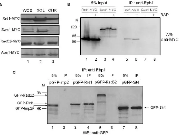
2.5.1 Rrd1 is associated with the chromatin and interacts with Rpb1 ... 47
2.5.2 Rrd1 associates with the CTD of Rpb1 and alters its structure in response to rapamycin ... 50
2.5.3 Rrd1 alters the GST-CTD structure in response to 4-NQO, but not MMS ... 56
2.5.4 Rrd1 directly alters the structure of the CTD in vitro ... 57
2.5.5 Comparison of RNA pol II occupancy at rapamycin-responsive genes ... 59
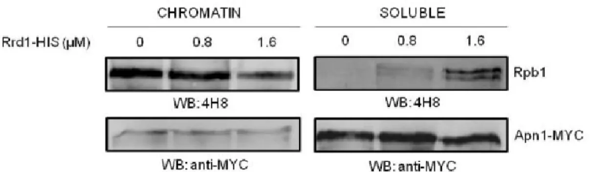
2.5.6 Purified Rrd1 stimulates the release of chromatin-bound RNA pol II in vitro ... 60
2.6 Discussion ... 62
2.7 Conclusion ... 66
2.8 Acknowledgments ... 67
3 ARTICLE #2 ... 73
3.1 Authors’ contributions ... 73
3.2 Abstract ... 74
3.3 Introduction ... 75
3.4 Materials and methods ... 77
3.4.1 Strains, media, plasmids and antibodies ... 77
3.4.2 Analysis of degradation of Rpb1 from whole cell extracts ... 77
3.4.3 Co-immunoprecipitation ... 78
3.4.4 Spot test analysis ... 78
3.4.5 Extraction of chromatin-associated proteins ... 78
3.4.6 In vitro chromatin assay ... 79
3.5 Results and discussion ... 80
3.5.1 Rrd1 is required for efficient RNAPII degradation in response to rapamycin ... 80
3.5.2 Rapamycin induces degradation of Rpb1 independent of ubiquitylation ... 85
3.6 Acknowledgment ... 90 3.7 References ... 91 4 ARTICLE #3 ... 95 4.1 Authors’ contribution ... 95 4.2 Abstract ... 96 4.3 Introduction ... 97
4.4 Material and methods ... 99
4.4.1 Cells, culture conditions and siRNA transfection ... 99
4.4.2 Western blotting ... 99
4.4.3 Colony formation assays. ... 100
4.4.4 Chromatin assay ... 100
4.4.5 Isolation of RNA and RT-PCR ... 101
4.4.6 Mitochondrial fractionation ... 102
4.4.7 Cell cycle analysis ... 102
4.4.8 Purification of PTPA-associated proteins and co-immunoprecipitation ... 102
4.4.9 Plasmids ... 103
4.4.10 Generation of knockout mice and genotyping ... 103
4.5 Results ... 105
4.5.2 PTPA siRNA diminishes colony formation ... 106
4.5.3 RNA pol II accumulates on chromatin after PTPA knockdown ... 107
4.5.4 The growth defect in PTPA knockdown cells is p53-independent ... 109
4.5.5 PTPA knockdown cells accumulate in G1 ... 111
4.5.6 PTPA shRNA knockdown cells do not exhibit an rrd1∆-like phenotype ... 112
4.5.7 PTPA does not stably interact with other proteins and shows no evidence of post-translational modifications ... 115
4.5.8 Distribution of PTPA in the mouse organs ... 120
4.5.9 Attempt to generate a PTPA knockout mouse ... 120
4.6 Discussion ... 123
4.7 References ... 127
5 DISCUSSION ... 130
5.1 Yeast Rrd1 ... 130
5.1.1 Rrd1 and RNA pol II ... 130
5.1.2 RNA pol II loss ... 134
5.1.3 Rrd1 and TOR ... 135 5.1.4 Model ... 135 5.2 PTPA in mammals ... 138 5.2.1 PTPA knockdown ... 138 5.2.2 PTPA substrates ... 139 5.2.3 PTPA in mice ... 140 5.3 Conclusion ... 141 REFERENCES ... 143
LIST OF FIGURES
Chapter 1
Figure 1.1: Multiple sequence alignment between PTPA proteins ... 12 Figure 1.2: PP2A holoenzyme composition ... 15 Figure 1.3: Peptide bond preceding a proline adopts the cis or trans conformation ... 19 Figure 1.4: Possible modifications of the CTD ... 27
Chapter 2
Figure 2.1: Rrd1 is associated with the chromatin and interacts with Rpb1. ... 48 Figure 2.2: Analysis of the GST-CTD and its interaction with Rrd1 ... 51 Figure 2.3: rrd1Δ mutants are unable to induce conformational changes to the
GST-CTD in response to rapamycin... ... 55 Figure 2.4: 4-NQO, but not MMS, induces structural changes onto the GST-CTD ... 57 Figure 2.5: Purified recombinant Rrd1 alters the structure of purified GST-CTD in
vitro...………….58 Figure 2.6: Comparison of RNA pol II occupancy at the indicated target genes in the parent and rrd1Δ mutant strain in response to rapamycin treatment. ... 60 Figure 2.7: Purified recombinant Rrd1 dissociates Rpb1 from the chromatin in
vitro... ... 61
Chapter 3
Figure 3.1:rrd1Δ or tor1-1 mutants are unable to efficiently degrade RNAPII in response to rapamycin... ... 81 Figure 3.2: Rrd1 is required for the release of RNAPII from chromatin. ... 84 Figure 3.3: Ubiquitylation of RNAPII is not required for its response to
Chapter 4
Figure 4.1: Reduction of PTPA by siRNA knockdown in HCT116 cells. ... 106
Figure 4.2: PTPA siRNA diminishes colony formation. ... 107
Figure 4.3: RNA pol II accumulation in PTPA knockdown HCT116 cells. ... 108
Figure 4.4: p53-independent cell death in PTPA knockdown cells. ... 110
Figure 4.5: PTPA knockdown cells accumulate in G1 phase. ... 112
Figure 4.6: PTPA knockdown cells by shRNA does not lead to sensitivity to 4-NQO, MMS or H2O2 or resistance to rapamycin. ... 113
Figure 4.7: PTPA knockdown in various cell lines. ... 114
Figure 4.8: FLAG-HA-hPTPA overexpression in HeLa cells. ... 116
Figure 4.9: PTPA protein does not stably interact with any protein. ... 118
Figure 4.10: PTPA do not interact with the β subunit of the CCT protein. ... 119
Figure 4.11: PTPA protein distribution in mouse organs. ... 120
Figure 4.12: No germline transmission after mouse genotyping. ... 122
Chapter 5 Figure 5.1: Proposed model of Rrd1 activity on RNA pol II ... 137
LIST OF ABBREVIATIONS
4EBP1: 4E binding protein 1 4-NQO: 4-nitroquinoline 1-oxide 6-AU: 6-azauracil
6-4PP: 6-4 photoproduct AP: Apurinic /apyrimidinic ATP: Adenosine triphosphate
Avo1/2/3: Adheres voraciously 1/2/3 BER: Base excision repair
Ca2+: Calcium
CD: Circular dichroism
CDK: Cyclin-dependent kinase
ChIP: Chromatin immunoprecipitation CPD: Cyclobutane pyrimidine dimers
CPSF: Cleavage and polyadenylation specific factor CSA/B: Cocayne syndrome factor A/B
CTD: C-terminal domain Cyps: Clyclophilins
DNA: desoxyribonucleic acid DSB: double-strand break
DSIF: DRB sensitivity-inducing factor ELL: eleven–nineteen lysine-rich leukemia EGF: Epidermal growth factor
FDA: Food and drug administration FKBP: FK-506 binding protein
GEF: General elongation factor GFP: Green fluorescent protein GG-NER: Global genome NER GST: Glutathione S-transferase GTF: General transcription factor GTP: Guanine triphosphate HAT: Histone acetyltransferase HDAC: Histone deacetylase HR: Homologous recombination Inr: Initiator element
KDa: Kilodalton
Kog1: Kontroller of growth 1
LCMT1: Leucine carboxyl methytransferase 1 MAPK: Mitogen-activated protein kinase Mg2+: Magnesium
miRNA: micro RNA
MMS: Methyl-methane sulfonate MMR: Mismatch repair
mRNA: messenger RNA
Msn2/4: multicopy suppressor of SNF1 mutation 2/4 NaAs: Sodium arsenite
NELF: Negative elongation factor NER: Nucleotide excision repair NHEJ: Non-homologous end-joining NMR: Nuclear magnetic resonance
Npr1: Nitrogen permease reactivator 1 Pcf1: Polyadenylation cleavage factor 1 PDK1: Phosphoinositide-dependent kinase 1 PI3K: Phosphatidylinositol 3-kinase
PIC: Pre-initiation complex PKC: Protein kinase C
PME-1: PP2A-specific methylesterase 1 PP2A: Protein phosphatase 2A
PP2AA: PP2A scaffolding subunit PP2AB: PP2A regulatory subunit PP2AC: PP2A catalytic subunit PP2AD: PP2A core dimer
PPIase: Peptidyl prolyl isomerase PSF: Protein-associated splicing factor
P-TEFb: Positive transcription-elongation factor b PTPA: PP2A phosphatase activator
Rheb: Ras homologous enriched in brain
Rictor: Rapamycin-insensitive companion of TOR Rtg1/2/3: Retrograde regulation 1/2/3
RNA: ribonucleic acid RNAi: RNA interference
RNA pol II: RNA polymerase II ROS: Reactive oxygen species
Rrd1/2: Rapamycin resistant deletion 1/2 rRNA: Ribosomal RNA
Ser/Thr: Serine/Threonine SSD: Single-strand damage shRNA: Small hairpin RNA siRNA: Small interfering RNA snRNA: Small nuclear RNA snoRNA: Small nucleolar RNA SUMO: Small ubiquitin-like modifier Swi/SNF: Switch/Sucrose nonfermentable Tap42: Type 2A associated protein-42kDa Tip: Tap42-interacting protein
TBP: TATA box binding protein TC-NER: Transcription-coupled NER TOR: Target of rapamycin
tRNA: Transfer RNA TSC: Tuberous sclerosis UV: ultraviolet
ACKNOWLEDGMENTS
First and foremost, I would like to thank my director; Dr Dindial Ramotar for giving me an opportunity to be a part of his dynamic team and also for providing me with the resources and encouragement throughout my project. I learned more than I expected and this knowledge will always follow me.
I am especially grateful to Dr Jim Daley for his support throughout my Ph.D. program and great help with this thesis. Thank you for transmitting me your passion for science; your advices will always be an immense inspiration in my life. Your silliness made me feel more ″comfortable″ in the lab. All those memorable moments will always remain in my memory. A heartfelt ‘’Thank you’’ to the international Dr Jeremie Poschmann for those stimulating discussions as well as for your ideas, legendary smell and precious friendship.
I would like to thank Dr Julie Douville and Anick Leduc for teaching me the basics of science and enlightening me with the first glance of research. I also want to express my thank you to all the present and past members of the lab for creating a dynamic work atmosphere. Also, I wish to thank my lab-neighbours Dr Jasmine Lefebvre and Dr Marie-France Lusignan for those great discussions over the lunches and their friendly support. Thank you to Dr Julie Ross and Charles-Étienne Lebert-Ghali for the productive exchange of dialogues and great help with the mice work.
My sincere thanks to Dr Elliot Drobetsky for his support during my stay at the research centre. Thank you for those animated discussions and for taking time off your busy schedule to write me all those reference letters. Thank you to Dr Bachir El
Affar and Helen Yu for their expertise and help with the protein purification project. I would like to express my special thanks to the members of the animal facility department for their patience while teaching me all the basics of working with mice.
Thank you to my jury members: Dr Martine Raymond, Dr Janos Filep, Dr Jacques Côté and Dr Pierre Belhumeur for reading and analyzing this thesis and for their valuable comments and constructive criticism.
In a more personal way, thank you to my mother Lorraine and her boyfriend Yves for their patience, support and great dinners. Thank you to my father Denis for moral support as well as my brother David for being so silly. A note of thanks to the rest of my family especially my grandfather who has taught me to always look ahead in life.
Finally, a special thank you to Sonu for sharing my ups and downs by being a part of my life every single day. I also want to thank all my close friends for being supportive and loyal. A coffee, a dinner or a discussion was always helpful in those difficult moments. I would like to say that I am very fortunate to know that I can always rely on you and that your friendship means a lot to me.
Last but not the least my gratitude is also extended to the molecular biology program (University of Montreal) for supporting my work through several scholarships.
CHAPTER 1
1 INTRODUCTION
According to the Canadian Cancer Society, cancer represents the leading cause of death in Canada since 2007. Studying cell metabolism is crucial both for a better understanding of the disease and for generating possible treatments. Cancer is a very complex disease and many factors can contribute to its development. Lifestyle, environmental events and heredity are all possible causes for cancer although it is often a combination of multiple factors. In order to circumvent these events, cells have developed a plethora of mechanisms to ensure regulated proliferation and growth and to maintain genomic integrity.
Deregulation of two classes of genes, named oncogenes and tumor suppressor genes, can lead to the onset of cancer. Inappropriate upregulation of oncogenes induces unregulated cell proliferation, leading to cancer. For example, activating mutations of the proto-oncogene Ras, a gene normally quiescent, are found in about 20% of all tumours and lead to uncontrolled growth of the cells [1, 2]. Current drugs used in cancer treatment target these genes or their products [3, 4]. Tumor suppressor genes function in pathways that protect against mutation or unregulated growth. Inactivating mutations in these genes can lead to tumorigenesis. Examples of tumor suppressors include genes involved in DNA repair, apoptosis, and transcription factors activated by cellular stress that induce cell cycle arrest in order to ensure DNA integrity [5]. Half of all cancers involve alteration of the important and best described tumor suppressor gene p53 [6]. Mutations in oncogenes and tumor suppressor genes arise when DNA damage is not repaired correctly, leading to irreversible mutations that alter protein function and regulation. Two categories of sources can cause DNA
damage: exogenous and endogenous. Exogenous DNA damaging agents include ultraviolet (UV) light from the sun, radiation such as x-rays or γ-rays, viruses, toxins or chemicals, and the main endogenous source is reactive oxygen species (ROS) [7, 8]. Cells have developed specific mechanisms to repair each type of DNA lesion in order to prevent carcinogenesis. Single-strand damage (SSD) is characterized by damage on only one strand of the DNA double helix [9]. Excision repair mechanisms use the intact strand as a template to repair the defective one. In base excision repair (BER), a damaged base is removed by a DNA glycosylase, resulting in an apurinic/apyrimidinic (AP)-site that is cleaved by an AP-endonuclease. Synthesis of the DNA is performed by a DNA polymerase and a DNA ligase seals the nick to complete repair [10]. Nucleotide excision repair (NER) repairs lesions caused by ultraviolet light and can be divided into global genome NER (GG-NER) or transcription-coupled repair (TC-NER). These two pathways differ in how the lesion is recognized, but share the later steps in which the lesion is excised and the resulting gap is filled [11]. Finally, mismatch repair (MMR) recognizes erroneous insertion, deletion or mis-incorporation of bases and repairs the wrong nucleotides with the correct ones [12].
Double-strand breaks (DSBs) are lesions of both strands of the DNA and can be repaired using either non-homologous end-joining (NHEJ) or homologous recombination (HR) [13]. In NHEJ, cells directly rejoin both ends of the break. This pathway is mostly used before DNA replication, when a sister chromatid is not available to serve as a template [14, 15]. On the other hand, HR uses the identical sequence from replication as a template to accurately repair the break.
Better understanding of proteins involved in the oxidative stress response is important for prevention of cancer and other diseases. Oxidative stress is caused by an imbalance between antioxidants and ROS such as free radicals or peroxides. Free radicals are unstable molecules with an unpaired electron that will rapidly react with proteins, lipids or DNA. Several types of DNA damage result from ROS including oxidized bases and strand breaks [7]. A main objective of our lab was to discover new genes important in the oxidative stress response. This was investigated in the budding yeast Saccharomyces cerevisiae, since this eukaryotic cell is genetically easy to manipulate. Homology between key proteins from yeast and mammalian cells also strengthens the notion of using yeast as model [16].
A yeast screen revealed that cells with a mutation in the RRD1 gene were hypersensitive to 4-nitroquinoline 1-oxide (4-NQO) but resistant to ultraviolet C (UVC). 4-NQO causes a variety of DNA damage such as bulky adducts and oxidative stress. The metabolic activated form 4-hydroxyaminoquinoline interacts with DNA to form stable quinoline-purine monoadducts repaired by NER [17-19]. UVC (280-100 nm) engender bulky adducts on DNA [20] also repaired by NER [21]. Further characterization of the rrd1 deletion in different genetic backgrounds showed the same phenotype [22]. rrd1Δ mutants are sensitive to 4-NQO [22, 23], vanadate, cycloheximide, ketoconazole and high concentration of Ca2+, they are resistant to caffeine [24] and show defect in cell cycle progression and morphology [25]. Surprisingly, rrd1Δ mutant cells were later characterized to be resistant to rapamycin, a drug that inhibits the Target of rapamycin (TOR) signalling pathway (discussed in detail below) [24]. Our lab became interested in understanding RRD1 function in the
cell since it seemed important for both genomic integrity and cell growth. The first chapter of my thesis will review literature on RRD1 and related topics important for understanding its function in the cell.
1.1 RAPAMYCIN AND TOR
In Saccharomyces cerevisiae, rrd1Δ mutants are resistant to rapamycin, hence the gene name rapamycin resistant deletion 1 [24]. Rapamycin treatment is known to mimic starvation conditions in yeast by inhibiting the master kinase Target of Rapamycin (TOR). Genome array studies have revealed that expression of multiple genes is altered by rapamycin treatment, including repression of ribosomal genes and activation of nitrogen source utilization genes [26].
1.1.1 Rapamycin
Rapamycin is a bacterial product found on Easter Island that interacts with the isomerase FKPB12 (Fpr1 in S. cerevisiae) to inhibit TOR [27]. Rapamycin is widely used in transplant therapy since it inhibits the proliferation of T cells. Rapamycin has several effects on the immune system such as inhibiting type I interferon production in plasmacytoid dentritic cells [28], modulation of T cell trafficking [29] and regulating Foxp3 expression in regulatory T cells [30]. On the other hand, recent studies have snown that rapamycin can increase the generation of CD8+ memory T cells [31-33]. Late administration of rapamycin can increase lifespan in mice [34]. Understanding the exact mechanism of TOR inactivation in T cells represents the next challenge of future work.
Rapamycin has also been used as an anticancer treatment acting as a cytostatic agent on several cancer cell lines. It can also sensitize cells to apoptosis when treated in combination with other chemotherapeutics agents [35-37]. In past years, analogs of rapamycin have been synthesized to circumvent the poor water solubility and low bioavailability of rapamycin [38]. New molecules such as temsirolimus and everolimus have recently been approved by the Food and Drug Administration (FDA) to treat advanced/metastatic renal cell carcinoma [39, 40].
1.1.2 TOR
The target of rapamycin (TOR) is a conserved Ser/Thr protein kinase which represents the catalytic activity of two complexes in the cell, TOR complex 1 (TORC1) and TOR complex 2 (TORC2) [41, 42]. The two complexes are formed of distinct and shared proteins and are responsible for regulating cellular processes in response to the environment. In mammalian cells, one TOR gene is present whereas in yeast Tor1 and Tor2 form TORC1 and TORC2 respectively [42, 43]. In mammalian cells, acute treatment with rapamycin inhibits mTORC1 only [27] through inhibition of its interaction with Raptor (regulatory associated protein of mTOR) [44]. In sustained treatment, both mTORC1 and mTORC2 are inhibited.
1.1.3 Yeast TOR
In yeast, TOR is a large protein present in two distinctive complexes. The TOR complex 1 (TORC1) includes Tor1 or Tor2, the scaffolding protein kontroller of growth 1 (Kog1) and the nutrient sensitive permease sorting factor Lst8 [45, 46]. On
the other hand, the TOR complex 2 (TORC2) includes Tor2, Avo1 (adheres voraciously), Avo2, Avo3 and Lst8 [47].
The TOR signalling pathway responds to nutrients such as carbon or nitrogen in order to promote cell growth [48]. TOR regulates gene expression depending on the availability of the nitrogen source. For example, in the presence of poor nitrogen sources such as urea or proline, a subset of genes involved in processing these sources are upregulated following inactivation of TOR [49]. Treatments with rapamycin or inhibition of TOR proteins results in reduced ribosome biogenesis, upregulation of autophagy, transcriptional modifications and increased mRNA turnover [45]. TORC2 regulates the cell-cycle-dependent polarization of the actin cytoskeleton, and this function of TORC2 is rapamycin-insensitive [50].
Both yeast Tor1 and Tor2 upregulate protein synthesis through activation of the translation initiation factor eIF4E as well as through transcriptional activation of ribosomal proteins. Inhibition of autophagy mediated by TOR phosphorylation of Apg1 is a mechanism to promote protein stability [51]. TOR also controls protein ubiquitylation by keeping nitrogen permease reactivator 1 (Npr1) in an inactive form [52]. The function of Npr1 is to stabilize plasma membrane amino acid transporters such as Bap2 [53], Mep2 [54], Tat2 [52] or Gap1 [55] against ubiquitylation-dependent degradation. Finally, TOR is involved in regulating transcription through the inhibition of starvation specific genes. For example, TOR phosphorylates the GATA-type transcription factor Gln3 in order to maintain it in the cytoplasm. When TOR is inhibited, during rapamycin treatment or starvation conditions, unphosphorylated Gln3 translocates to the nucleus and activates the transcription of
genes involved in the metabolism of secondary nitrogen sources [26, 56]. TOR also inhibits general stress transcription factors by keeping them in the cytoplasm, such as the multicopy suppressor of SNF1 mutation 2-4 (Msn2 and Msn4) [57] and the heterodimeric retrograde regulation 1-3 (Rtg1-Rtg3) [58].
TOR is important for the rapid regulation of a variety of protein phosphatase 2A (PP2A) complexes. The type 2A associated protein-42kDa (Tap42) binds to Sit4 phosphatase, a PP2A-related catalytic subunit in order to inhibit the dephosphorylation of both Npr1 and Gln3. The interaction between Tap42 and Sit4 is promoted by TOR phosphorylation of Tap42-interacting protein (Tip41). Treatment with rapamycin or inhibition of TOR leads to inactivation of Tip41, resulting in its binding to Tap42, which releases Sit4. Free Sit4 is then able to dephosphorylate transcription factors as well as Tip41, creating a fast response to stress conditions [59]. Our lab and others have shown that Rrd1 interacts with Sit4 phosphatase, and this interaction will be discussed in a later section [23, 60].
1.1.4 Mammalian TORC composition
In mammalian cells, both TOR complexes contain mTOR, mLST8/GβL and deptor [41, 42]. Deptor acts as an inhibitor of both mTORC1 and mTORC2 [61, 62], whereas mLST8/GβL binds to the mTOR kinase domain [63]. Raptor, a scaffold protein that links mTOR kinase to mTORC1 components [64], and PRAS40, an inhibitor or competitive substrate of mTORC1, are both specific to the mTORC1 complex. On the other hand, Rictor (rapamycin-insensitive companion of mTOR) and
mSin1, which are important for mTORC2 assembly and signaling and PRR5/protor, are part of the mTORC2 complex [65].
1.1.4.1 mTORC1
Anabolism is promoted through integration of both extra- and intracellular signals by the mTORC1. Nutrients and growth factors modulate mTORC1 activity in order to increase protein synthesis, cell growth, cell proliferation and cell metabolism [66]. Inactivation of mTORC1 leads to macroautophagy, or the degradation of cell proteins or organelles into amino acids or simple molecules [67].
Growth factors signal through phosphatidylinositol 3-kinase (PI3K) to activate phosphoinositide-dependent kinase 1 (PDK1), leading to phosphorylation of Akt and subsequent inhibitory phosphorylation of the Tuberous Sclerosis Complex (TSC) (including TSC1 and TSC2). TSC2 contains a GTPase-activating protein [31] and has been shown to stimulate the Ras homologue enriched in brain (Rheb). TSC1 stabilizes the TSC2 protein [68]. Inactivation of TSC complex allows the GTP-bound form of Rheb to interact and activate mTORC1 [69]. Amino acids, such as leucine, also activate mTORC1 through Rag proteins. These proteins bind to raptor and promote the interaction between Rheb and mTORC1 [70].
Finally, mTORC1 is important for ribosomal protein synthesis [71]. The ribosomal S6 kinase 1 (S6K1) can be phosphorylated on Thr389 and the eukaryotic initiation factor 4E binding protein 1(4EBP1) can be phosphorylated on multiple sites (Thr37/46, Thr70 and Ser65) by mTORC1 kinase activity to promote cell growth and
proliferation. mTOR activation is usually measured by assaying these downstream substrates [72].
1.1.4.2 mTORC2
The main substrate of mTORC2 is the serine/threonine protein kinase Akt, which promotes cell proliferation, survival and migration [73]. It is interesting that Akt regulation is both upstream (mTORC1) and downstream (mTORC2) of mTOR [41]. mTORC2 activates Akt through phosphorylation on serine 473 which allow the kinase to phosphorylate other substrate such as mTORC1 [74]. mTORC2 is also known for controlling actin cytoskeleton organization during cell growth through the activation of protein kinase C α (PKC-α) [75, 76].
1.1.5 Rrd1 and TOR
Rrd1 forms a complex with the TOR pathway members Tap42 and Sit4 [23, 60, 77]. Overexpression of Tap42 suppresses the rapamycin resistance seen in rrd1Δ mutant strains [77]. Moreover, both Sit4 and Rrd1 work in the same pathway to mediate resistance to oxidative stress induced by 4-nitroquinoline 1-oxide (4NQO) and UVA [23].
As mentioned previously, rapamycin also induces a reorganisation in the transcription profile of genes [56]. Our lab showed that rrd1∆ mutant cells fail to downregulate expression of genes encoding ribosomal proteins such as RPS26A, RPL30 and RPL9 when treated with rapamycin. Preliminary data also revealed that
RNA pol II was not degraded as efficiently as in a WT strain following rapamycin treatment [78]. So far, the exact mechanism by which Rrd1 deletion leads to rapamycin resistance remains unknown, and investigating this phenomenon is a main goal of this thesis.
1.2 RRD1/PTPA OVERVIEW
RRD1 homologs exist in a variety of species such as Xenopus laevis [79], Drosophila melanogaster, and Schizosaccharomyces pombe [80]. It is highly conserved from yeast to human, showing 40% sequence identity with its human homolog PTPA (phosphatase two A phosphatase activator). An alignment is shown in Figure 1.1.
S. cerevisiae also contains an RRD1 homolog called RRD2 showing 25% sequence identity. rrd2Δ mutant strains are also resistant to rapamycin and caffeine, but the phenotypes are weaker than rrd1Δ [24]. Deletion of both genes is lethal [24, 25], suggesting functional redundance and revealing their critical importance for the cell. Expression of the mammalian counterpart PTPA can rescue lethality in the
rrd1Δ rrd2Δ double mutant [24, 81]. We chose to focus on Rrd1 because the rrd2∆
rapamycin phenotype is less severe, rrd2∆ cells are not sensitive to oxidative stress, and a mammalian homolog for RRD2 has not yet been identified.
Figure 1. Alignmen JALVIEW (hPTPA) Schizosac (s.cerevis .1: Multiple nt was perf W [83]. Pr , Xenopus ccharomyces siae) are pres
e sequence a formed with roteins from laevis (xP s pombe sented. alignment b h CLUSTAL m Mus mus PTPA), Dro (s.pombe) between PTP L X2 [82] sculus (mPT osophila me and Sacch PA proteins and represe TPA), Hom elanogaster haromyces s ented with mo sapiens (dPTPA), cerevisiae
1.2.1 PTPA
PTPA is encoded by a single gene on chromosome 9q34 in human (chromosome 2 in mouse) and consists of 10 exons and 9 introns [84]. The transcription factor yin yang 1 (YY1) is involved in the regulation of transcription of the PTPA gene [85]. PTPA also possesses multiple splicing sites resulting in seven distinctive products leading to the expression of 4 protein isoforms. However, only 2 of these proteins are detectable in vivo [86].
1.2.2 Rrd1/PTPA structure
Crystal structures of yeast Rrd1 and Rrd2 and human PTPA were solved using truncated peptides where non-structured regions were removed. Interestingly, the overall structures of all three proteins were very similar and were organized into an α-helical compact structure [87]. Comparison with known structures revealed no obvious similarity with any other previously analyzed proteins [88].
Mammalian truncated PTPA protein contains 17 α helices and 4 short β strands. The structure is organized in 3 main domains: the core, the lid and the linker. The core is linked to the lid, located at the C-terminus, by the linker forming a large cleft. The structure also revealed a deep pocket of conserved amino acid residues between the core domain and the linker possibly representing a protein interaction domain [88]. It was previously reported that the conserved region 200GVWGLD205 is essential for the peptidyl prolyl isomerase activity (discussed in detail below) as well as activation of PP2A [80, 89].
1.2.3 PP2A
As indicated by its name, PTPA was first described as an activator of protein phophatase 2A (PP2A) in rabbit skeletal muscle and Xenopus laevis oocytes [79]. The weak phosphatase activity of PP2A could be stimulated by PTPA in an ATP and Mg2+-dependent manner in vitro [90]. Mutational analysis has also shown that specific amino acids (V209D, E270A,V281D, G290D and M294D) are important for both the interaction between PP2A and PTPA and the ATPase activity of the complex [88]. The mechanism by which this occurs is still poorly understood [79, 90]. PP2A complexes are conserved serine/threonine phosphatases ubiquitously expressed in the cell and are important for the regulation of numerous signalling pathways [91]. Deregulation of PP2A is associated with cancer and Alzheimer’s disease [92-95]. The PP2A heterodimeric complex (Figure 1.2) is formed of a core dimer (PP2AD) containing a structural or scaffolding A subunit (PP2AA) and a catalytic C subunit (PP2AC), and each subunit can be found in 2 distinct isoforms (α or β). The core dimer can also associate with a regulatory B subunit (PP2AB) to form the heterotrimeric holoenzyme. There are 4 structurally different families of PP2AB : B (PR55), B’ (PR61), B’’ (PR48, PR72 or PR130, G5PR) and B’’’ (PR93 or PR110) and each family consists of 2 to 5 different isoforms [96].
Figure 1. Th elongatio the conse have been carcinoge blocking Th and the lo B subuni scaffoldin subunits independ .2: PP2A ho he scaffoldi on-A subunit erved ridge n found in s enic toxin o active site o he diversity ocalization o its interacts ng subunit, t give rise dent function oloenzyme c ing subunit t-TOR) repe of HEAT re some tumor okadaic acid of the catalyt of the regu of the holoe differently w the catalytic to over 70 ns in the cell composition PP2AA is eats, and the
epeats 11-15 s [98, 99]. T d [100] that tic subunit [9 ulatory subu nzyme in th with the cor subunit or b 0 different [91]. n composed e catalytic s 5 [97]. Muta The core dim
inhibits PP 97].
unit is impor he cell [101, re dimer, som both. The dif
heterotrimer of 15 HE subunit PP2A ations in thi mer is the m P2A by inte
rtant for sub 102]. Each metimes int fferent comb ric holoenz EAT (huntin AC interacts is interaction main target o eracting with bstrate speci of the regul teracting wit binations of zymes displ ngton-s with n site of the h and ificity latory th the these aying
1.2.3.1 Post-translational modifications
Methylation of the PP2A holoenzyme is an essential mechanism of regulation [103-106]. Methylation of the carboxy-terminal Leu309 of the catalytic subunit (PP2AC) is important for recognition by some of the regulatory subunits (PP2AB) such as the regulatory subunit Bα [107, 108]. There is also evidence that inhibition of the methylation site in yeast leads to decreased formation of the holoenzyme [100]. Methylation is mediated by a conserved protein, a PP2A-specific leucine carboxyl methyltransferase (LCMT-1), and demethylation is catalysed by a PP2A-specific methylesterase (PME-1) [103, 109, 110]. Levels of this methyltransferase vary during the cell cycle, suggesting a role in cell-cycle regulation [91, 111]. The methylation status is critical for the differentiation of neuroblastoma cells and could possibly play an important role in Alzheimer disease [112, 113].
The catalytic subunit C of PP2A is also targeted for phosphorylation on Tyrosine 307 by tyrosine kinases such as pp60v- src, pp56lck, epidermal growth factor (EGF) and insulin receptors. Phosphorylation results in inactivation of the enzyme [114] and it has been associated with Alzheimer disease [115]. Phosphorylation of a regulatory B’ subunit (B56α) by the serine/threonine kinase PKR [116] seems to be important for apoptosis. In short, phosphorylation of B56α on Serine 28 activates the phosphatase activity required for dephosphorylation of Bcl2 and inhibition of apoptosis [117, 118].
1.2.4 Rrd1/PTPA and PP2A
In yeast, Rrd1 was found to interact with the PP2A-like phosphatases Pph3 and Ppg1 [77]. It was also shown that Rrd1 and Pph3 act synergistically to induce rapamycin resistance in yeast [78]. The interaction with Pph3 was confirmed using affinity purification coupled with mass spectrometry analysis in mammalian cells [119].
Deletion of PTPA homologs in yeast results in accumulation of PP2A and PME-1 complex and decreased methylation of the catalytic subunit (PP2AC) [120]. Also, inactive PP2A can be re-activated by PTPA in a Mg2+/ATP dependent manner in vitro [79], suggesting that PTPA inhibits the methyltransferase activity of PME-1 [110]. It remains unknown how PTPA performs this activation, but an attractive possibility is that it acts as a cis/trans peptidyl prolyl isomerase on Proline 190 close to the active site of the catalytic C subunit of PP2A and this function will be discussed later [87, 89].
Finally, a recent study suggested that depletion of PTPA with RNAi results in cell transformation caused by a defect in PP2A catalytic subunit C methylation. This defect altered the assembly of the catalytic subunit C with the scaffolding subunit A and suggests a novel role for PTPA as a tumor suppressor [121].
1.2.5 PTPA and apoptosis
To gain more insight into a possible function of PTPA, our laboratory previously monitored the biological response following transient overexpression of
PTPA labelled with the green fluorescent protein (GFP). The results showed cell death of the PTPA-overexpressing cells via p53-independent apoptosis in a time-dependant manner. This apoptosis was independent of the mitogen-activated protein kinase (MAPK). Surprisingly, inhibition of PP2A with okadaic acid did not prevent the PTPA overexpressing cells from dying through apoptosis. The exact mechanism leading to apoptosis remains unknown, but nonetheless these data reveal that a specific level of PTPA is required for normal homeostasis of the cells [122].
1.2.6 Rrd1/PTPA as a peptidyl prolyl isomerase (PPIase)
A key breakthrough was the discovery that PTPA and its yeast homolog, Rrd1, possesses peptidyl prolyl cis/trans isomerase (PPIase) activity. PPIases are enzymes that convert proline residues between their two distinct isoforms, cis or trans (Figure 1.3). PPIases are highly conserved from yeast to human [123]. These ubiquitous enzymes can be divided into four structurally different families: cyclophilins (Cyps), FK-506 binding proteins (FKBPs), parvulins and the Ser/Thr phosphatase 2A (PP2A) activator PTPA. The catalytic domain of PTPA is an α-helix fold whereas the other PPIases are characterized by a central β-sheet [87, 124, 125].
Typically, the peptide bond linking amino acid residues in a protein adopts the trans isoform since this is the less energetic conformation as compared to the cis conformation. Interestingly, proline residues are the only amino acids in which both conformations are relatively energetically equivalent [126]. The possibility of having two distinct structures (cis and trans) can act as a molecular switch, similar to other post-translational modifications, enabling proteins to perform different functions
within the dynamic Figure 1 conform Th drug cyc immunos these dru prevent t pathway. budding y lethal [13 FKBPs o folding, a e cell (Figur processes in 1.3 : Pept ation he Cyps and closporine suppressant ugs do not the formatio Moreover, yeast since t 30]. Analys only perform and therefore re 1.3). PPIa n the cell [12 tide bond d FKBPs hav A uses th and anti-can directly inh on of a tern these 2 fa the disruptio sis througho m non-essent e interest in t
ases are know 27].
preceding
ve been wide he cyclophi
ncer rapamy hibit the iso nary comple amilies of P on of each e out the year
tial and redu these molecu wn to be acc a proline ely studied s ilins as a ycin targets omerase acti ex leading t PPIase are enzyme indiv rs has sugg undant roles ules as thera celerating ag adopts th since the im target [12 FKBPs [12 ivity of PPI to dysfuncti dispensable vidually or a gested that s as chapero apeutic targe
gents that reg
he cis or mmunosuppre 28] whereas 29]. Interesti Iases but in ion of a sp for viabili all together i cyclophilins ones or in pr ets has decrea
gulate trans essant s the ingly, nstead ecific ity in is not s and rotein ased.
Another PPIase, Pin1, is a member of the parvulin family and has been found to play roles in a variety of cellular processes. Pin1 is the only enzyme that can isomerise a specific motif formed of phosphorylated Ser/Thr-Pro [131]. Pin1 cis/trans isomerisation is involved in the control of cell growth regulation, genotoxic stress, and the immune response. Deregulation of Pin1 has been linked to Alzheimer’s disease, cancer and aging [132]. Interestingly, it has been shown that Pin1 binds to a pSer-Pro motif on the C-terminal domain (CTD) of the large subunit of RNA pol II and regulates the phosphorylation status of this domain through inhibition of the phosphatase FCP1 and stimulation of the kinase cdc2/cyclin B [133-135].
The PPIase activity of PTPA was discovered when PTPA was shown to isomerize synthetic PP2A catalytic subunit peptides in vitro [87, 89]. This led to a model in which isomerisation by PTPA could activate the phosphatase activity of PP2A. It is likely that PTPA and its yeast homolog Rrd1 isomerize other substrates, and identification of such substrates could explain its role in 4-NQO sensitivity and rapamycin resistance. Our lab previously showed that transcription of a subset of genes is deregulated in rrd1∆ cells [78]. This, along with the knowledge that Pin1 isomerizes the CTD of RNA pol II, led us to investigate whether the CTD is also an Rrd1 substrate. We found that isomerisation of the CTD by Rrd1 does indeed play a major role in transcription regulation, and these data will be presented in Chapter 2. To provide the necessary background, a review of the transcription mechanism in eukaryotic cells is presented in the next section.
1.3 TRANSCRIPTION 1.3.1 Overview
Transcription is the mechanism by which RNA is synthesized from the DNA template. During this process, RNA polymerase reads from one strand of the double-stranded DNA, called the template strand, whereas the other strand is termed the coding strand. The template strand is read in a 3’ to 5’ direction and the RNA is synthesized from 5’ to 3’, comparable to DNA replication. The resulting RNA is single-stranded and its sequence is identical to the coding strand except that uracil is substituted for thymine. Transcription can be divided into three major steps: initiation, elongation and termination. Since it leads to gene expression and is one of the most frequent events in a cell, it is highly regulated.
In eukaryotic cells, there are three different RNA polymerases, which each transcribe a specific type of RNA. Each RNA polymerase is composed of 4 to 14 polypeptides and requires the aid of distinct additional factors to perform its function. Ribosomal RNAs (rRNA) are synthesized by RNA pol I in the nucleus and are part of the 18S, 5.8S and 28S ribosomal subunits. These subunits are required for the assembly of the full ribosome, which is involved in the translation of messenger RNA (mRNA). Transcription of the rRNA represents around 60% of the transcription in a cell. Transfer RNA (tRNA) and the 5S subunit of rRNA are transcribed by RNA pol III and account for about 10% of the total transcription [136]. tRNAs are involved in transferring each amino acid to the polypeptide chain at the ribosome during translation, and 5S rRNA is another constituent of the ribosomal complex. Finally,
RNA pol II transcribes the mRNA in the nucleus. Most mRNAs code for genes and are translated into proteins. RNA pol II also transcribes non-coding small RNAs such as small nuclear RNA (snRNA), small nucleolar RNA (snoRNA) and micro RNA (miRNA) [137]. Small non-coding RNAs are involved directly in many different cellular pathways, although the specific function of the majority of them remains unknown [138]. My thesis will focus on the regulation of RNA pol II.
1.3.2 Transcription initiation
Initiation of transcription occurs at the core promoter, which is usually located upstream of the gene and contains specific sequences that recruit initiation factors. The TATA box, which is involved in about 30% of gene transcription, is found around 25 bases upstream of the transcription start site and possesses the consensus sequence TATAWAAR [139]. The TATA box is often associated with an initiator element (Inr) and both can act synergistically to activate transcription of abundantly expressed genes [140]. Another element called the downstream promoter element (DPE) activates transcription coupled to the Inr in TATA-less promoters [141]. All these markers are important to correctly direct the pre-initiation complex [142] to the site of transcription initiation. There are also cis-acting DNA sequence such as enhancers, silencers and insulators [143] and trans-acting elements such as the RNA pol II pre-initiation complex [142], transcription factors and chromatin remodeling proteins [144]. Transcription is highly regulated and these different elements can either activate or repress transcription.
Various transcription factors join RNA pol II at gene promoters in an organized order. General transcription factors (GTFs) that are necessary for initiation include TFIIA, TFIIB, TFIID, TFIIE, TFIIF and TFIIH. The formation of the PIC occurs by stepwise recruitment of the different GTFs to the promoter region and is initiated by TFIID binding to the core promoter. TFIID is a multi-subunit complex containing 14 different factors including TBP (TATA-binding protein) [145]. TBP is known to bind to this AT-rich region and unwind the DNA, forming a single-stranded “bubble” for the transcription machinery. TFIIB and TFIIA are then recruited and stabilize TFIID at the promoter region, followed by the recruitment of the RNA pol II complex already bound to TFIIF. At this point, transcription can only initiate when TFIIE and TFIIH join the complex. The ATP-dependant helicase activity of TFIIH melts the promoter, forming an “open” initiation complex and leading to the release of RNA pol II. This is called promoter clearance and represents the beginning of transcription elongation [146].
1.3.3 Transcription elongation
Elongation is a highly regulated and critical step that is mandatory for the correct organization and integrity of the genome. This step begins when promoter escape is complete, when the new RNA associates stably with the transcription complex. TFIIB is important to stabilize this association as well as to allow elongation initiation [147, 148]. In yeast, DRB sensitivity-inducing factor (DSIF), consisting of Spt4, Spt5, and negative elongation factor (NELF) facilitates RNA Pol II pausing in the promoter-proximal region, and TFIIS also associates with the paused
polymerase [149]. This pause is necessary for capping enzymes to bind to the C-terminal domain (CTD) (discussed in depth below) of Rpb1 phosphorylated on serine 5 and Spt5 and to allow nascent RNA capping [150, 151]. Positive transcription-elongation factor-b (P-TEFb) is involved in the phosphorylation of DSIF, NELF and serine 2 on the CTD leading to productive elongation [152]. TFIIF, eleven-nineteen
lysine-rich in leukemia (ELL) and Elongin are the main factors that stimulate RNA
pol II elongation and inhibit pausing [153]. TFIIF seems to be important for RNA pol II release from a stalled state [154]. Elongin is not active until the RNA transcript is 8-9 nucleotides long and until TFIIF leaves the complex [155]. Finally, the last component of transcription elongation is topoisomerase I, which allows unwinding of the DNA throughout the process [156].
Transcribing RNA pol II adopts three different states: a pretranslocation state where the nucleotide added to the RNA chain is still in the addition site, a posttranslocation state where the addition site becomes free and a backtracked state where RNA pol II performs a retrograde motion [157, 158]. RNA pol II backtracking of one residue is favorable whereas longer backtracking leads to a possible irreversible arrest [158] and this is detailed in a later section.
Nucleosomes protect the DNA by keeping it in a tightly closed form, and this process must be reversed to let RNA pol II access the gene to be transcribed. Several factors involved in remodeling the chromatin have been identified and will be discussed briefly here. The first category includes ATP-dependent chromatin remodelling complexes that use ATP hydrolysis to modify the chromatin structure [159, 160]. In yeast, the Swi/Snf (Switch/Sucrose nonfermentable) complex moves
nucleosomes around and can repress or activate transcription depending on the situation [161, 162]. Histones chaperones, such as FACT and Spt6, regulate intracellular histone dynamics, histone storage and replication-associated chromatin assembly [163, 164].
Acetylation of histones H3 and H4 is important during transcription elongation, and histone acetyltransferases (HATs) and deacetylases (HDACs) have been found in the coding region of multiple genes [165] although they are more commonly associated with the promoter and 5’ region [166]. It has also been documented that histone methylation, mostly on H3, is important for progression of transcription [167]. One example is Set2, which interacts with active genes through its affinity for RNA pol II phosphorylated on both serine 2 and 5 of the CTD [168-170]. Finally, ubiquitylation of either histone H2B [171] or phosphorylation of histone H3 [172] are possible modification required for transcription elongation regulation.
1.3.4 Transcription termination
There are two proposed models for the termination of transcription: the torpedo model and the anti-terminator model. In the first model, cleavage of the polyadenylated site creates a new 5’-end, and exonuclease or helicase activity leads to the dissociation of RNA pol II [173]. The second model states that the appearance of the polyadenylation sequence on the RNA strand triggers a reorganisation in the binding factors, leading to a decrease in RNA pol II elongation [174]. Pausing of RNA pol II is also important for termination and occurs at the 3’-end of the gene,
10-30 nucleotides downstream from the hexanucleotide AAUAAA. This specific sequence is recognized by the CPSF (cleavage and polyadenylation specific factor) group of proteins [175-178]. RNA pol II CTD regulation is also involved in 3’-end processing, but this will be discussed in a later section.
Recent research supports a combination of both models. As mentioned previously, the torpedo model involves the activity of the yeast 5’-3’ exonuclease Rat1 (Xrn2 in mammals) in order to recruit the 3’-end processing factors, but this nuclease does not seem to be essential for cleavage at the poly(A) site [179, 180]. Xrn2 interacts with p54nrb and PSF (protein-associated splicing factor), which are involved in transcription, splicing and polyadenylation [181, 182]. Xrn2 action on the cleavage of the RNA product precedes the release of RNA pol II, as predicted by the second model [183].
1.3.5 RNA polymerase II structure
The RNA pol II holoenzyme is composed of 12 conserved subunits (termed Rpb1-12) resulting in a large complex of about 550 kDa [184]. All subunits are necessary for yeast cells to grow normally [185]. Three of the subunits are unique to RNA pol II: Rpb4, Rpb7 and Rpb9, whereas the rest are common to all RNA polymerases. The two major subunits Rpb1 and Rpb2 form the catalytic center and are homologous to subunits of the bacterial RNA polymerase [185]. Mutational analysis revealed that Rpb1 and Rpb2 are both required throughout the entire transcription process, from initiation to termination [186]. The largest subunit Rpb1 contains a unique C-terminal domain (CTD) containing a heptapeptide repeated
26-27 times Tyr-Ser-P organism 1.3.5.1 R RN occurs th Through manner. T tyrosine ( phosphata glycosylt revealed proline ( Therefore acid lead [189]. Th Figure 1. in yeast [18 Pro-Thr-Ser-ms [188]. RNA pol II C-NA pol II CT hrough direc each step o These modi (Tyr1), threo ases and 2) transferase a a third categ (Pro3 or 6) e, the possib ds to a vast his is summa .4: Possible 87]and 52 ti -Pro-Ser is h -terminal do TD regulatio ct or indirect of transcript fications inc onine (Thr4) glycosylatio and deglyco gory of CTD ) by peptid bility to regu possibility arized in the modificatio imes in mou highly conse omain on is critica t interaction tion, the CT clude: 1) ph ) and all thr on of serines osylases. R D modificati dyl prolyl c ulate the CT of combina diagram bel ons of the C
use and hum erved in euk al for correc n with differ TD is rever hosphorylatio ree serines ( s (Ser2, 5 o Research on ion, isomeris cis/trans iso TD through ations and r low (Figure CTD
man. This con aryotes and ct transcripti rent RNA pr sibly modif on and deph (Ser2, 5 or 7 r 7) and thr n Pin1 as w sation of the omerases (P modification regulates int 1.4). nsensus sequ is essential ion of gene rocessing fa fied in a sp hosphorylatio 7) by kinase reonine (Thr well as mine e peptide bo PPIases) act ns of each a teracting par uence in all s and actors. ecific on on es and r4) by e has ond of tivity. amino rtners
Rpb1 can be found in two main forms during transcription: IIa, the hypophosphorylated form and IIo, the hyperphosphorylated form. The IIa form is preferentially found at the pre-initiation site whereas the IIo form is found later in the elongation process where each repeat is phosphorylated at least once [190]. Chromatin immunoprecipitation (ChIP) analysis revealed that Ser5 phosphorylation by the cyclin-dependant kinase (Cdk)7 (Kin28 in yeast), was higher at the 5’ region of a gene [191]. This phosphorylation is required to release the mediator complex from pol II, allowing recruitment of capping enzymes [192, 193].
Once the transcription machinery leaves the initiation site, Ser2 phosphorylation by Cdk9 (Ctk1 in yeast) is initiated and is important for binding of the 3’-RNA processing machinery. This phosphorylation is most abundant at the 3’ end of the transcribed gene [194, 195]. Phosphatases such as SCP1 (Ssu72 in yeast) and Fcp1 dephosphorylate Ser5 and Ser2, respectively, and are required for the recycling of RNA pol II following transcription of a gene. Once RNA pol II reaches the polyadenylation signal, most of the Ser5 phosphorylation is gone and only Ser2 remains. In this state, pol II interacts with the polyadenylation cleavage factor (Pcf)1. Interaction of the CTD with the 3’ processing factors is important for transcription termination. More recently, several studies have shown that Ser7 play a role in transcription of snRNA genes [196] as well as some protein coding genes, and that this phosphorylation site seems to be important for termination [197].
Throughout transcription, histone modifications are related to the phosphorylation status of the RNA pol II CTD. For example, the histone methyl-transferases Set1 and Set2 are recruited by phosphorylated Ser5 and Ser2/Ser5,
respectively [198]. Taken together, the phosphorylation status of the CTD of RNA pol II is a useful tool to monitor each step in the transcription of a gene and a better knowledge of this mechanism represents the challenge of future investigation. Determining the precise phosphorylation status of each heptapeptide will be an important future goal for the transcription field.
Each repeat of the RNA pol II CTD contains 2 possible prolines that could be isomerised. The mammalian Pin1 (Ess1 in yeast) PPIase isomerizes prolines preceded by phospho-serine or phosphor-threonine residues and inhibits Fcp1 phosphatase activity leading to inhibition of transcription [199]. It has been shown that Ess1 and Pin1 preferentially recognize both the Ser2 and Ser5 phosphorylated form of RNA pol II in vitro [200].
Finally, reversible addition of a monosaccharide N-acetylglucosamine (O-GlcNAc) has been found on serine and threonine residues, but it is still unknown how glycosylation regulates transcription [201]. Interestingly, both glycosylation and phosphorylation cannot be found simultaneously on the CTD [202].
1.3.6 RNA pol II arrest
Transcription elongation can be interrupted at any point and various consequences can result from this arrest. As mentioned above, programmed transcriptional pausing is important for factors to join the complex, but unscheduled pausing can become problematic and lead to transcriptional arrest. DNA compaction into chromatin, DNA-binding proteins and DNA lesions can all represent possible
obstacles to transcription elongation. The interaction between RNA pol II and the DNA is highly stable and a blockage in transcription leading to an arrest can be lethal. Consequently, cells have developed a plethora of mechanisms to counteract these events.
TFIIS is a general elongation factor (GEF) known to be important for releasing RNA pol II from transcriptional arrest. This arrest is recognized by the loss of contact between the 3’-end of the elongating RNA transcript and the RNA pol II active site following retrograde motion of RNA pol II. At this point, TFIIS re-established this link by endonucleolytically cleaving the RNA, known as cleavage-resynthesis [157]. Elongation factors are recruited and released dynamically throughout the elongation process and the association of RNA pol II with them is usually difficult to detect because of their transient nature. In the case of TFIIS, treatment with 6-azauracil (6AU), which decreases GTP and UTP intracellular levels and leads to an inhibition of elongation [203], can allow detection of TFIIS associated with RNA pol II by ChIP analysis [204].
1.3.6.1 Transcription-coupled nucleotide excision repair (TC-NER)
Transcription-coupled nucleotide excision repair (TC-NER) is an efficient mechanism to recognize transcription-blocking lesions on DNA and ultimately repair them. TC-NER is able to efficiently remove the two main photolesions induced by UV-C: cyclobutane pyrimidine dimers (CPD) and 6-4 photoproducts (6-4PP) [205]. The interaction between the elongating RNA pol II and the Cockayne’s syndrome B (CSB) protein (Rad26 in yeast) becomes more stable when the complex is blocked at
a lesion [206]. CSB allows the interaction of CSA with DNA which in turn recruits XPA-binding protein 2 (XAB2), the high-mobility-group nucleosomal binding protein (HMGN1) and TFIIS [207]. Other factors, such as SWI/SNF histone acetyl transferase and p300/CBP, modify the chromatin around the lesion to allow access to the repair machinery [208]. Defects in TC-NER result in Cockayne syndrome characterised by growth failure, impaired development of the nervous system, photosensitivity and premature aging [209]. It was shown that when the TC-NER complex is unable to repair the damage, Def1 recruits the ubiquitylation machinery and RNA pol II is degraded to allow repair of the lesion by global genomic repair (GGR), a separate branch of NER that is independent of transcription [210].
1.3.6.2 RNA pol II ubiquitylation
When RNA pol II becomes irreversibly stalled during transcription, as occurs in Cockayne syndrome patients, its removal from chromatin is the only option. The main mechanism for displacement of RNA pol II is ubiquitylation, leading to proteasomal degradation of the protein. Ubiquitin is a highly conserved (96% identity between yeast and human) protein of 76 amino acids found in all eukaryotes. Ubiquitylation is a protein post-translational modification process where ubiquitin is covalently linked to a lysine residue of the targeted protein. Ubiquitin is first activated by an E1 ubiquitin-activating enzyme using ATP, and is then transferred to an E2 ubiquitin-conjugating enzyme. The E2 enzyme transiently carries the activated ubiquitin to an E3 ubiquitin ligase. The E3 transfers the ubiquitin from the E2 to the lysine of a specific substrate. The addition of multiple ubiquitins onto a substrate,
called polyubiquitylation, directs this substrate for degradation by the proteasome [142, 211].
Rpb1 can be ubiquitylated on only two lysines: K330 and K695 [212]. In yeast, the only known E1 is Uba1 and the E2 can be either Ubc5 or Ubc4 [213]. Rsp5
(NEDD4 in mammals) is the E3 required for monoubiquitylation of Rpb1, whereas Elc1 is required for polyubiquitylation in response to DNA damage [214, 215]. The Rpb1 CTD is required for ubiquitylation, but it is unknown how the protein is recognized [213]. Finally, it has been shown that ubiquitylation of RNA pol II is also present without DNA damage, indicating that Rpb1 degradation occurs during normal, unperturbed transcription. The 19S regulatory particle of the proteasome is required for active transcription, supporting this idea [216].
1.3.6.3 RNA pol II sumoylation
It has been shown that RNA pol II elongation arrest following DNA damage leads to ubiquitylation-dependant degradation of the protein in order for TC-NER to take place [217]. RNA pol II degradation is still effective in CS cells, however, in which the ubiquitylation pathway is altered, indicating that RNA pol II can be degraded by a separate ubiquitin-independent mechanism [218]. Recent findings have shown that sumoylation of Rpb1 following UV radiation is necessary for this degradation [219]. Thus, sumoylation may be involved in Rpb1 degradation in some circumstances. In yeast, the small ubiquitin-like modifier (SUMO) protein works in a three step enzymatic reaction similar to ubiquitin.
Based on the literature, we hypothesized that Rrd1 plays a role in regulating RNA pol II transcription through its peptidyl prolyl cis/trans isomerase activity and that this mechanism is conserved in mammals. The first objective is to determine whether Rrd1 localizes on the chromatin and interacts with RNA pol II. The second objective is to investigate the role of Rrd1 in the CTD isomerisation in response to rapamycin both in vivo and in vitro. Finally, the last objective is to understand the mechanism of RNA pol II degradation following rapamycin treatment. A better understanding of its role in transcription could explain the 4-NQO sensitivity as well as the rapamycin resistance of cells lacking Rrd1. The following two chapters (Chapter 2 and 3) of my thesis focus on papers published on a role for Rrd1 as a regulator of RNA pol II. The main finding of this work is that Rrd1 is associated with the chromatin and interacts with RNA polymerase II. In vitro and in vivo analysis with circular dichroism revealed that Rrd1 mediates structural changes of the C-terminal domain of the large subunit of RNA pol II, Rpb1, in response to rapamycin and 4-NQO. Consistently, we demonstrated that Rrd1 is required to alter RNA pol II occupancy on rapamycin responsive genes. We also showed that upon rapamycin exposure Rrd1 mediates the degradation of RNA polymerase II and that this mechanism is ubiquitin-independent.
We also hypothesised that Rrd1 function is conserved in mammalian cells. We first investigated PTPA function by performing its knockdown in mammalian cells using RNA interference (RNAi). The second objective of this work is to identify proteins interacting with PTPA. Finally, the last objective is to analyse the effects of PTPA knockout in mice. Chapter 4 presents results where PTPA knockdown did not
affect sensitivity to rapamycin, 4-NQO or H2O2. We also tried to find protein interaction partners for PTPA using tandem affinity purification, but no stable partners for PTPA were found. Finally, we attempted to study PTPA in a mouse model. We first determined that PTPA was expressed in a tissue-specific manner and was most abundant in the bone marrow, thymus and brain. We pursued creation of a knockout mouse and successfully generated chimeras, but the mutated allele was not transmitted to the germline.
CHAPTER 2
Article 1
Rrd1 isomerizes RNA polymerase II in response to rapamycin
Nathalie Jouvet*, Jeremie Poschmann*, Julie Douville, Lisa Bulet and Dindial Ramotar
* authors contributed equally
![Figure 1. Alignmen JALVIEW (hPTPA) Schizosac (s.cerevis .1: Multiplent was perfW [83]](https://thumb-eu.123doks.com/thumbv2/123doknet/2157458.9493/31.918.203.713.114.641/figure-alignmen-jalview-hptpa-schizosac-cerevis-multiplent-perfw.webp)