Université de Montréal
Comparative safety respiratory pharmacology:
Validation of a head-out plethysmograph – pneumotachometer
testing device in male Sprague–Dawley rats, Beagle dogs and
Cynomolgus monkeys
par
Margarita S. Legaspi
Département de biomédecine vétérinaire
Faculté de médecine vétérinaire
Mémoire présenté à la Faculté de médecine vétérinaire en vue de l’obtention du grade de
maître ès sciences (M. Sc.) en sciences vétérinaires
option biomédecine
Avril 2010
Université de Montréal Faculté de médecine vétérinaire
Ce mémoire intitulé
Comparative safety respiratory pharmacology:
Validation of a head-out plethysmograph – pneumotachometer testing device in male Sprague–Dawley rats, Beagle dogs and Cynomolgus monkeys
présenté par
Margarita S. Legaspi
a été évalué par un jury composé des personnes suivantes :
Sophie Cuvelliez, présidente-rapporteuse
Éric Troncy, directeur de recherche
RÉSUMÉ
Le but de cette étude était d’évaluer les qualifications de performance du système FlexiWare® chez le rat male Sprague Dawley et le singe Cynomolgus éveillés, ainsi que chez le chien Beagle éveillé et anesthésié, suite à l’administration de produits ayant une activité pharmacologique connue. Les produits utilisés incluaient l’albutérol administré par inhalation, la méthacholine, et le rémifentanil administrés par voie intraveineuse. Une solution saline administré par voie intraveneuse, a été utilisée comme substance témoin. Différentes variables ont servi à évaluer la réponse des animaux (rats, chien, singe). Ces dernières comprenaient la fréquence respiratoire (RR), le volume courant (TV), la ventilation minute (MV). Des paramètres additionnels ont été évalués chez le rat, soit les temps d’inspiration (IT) et d’expiration (ET), le temps du pic de débit expiratoire, les pics de débits inspiratoire et expiratoire, le ratio inspiratoire:expiratoire (I:E), le ratio inspiratoire sur respiration totale (I:TB), et l’écoulement expiratoire moyen (EF50).
Les résultats obtenus ont démontré que le système FlexiWare® était suffisamment sensible et spécifique pour dépister, chez les espèces animales utilisées, les effets bronchodilateur, bronchoconstricteur et dépresseur central des substances testées. Il pourrait faire partie des méthodes (ICH 2000) utilisées en pharmacologie de sécurité lors de l’évaluation de substances pharmacologiques sur le système respiratoire des animaux de laboratoire. Les espèces animales utilisées ont semblé s’adapter aisément aux procédures de contention. Les paramètres évalués, RR, TV et MV ont permis de caractériser la réponse des animaux suite à l’administration de produits pharmacologiques à effets connus, judicieusement complétés par les variables de débit. L’ajout de paramètres du temps n’était pas primordiale pour détecter les effets des drogues, mais offre des outils complémentaires d’interpréter les changements physiologiques. Cependant, chez le rat conscient, la période d’évaluation ne devrait pas s’étendre au-delà d’une période de deux heures post traitement.
Ces études constituent une évaluation des qualifications de performance de cet appareil et ont démontré de manière originale, la validation concurrentielle, en terme de précision (sensibilité et spécificité) et fiabilité pour différentes variables et sur différentes espèces.
MOTS CLÉS :
ABSTRACT
The aim of this study was to evaluate the performance qualifications of the FlexiWare® system in conscious Sprague-Dawley rats, Cynomolgus monkeys, as well as awake and anesthetized Beagle dogs following the administration of pharmacological substances with known effects on the respiratory system. The pharmacological substances included albuterol administered by inhalation; methacholine and remifentanil, both administered intravenously. A preparation of saline solution administered intravenously was used as control. Respiratory monitoring included: respiratory rate (RR), tidal volume (TV), minute ventilation (MV), in rats, dogs and monkeys. Additional time-, flow-, and ratio-derived variables were used in the rat model. Those variables included inspiratory (IT) and expiratory (ET) times, time to peak expiratory flow, peak inspiratory and expiratory flows, mid-tidal expiratory flow (EF50), inspiratory:expiratory (I:E) and inspiratory to total breath (I:TB) ratios.
The results of this study have proven that the FlexiWare® was a reliable method and should be considered in the core battery recommended in safety pharmacology studies (ICH 2000) to assess the broncho-dilative, -constrictive, and central depressant effects of drugs on the respiratory system of the common laboratory animal species. The animals appeared to adapt well to the restraint unit. The variables evaluated, particularly RR, TV and MV, were adequate and allowed to characterize the response of the animals following the administration of the pharmacological substances. They are judiciously completed with flow-derived variables. The addition of within-breath time parameters was not primordial to detect drug effects but offered complementary tools to interpret physiological changes. However the evaluation period should be limited to the first 2 hours post treatment.
These studies represent a performance qualifications evaluation of the system and have originally demonstrated the precision (sensitivity and
specificity) as well as repeatability for different variables and on different species of interest.
KEYWORDS:
TABLE OF CONTENTS
RÉSUMÉ ... iii
MOTS CLÉS : ... iv
ABSTRACT ... v
KEYWORDS: ... vii
TABLE OF CONTENTS ... viii
LIST OF TABLES ... x
LIST OF FIGURES ... xi
LIST OF ABBREVIATIONS ... xii
DEDICATION ... xiv
ACKNOWLEDGEMENTS ... xv
INTRODUCTION ... 1
LITTERATURE REVIEW ... 3
1. ANATOMY OF THE RESPIRATORY SYSTEM ... 3
1.1. The upper respiratory tract ... 3
1.2. The lower respiratory tract ... 6
1.3. Pulmonary blood flow ... 10
1.4. Muscles of respiration ... 12
2. PHYSIOLOGY OF THE RESPIRATORY SYSTEM ... 13
2.1. Inspiration process ... 14
2.2. Expiration process ... 14
2.3. Gas exchange in the lungs ... 15
2.4. Blood flow and O2 transport ... 16
2.5. Carbon dioxide (CO2) transport ... 17
2.6. Respiratory receptors ... 18
2.7. Respiratory mechanics ... 22
2.7.2. Compliance ... 23
2.7.3. Ventilation and Lung Mechanics ... 24
2.7.4. Elastic properties of the lungs ... 24
2.7.5. Pulmonary Volumes and Capacities ... 25
2.7.6. Dead space ... 27 3. SAFETY PHARMACOLOGY ... 30 3.1. Non-invasive methods ... 32 3.1.1. Spirometry ... 32 3.1.2. Body plethysmography ... 32 3.1.3. Pneumotachometry ... 34 3.1.4. Oscillometry ... 34 3.2. Invasive methods ... 36
4. POSITIVE CONTROL PHARMACEUTICAL SUBSTANCES ... 36
4.1. Albuterol ... 37
4.2. Methacholine ... 37
4.3. Remifentanil ... 37
5. PUBLICATIONS... 39
6. DISCUSSION AND CONCLUSION ... 104
LIST OF TABLES
Article #1
Table 1. Effect of positive and negative control substances………...64
Article #2
Table 1. Dose levels of positive control drugs………96 Table 2. Respiratory parameters following remifentanil……….96
LIST OF FIGURES
Figure 1. Schema of the upper respiratory tract of dogs ………...6
Figure 2. Schematic representation of rat lungs anatomy………...10
Figure 3. Surface view of capillaries in alveolar wall………..…11
Figure 4. Pulmonary blood circulation………12
Figure 5. O2 dissociation curve………17
Figure 6. Respiratory volumes and capacities……….26
Figure 7. Anatomic and alveolar dead space………..27
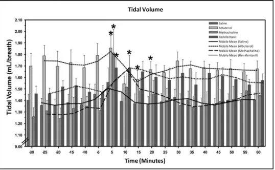
Article #1 Figure 1. Tidal volume evolution in each treated group..………..….66
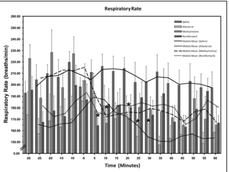
Figure 2. Respiration rate evolution in each treated group……….67
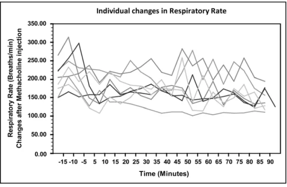
Figure 3. Individual changes observed in respiratory rate………..68
Figure 4. Individual changes observed in peak expiratory flow……….69
Article #2 Figure 1. Tidal volume after albuterol administered by inhalation…………..…97
Figure 2. Respiratory rate following albuterol administered by inhalation…….98
Figure 3. Minute volume following albuterol administered by inhalation……..99
Figure 4. Respiratory monitoring following methacholine bolus………..100
Figure 5A. Tidal volume following methacholine bolus administration……...101
Figure 5B. Respiratory rate following methacholine bolus administration…...101
Figure 5C. Minute volume following methacholine bolus administration……102
Figure 6. Respiratory monitoring (5 min average at pharmacological onset)…...103
LIST OF ABBREVIATIONS
AAALAC: Association for Assessment and Accreditation of Laboratory Animal Care International.
CO2: Carbon dioxide DL: Deciliter
Vd: Dead space
EF50 : Mid-tidal expiratory flow ERV: Expiratory Reserve Capacity ET: Expiratory Time
FDA: Food and Drug Administration FEV: Force Expiratory Volume FRC: Functional residual capacity FVC: Forced vital capacity
H+ : Hydrogen Ion H2CO3: Water carbonic acid HCO3- : Bicarbonate ion
Hg: Mercury
IACUC: Institutional Animal Care and Use Committee IC: Inspiratory Capacity
ICH: International Conference of Harmonization I:E: Inspiratory:Expiratory ratio
IND: Innovative New Drug IRV: Inspiratory Reserve Volume IT: Inspiratory Time
I:TB: Inspiratory to total breath ratio KPA: KiloPascal
Ml: Mililitre MV: Minute volume N2: Nitrogen dioxide
O2: Oxygen
PaCO2: Partial pressure of carbon dioxide in arterial blood Pae: Airway Opening Pressure
PaO2: Partial pressure of oxygen in arterial blood PCO2: Partial pressure of carbon dioxide
PEK: Peak Expiratory Flow Ppl: Pleural cavity pressure Pes: Esophagus Pressure PO2 : Partial pressure of oxygen RBC: Red blood cells
RR: Respiratory rate RV: Residual volume TLC: Total lung capacity TV: Tidal Volume VC: Vital Capacity
DEDICATION
To Fernando,
For his support when I felt vulnerable, for his wonderful and remarkable pure soul. For always be hand in hand and met life’s changes and challenges. For built this beautiful life, for the true love we share.
To my parents,
Who showed me the true meaning of life, and truly gave me love and guidance.
To my sister Mariana,
Thank you for always being and standing beside me even when we are far.
To my family,
For their support and patience. Thank you for always believe in me and love me the way I am. Thank you to always be there.
To life,
Which lets me experience good and bad moments, and gives me the opportunity every day to try to become a better person.
ACKNOWLEDGEMENTS
The author would like to thank:
Pr. Eric Troncy, for his support and guidance during this process.
Dr. Simon Authier, for his motivation and coaching.
Dr. Jean-Paul Descôteaux, for his confidence in my capacities.
Pr. Sophie Cuvelliez, for having accepted to chair this jury.
LAB Research Inc, for the support in the realization of this study.
LAB Veterinary Services personnel for their assistance and support in the process of the experiments.
INTRODUCTION
In the drug development process, the testing of new drugs in laboratory animals is a prerequisite (Jasso 2002). The objective of this evaluation is to quantify the effects of potentially new drugs on different animal physiological functions in order to assess their safety. It is recommended to test on two different animal species, one rodent species, normally the rat, and one non-rodent species, either the dog or a non-human primate (Wallas 2001).
The International Conference of Harmonization (ICH) (2000) was responsible to define the guidelines used in safety pharmacology studies. Those studies are part of pharmacology studies, which are divided into three categories: primary pharmacodynamic, secondary pharmacodynamic and safety pharmacology studies (FDA 1997). The term “Safety Pharmacology” was first defined at the ICH in 2001 (ICH 2001). The aim of this discipline is to characterize the pharmacokinetic / pharmacodynamic relationship (PK / PD) of drug’s effects using continuously evolving methodology (Pugsley et al. 2008). Pharmacology studies have been considered an important component in drug safety assessment. The reason is that numerous examples are reported in the literature indicating that conventional preclinical toxicity testing methods could not predict problems occurring following the administration of new dugs to patients. Some of the problems are hepatotoxicity (Peters 2005) and central nervous system effects (Tsaioun et al. 2009).
The safety pharmacology studies are defined as those that investigate the potential undesirable pharmacodynamic effects of a substance on physiological functions, in relation to exposure in the therapeutic range. The respiratory system is an essential part of the core battery recommended in safety pharmacology studies (ICH 2000). The effects of the test substance on the respiratory system should be assessed appropriately. Clinical observations of animals are generally not adequate to assess respiratory function. Respiratory rate (RR) and other measures of respiratory function such as airway resistance, compliance, pulmonary arterial blood pressure, blood gases, and pH are recommended. Those
parameters should be quantified by using appropriate methodologies (ICH 2000).
This literature review will include the anatomy and physiology of the respiratory system of the different species, rats, dogs and monkeys used in this study. The rules and use of safety pharmacology will also be included.
LITTERATURE REVIEW 1. ANATOMY OF THE RESPIRATORY SYSTEM
The anatomy of the respiratory tract differs among species. The shape of both the upper and lower respiratory tract; the extent, shape, and pattern of the turbinate bones; the branching patterns of bronchi; the anatomy of terminal bronchioles, including collateral ventilation; the lobation and lobulation of the lungs; the thickness of the pleura; the completeness of the mediastinum; relationship of pulmonary arteries to bronchial arteries and bronchioles; the presence of vascular shunts and the blood supply could be different from one species to the other. Each variation in the anatomic structure could imply possible variations in the respiratory function (Reznick 1990).
However, some animal species have anatomically similar respiratory tracts. For instance, the dogs, monkeys and rats are similar while horses are closer to the human respiratory tract (Merck 2008). The respiratory system can be subdivided into the upper and the lower respiratory tracts based on anatomical features (Carter 2007).
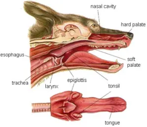
1.1. The upper respiratory tract
The upper respiratory tract includes the nose, nasal passages, pharynx, larynx, and trachea (fig. 1), In addition to the obviously wide range of size and external shapes of the nose, there are also interspecies differences in the internal anatomy and physiology of the upper respiratory tract.
The upper respiratory structures of the tract act to tunnel fresh air down, from outside to inside the body. The upper airways are important because they must always stay open to be able to breath.
The nose assists in the production of sound and in olfaction. The most important function is to warm and to humidify the air through a highly vascularized mucus membrane covering the inside of the nose. The upper one-third of the nasal cavity is lined with olfactory epithelium which sits on the
lamina propria containing numerous serous and mucus glands while the lower
two-thirds are lined with pseudostratified ciliated columnar epithelium containing high amount of goblet cells. Goblet cells, with microvilli on the surface, are present through the respiratory tract. Those cells are responsible for mucus secretion. The microvilli retain particles in the pharynx where they are swallowed to prevent foreign bodies to penetrate the lungs (Rusell 2004). The rat has a smaller percentage of goblet cells than man. Also, secretory cells are more frequent in rats compared to humans, except in the terminal bronchioles where they are comparable (Miller et al. 1993). In the resting animal, the nasal cavity, pharynx and larynx, provide approximately 60% of the frictional resistance to breathing (Cunningham & Klein 2007). In animals, nasal resistance can be decreased by dilation of the external nares and by vasoconstriction which reduces the volume of blood in the vascular sinuses within the nasal mucosa and, as a consequence, the mucosal thickness decreases and the space available for air within the nose increases(Cunningham & Klein 2007).
The pharynx extends from the base of the skull to the inferior border of the cricoid cartilage. It is divided into 3 parts: nasopharynx, oropharynx and laryngopharynx. The oropharynx also has a digestive function. It is delimited by the soft palate superiorly, the basis of the tongue inferiorly, the palatoglossal and the palatopharyngeal arches on lateral sides. The laryngopharynx or hypopharynx also has a digestive function and lies caudally to the larynx, extending from the superior border of the epiglottis and the pharyngo-epiglottic folds to the inferior border of the cricoid cartilage, when it narrows and becomes in continuity through the esophagus (Moore et al. 2006). Both are lined with non-keratinized stratified squamous epithelium. The nasopharynx includes the respiratory area, which is adapted to its main functions of filtering, warming, and humidifying (McGowan et al. 2003). It lies superior to the soft palate and is the posterior extension of the nasal cavities (Moore et al. 2006).
The larynx is located at the crossroads of the air and food passages. It includes the epiglottis, the thyroid, as well as the arytenoid and cricoid
cartilages. The larynx, by its position, prevents the accidental introduction of foreign bodies in the trachea. Its cranial end is attached to the hyoid bone and lies below the epiglottis, while its caudal end is in continuity with the trachea. One of the larynx main functions is to assist in warming and humidifying incoming air. The epiglottis is a structure, which serves to guide the larynx upwardly behind the soft palate so it can lock into the nasopharynx.
In rats, the larynx began at the opening bounded anteriorly by the free border of the epiglottis. The length of the larynx is 4–5 mm. The cricoid cartilage formed a complete ring at the inferior aspect of the larynx and is fixed to the tracheal rings (Nayci 2004). The thyroid cartilage, is a wedge shaped structure of which sides are called laminae. The thyroid cartilage articulates with the cricoid cartilage at the inferior side, cornually and bilaterally. The arytenoid cartilages are bilaterally situated and shaped like small three side pyramids, the bases of which are concave, representing the articular facets that glide upon the corresponding facets of the posterosuperior aspect of the cricoid lamina (Harvey 1993). The cricoid cartilage sits next to the thyroid; its function is to provide attachments for the various muscles, cartilages, and ligaments involved in opening and closing the airways and in vocalization production. The cricoid cartilage is roughly circular in shape inferiorly, and above, it follows the outline of the glottis. In other non-human mammals, cricothyroid joints vary in size and may be absent (Norris 1995). The principal role of airway smooth muscle is to control airway calibre, so the balance between resistance to airflow and physiological dead space is optimised (Matera et al 2002).
Figure 1. Schema of the upper respiratory tract of dogs (Printed from
http://www.vetmed.wsu.edu/cliented/anatomy/dog_resp.aspx)
1.2. The lower respiratory tract
The lower respiratory tract includes the bronchi, the bronchioles, and the lungs. The lower respiratory tract structure varies both within a species and among specie. Irregular dichotomous and trichotomous airway branching patterns are present in human and nonhuman primate lungs. In contrast, the dog and common laboratory rodents exhibit a predominantly monopodial branching system (Miller et al 1993). In most laboratory rodents, the conducting airway terminates abruptly, therefore non-cartilaginous non-alveolarized airways, terminal brochioles open into a completely alveolarized airway (Bal & Ghoshal 1988). The trachea is nearly but not quite cylindrical, being flattened on the posterior. The trachea helps to direct inspired air to the gas exchanging regions of the lung (West 2005).
In dogs, the trachea extends from a transverse plane through the middle of the axis to a plane between the fourth and fifth thoracic vertebrae. It is composed of approximately 35 C-shaped tracheal cartilages. They are open dorsally and the space is bridged by the tracheal muscle (Evans et al 1988). At examination the trachea will be felt as a firm, tubular structure extending
throughout much of the cervical length on the vertical surface of the neck near the midline, the cartilaginous nature of the trachea allows it to be grasped between the fingers. Incomplete cartilaginous rings are bridged by the trachealis muscle (Smith 1999).
In rats, the length and internal diameter of the trachea is 25–27 mm and 3–4 mm, respectively. The cranial surface of the trachea is convex, and covered, in the neck area, in a cranio-caudal direction, by the isthmus of the thyroid gland, the inferior thyroid veins, the arteria thyroidea ima, the sternothyreoideus and sternohyoideus muscles, and the cervical fascia. Caudally, it is in contact with the esophagus. Laterally, in the neck, it is in relation with the common carotid arteries, the right and left lobes of the thyroid gland, the inferior thyroid arteries and, the recurrent nerves. In the thorax, it lies in the cranial mediastinum, and is in relation on the right side with the pleura and right vagus, and near the root of the neck, with the innominate artery. On its left side are the left recurrent nerve, the aortic arch, and the left common carotid and subclavian arteries (Gray 2000). The muscular tissue consists in two layers (longitudinal and transverse) of non-striated muscle. The longitudinal fibers are external, and consist of a few scattered bundles. The transverse fibers are internal and form a thin layer, which extends transversely between the ends of the cartilages (Goldbloom et al. 1960). The mediastinum in primates and dogs are comparable (Miller et al. 1996). This structure can be divided into a cranial part, lying cranial to the heart; a middle part, containing the heart; and a caudal part, lying caudal to the heart. The caudal mediastinum is thin. It attaches to the diaphragm far to the left of the median plane. Cranially it is continous with the middle mediastinum (Evans 1988).
In rodents, the potential space between the medial walls of the two visceral pleurae is called the mediastinum. It is nearly filled with structures including an endocrine gland called the thymus and the parietal pericardium (Wingerd 1988). The tracheobronchial tree is a branching system that delivers air to the alveoli. The number of branches depends on the animal’s size. For instance, mice have about 10 and horses 40 or more. The tracheobronchial tree is
lined by a secretory, ciliated epithelium. The bronchi are supported by cartilage and supplied with bronchial glands and goblet cells, the secretions of which contribute to the mucous lining of the airways. The smaller airways, known as bronchioles, lack cartilages, glands and goblet cells. With the exception of the trachea and the cranial part of the mainstream bronchi, the airways are intrapulmonary. Alveolar septa attach to the outer layers of the airways so that tension within the septa pulls the airways open and helps to maintain their patency (Cunningham & Klein 2007).
The alveolar ducts and alveoli primarily consist of simple squamous epithelium that permits rapid diffusion of oxygen (O2) and carbon dioxide (CO2). Gas exchange between the air in the lungs and the blood in the capillaries occurs across the walls of the alveolar ducts and alveoli (Bray et al. 1999).
The alveolar duct branching system of the rat is complex (Miller et al. 1993). It is a three dimensions system in which multiple branches may occur within the distance of a single alveolus. The alveolar duct system of the rat is only about 100 µm.
In primates, the bronchioles are arranged stereotaxically like those of other mammalian lungs. The four bronchiole systems, dorsal, ventral, medial, and lateral, arise from both bronchi, respectively, although some bronchioles are lacking. In the right lung, the bronchioles form the upper, middle, accessory, and lower lobes, while in the left lung, the upper and accessory lobes are lacking and bi-lobed middle and lower lobes are formed (Nakakuki 1986).
The lungs are a pair of sponge-like organs located inside the thoracic cavity. They are not directly attached to the ribs. They are suspended by a double-walled sac called pleura (Wilmore et al. 2008). The inner layer of the sac (visceral pleura) tightly adheres to the lungs, and the outer layer (parietal pleura) is attached to the wall of the chest cavity. The two layers are separated by a thin space, called the pleural cavity, filled with pleural fluid allowing the inner and outer layers to slide over each other, and prevent them from being easily separated. The liquid allows the two pieces to slide over one another but makes
them difficult to separate. In the thorax, therefore, the pleural fluid mechanically links the lungs to the thorax so that the respiratory system behaves as a single unit. The lungs of most species have a total of six lobes, each supplied by a lobar bronchus, which gives rise to daughter bronchi. Even in specie such as the horse that lack lobation, the same pattern of six lobar bronchi is still present. At each division of a parent bronchus, the diameter of the other is similar to that of the parent (Cunningham & Klein 2007).
The primarily function of the lungs is to allow O2 to move from the air into the venous blood and CO2 to move out (West 2005). The air enters through the trachea going to the left and right bronchi and branch many times throughout the lungs, until they eventually form a little thin walled air sacs or bubbles, known as the alveoli. The alveoli are the sites of gas exchange with the blood (Widmaier et al. 2006).
The lungs present few differences between laboratory animal species. In dogs, each lung is roughly triangular and has apex, costal, medial, diaphragmatic surfaces, dorsal, ventral, and caudal borders. Each lung is divided by deep fissures into distinct lobes. The caudal fissures of the dog’s lungs correspond to the oblique fissures of the human lungs (Ishaq 1980). Dogs like humans, have right and left lungs. Both sides of the lungs are further divided into lobes. Inside the lungs, the bronchi divide into smaller and smaller tubes, called bronchioles, much like branches of a tree divide into smaller and smaller branches.
In primates, the lungs of the rhesus monkeys are generally separated into six lobes. The left lung has two lobes, cranial and caudal, of which the cranial is further segmented into a cranial and caudal portion. The right lung has four distinct lobes in the rhesus monkeys. The lungs are lined by connective tissue band with a mesothelial surface facing the pleural space (Wolfe-Coote 2005).
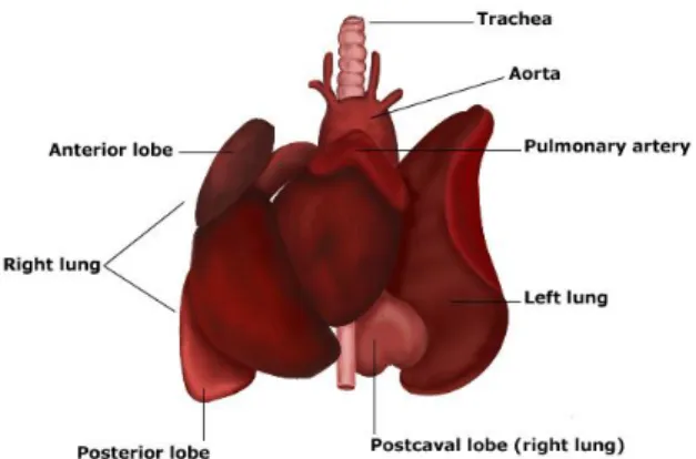
In the rat, the lungs are large spongy structures. The right lung is divided into four lobes: cephalic, medial, caudal and postcaval (Fig. 2) (Wingerd 1988).
Figure 2. Rat lungs anatomy (Printed from www.tutorvista.com)
1.3. Pulmonary blood flow
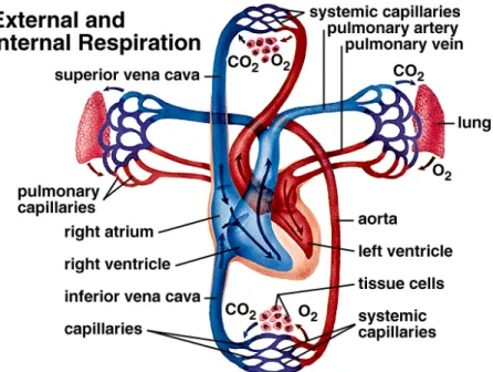
The lung receives blood flow from two circulatory systems: the pulmonary circulation and the bronchial circulation. The pulmonary circulation receives the total output of the right ventricle, perfuses the alveolar capillaries and participates in gas exchange. The bronchial circulation, a branch of the systemic circulation, provides a nutritional blood supply to airways and other structures within the lung (Cunningham & Klein 2007).
The pulmonary circulation carries roughly the same flow as the systemic circulation. The arterial pressure and the vascular resistance are normally only one-sixth as great. The media of the pulmonary arteries is about half as thick as in systemic arteries of corresponding size. In the larger vessels, it mainly consists of elastic tissue but in the smaller vessels, it is mainly muscular. The transition in vessels is close to 1 mm diameter. Pulmonary arteries lie close to the corresponding air passages in connective tissue sheaths (Lumb 2006). The bronchial circulation provides nutrient blood flow to airways, large blood vessels, and the pleura (Mc Laughlin et al. 1961).
The pulmonary circulation begins at the main pulmonary artery, which receives the mixed venous blood pumped by the right ventricle. The main pulmonary arteries that accompany the bronchi are elastic, but the smaller arteries adjacent to the bronchioles and the alveolar ducts are muscular
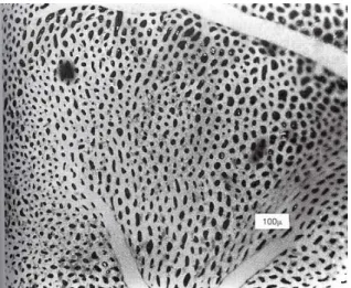
(Cunningham & Klein 2007). This, then branches successively like the system of airways and, indeed, the pulmonary arteries accompany the airways as far as the terminal bronchioles. Beyond that, they break up to supply the capillary bed which lays in the alveoli walls. The pulmonary capillaries form a dense network in the alveolar wall which makes an exceedingly efficient arrangement for gas exchange (Fig. 3). The oxygenated blood is then collected from the capillary bed by the small pulmonary veins that run between the lobules which drain into the left atrium (West 2005).
Figure 3. Surface view of capillaries in alveolar wall(Printed from: Guyton,
A.C. & Hall, J.E., 2006. Textbook of Medical Physiology., p 497)
The amount of smooth muscle in the media of pulmonary arteries determines the reactivity of the vasculature. The small pulmonary arteries lead into pulmonary capillaries, which form an extensive branching network of vessels within the alveolar septum, covering almost the alveolar surface. Not all capillaries are perfused in the resting animal. As a result, vessels that are unperfused can be recruited when pulmonary blood flow increases. Pulmonary veins with thin walls conduct blood from capillaries to the left atrium and also form a reservoir of blood for the left ventricle (Fig 4) (Cunningham & Klein 2007).
In the bronchioles, oxygenated arterial blood flows to the lungs through small bronchial arteries that originate from the systemic circulation. After this
bronchial and arterial blood has passed through the supporting tissues, it empties into the pulmonary veins and enters the left atrium. Therefore, the flow into the left atrium and the left ventricular output are about 1 to 2 per cent greater than the right ventricular output (Guyton et al. 2006).
Figure 4. Pulmonary blood circulation (Printed from www.mhhe.com)
1.4. Muscles of respiration
Energy provided by muscles causes air to enter the lungs during inhalation. During exhalation, much of the energy causing air to leave the lungs is provided by the elastic energy stored in the stretched lung and the thorax (Cunningham and Klein 2007). There are several muscles which play an important role in the inspiration and expiration process during breathing (Adams 1998).
The inspiration muscles are composed of the diaphragm, the external intercostal muscles, and the accessory muscles. The accessory muscles include the scalene muscles and the sternomastoids. Other muscles playing a minor role include the alae nasi, which causes flaring of the nostrils and, small muscles in
the neck and head. The primary inspiration muscle is the diaphragm, which is a domed musculotendinous sheet separating the abdomen and thorax and innervated by the phrenic nerve, responsible for an active inhalation. The external intercostals muscles also are active during inhalation. The fibers of these muscle contraction moves the ribs rostrally and outward. The relative contributions of diaphragmatic and costal movements to ventilation under different metabolic demands are not well defined in animals. Because the cranial ribs support the forelimbs in quadrupeds, they participate less in ventilation than do the more caudal ribs. Other inspiratory muscles include those connecting the sternum and head. These muscles contract during stenous breathing and move the sternum rostrally (Cunningham & Klein 2007).
During expiration, the most important muscles are those of the abdominal wall, including the rectus abdominis, internal and external oblique muscles and
transversus abdominis. The internal intercostal muscles also assist during the
process (West 2005). Contraction of the abdominal muscles increases abdominal pressure, which forces the relaxed diaphragm forward and reduces the size of the thorax. The fibers of the internal intercostals are directed cranio-ventrally, from the cranial border of one rib to the caudal border of the next cranial rib, so their contraction decreases the size of the thorax by moving the ribs caudally and ventrally. As the thorax becomes smaller, the thoracic pressure increases and forces air out of the lungs (Cunningham & Klein 2007).
2. PHYSIOLOGY OF THE RESPIRATORY SYSTEM
In mammals, the function of the respiratory system is essential to maintain homeostasis (National Research Council 2003). Its major function is gas exchange between the external environment and the organism's circulatory system. This exchange facilitates oxygenation of the blood with concomitant removal of CO2 and other gaseous metabolic wastes from the circulation as water, sulfates, phosphates and nitrogen, (Widmaier et al. 2006).
The role of the inspiration and expiration is essential for breathing helping to change the intrathoracic pressure involving different muscles and to permit the exchange of O2 and CO2 (Schwartzstein & Parker, 2005).
2.1. Inspiration process
Muscular activity is involved in the inspiration process. Various muscles are involved, the diaphragm being the main one. Those muscles act to increase the thoracic volume, causing intra-pleural and alveolar pressure to fall creating an alveolar-mouth pressure gradient, drawing air into the lungs.
Resting animals breathe slowly and have low flow rates. In this situation, the primary work of the respiratory muscles is against the compliance of the lungs (Cunningham & Klein 2007). However, during exercise or in response to a stressful situation, they may contract vigorously (West 2005) and respiratory muscle activity increases in order to generate an increase in minute ventilation (MV). In cursorial (running) mammals, ventilation is synchronized at canter and gallop, but not at the trot. Inhalation occurs as the forelimbs are extended and the hind limbs are accelerating the animal forward. Exhalation occurs when the forelimbs are in contact with the ground. In the galloping horse and perhaps in other galloping quadrupeds, much of the increase in size of the thorax during inhalation is a consequence of elongation of the trunk rather than an increase in the diameter of the thorax (Cunningham & Klein 2007).
2.2. Expiration process
Expiration is present at the end of inspiration. Expiration in a resting animal is a passive process that does not require muscle contractions (except in horses, where expiration is a passive – active process). Relaxation of the muscles contracted during inspiration permits the intrinsic elastic properties of the lungs and the thoracic wall to recoil to the original volume. The return to the original volume increases the intra-pulmonary pressure so it is greater than atmospheric, and air is forced out of the lungs. Forced expiration is an active process that
forces more air from the lungs that would occur during normal passive expiration. Forced expiration requires contraction of abdominal muscles to force viscera against the diaphragm and contraction of other muscles to pull the ribs caudad. Both of these actions reduce the size of the thoracic cavity and permit recoil of the lungs to a smaller volume than typical for resting expiration (Frandson et al. 2003).
At the end of a normal exhalation, some air remains in the lungs. This air volume is known as functional residual capacity (FRC). At FRC, the pressure in the pleural cavity (Ppl) surrounding the lung is approximately 5 cm H2O below atmospheric pressure. Ppl decreases during inhalation as the thorax enlarges and the respiratory muscles perfom work to stretch the elastic lung. Resting animals breathe slowly and have low flow rates. In this situation the primary work of the respiratory muscles is against the compliance of the lungs. When the respiratory rate increases (e.g. during exercise), flow rates increase, and more energy is used to generate flow against the frictional resistance of the airways (Cunningham & Klein 2007).
2.3. Gas exchange in the lungs
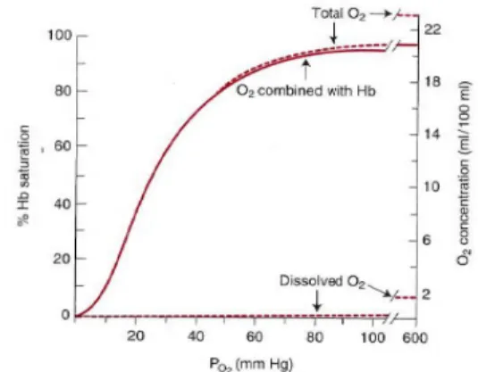
The lung is the organ of gas exchange, providing the means of transferring O2 from the air to the blood for subsequent distribution to the tissues. At the same time, it enables CO2 removal from the blood which is then exhaled to the atmosphere. Gas exchange occurs by passive diffusion for CO2 but for O2, haemoglobin (Hb)-induced attraction helps a lot (Warrell et al. 2005). At rest, 100 mm Hg pressure of O2 in the alveoli exceeds by about 60 mm Hg the 40 mm Hg O2 pressure in blood that enters the pulmonary capillaries. Consequently, O2 dissolves and diffuses through the alveolar membranes into the blood (McArdle
et al. 2006). Oxygen is poorly soluble in water and therefore in plasma. Because
of this low solubility, most animals need an oxygen carrying pigment to transport sufficient O2 to the tissues. When blood in the pulmonary capillaries flows past the alveoli, O2 diffuses from the alveoli into the blood until the partial
pressures equilibrate, and there is no further driving pressure difference. Without Hb, which transports the majority of the O2, the cardiac output would have to be inordinately high to maintain the O2 supply to the body organs (Cunningham & Klein 2007). In contrast, CO2 exists under a slightly greater pressure in returning venous blood than in the alveoli causing a net diffusion of CO2 from the blood into the lungs. Gas exchange occurs so rapidly in the healthy lung that alveolar-gas / blood alveolar-gas equilibrium takes place in about 0.25 second or within one third of blood transit time through the lungs. Even in high intensity exercise, red blood cell velocity through a pulmonary capillary generally does not exceed by more than 50% its velocity at rest (McArdle et al. 2006).
2.4. Blood flow and O2 transport
Systemic arterial blood enters capillaries throughout the body; it is separated from the interstitial fluid by only the thin capillary wall which is highly permeable to both O2 and CO2. The interstitial fluid, in turn, is separated from the intracellular fluid by the plasma membranes of the cells which are also quite permeable to O2 and CO2. The supply of the new O2 to the alveoli and the consumption of O2 in the cells create partial pressure in O2 (PO2) gradients that produce net diffusion of O2 from alveoli to blood in the lungs and from blood to cells in the rest of the body (Widmaier et al. 2006).
In the blood circulation, O2 is present in two forms: dissolved in the plasma and red blood cells (RBC) cytoplasm and combined to Hb molecules in the RBC (Wnek and Bowlin, 2008). The amount of O2 dissolved in blood is directly proportional to blood PO2. Relatively insoluble in water, only 3 ml of O2 can be dissolved in 1 L of blood at normal arterial PO2 (100 mm Hg) (Widmaier
et al. 2006). The pulmonary capillary blood equilibrates with the alveolar O2
tension of 100 mm Hg, therefore, 0.3 ml of O2 dissolves in each decilitre of blood. If an animal breathes pure oxygen, the alveolar oxygen tension increases to approximately 600 mg Hg, and 1.8 mL of oxygen dissolves in each decilitre of plasma (Cunningham & Klein 2007).
Oxygen transport is the volume of O2 moved through the circulation in a unit of time. Cardiac output or blood flow to a specific site is important to O2 transport and tissue delivery. Blood O2 content depends on the pulmonary capillary PO2 produced by gas exchange, the amount of Hb present, and the slope of O2-Hb dissociation curve (Stein, 1998). The binding of O2 is a four-step process, and the O2 affinity of a particular haem is influenced by the oxygenation of the others. This means that when the first haem unit is oxygenated, O2 affinity of the second haem unit is increased and so on. These haem-haem interactions are responsible for the sigmoid shape of the oxy-Hb dissociation curve (Cunningham & Klein 2007).
Figure 5. O2 dissociation curve. (Printed fromo: West, J.B., 2005. Respiratory Physiology: The Essentials. 7th ed. Lippincott Williams & Wilkins., p76)
2.5. Carbon dioxide (CO2) transport
During steady-state conditions, approximately 80 molecules of CO2 are exhaled from the lungs for every 100 molecules of O2 taken up from the alveoli into pulmonary capillary blood (148-150 molecules). The CO2 is produced in the tissues and carried to the lungs through the venous blood in three forms: dissolved CO2, a chemical combination of CO2 and Hb and as bicarbonate
(HCO3-) which is the major form (90%) (Costanzo 2006). When considering tissue CO2 content, the difference between CO2 and O2 amounts may be as great as tenfold because CO2 is 23 times more soluble than O2 in plasma (Brown et al 2006). The total atmospheric pressure is equal to 760 mm Hg; CO2 is almost absent in the atmospheric air, this explains the effective diffusion gradient for CO2 from the body to the air (Reece 2004).
At a normal arterial PCO2 of 40 mm Hg, the amount of CO2 transported in solution is equal to 28 mL/L blood or equivalent to 100 mm Hg PO2. Because of its higher solubility, approximately 6% of total blood CO2 is in physical solution compared to 1.5 % of O2 dissolved in arterial blood. The concentration of dissolved CO2 is greater in venous blood than in arterial blood. Rapidly CO2 diffuses out of the cell due to its higher intracellular versus blood partial pressure . Once CO2 diffuses out of the cell, it quickly moves into the RBC. Once inside RBC, CO2 is chemically converted in the presence of water by the enzyme carbonic anhydrase to carbonic acid (H2CO3-) and finally into a hydrogen ion (H+) and HCO3- (Brown et al. 2006).
2.6. Respiratory receptors
Lungs contain sensory receptors that communicate with the respiratory centers through vagal nerve afferents. At rest, the chemical state of the blood exerts the greatest control on pulmonary ventilation. Variations in arterial PCO2, pH, PO2, and temperature activate sensitive neural units in the medulla and arterial system to adjust ventilation and maintain arterial blood chemistry within narrow limits (McArdle et al. 2006).
Different patterns during the respiratory process need to be coordinated in order to avoid abnormal breathing patterns (Mc Ardle et al. 2006). Some of abnormal breathing patterns are tachypnea, bradypnea, apnea, and hyperapnea (Springhouse, 2008). Many types of receptors are involved in the respiratory control: chemoreceptors (central and peripheral), lung receptors (irritant,
pulmonary stretch, and juxtapulmonary), receptors in the chest wall, several receptors in the trachea, laryngeal, arterial baroreceptors, and pain receptors.
The chemoreceptors monitor blood gas tension in arterial PaCO2, PaO2 and pH, and helps keep MV appropriate to metabolic demands of the body. The chemoreceptors respond to hypercapnia (↑ CO2), hypoxia (reduction of O2), anoxia (absence of O2) and acidosis (↓ HCO3- or ↑ PCO2). The most sensitive system is the one responding to PaCO2. The central chemoreceptors are tonically active and vital for respiration maintenance. Eighty percent of ventilation drive is a result of central chemoreceptors stimulation. When they are inactivated, the respiration ceases. These receptors are located in the brainstem on the ventrolateral surface of the medulla, close to the exit of cranial nerves IX and X. They are anatomically separated from the medullary respiratory control center. These chemoreceptors respond to H+ concentrations. A rise in [H+] increases ventilation, while a lowering decreases it (Bijlani 2004). The peripheral chemoreceptors are located around the carotid sinus and aortic arch. Stimulation of peripheral chemoreceptors has both cardiovascular and respiratory effects. Peripheral chemoreceptors provide the primary site to detect arterial hypoxia and by reflex initiate a ventilatory response (McArdle et al. 2006).
The carotid bodies are chemoreceptors sensitive to CO2 and O2 concentrations in the blood (Unchoa & Cameiro, 2005). They are located close to the bifurcation of the internal and external carotid arteries. The latter appear to be most active in the foetus and of little importance in the adult. The carotid bodies are small, pink nodular structures with extremely high blood flow per gram. This high blood flow and metabolism ratio allows the carotid bodies to obtain their O2 needs from dissolved O2. Carotid bodies contain several cell types. Type I cells, or glomus cells, synapse with afferent nerves that transmit information back to the brain. These glomus contain a variety of neurotransmitters, including catecholamines, especially dopamine. Glomus cells are probably responsible for the chemosensitivity of the afferent nerve terminals. Type II cells support the axons and blood vessels that ramify within the carotid
body. When the carotid bodies are perfused with blood that has a low PaO2, high PaCO2, or low pH, firing rates in the carotid sinus nerve afferent fibers increase. As PaCO2 increases and pH decreases, there is an almost linear increase in ventilation, the response to PaO2 is non linear (Cunningham & Klein 2007). Peripheral chemoreceptors are sensitive to PaO2, PaCO2, pH, blood flow, and temperature (McGowan et al. 2003). The activity in the nerve fibers is directly related to the degree of hypoxia at the chemosensory cells (Kline et al. 2005).
The carotid bodies respond to the autonomic nervous system (Koyama et
al. 2000). The sympathetic action vasoconstricts and increases sensitivity to
hypoxia while the parasympathetic action vasodilates and reduces sensitivity to hypoxia. Unlike central chemoreceptors, peripheral chemoreceptors are directly stimulated by blood pH (McGowan et al. 2003). The vagus contains myelinated afferents slowly adapting pulmonary stretch receptors which are stimulated by changes in lungs volumes. Stretch depolarizes these receptors, sending action potentials to respiratory centers in the brain via the vagal nerves (Johnson & Byrne, 2003). The stretch receptors are situated in the smooth muscle of the bronchial walls and respond to changes in transmural pressure (McGowan et al. 2003). When these areas are excessively stretched, the information is relayed to the expiratory center. The center responds by shortening the duration of the inspiration, which decreases the risk of overinflating the respiratory structures. This response is known as Henri-Breuer reflex (Wilmore et al. 2008).
The respiratory receptors do not act alone in controlling breathing. Breathing is also regulated and modified by the changing chemical environment of the body. For example, sensitive areas in the brain respond to changes in CO2 and H+ levels. Central chemoreceptors are stimulated by an increase of H+ ions in the cerebrospinal fluid. The goal of respiration is to maintain appropriate levels of blood and tissue gases and to maintain proper pH for normal cellular function. Small changes in any of these, if not carefully controlled, could impair physical activity and jeopardize health (Wilmore et al. 2008).
During exercise, resting arterial Hb saturation in O2 is close to 100% in normal conditions (no pathology to Hb or RBC) and O2 content cannot be raised significantly. Oxygen delivery to exercising muscle is augmented by increasing muscle blood flow, made possible by metabolic vasodilatation. Oxygen extraction from the delivered blood is increased. Mixed venous blood has reduced PvO2 and increased PvCO2. Passive limb moving stimulates ventilation in both anaesthetized and awaken animals. This is a reflex due to receptors presumably located in joints or muscles. It may be responsible for the abrupt increase in ventilation that occurs during the first seconds of exercise (West 2007).
At high altitude, barometric pressure falls progressively. For instance, it falls from around 101 kPa (760 mmHg) at sea level to 33.6 kPa (252 mmHg) on the summit of Everest; however, O2 fraction remains constant at 0.209. If ventilation remains unchanged, reduced inspired PO2 inevitably leads to reduced PaO2 but PaCO2 will be unaltered (Ward et al. 2006). At such altitude, metabolic rate is unchanged in resting animals. Therefore increased ventilation causes a decrease in PaCO2. This decrease in PaCO2 inhibits ventilatory drive, which limits the hypoxic ventilator response by the carotid body chemoreceptors (Johnson et al. 2003). The RBC concentration increases and the oxyHb dissociation curve is shifted to the right. However, at very high altitude the very low PaCO2 shifts the curve to the left and the beneficial effect of increased O2 binding in the lungs seems to outweigh the impaired O2 release in the tissues. The widespread hypoxia vasoconstriction in the lungs is helpful (Ward et al. 2006). Hypoxic vasoconstriction is unique to pulmonary circulation. The pulmonary response is part of a self-regulatory mechanism by which pulmonary capillary blood flow is automatically adjusted to alveolar ventilation for maintaining the optimal balance of ventilation and perfusion (Dumas et al. 1999). It increases pulmonary vascular resistance and those living at high altitude may develop right ventricular strain and failure (Ward et al. 2006). The
pulmonary vascular resistance may persist through childhood and adult life if the individual remains at high altitude (Rudolph 2001).
2.7. Respiratory mechanics
Respiratory mechanics explains how the forces generated by respiratory muscles cause effective ventilation of the alveoli. Resistance and compliance are two physical factors important in determining the relationship between pressure and flow in the lungs, and in the distribution of ventilation to different regions of the lungs. The effectiveness of ventilation not only depends on the amount of fresh air reaching the alveoli, but also on the matching of ventilation to blood flow in different regions of the lung (Johnson et al. 2003).
2.7.1. Resistance
Respiratory resistance is a combination of resistance to gas flow in the airways and resistance to deformation of tissues of both the lungs and the chest wall. In normal lungs, respiratory resistance is controlled by changes in airway diameter, mainly in small airways and bronchioles. Gas flows from a region of high pressure to one of lower pressure. The rate at which it does so is a function of the pressure difference and the resistance to gas flow. In the healthy subjects, the small airways only make a small contribution to total airway resistance because their aggregate cross-sectional area increases to very large values after about eight generations. Gas flow velocity and airway diameter decrease in successive airway generations. The airway lining will influence frictional resistance more with turbulent than with laminar flow, with changes in mucus consistency that occur in many airway diseases (Lumb 2006). The factors determining airway resistance are analogous to those determining vascular resistance in the circulatory system. Those factors include tube length, tube radius, and interactions between moving gas molecules. Physical, neural and chemical factors affect airway radii and therefore resistance. One important physical factor is the trans-pulmonary pressure which exerts a distending force on the airways, just as on the alveoli. This is a major factor keeping the smaller
airways, those without cartilage, to support them from collapsing. Trans-pulmonary pressure increases during inspiration, airway radius becomes larger and airway resistance lower as the lungs expand during inspiration. The opposite occurs during expiration (Widmaier et al. 2006).
2.7.2. Compliance
Lung compliance is defined as the change in lung volume per unit change in trans-mural pressure gradient. There are two major determinants of lung compliance; first, the stretchability of lung tissues (particularly their elastic connective tissues, thus, a thickening of the lung tissues decreases lung compliance) and the other is the surface tension at the air-water interfaces within the alveoli. The surface of the alveolar cells is moist, so the alveoli can be pictured as air-filled sacs lined with water. At an air-water interface, the attractive forces between the water molecules, known as surface tension, make the water lining like a stretched balloon that constantly tries to shrink and resists further stretching. Thus, expansion of the lung requires energy not only to stretch the connective tissue of the lung, but also to overcome the surface tension of the water layer lining the alveoli (Widmaier et al. 2006). Compliance is usually expressed in L or mL per KPa or cmH2O. The normal value of compliance is approximately 1.5 L.kPa-1 or 150 mL.cmH2O-1. When an increase of peribronchial and perivascular fluid and thickening of the lung tissues occurs for instance, a decrease in lung compliance is observed (Lumb 2006), a condition called as stiff lungs.
Elastic recoil is expressed as elastance, which is the reciprocal of compliance. Only a quarter to a third of the lungs elastic recoil is caused by the elastic fibres of its alveolar walls, most is caused by surface tension at the alveolar tissue (Bray et al. 1999). Respiratory system elastance is known to increase with frequency breathing, and tissue resistance is known to decay hyperbolically over the range of physiological breathing frequencies, whereas
airway resistance remains fairly constant both in healthy and mildly constricted lungs (Lutchen et al. 1994).
2.7.3. Ventilation and Lung Mechanics
Breathing is caused by rhythmic contractions of skeletal muscles that are entirely dependent upon intact nervous connections from the medulla through the spinal cord, the phrenic and intercostal nerves. Breathing is thus the product of chemical and neural influences on a network of neurones, motor nerves, and muscles; the result being dependent on the mechanical properties of the chest, the lungs, and the airways (Whipp 1987). The act of breathing is performed against several impediments: elastic, resistive, visco-elastic, plasto-elastic, inertial and gravitational forces, compressibility of intra-thoracic gas, and distortion of the chest wall (Polese et al. 2005).
Ventilation is defined as the exchange of air between the atmosphere and alveoli. Like blood, air moves by bulk flow, from a region of high pressure to one of low pressure. In the respiratory system, pressures are expressed in relation to atmospheric pressure, which is 760 mmHg at sea level (West 2008). During ventilation, air moves into and out of the lungs because the alveolar pressure is alternately less than and greater than atmospheric pressure (Widmaier et al. 2006).
The intrapleural pressure causes the lung and thoracic wall to move in and out together during normal breathing (Widmaier et al. 2006). When the thorax expands and pull the lungs, they must comply and expand. The air must comply and smoothly flow to the alveoli. If the thorax and lungs are stiff or the airways are narrow, so the air does not move in and out of the lungs freely, pulmonary ventilation will be impaired (Sabyasachi 2007).
2.7.4. Elastic properties of the lungs
The elastic properties of the lung and of the chest wall to airflow resistance are part of the mechanic of breathing. The lung is a volume-elastic
container that tends to deflate itself. The deflation force exerted by the lungs is call elastic recoil pressure. This pressure increases with lung volume and the pressure requires maintaining inflation equals to the elastic recoil. Lung expansion is maintained either by negative pleural pressure at the outer surface of the lung or by positive pressure applied by the airway opening to the inner surface of the lung (Shields et al. 2004).
The movements of the lungs are entirely passive and result from forces external to the lungs. The pattern of lung response is governed by the physical impedance of the respiratory system. The most important impedances are: elastic resistance of lung tissue and chest wall, resistance from surface forces at the alveolar area, frictional resistance to gas flow through the airways, frictional resistance from deformation of thoracic tissues, and inertia associated with movement of gas and tissue. The surface forces have a predominant influence on the elastic properties of the lung at volumes higher than functional residual capacity, which implicates alveolar size as the major determinant of specific pulmonary elastance for a constant number of alveoli (Haber et al. 1983).
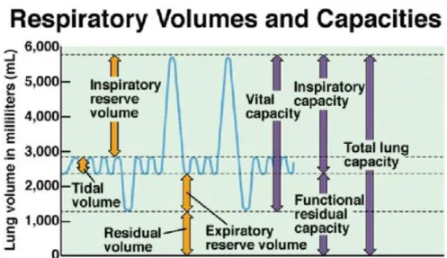
2.7.5. Pulmonary Volumes and Capacities
The volume of gas in the lungs can be divided into the following components:TLC, VC, IC, ERV, IRV, TV, RV and FRC (Fig 5). A capacity is defined as the summation of two, or more, volumes. From these variables, the most commonly used to test respiratory function are TV (amount of air breathed in or out during a quiet breathing), RV, VC and FRC. Those can be useful in diagnosing certain pulmonary diseases (Johnson et al 2003). Pulmonary volumes are either associated with the amount of air within them at any time or with the amount associated with breath (Reece 2004).
Figure 6. Respiratory volumes and capacities. Printed from
(http://images.google.ca/imgres?imgurl=http://faculty.stcc.edu)
The O2 needs of metabolism require that an animal take a certain volume of air into its lungs, essentially its alveoli, each minute. The total volume of air breathed per minute or MV (minute ventilation), is determined by the volume of each breath, known as the tidal volume (TV), and the number of breaths per minute, know as respiratory frequency or RR (respiratory rate).
MV = TV × RR
The increase in MV, which must occur when an increase in metabolic rate demands more oxygen, can be brougth about through an increase in TV, RR or both. The IRV is the amount of air that can still be inspired after inhaling the TV, and ERV is the amount of air that can still be expired after exhaling the TV (Johnson et al. 2003). The RV is the amount of air remaining in the lungs after the most forceful expiration. The TLC is the sum of all volumes while VC is the sum of all volumes over and above the RV, which is also the maximum amount of air that can be inspired after the most forceful expiration; IC is the sum of TV and IRV, and FRC is the sum of ERV and RV (Reece 2004). Normal lung volumes provide lessons about respiratory physiology in healthy individuals.
The TLC is more than ten times larger than the normal TV, so there is a tremendous reserve capacity for increased ventilation with increased O2 demand or reduced supply. This is part of the reason why pulmonary gas exchange is usually not a limiting factor in O2 uptake at sea level in anyone except highly trained elite athletes (Johnson et al. 2003). Finally, FRC is the volume of air present in the lungs at the end of passive expiration (Sabyasachi 2007). A lowered or elevated FRC helps to diagnose some forms of respiratory disease.
2.7.6. Dead space
The anatomic dead space is the volume of the upper respiratory system where gas exchange do not occur. Dead space (Vs) can also be present within the alveoli. This alveolar dead space is caused by alveoli that are poorly perfused with blood, so that gas exchange cannot occur optimally. Physiological dead space is the sum of the anatomic and the alveolar dead space (Cunningham & Klein 2007). The strict definition of the anatomical dead space is the volume of an inspired breath which has not mixed with the gas in the alveoli. Because gas exchange effectively only takes place in the alveoli there is no CO2 excreted into the dead space (Davies & Moore 2003). Essentially, all the gas exchanges between air and blood take place at the alveolar surface. At the end of the inspiration, the content of the alveoli has been diluted by inspired room air, which now also fills the anatomical dead space. At the end of expiration, the anatomical dead space is filled with alveolar air, and this partly used air is inhaled first in the next inspiration (Fig. 6) (Davies & Moore 2003). Not all of the air entering the respiratory system actually reaches the alveoli and takes part in gas exchange.
Figure 7. Anatomic and alveolar dead space
The structures included in the dead space are: nose and mouth, pharynx, larynx, trachea, bronchi and bronchioles, down to and including the terminal bronchioles. The fraction of each breath ventilating the dead space is known as the VD/TV ratio. The VD/TV varies considerably among species. In smaller species, such as dogs, it approximates 33%, whereas in some larger species, such as cattle and horses, it approximates 50% to 75%. Because the volume of the anatomic dead space is relatively constant, changes in TV, RR or both can alter the relative amounts of air that ventilate the alveoli and the dead space. These changes in TV and RR occur in animals during exercise and thermoregulation. For example, the small TV and high RR characteristic of panting in dogs cause more air to ventilate the dead space in order to cause water evaporation and heat loss. Cattle, pigs and mules subjected to heat stress also increase RR and dead space ventilation when trying to lose heat. In contrast to the effects of heat stress, cold stressed animals have a higher metabolic rate, which is necessary to maintain body temperature in cold conditions. This leads to an increase in both O2 consumption and CO2 production, making it necessary for the animal to increase alveolar ventilation and decrease dead space ventilation. Reducing the RR and increasing TV accomplish the latter adaptations (Cunningham & Klein 2007).
Anatomical dead space can be measured using Fowler’s method which is based on the single breath nitrogen test (McGowan et al. 2003). This test measures inequalities of ventilation, useful to evaluate ventilatory capacity (West 2007). If some regions of the lung expand before others in the process of inspiration, they will receive inappropriately large parts of this dead space, and the regions receiving air later in inspiration will receive more fresh air. This type of dead space is called anatomical dead space because it measures the anatomical volume of the conducting airways. If each acinus or end respiratory unit was perfect, the amount of air received by each alveolus would be matched by the flow of blood through the pulmonary capillaries. In a normal healthy
person, anatomical and physiological dead space are almost equal, alveolar dead space being very small (<5 mL). However, the volume of alveolar dead space increases when lung disease alters ventilation / perfusion matching,. Physiological dead space is calculated, using the Bohr Equation (McGowan et al 2003). The Bohr Equation permits the determination of the sum of the anatomic and the alveolar dead space. It evaluates the measurable volume of CO2 found in the mixed expired gas coming from the alveoli. The PCO2 of the collected mixed expired gas can be determined with a CO2 meter. The CO2 meter is also often used to estimate the alveolar PCO2 by analyzing the gas expelled at the end of a normal tidal expiration, the end tidal CO2 (Levitzky 2007).
3. SAFETY PHARMACOLOGY
Safety Pharmacology is the discipline trying to predict whether a drug, if administered to human (or animal) population, is likely to be found safe (Pugsley
et al. 2008). It is, with the primary pharmacodynamic studies which are those
evaluating the mode of action and/or effects of a substance in relation to its desired therapeutic target and secondary pharmacodynamic studies which evaluate the mode of action and/or effects of a substance not related to its desired therapeutic, a part of the pharmacology discipline (FDA 1997).
The origins of Safety Pharmacology are grounded upon observations that organ functions can be toxicological targets in humans but the effects cannot be readily detected by standard toxicological testing (Mortin et al. 1997; Williams 1990; Zbinden, 1984). The objectives of safety pharmacology are: 1) to identify undesirable pharmacodynamic properties of a substance that may have relevance to its human safety; 2) to evaluate adverse pharmacodynamic and/or pathophysiological effects of a substance observed in toxicology and/or clinical studies; and 3) to investigate the mechanism of the adverse pharmacodynamic effects observed and/or suspected (ICH 2001).
The term first appeared in drafts of the ICH in 1997 (Bass et al 2004). Following that, in 1999, an Expert Working Group was formed and began their work to define the guidelines of Safety Pharmacology. A harmonized Safety Pharmacology guideline was finalized and adopted by the regional regulatory authorities over 2000–2001 (ICH 2001). They describe their implications in studies of the three vital systems: central nervous, cardiovascular and respiratory systems (Porslot et al. 2002).
Those guidelines were accepted in United States, Europe and Japan. Prior to 1990’s, toxicological testing done on lead compounds was limited and did not allow to identify all adverse effects produced by those compounds. The U.S. and European regulations provided only general references to the evaluations of drug effects (Kinter et al 1994;Lumley, 1994).
Organ function assessments were inconsistent and often viewed as unimportant
(Green, 1995; Proakis, 1994). In fact, during that period, it was discovered that some of those compounds showed rare and potentially lethal adverse effects later in the testing process (Pugsley et al. 2008).
The organ systems and functions most frequently responsible in these events were the cardiovascular, central nervous and renal systems, The result was almost always a critical care emergency (Kinter et al 1994). Such episodes contributed to the development of Safety Pharmacology.
The ICH guidelines have brought uniformity to the evaluations of new drugs for effects on organ functions (Bass et al. 2004). Within a short period, Safety Pharmacology had evolved into a discipline designed to bridge the gap between preclinical toxicology and preclinical and clinical drug development (Bass et al. 2004). However the creation of Safety Pharmacology has not resolved all problems with respect to detection of rare and lethal adverse effect liability and more problems have to be solved in order to quantify, with high certainty, a risk/benefit assessment of a new drug (Pugsley et al. 2008).
Its future will depend, in part, upon the scientific and technological advances and regulatory challenges that envelop pharmaceutical development The scientific challenge facing Safety Pharmacology is to keep pace, to adapt, and to incorporate new technologies in the evaluation of new drugs in nonclinical models and identifying the effects that pose a risk to human volunteers and patients (Bass et al. 2004).
It was suggested to evaluate the respiratory system using different methodologies assessing RR and other measures of respiratory function (e.g. TV) or Hb saturation in O2 (ICH 1997). To obtain reliable results, it is important to reduce to a minimum the potential effects of exterior factors that may affect the response of the animals. It was suggested by Savoie (1982), to use non-invasive methods since these methods are less stressful on the animals and do not require anaesthesia, analgesia or tranquilizer.
3.1. Non-invasive methods 3.1.1. Spirometry
Spirometry is the measurement of airflow into and out of the lungs (Wagner et al. 2006). It measures air movement and results are expressed in volume (Lumb 2006). It is the most common method used to evaluate lung function (Wagner et al. 2006). It is inexpensive, convenient, and easy to perform. It is used to determine among other things VC, the forced vital capacity, the forced expiratory volume (FEV), the peak expiratory flow rate, and the FEV at 25-75% of the volume expired.
The technique is widely used in humans for the diagnosis of lung disease, monitoring, evaluation of disability, or for public health surveys (Lockey et al. 2000). It is less suitable for animals as it requires subject cooperation (Coffman and Hessel, 2005). In spite of that difficulty, it has been used in animals for many years as a research tool. As early as 1923, spirometry was used in studies of energy metabolism in farm animals (Orr & Magee, 1923). It was also used in sheep (Edward, 1966), calf (Blaxter et al. 1953, Bureau et al. 2001), dogs (O’Toole et al. 2001) and cats and rabbits (Bartlett, 1957).
More recently it has been used in rodent to study for the development of rodent models of sulfur mustard-induced pulmonary injury (Weber et al. 2010). In anesthetized rodents, the possibility of using a two-sidearm trachealcannula for measurements of respiratory airflow was investigated by Mortola and Noworaj (1983). This procedure appeared to be a suitable and verypractical way of measuring mean airflow and tidal volume in anesthetized small animals, ranging in size from newborn rats to newborn puppies.
3.1.2. Body plethysmography
Recent emphasis on the benefits of noninvasive technology has renewed interest for the use of body plethysmography, among other things to analyze expiratory tidal flow patterns as a tool to assess airway obstruction. The main advantage of this approach is its non-invasiveness, allowing simple, rapid, and