Mécanisme de ciblage des prohormones convertases vers
les granules de sécrétion denses
par
Dimitrios Dikeakos
Département de Biochimie Faculté de Médecine
Thèse présentée à la Faculté des études supérieures en vue de l’obtention du grade de Ph.D.
en Biochimie
Juin, 2008
Faculté des études supérieures
Cette thèse intitulée:
Mécanisme de ciblage des prohormones convertases vers les granules de sécretion denses
présentée par : Dimitrios Dikeakos
a été évaluée par un jury composé des personnes suivantes :
Guy Boileau, président-rapporteur Timothy L. Reudelhuber, directeur de recherche
James G. Omichinski, co-directeur Sylvie Mader, membre du jury Geoff Hendy, examinateur externe Serguei Chteinberg, représentant du doyen
Résumé
Les cellules endocrines et neuroendocrines contiennent des organelles spécialisées nommées les granules de sécrétion denses. Ces organelles renferment des protéines et des peptides qui sont sécrétées uniquement lorsque la cellule reçoit un stimulus physiologique. Le ciblage des protéines vers les granules de sécrétion est indispensable à la production de certaines hormones peptidiques tels l’insuline et le glucagon. Tandis que des études ont démontrés que les protéines contiennent des signaux pour être ciblés dans divers compartiments cellulaires, aucun signal canonique n’a été découvert pour le ciblage vers les granules. Nous avons déjà démontré que des hélices alpha associées à la membrane et situées dans la région C-terminale de la prohormone convertase PC1/3 jouent un rôle important dans la capacité de cette protéine d’être ciblée vers les granules.
Notre hypothèse est que les hélices alpha sont responsables de cibler les protéines dans les granules de sécrétion et que ce ciblage est médié par des interactions membranaires.
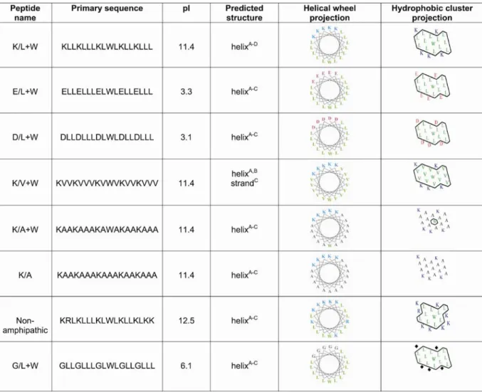
Le but de cette étude était de déterminer le mécanisme utilisé par les hélices alpha pour cibler des protéines vers les granules. Premièrement, nous avons déterminé les caractéristiques biophysiques d’hélices alpha aptes à cibler des protéines vers les granules en testant une série d’hélices synthétiques qui varient en termes de la composition des résidus, la charge, l’amphipathicité et l’hydrophobicité. Deuxièmement, nous avons testé si une hélice alpha était nécessaire pour le ciblage vers les granules des trois enzymes PC résidentes des granules (PC1/3, PC2 et PC5/6A). Nous avons également vérifié l’efficacité du ciblage vers les granules et comparé les différences entre les trois enzymes PC résidentes des granules. Troisièmement, nous avons résolu la structure tridimensionnelle d’un des domaines de ciblage de PC1/3. La fonction individuelle de chaque résidu a été déterminée par mutagénèse dirigée.
Nos résultats démontrent que la présence de résidus chargés (négativement ou positivement) et qui sont ségrégés d’une surface hydrophobe dans les hélices alpha jouent un rôle critique dans la fonction des hélices alpha de cibler des protéines vers les granules.
Des hélices alpha ont ciblée PC1/3, PC2 et PC5/6A vers les granules. Une analyse détaillée des structures prédites formées par les domaines de ciblages des enzymes PC démontre une corrélation entre la capacité des hélices de cibler une protéine vers les granules et la présence d’une surface hydrophobe. De plus, la détermination de la structure tridimensionnelle d’un des domaines de ciblage de PC1/3 (PC1/3711-753) a nécessité la
présence d’une micelle composée d’éléments membranaires. La structure et un essai fonctionnel ont démontré la nécessité de la Leucine 745 (L745) pour assurer le ciblage vers les granules.
En résumé, nos résultats démontrent la nécessité de résidus hydrophobes situés dans une hélice alpha pour cibler des protéines vers les granules de sécrétion et ce possiblement à l’aide d’interactions membranaires.
Mots-clés: granules de sécrétion, trafic de protéines, trafic membranaire, résonance magnétique nucléaire
Abstract
Endocrine and neuroendocrine cells contain specialized secretory organelles called dense core secretory granules (DCSGs). These organelles are the repository of proteins and peptides that are secreted in a regulated manner when the cell receives a physiological stimulus. The targeting of proteins to DCSGs is crucial for the generation of peptide hormones including insulin and adrenocorticotropic hormone. While previous work has demonstrated that proteins destined to a variety of cellular destinations contain targeting sequences, no single consensus sequence for secretory granule sorting signals has emerged. It has been previously shown that membrane associated alpha helical domains in the carboxy-terminal tail of the prohormone convertase PC1/3 play an important role in the ability of this region to direct DCSG targeting.
Our hypothesis is that alpha helices are responsible for redirecting proteins to DCSGs and that this targeting is mediated by protein-lipid interactions.
The goal of this study was to determine the mechanism used by alpha helices to direct sorting of proteins to DCSGs. First, we determined the biophysical characteristics of sorting helices by testing a series of engineered alpha helices that vary in residue composition, charge, amphipathicity and hydrophobicity. Second, we tested whether an alpha helix was necessary for the DCSG targeting of the three DCSG granule resident PC enzymes (PC1/3, PC2 and PC5/6A). We also assessed the efficiency of entry into granules and compared differences between the three granule-resident PC enzymes. Lastly, we solved the three-dimensional solution structure of one of the PC1/3 helical DCSG-sorting domains. The function of the individual amino acids making up this DCSG-sorting domain was tested by site-directed mutagenesis.
Our results demonstrate that the presence of charged (either positive or negative) amino acids, spatially segregated from a hydrophobic patch in the alpha helices of secretory
proteins plays a critical role in the efficiency of alpha helices to direct secretory granule sorting. Alpha helices were also critical to target PC1/3, PC2 and PC5/6A to the regulated secretory pathway. Analysis of the predicted structures formed by these three granule sorting helices showed a correlation between their granule sorting efficiency and the clustering of hydrophobic amino acids in their granule targeting helices. Moreover, the determination of the three-dimensional solution structure of one of the DCSG-sorting domains of PC1/3: PC1/3 711-753 required the presence of a micelle. The structure in
conjunction with a functional DCSG-sorting assay revealed the importance of leucine 745 (L745) located within the alpha helix in mediating DCSG sorting.
In summary, our results demonstrate the requirement of hydrophobic residues situated in alpha helices to direct proteins to DCSGs possibly through membrane interactions.
Keywords: secretory granules, prohormone convertases, protein trafficking, membrane trafficking, nuclear magnetic resonance
Table of contents
Résumé... iii
Abstract ... v
List of tables... xi
List of figures ... xii
List of abbreviations... iii
Acknowledgements ... vii
Chapter 1 ... 1
INTRODUCTION ... 1
1.1 Protein codes: The advent of the signal sequence... 2
1.2 Dense core secretory granules within the secretory pathway ... 4
1.2.1 Early secretory pathway... 4
1.2.2 The trans Golgi network as a molecular sorting station... 5
1.3 Three truths and three postulates ... 11
1.4 A plethora of signals a paucity of consensus ... 13
1.4.1 Membrane associated tethers ... 15
1.4.2 Membrane lipids implicated in vesicular traffic ... 16
1.4.3 Cargo interactions via membrane tethers... 22
1.4.3.1 Carboxypeptidase E ... 22
1.4.3.2 Paired Basic Amino Acids ... 22
1.4.3.3 Interaction with DCSG transmembrane proteins ... 23
1.5 Aggregation... 24
1.5.1 The granular milieu ... 24
1.5.2 Aggregation chaperones... 26
1.5.3 A role for cargo in directing DCSG biogenesis? ... 27
1.6 In vivo models of granule biogenesis ... 29
1.8 Granule active Prohormone Convertases as a model to study sorting mechanisms of a
DCSG cargo/tether ... 32
1.8.1 The cell biology of the granule-localized PC enzymes... 34
1.8.1.1 PC1/3 and PC2 ... 34
1.8.1.2 PC5... 36
1.9 Objectives of the dissertation ... 38
Chapter 2 ... 39
A Hydrophobic Patch in a Charged Alpha Helix is Sufficient to Target Proteins to Dense Core Secretory Granules ... 39
ABSTRACT... 41
INRODUCTION ... 42
EXPERIMENTAL PROCEDURES ... 44
Recombinant plasmid construction ... 44
Mammalian cell culture, transfection and secretion analysis... 45
Protein secondary structure predictions. ... 46
Immunocytochemistry and confocal microscopy ... 46
RESULTS ... 48
Exposed alpha helices direct secretory proteins to granules... 48
Biochemical characteristics of granule-targeting alpha helices ... 55
DISCUSSION ... 62
ACKNOWLEDGMENTS ... 64
REFERENCES... 65
Chapter 3 ... 68
PC1/3, PC2 and PC5/6A are Targeted to Dense Core Secretory Granules by a Common Mechanism ... 68
SUMMARY ... 70
INTRODUCTION ... 71
Recombinant plasmid construction ... 73
Mammalian cell culture, transfection and secretion analysis... 73
Immunocytochemistry and confocal microscopy ... 75
Statistical analysis ... 75
RESULTS ... 76
The secretory granule sorting domain of PC5/6A is contained in the last 38 amino acids of the C-terminus ... 76
The PC5/6A secretory granule sorting domain is predicted to form an alpha helix .... 80
The minimal granule sorting domains of PC1/3, PC2 and PC5/6A selectively re-direct a constitutive protein to granules ... 82
Structural correlates of sorting efficiency ... 86
DISCUSSION ... 88
ACKNOWLEDGMENTS ... 89
REFERENCES... 90
Chapter 4 ... 94
Functional Anatomy of a Secretory Granule Sorting Domain: Solution Structure of the C-terminal Helical Domain of the Protease PC1/3 ... 94
ABSTRACT... 96
INTRODUCTION ... 97
MATERIALS AND METHODS... 100
15 N and 15N/13C-labeled protein expression and purification... 100
NMR sample preparation and data collection... 100
Structure calculations ... 101
Recombinant plasmid construction ... 102
Mammalian cell culture, transfection and secretion analysis... 102
RESULTS ... 104
The NMR solution structure of the PC1/3 DCSG-sorting domain reveals two alpha
helical regions. ... 106
Correlation between the structure of PC1/3 711-753 and its ability to act as a DCSG sorting domain... 111
Mutational analysis of the second helix of PC1/3 711-753... 114
Association of the PC1/3 DCSG sorting domain with micelles ... 117
Identification of a calcium-binding site in PC1/3 ... 119
Mutational analysis of the calcium-binding site of PC1/3 711-753... 123
DISCUSSION ... 125
ACKNOWLEDGEMENTS ... 130
REFERENCES... 131
Chapter 5 ... 139
Conclusion ... 139
5.1 The Carboxy-terminal domain of PC1/3 has a dual function ... 140
5.2 Models on how the late secretory pathway environment contributes to DCSG sorting ... 141
5.3 Concluding remarks ... 147
5.4 Long-term Perspectives... 147
5.5 Where do we go from here? ... 149
Bibliography... 151 Appendix ... I Figure License... I
List of tables
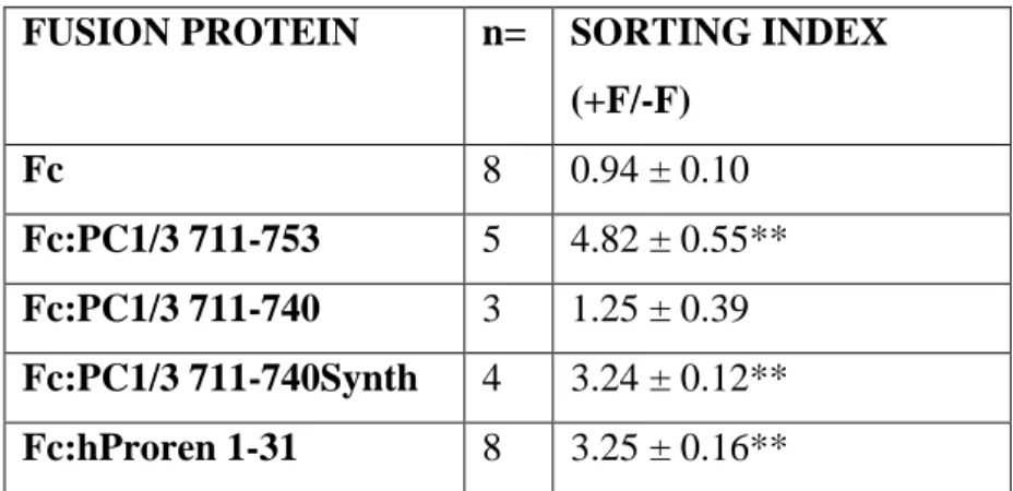
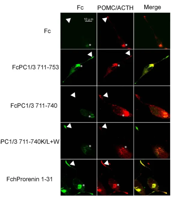
Table 1.1: Examples of canonical sorting signals identified for specific cellular compartment... 3 Table 2.1: Secretory granule sorting efficiency of fusion proteins in transfected AtT-20 cells. ... 51 Table 2.2: Biochemical characteristics of the synthetic helices tested for granule sorting activity:... 58 Table 2.3: Secretory granule sorting efficiency of Fc fusion proteins containing various alpha helical peptides with differing biochemical properties. ... 60 Table 4.1 Structural statistics for of PC1/3 711-753... 107
List of figures
Figure 1.1: Schematic representation of the secretory pathway in endocrine and
neuroendocrine cells. ... 6
Figure 1.2: Dense core secretory granule maturation. ... 10
Figure 1.3 Proteins sorted to dense core secretory granules (DCSGs) can be functionally divided into three groups... 14
Figure 1.4: Membrane lipids separated as structural or signaling molecules. ... 20
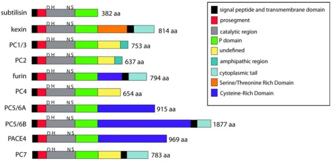
Figure 1.5: The Prohormone Convertases... 33
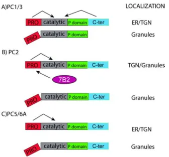
Figure 1.6: The Granule Resident members of the PC family... 37
Figure 2.1: A variety of alpha helices are able to target proteins to secretory granules. ... 50
Figure 2.2: A variety of alpha helix-containing domains target fusion proteins to secretory granules. ... 53
Figure 2.3: Granule-targeting efficiency of alpha helices with differing biochemical properties... 59
Figure 2.4: The G/L+W fusion protein is localized to the cell surface... 61
Figure 3.1 The PC5/6A granule-sorting domain is contained in the last 38 amino acids of its C-terminus... 78
Figure 3.2: The PC5/6A C-terminus contains a granule-sorting domain predicted to form an alpha helix. ... 81
Figure 3.3 (A) and (B): Comparison of the sorting capacity of fusion proteins containing various PC family C-termini: ... 83
Figure 3.4: Comparison of the sorting capacity of fusion proteins containing various PC family C-termini:... 84
Figure 3.5: Predicted biophysical properties of C-terminal granule sorting helices in PC enzymes... 87
Figure 4.1: PC1/3 711-753 requires a micelle for structure determination ... 105
Figure 4.3: PC1/3 711-753 contains two amphipathic helices. ... 109
Figure 4.4: PC1/3 711-753 phylogenetic analysis ... 110
Figure 4.5: Functional assay of the two helices in PC1/3 711-753... 112
Figure 4.6: Site-directed mutagenesis of the second helix of PC1/3711-753... 115
Figure 4.7: PC1/3 711-753 interactions with a CHAPS micelle ... 118
Figure 4.8: PC1/3 711-753 interacts with calcium ... 120
Figure 4.9: Calcium-binding site in PC1/3 711-753... 121
Figure 4.10: PC1/3 711-753 backbone dynamics ... 122
Figure 4.11: Mutational analysis of the calcium binding site on the sorting of PC1/3711-753 to secretory granules. ... 124
Figure 4.12: Hydrophobic molecular surface representation of PC1/3 711-753... 129
Figure 5.1: Model for the sorting of PC1/3 to DCSGs ... 144
List of abbreviations
ACTH: adrenocorticoptropic hormone ACTH: adrenocorticoptropic hormone
Alpha-SNAP: alpha soluble NSF attachment protein ANF: Atrial natriuretic factor
AP-1: adaptor protein-1 AP-2: adaptor protein-2
BDNF: Brain-derived neurotropic factor C-ter: C-terminal domain
Cer: ceramide
CgA: chromogranin A CgB: chromogranin B
CHAPS: 3-[(3-Cholamidopropyl) dimethylammonio]-1-propanesulfonate COP-II: coat protein complex-II
COS: CV-1 in origin
COSY: correlation spectroscopy CPE: carboxypeptidase E CT-HSQC: constant time HSQC DAG: diacylglycerol
DCSG dense core secretory granules Dhc: dehydrocholesterol
DPC: dodecyl phosphocholine
EIF4e: eukaryotic translation initiation factor 4E ER: endoplasmic reticulum
GFP: green fluorescent protein GH4: growth hormone 4 cells
GlcCer: glucosylceramide GST: glutathione-S-transferase
H-ATPase: proton adenosine triphosphate -ase
HDEL: mammalian endoplasmic reticulum retention signal (histidine-aspartic acid- glutamic acid-leucine)
HMQC: heteronuclear multiple quantum coherence-total correlation HSQC: Heteronuclear Single Quantum Coherence
HPLC: high pressure liquid chromatography INS-1: insulinoma-1
ISGs: immature secretory granules
KDEL: mammalian endoplasmic reticulum retention signal (lysine-aspartic acid- glutamic acid-leucine)
NMR: nuclear magnetic resonance NOEs Nuclear Overhauser Effects
NOESY: Nuclear Overhauser Effect Spectroscopy NSF: N-ethylmaleimide sensitive fusion protein PA: phosphatidic acid
PAM: Petidyl-α-amidating monooxygenase PBS: phosphate buffered saline
PC: proprotein convertases PC12: pheochromocytoma 12 cells PE: phosphotidylethanolamine PG: phosphatidylglycerol PI: phosphatidylinositol PM: plasma membrane PN-1: protease nexin-1 POMC: proopiomelanocorticotropin Pro-SAAS: non acronymic
PS: phosphatidylserine
PTB: polypyrimidine binding protein PtdCho: phosphatidylcholine
RER: rough endoplasmic reticulum REST: RE-1 silencing transcription factor RMSD: random mean standard deviation SEAP: Secreted alkaline phosphatase SgII: secretogranin II
SgIII: secretogranin III SM: sphingomyelin
SNARES: N-ethylmaleimide sensitive fusion protein attachment receptors Syt IV: synatoptotagmin IV
SEM: standard error on mean
T-SNARE: target N-ethylmaleimide sensitive fusion protein attachment receptors TAD: torsion angle dynamics
TGN: trans Golgi network Type I membrane protein:
a single transmembrane stretch of hydrophobic residues, with the portion of the polypeptide on the NH2-terminal side of the TM domain exposed on the exterior side of the membrane and the COOH-terminal portion exposed on the cytoplasmic side
V-SNARE: vesicle N-ethylmaleimide sensitive fusion protein attachment receptors VAMP-2: vesicle-associated membrane protein-2
Acknowledgements
First and foremost, I am indebted to my supervisor Timothy Reudelhuber. He inspired me from the beginning and constantly pushed me to become a rigorous scientist. I owe him the passion I now have for research. His supervision style was very simple: make sure everything is perfect and then I will guide you. In return, I received training on the bench, manuscript preparation, poster presentation and everything else all my fellow graduate students from neighboring labs never claim to have received.
My co-supervisor, Jim Omichinski also gratefully accepted to have a biochemist without any structural biology experience such as myself attempt an NMR project. Jim’s daily advice and the nightly emails to boost my confidence when all experiments failed permitted me to complete the structural portion of my research project. Without a doubt Tim and Jim will be lifelong friends a true testament to the respect I have for my supervisors.
The help received from the entire lab is also greatly appreciated. Marie-Josée Lacombe (Forskolin) never refused to make a construct or to complete a pulse-chase assay. Her precision is something I will never equal. In the end, it was always best to just leave some things to Marie-Jo. Her friendship also was golden during my years at the lab and I will never forget the first day she brought me up to the lab. Chantal Mercure: experience and knowledge. Need I say more? She basically knows everything there is to know about the lab. Our discussions around the table in the office rivaled great political debates and some days we could have solved the whole world’s problems over tea and an espresso. Nadheige Lochard, a graduate student was always there for me. We constantly pushed each other to excel in science and in the end I hope to have become half as good a scientist as her. Through the good or the bad and everything in between Nadheige was always there for
me. Matei Mireuta was my partner in crime. Summer student extraordinaire Matei never needed to be told things twice. Science related things that are. As much as we worked, we had great times. Dismantling an FPLC in the cold room until it worked, nothing really stopped us. I will never see picture framing in the same way after having worked with Matei. During the last years of my thesis, I met a ‘spectacular postdoc’ Paola Di Lello (Pat). She basically had the patience to train me as a spectroscopist even though on some days I could not even pick out alanines and isoleucines. I owe her a lot as Paola donated (charity time) many hours at the expense of her own project to make sure I got something out of mine. Paola is a true sister that I can never ever forget and I respect her immensely.
While the lab was great, on harder days it is my wife and love of my life Genny that kept me going. Recently she gave me the greatest gift of all: our son Elias. I adore you guys and apologize for not spending more time at home. Genny was there since the beginning and she deserves a medal for putting up with it all.
My mom, dad and sister were also always supportive of my career choices and never doubted any of my moves. I basically have the best wife and family in the world and am not afraid to say that.
I also thank both the FRSQ and University of Montreal for funding and all of the support staff at the IRCM. Many collaborators made my project much easier and I would like to thank Nabil Seidah, Claude Lazure, Pascale Legault, Michel Chrétien, Ajoy Basak, Gaétan Thibault, Christian Deschepper, Dror Warshawski, Alexandre Arnold and Isabelle Marcotte for excellent science.
Chapter 1
INTRODUCTION
The intracellular sorting of peptide hormone precursors to dense core secretory granules (DCSGs) is essential for their bioactivation. Despite the fundamental importance of this cellular process, the nature of the sorting signal(s) for entry of proteins into DCSGs remains a source of vigorous debate. In this chapter, I will highlight recent discoveries that are consistent with a model in which several protein domains, acting in cell-specific fashion and at different steps in the sorting process, act in concert to regulate the entry of proteins into DCSGs.
1.1 Protein codes: The advent of the signal sequence
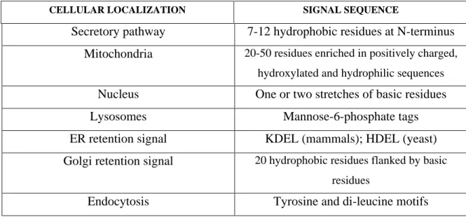
The accurate sorting of proteins to their cellular destinations is of fundamental importance in biology and must occur with high precision in the context of a highly concentrated and extremely complex mixture of proteins. The identification of the “codes” carried by proteins that ensure their proper intracellular sorting has been a topic of intense and fruitful research for more than 40 years. As a result, most introductory textbooks now include descriptions of the canonical signals, which direct the sorting of proteins to specific cellular destinations (Summarized in Table 1.1). Thus, proteins are imported in the secretory pathway based on the presence of an N-terminal cleavable signal peptide containing 7-12 hydrophobic amino acids (1). Mitochondrial-destined proteins, contain an N-terminal code termed the ''presequence'' consisting of 20-50 residues enriched in positively charged (lysine or arginine), hydroxylated (tyrosine, threonine or serine) and hydrophilic sequences (2). The mitochondrial ''presequence'' also has the ability to form amphiphilic alpha helices. As for nuclear proteins, they contain the ''nuclear localization signal'' consisting of either one (monopartite) or two (bipartite) stretches of basic amino acids (3). The sugar residues of proteins destined to lysosomes contain mannose-6-phosphate which bind to the mannose-6-mannose-6-phosphate receptor to direct lysosomal sorting (4). Moreover, proteins can also contain ''codes'' enabling them to be retained in a specific
cellular location. The lysine-aspartic acid- glutamic acid-leucine (KDEL) sequence in mammals (5) and histidine-aspartic acid-glutamic acid-leucine (HDEL) sequence in yeast (6) serve as endoplasmic reticulum (ER) retention signals while a stretch of 20 hydrophobic residues flanked by basic residues results in a Golgi retention signal (7). Finally, proteins that are endocytosed contain tyrosine and di-leucine motifs (8). All of the above sequences are sufficient to redirect proteins to the specific organelle defined by the code.
However, a similar canonical ''code'' has not been established for directing proteins to dense core secretory granules (DCSG). These cytoplasmic organelles, found uniquely in endocrine and neuroendocrine cells, store hormones, proteases and signaling molecules until the cell receives a signal for their release. For example, the inactive proinsulin prohormone is activated within DCSGs by specific proteases and will be released when blood glucose levels are elevated (9). As such, DCSGs are the key component in the regulated secretory pathway. Why has the identification of DCSG sorting signals been such an elusive goal?
CELLULAR LOCALIZATION SIGNAL SEQUENCE
Secretory pathway 7-12 hydrophobic residues at N-terminus Mitochondria 20-50 residues enriched in positively charged,
hydroxylated and hydrophilic sequences Nucleus One or two stretches of basic residues
Lysosomes Mannose-6-phosphate tags
ER retention signal KDEL (mammals); HDEL (yeast) Golgi retention signal 20 hydrophobic residues flanked by basic
residues
Endocytosis Tyrosine and di-leucine motifs
Table 1.1: Examples of canonical sorting signals identified for specific cellular compartment
1.2 Dense core secretory granules within the secretory pathway
1.2.1 Early secretory pathway
There has been a lot of debate not only about how DCSG sorting occurs, but also just where in the cell this triage takes place. All cells have the capacity to rapidly secrete proteins after their transit through the constitutive secretory pathway. Thus, proteins destined for the secretory pathway will first transit to the ER. It is here that the N-terminal secretory pathway signal peptide sequence (Table 1.1; secretory pathway) will be recognized by the signal recognition particle located on the signal recognition receptor found on the ER membrane (10). In eukaryotes, the Sec 61/Sec Y complex forms a conducting channel ensuring the co-translational translocation of secretory pathway proteins to the ER lumen (11). Upon exiting the ER at specific areas lacking ribosomes (12), properly folded secretory proteins will be transported to the Golgi in vesicles coated with coat protein complex-II (COP-II) (13) with the proper transfer of cargo from vesicles ensured by specific vesicular adaptors termed soluble N-ethylmaleimide sensitive fusion protein attachment receptors (SNARES) (14). SNARES are located on the cytosolic face of vesicles (v-SNARE) and will dock a vesicle via an interaction with a target vesicle’s t-SNARE. In the Golgi, proteins are glycosylated in the various cisternae and stacks, which range from the cis, the medial and ending with the trans Golgi networks (TGN) in the anterograde trafficking of secretory proteins. During the 1980s, there was considerable debate on whether trafficking through the Golgi occurred using the same vesicle (cisternal maturation) or using cargo transferred via SNARE adaptors to different vesicles (vesicular trafficking model, For Review see (15)). In 2006, both Losev et al. (16) and Matsuura et al. (17) showed immunofluorescent images of differentially tagged Golgi cisternae. Using
time-lapse video microscopy, both groups demonstrated that the Golgi cisternae would change color over time indicating that the same vesicle was being trafficked along the Golgi. These findings are consistent with a cisternal maturation model for Golgi trafficking and effectively resolved (for the time being) the vigorous debate.
1.2.2 The trans Golgi network as a molecular sorting station
Upon reaching the terminal Golgi stacks in the TGN, proteins are transported in budding vesicles to specific various destinations. In all mammalian cells, proteins can be trafficked to various destinations. Proteins will either bud off into low density vesicles and be secreted via a non-regulated constitutive pathway or be routed towards the intracellular endosomal pathway (18;19). Moreover, certain proteins can be re-routed to earlier compartments via recycling mechanisms (20). In polarized cells, there is a differential sorting originating from the TGN to either the apical or basolateral surface of the cell ensuring the asymmetrical distribution of certain proteins (21). In contrast, a unique regulated pathway of secretion exists in endocrine and neuroendocrine cells where exocytosis of high density DCSGs is tightly regulated (Figure 1.1). A great deal of evidence supports the view that in the appropriate cell type, DCSG sorting signals can re-direct proteins from the constitutive secretory pathway to DCSGs confirming that it is not a default secretory pathway, but rather a pathway that requires specific biophysical properties of the stored protein or the recognition of a “sorting signal” by the cellular machinery. The nature of this DCSG sorting signal has not been clearly defined to date.
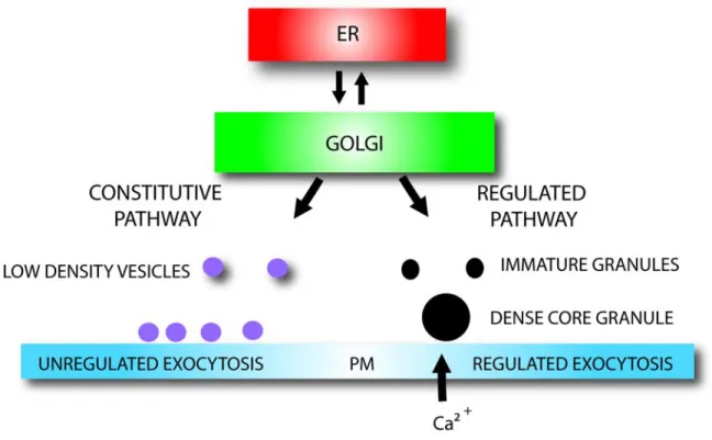
Figure 1.1: Schematic representation of the secretory pathway in endocrine and neuroendocrine cells.
In the anterograde trafficking pathway, proteins pass through the endoplasmic reticulum (ER), then through the cis, medial and trans Golgi networks. The trans Golgi network serves as a sorting station where non-granule proteins are packaged in low-density vesicles and are secreted in an unregulated manner across the plasma membrane (PM). Granule proteins are first packaged in immature secretory granules that fuse and form dense core secretory granules. The dense core secretory granules are secreted when the cell receives an influx of calcium.
Some groups have proposed that DCSG sorting occurs through the action of a sorting “receptor” present in the trans-Golgi network (TGN), which latches onto granule-destined proteins at sites where nascent granules will bud. This model has been referred to as the “sorting by entry” model and implies that only DCSG proteins can be present in this organelle. Studies performed by Chung et al. in 1989 provided the first support for the “sorting by entry” hypothesis. They used peptide hormones as affinity ligands to find partner proteins in canine pancreatic tissue. Using this approach, they identified a 25-kDa protein considered as a potential DCSG sorting receptor (22). Further experiments revealed that this protein was identical to chymotrypsinogen and was not detectable in every endocrine tissue. Furthermore, chymotrypsinogen bound granule proteins with very low affinity (23). One of the limitations of Chung’s studies was that it was difficult to distinguish between proteins present in the TGN and DCSG proteins themselves. The inherent difficulty of separating the closely spaced secretory pathway compartments is a complicating factor when identifying DCSG and non-DCSG proteins and this analytical limitation has made the identification of compartment-specific partners difficult. In addition, the presence of the constitutively secreted glycosaminoglycan protein in DCSGs suggests that proteins other than those destined for regulated secretion may enter DCSGs (24).
About 10 years later, Cool et al. (25) postulated that carboxypeptidase E (CPE) could be the bona fide DCSG sorting receptor. All of their observations stemmed from an obese mouse line with a spontaneous mutation in CPE: the Cpe fat/fat mouse. Isolation of CPE from the Cpe fat/fat mouse revealed a decreased binding of various peptide hormones to the mutated CPE when compared to native CPE. In addition, the authors reported that the DCSG protein proopiomelanocorticotropin (POMC) was no longer stored in DCSGs in the Cpe fat/fat mouse. However, competitors were quick to point out in independent experiments that hormone secretion was normal in the Cpe fat/fat mouse (26). A common argument against CPE as a sorting receptor is that no single receptor can exist to ensure the 1:1 ratio needed to ensure the functionality of the “sorting by entry” theory as the flow of DCSG
proteins traversing the regulated secretory pathway is too large. However, it should be noted that the signal recognition particle receptor is sufficient to handle the large traffic of proteins being co-translationally translocated across the ER membrane.
With all of this conflicting data, “the sorting by entry” view was losing support. Simultaneously, convincing evidence was presented that in cells that generate DCSGs, all of the contents of the TGN are initially encapsulated into nascent granules termed immature secretory granules (ISGs). This finding led to the development of the “sorting by retention” model, which proposes that those proteins destined to be secreted constitutively are progressively extruded in low density vesicles as the granule matures, ultimately leaving only the correct cargo protein in the mature DCSG. A detailed mechanism explaining this extrusion has been clearly identified (Figure 1.2) as well as the mechanism of granule maturation. Thus, the maturation of ISGs involves the progressive acidification of the ISG lumen (27) and the homotypic SNARE-dependent fusion between the various ISGs to form the mature DCSG; a process requiring the soluble cytosolic factors N-ethylmaleimide sensitive fusion protein (NSF) and alpha soluble NSF attachment protein (alpha-SNAP) (28). A member of the synaptotagmin calcium-sensing family of proteins, synatoptotagmin IV (syt IV), is critical to ensure the ISG homotypic fusion event (29). It is adaptor protein-1 (AP-protein-1), a clathrin protein adaptor that assists in the budding of all non-DCSG proteins from the lumen of the ISGs via clathrin coated vesicles. The non-granular proteins in their clathrin coated vesicles are believed to be transported to the endosomal compartment (30). The resulting DCSGs produced are then ready to exocytose on the plasma membrane through the interaction of their granule specific vesicle-associated membrane protein-2 (VAMP-2) v-SNARE located on the cytosolic face of the DCSG and the plasma membrane’s t-SNARE (31). While the ''sorting by retention'' model provides clues concerning granule formation and the trafficking of the granule from the TGN to the plasma membrane, the proteins identified to date are all cytosolic and do not explain the selective retention of cargo within the granule core. Perhaps the best such example is provided by a recent proteomics analysis of bovine adrenal medulla granules using a
phosphatidyl inositol (4, 5) bisphosphate pull-down assay (32). Phosphatidyl inositol (4,5) bisphosphate is a lipid implicated in the exocytosis of granules and is situated on the cytosolic face of the plasma membrane. The proteomics analysis did not reveal any granule cargo proteins but unsurprisingly identified cytosolic proteins involved exclusively in exocytosis. What about the inside of the granule?
Regardless of which theory is correct, a subset of proteins is somehow correctly stored in DCSGs. These granule resident proteins must contain “codes” allowing efficient targeting to this organelle. Moreover, these proteins may be interacting with partner proteins in order to enter granules while the granular environment itself may also influence the efficient targeting of granule proteins. Can a mechanism be proposed to help explain this critical event?
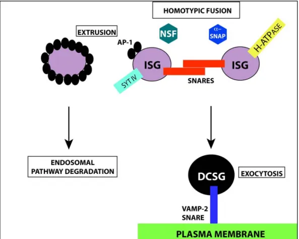
Figure 1.2: Dense core secretory granule maturation.
Equivalent immature secretory granules (ISGs) go through a homotypic fusion event facilitated by SNARE adaptors located on the ISG surface. The homotypic fusion is assisted by the cytosolic factors NSF and alpha-SNAP, the vesicle bound synaptotagmin IV (syt IV) protein and a proton adenosine triphosphate -ase (H-ATPase) assuring the acidification of the ISG lumen. Proteins not destined for DCSGs are extruded via AP-1coated vesicles that attract clathrin and are targeted to the endosomal pathway for degradation. The mature DCSG will fuse with the plasma membrane (PM) and be exocytosed via a specific interaction with the DCSG’s VAMP-2 SNARE.
1.3 Three truths and three postulates
The first truth is that regardless of the site at which sorting occurs, a mechanism has to exist that establishes and then maintains the segregation of DCSG cargo proteins from those that are constitutively secreted. Thus, it is a reasonable postulate that some mechanism exists to anchor the appropriate cargo proteins to the DCSG as it forms or matures.
A second truth is that the sorting of proteins to DCSGs is a prerequisite for certain post-translational processing steps in hormone and protease activation. For example, the conversion of proinsulin to active insulin only occurs in the acidic (pH=5.5) DCSG compartment (9). Orci et al. showed that ionophores disrupting the proton exchange mechanism in the late secretory pathway also blocked the activation of proinsulin (9). The cleavage of POMC to its many active peptides including adrenocorticoptropic hormone (ACTH) is equally influenced by the specific ion composition present in the DCSG (33). In addition, the proteolytic activation of prorenin to renin can also only occur in DCSGs (34). All of the above examples indicate that the precursors are encapsulated in the nascent secretory granules. This is an efficient mechanism for the organism because it ensures that the secretion of the active hormones or proteases is under appropriate physiological control. However, for granule-restricted activation to occur it is necessary that both the protein precursors and the appropriate processing enzymes end up in the same DCSG. In the case of proinsulin, this means that the proprotein convertases 1/3 and 2 (PC1/3 and PC2) as well as CPE, all of which are required for generation of active insulin, have to be co-targeted with proinsulin in the budding granules. Thus, a second postulate is that a mechanism exists to ensure efficient co-targeting of protein precursors and their processing enzymes in the same organelle.
DCSGs also share by definition the distinguishing trait of a core that appears dark or dense in electron micrographs. However, in spite of this common appearance, there may be important functional and mechanistic differences in DCSGs. For example, the gonadotropes of the pituitary store luteinizing hormone and follicle stimulating hormone in separate DCSGs and their release is independently controlled (Reviewed in (35)). Likewise, there are two types of DCSGs in chromaffin cells containing either epinephrine or norepinephrine and these are morphologically distinct (36). Norepinephrine granulesare larger and electron opaque with a prominent halo between the granulemembrane and the dense core. The epinephrine granules are smaller, finely granular structures that fill the enclosingmembrane and have no halo. It appears that these differences can depend on the biophysical characteristics of the cargo itself (37). Accordingly, Sobota et al. demonstrated that proteins that can self-aggregate would be stored in distinct DCSG from proteins that lack tertiary structure (37). Thus, cargo itself can dictate the composition of a DCSG independently of the maturation of the granule.
The signals for targeting proteins to DCSGs also show tissue-specific variations: Removal of 90 amino acids from the C-terminus of the granin chromogranin A (CgA) prevents its sorting to DCSGs in pituitary growth hormone 4 cells (GH4), but has no effect on DCSG sorting in sympathoadrenal pheochromocytoma 12 cells (PC12) (38). Likewise, POMC is efficiently stored in DCSGs when transfected into cultured pituitary cells, but not in sympathetic neurons (39). Thus, a third truth is that not all DCSGs are alike and it is a reasonable postulate that DCSGs can be assembled, even within the same cell, through more than one mechanism.
Could some of these truths explain the difficulties in reaching consensus on the protein signals necessary for DCSG targeting?
1.4 A plethora of signals, a paucity of consensus
There has been no shortage in the variety of DCSG sorting mechanisms proposed in the last 2 decades: these include protein domains that interact with or that traverse membranes and that may or may not interact with additional proteins on the cytoplasmic side of the DCSG, proteins proposed to be a “master switch” for granule formation, universal granule cargo receptors, protein domains that mediate aggregation in the late TGN, certain paired basic protease cleavage sites or alpha helices in secretory proteins, disulfide constrained loops, acidifying proton pumps and other mechanisms. As a result, investigators have become progressively entrenched in defending their favorite mechanisms and commonly use the descriptors “controversial” and “difficult to repeat” to describe the work of others in their publications. Nevertheless, it is possible to accommodate most of these findings into a model that subdivides targeting function into three components (Figure 1.3): membrane associated (or traversing) tethers, tether-associated cargo and aggregation.
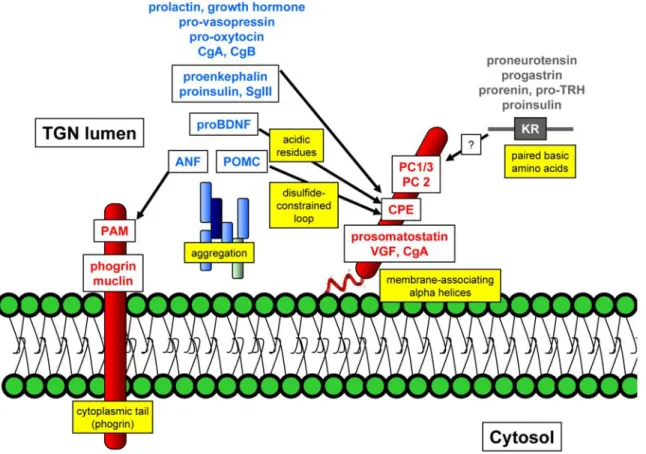
Figure 1.3: Proteins sorted to dense core secretory granules (DCSGs) can be functionally divided into three groups.
Tethers (shown in red) either traverse or associate with membranes. Many DCSG cargo proteins also aggregate to form the dense core (shown in blue) and these aggregates may contain more that one protein. Some DCSG proteins associate with membrane tethers (arrows). The highlighted yellow boxes indicate the various protein domains or mechanisms that have been implicated in DCSG sorting. Note that some proteins (such as insulin) may have multiple DCSG sorting mechanisms. See text for details and abbreviation
1.4.1 Membrane-associated tethers
The fact that many DCSG proteins are either membrane-associated or traverse the membrane is significant. Petidyl-α-amidating monooxygenase (PAM), muclin and phogrin are all type I membrane proteins. PAM is an enzyme catalyzing the alpha-amidation of extended peptides containing glycine residues (40). In the pituitary and brain, PAM is in the lumen of the DCSG and anchored via its carboxy terminal transmembrane domain (40). Muclin is anchored in the DCSG of pancreatic acinar cells where it binds O-sulfated proteins (41). The fact that both PAM and muclin are membrane anchored in granules ensures that both these proteins will not be secreted constitutively. Phogrin is a protein tyrosine phosphatase present in many neuronal and endocrine cell types with a granule sorting domain located in the cytoplasmic tail of the protein (42;43). Although the exact nature of the signal is still debated, it appears that this domain can bind the clathrin adaptor proteins AP-1 and adaptor protein-2 (AP-2) in vitro (44). When one considers that vesicular transport proteins are present on the cytosolic face of vesicles, the interactions described between phogrin and the AP-1 and AP-2 proteins provide a unique means of communication between the granule cargo proteins and the membrane domains or cytoplasmic proteins that will define the budding DCSG.
DCSG proteins can also interact with the membrane without traversing it. The membrane-binding domain of the granule-resident protein CPE is located in the final 22 residues of its C-terminal domain and is proposed to have a shallow membrane interaction (45). The CPE DCSG sorting domain adopts an alpha helical secondary structure (45). The prohormone convertases PC1/3 (46), and PC2 (47) are also targeted to DCSGs and as for CPE there is agreement that the granule sorting is mediated by short alpha helical domains. PC1/3 contains a region predicted to form an amphipathic alpha helix in its C-terminal domain between residues 711-753 (48). An alpha helical domain has also been
shown to be important for targeting prosomatostatin (49), CgA (50) and VGF (51) to DCSGs. Interestingly, both the alpha helices in the targeting sequences of prosomatostatin and CgA are amphipathic like PC1/3 apart from being located on the N-terminus. Specifically for prosomatostatin, two leucine residues located at positions 7 and 11 of the alpha helix respectively, form a hydrophobic pocket critical in targeting prosomatostatin to DCSGs (49) suggesting yet again a possible membrane interaction. In summary, this group of DCSG proteins could therefore be tethered to the membranes of the TGN or the maturing granule. Since many of the DCSGs proteins are membrane tethered could the membrane itself play an important role in the sorting process?
1.4.2 Membrane lipids implicated in vesicular traffic
Since a large number of DCSG sorting domains interact with membranes, the glycerophospholipids, sphingolipids and cholesterol present in eukaryotic cells may play a pivotal role in targeting proteins to DCSGs.
Membrane lipids can be divided in 3 large subclasses. First, glycerophospholipids contain a glycerol backbone and a polar phosphate group esterified to a choline (Figure 1.4; phosphatidylcholine (PtdCho), shown as PC in figure), an ethanolamine (phosphotidylethanolamine, PE; not shown in Figure 1.4), a serine (phosphatidylserine, PS; not shown in Figure 1.4) or an inositol head group (phosphatidylinositol, PI). Two non-polar side chains of varying length are attached to the glycerol backbone. Accordingly, these lipids can be modified resulting in phosphorylated inositol head groups (Fig. 1.4; phosphatidylinositol phosphate, (shown as PIP in figure) and phosphatidyl inositol 2-phosphate, shown as PIP2 in figure). In contrast, the second class of membrane lipids: sphingolipids contain a ceramide (Figure 1.4; ceramide, Cer) backbone instead of the glycerol backbone found in glycerophospholipids (52). Structurally, most of the glycerophospholipids adopt a perfect cylindrical geometry due to the diametrical
equivalency of both the head groups and non-polar side chains (Figure 1.4: Space-filling model structure of PtdCho shown as PC in figure). In contrast, the sphingolipids will form very narrow cylinders due to their saturated highly compact tails (Figure 1.4; Structure of sphingomyelin, SM). This results in very tightly packed lipids that give rise to a gel-like state, which does not abide by the classical fluid-like mosaic hypothesis postulated for membranes. As such, a third class of lipids: sterols such as cholesterol (Figure 1.4; Cholesterol) are localized in areas where sphingolipids are present giving a more fluid membrane (52). Membrane patches rich in sphingolipids and cholesterol are defined as lipid rafts (53).
Most lipids are synthesized at the ER and are then trafficked to various organelles. The ER is composed of PtdCho, PE, PI, PS, phosphatidic acid (PA), Cer, galactoceramide, cholesterol and triacylglycerol while the mitochondrion is composed of PE, phosphatidylglycerol (PG), cardiolipin and PA. Mono- and bi- and triphosphorylated inositol lipids are found on the cytosolic leaflet of the plasma membrane as are sphingolipids (52). At the Golgi, some specific lipids are localized on both the cytosolic and lumenal leaflets. In the lumenal leaflet of the TGN, where protein sorting to DCSGs occurs, a large number of sphingolipids are synthesized including sphingomyelin, galactocerebroside and lactosylceramide (52). These sphingolipids co-exist with cholesterol at the lumen of the TGN (53). As for glycerophospholipids, certain varieties are localized on the cytosolic leaflet of the Golgi such as PtdCho, PE, and PI-4 phosphate. A phosphotransferase capable of removing the phosphate group of PtdCho produces diacylglycerol at the cytosolic leaflet of the TGN (54). The diacylglycerol is interconvertable with PA via a diacylglycerol kinase also present on the cytosolic leaflet of the TGN (55). PA may also be obtained directly from PtdCho using phospholipase D 1 (56;57). The existence of lipid flippases, capable of transferring both diacylglycerol and PA from the cytosolic Golgi leaflet to the lumenal leaflet of the TGN, has been proposed (58). Compelling evidence has also been obtained for the implication of glycerophospholipids on sorting to DCSGs. For instance, protein kinase D is capable of
binding the diacylglycerol present at the TGN and regulate vesicular budding (54). Unfortunately, this observation has not been extended for the regulated pathway and is limited to constitutively secreted vesicles. Instead, work by Shields and colleagues demonstrated that inhibition of PA synthesis by 1-butanol altered the appearance of the Golgi apparatus and decreased secretion in an endocrine cell type (59). Thus, can Golgi lipids be involved in vesicular trafficking and specifically DCSG protein targeting?
CPE preferentially binds sphingolipids and cholesterol-rich membrane fractions (60) while both the prohormone convertases PC1/3 (46) and PC2 (47) bind liposomes enriched in sphingolipids and cholesterol. The lipid raft patches may serve as anchors in the TGN where the alpha helical sequences presented in the C-termini of PC1/3, PC2 and CPE are tethered. Moreover, cholesterol depletion by statin drugs diverted CPE from DCSGs to constitutively secreted vesicles (60). Specific inhibition of sphingolipid synthesis with the drug fuminosin resulted in the re-routing of PC2 to the constitutive pathway without affecting another granule protein CgA (61). This raises the possibility that different granule proteins may be interacting with different lipids but also that direct membrane interactions may not be sufficient for all proteins to be correctly targeted to DCSGs. Furthermore, cholesterol depletion in endocrine cells completely eliminated the regulated pathway (62). Thus, the present tools to study lipids in regulated secretion are limiting especially since membranes are an integral part of the cell. An animal model was used to circumvent the difficulty in blocking lipids by pharmacological agents as is done in cell culture models. The mouse model reiterates the necessity for cholesterol in granule formation (63). The Smith-Lemli-Opitiz dehydrocholesterol 7-/- (Dhc7 -/-) mice are deficient for the 7-Dhc reductase gene, the final step in cholesterol synthesis. The Dhc7 -/- mice contained abnormally sized granules compared to control mice. A biophysical characterization of liposomes containing minimal cholesterol quantities such as in the Dhc7
-/-
mice showed a decrease in membrane curvature leading the authors to believe that diminished cholesterol quantities prevent budding of vesicles from the TGN.
In summary, while specific lipid content has been shown to be important for the sorting of proteins to DCSGs, no specific lipid-protein interactions have yet been identified that would explain DCSG sorting.
Figure 1.4: Membrane lipids separated as structural or signaling molecules.
The main eukaryotic membrane lipids are the glycerophospholipids such as phosphatidylcholine (PtdCho; PC). Their diacylglycerol (DAG) backbone carries a
phosphate (phosphatidic acid; PA) esterified to a choline (forming PtdCho), ethanolamine (forming phosphatidylethanolamine (PE); not shown), serine (forming phosphatidylserine (PS); not shown), or inositol (forming phosphatidylinositol; PI). The phosphosphingolipid sphingomyelin (SM) and the glycosphingolipid glucosylceramide (GlcCer) have a ceramide (Cer) backbone, consisting of a sphingoid base (such as sphingosine; Sph), which is amide-linked to a fatty acid. Lipids can be interconverted by the actions of kinases and phosphatases. Adapted by permission from Macmillan Publishers Ltd: Nature Reviews Molecular Cell Biology, Volume 9, Issue 2 copyright 2008
1.4.3 Cargo interactions via membrane tethers
1.4.3.1 Carboxypeptidase E
A second group of granule-sorting domains may act by binding cargo proteins to the granule-tethered proteins described in the previous sections. For example, CPE has been proposed to interact with a number of granule cargo proteins including proenkephalin, proinsulin, POMC (64) and secretogranin III (SgIII) (65). POMC binds CPE via an amino-terminal disulfide constrained hydrophobic loop (64). Brain-derived neurotropic factor (BDNF) is also targeted to the regulated secretory pathway through an interaction with membrane-tethered CPE (66). Analysis of superimposed X-ray structural models identified key acidic residues implicated in a complex between CPE and BDNF. Interestingly, some of these cargo-CPE interactions promote retention in secretory granules, even though some of the cargos are not enzymatic substrates of CPE reinforcing the claim for a tether. Furthermore, no common mechanism for interaction of these cargo proteins with CPE has yet emerged.
1.4.3.2 Paired Basic Amino Acids
Paired basic amino acids have also been reported to direct DCSG sorting in some proteins including proneurotensin (67), prorenin (68), prothyrotropin releasing hormone (69) and progastrin (70) and to increase the sorting efficiency of proinsulin (71). In the analyses carried out to date, it appears that these paired basic amino acids must constitute a cleavage site for one of the granule-resident prohormone convertases (PC1/3 or PC2) to function as a granule sorting domain since changing the cleavage site to one recognized by
furin (another member of the family which cleaves its substrates in the early secretory pathway) causes the proteins to be secreted through the default constitutive pathway (68). These results raise the possibility that certain DCSG-targeted proteases can act as sorting chaperones for their substrates in addition to being processing proteases. The interaction of substrates with membrane-tethered proteases is thus another potential granule sorting mechanism.
1.4.3.3 Interaction with DCSG transmembrane proteins
DCSG transmembrane domains are not inert and they too are capable of assisting the correct entry of granule-resident proteins. In fact, muclin has been suggested to act as a granule cargo “receptor” in pancreatic cells through its binding of sulfate groups on O-linked glycosylated proteins (41). Atrial natriuretic factor (ANF) has also been shown to be tightly bound to the membranes of atrial myocyte secretory granules through its interaction with PAM (72), although it is not a substrate of PAM. Since there are no-known alpha-amidated peptides in the cardiac atrium (72), no enzymatic role can be postulated for PAM apart from a DCSG sorting role. Recently, PAM has been found to interact with the new partner proteins: Kalirin/Trio. Overexpression of the Rho guanine nucleotide exchange factors Kalirin/Trio diverted the granule protein POMC to the constitutive secretory pathway and resulted in the secretagogue-independent secretion of unprocessed POMC (73). Pharmacological inhibition of the guanine nucleotide exchange activity of Kalirin/Trio restored POMC localization to DCSGs and processing of POMC to its many peptides. Since Kalirin/Trio both interact with the DCSG integral membrane protein PAM, this study indicates how partner proteins can be affected by signaling molecules and subsequently regulate DCSG protein localization thereby ensuring substrate activation. Thus, a variety of interactions with “tethers” may serve to target proteins to secretory granules. Notably, if this mechanism is correct, it would in some cases provide a means to ensure that processing enzymes and their substrates end up in the same DCSGs.
1.5 Aggregation
A third category of granule targeting mechanisms involves formation of high molecular weight protein complexes or aggregates. Indeed, many granule targeted cargo proteins have the ability to multimerize or aggregate, leading in most cases to the formation of electron dense cores. What causes this aggregation and how does it affect the formation of DCSGs?
1.5.1 The granular milieu
The protein concentration in the late secretory pathway is highly elevated. For example, proinsulin has a measured concentration of 42 mM in pancreatic beta-cell granules (74) where it can form hexamers (75). Granule resident proteins do not exhibit aggregation in the early secretory pathway. Cellular fractionation studies of pituitary cells expressing the granule resident hormones prolactin and growth hormone demonstrate that the latter hormones are soluble in the cis and medial regions of the Golgi (76). Prolactin and growth hormone share the unique property of being soluble in Lubrol detergent to test their aggregation (77). Thus, Lubrol solubility was only required for TGN and DCSG fractions of growth hormone and prolactin (77). Furthermore, Lubrol solubility is reversible after 30 minutes demonstrating the inherent nature of these hormones to aggregate. Strikingly, expression of these hormones in non-endocrine CV-1 in origin (COS) fibroblast cells resulted in no aggregation (78) suggesting that there is something unique about the TGN/DCSG environment provided by endocrine cells permitting this “molecular crowding”. What make the TGN so unique in these cell types?
Initial experiments relied on the mildly acidic and divalent-cation rich DCSG environment to study aggregation. Purified secretogranin II (SgII), a granin family
granule-resident protein, aggregated in the presence of 10 mM calcium at pH = 5.2 (79). In vivo, packaging of SgII in DCSGs was inhibited by the addition of ammonium chloride which increases the pH (79). Furthermore, rough endoplasmic reticulum (RER) specific permeabilization of 1-10 mM calcium buffered at pH = 6.4 aggregated both SgII and a second granin chromogranin B (CgB) in the early secretory pathway where both CgB and SgII are normally non-aggregated (80). A direct correlation between the ability to aggregate in vitro and to be sorted to secretory granules in transfected cells has also been reported for another granin family member: CgA (81). Because granins are acidic proteins that can cluster in the slightly acidic environment present in DCSGs (For review see (82)) it has been suggested that aggregation may serve to prevent their extrusion from the maturing granule. Indeed, it has been shown that treatment of PC12 cells with bafilomycin A1, a specific inhibitor of vacuolar H-ATPase, resulted in a decrease in regulated secretion of CgA, with a concomitant decrease in visible DCSGs (83) suggesting that regulated secretion of CgA and acid-dependent dense core formation are linked in DCSGs. The pH effect on CgA may be modulated by CgA’s response to the calcium present in the late secretory pathway. In fact, CgA is a high capacity but low affinity calcium binding protein (84). While CgA is present as a dimer in the early secretory pathway (ER and cis Golgi) it can form higher order multimers when calcium-bound in the DCSG (85). Calcium mediated aggregation through direct binding to a protein has also been demonstrated for pro-ANF and this directly correlates with the ability of pro-ANF to be correctly targeted to DCSGs (86).
Other divalent ions present in the late secretory pathway of pituitary cells have also correlated with granule protein aggregation. Histochemistry identified a major difference between the TGN from pituitary cells and the TGN from fibroblasts, which do not contain any DCSGs. In fact, pituitary cells contained a higher concentration of the divalent ion zinc (87) and zinc is present in high enough concentrations to ensure a 1:1 complex with granule hormones (88). A second divalent ion, copper, has also been measured in high concentrations in the late secretory pathway of pituitary cells (89). Chelation of both zinc
and copper reduced the aggregation of both prolactin and growth hormone and their subsequent storage to DCSGs (78). Moreover, proinsulin can also bind zinc in the late secretory pathway resulting in proinsulin crystal hexamers in the DCSGs of pancreatic alpha cells (75)
1.5.2 Aggregation chaperones
Proteins can also act as chaperones to increase the aggregation of partner proteins. However, tags placed on proteins to facilitate their visualization in a cell or their purification can lead to an increase of aggregation for proteins that will be stored in DCSGs or to the misrouting of non-granules proteins to the regulated pathway. Secreted alkaline phosphatase (SEAP) is a constitutively secreted protein that specifically increased the aggregation of CgA in the presence of millimolar calcium concentrations (81). However, this was later found to be due to a hexa-histidine tag placed at the C-terminus of SEAP which bound the free calcium present in the secretory pathway resulting in a novel form of aggregation which did not involve hydrophobic interactions (90). Similar observations have been made with green fluorescent protein (GFP) which can form disulphide bonds and efficiently target GFP tagged proteins to DCSGs (91). Great caution must therefore be taken when analyzing results with hexa-histidine and GFP tagged proteins (92). Moreover, 7B2 is a granin family member which assists the granule targeting of PC2 (93). Expression of 7B2 also results in the aggregation and enhanced granule-localization of proenkephalin (92). Thus, while 7B2 does not cause PC2 to aggregate, it can cause the aggregation of a different partner raising the possibility that aggregation may be sequence specific as has been described for the coiled-coiled domains of CgA (94).
Thus, aggregation not only forms a visible core, it appears to play a role in the sorting retention of cargo protein as well.
1.5.3 A role for cargo in directing DCSG biogenesis?
DCSGs are essentially budded off regions of TGN that are densely packed with cargo. In 2002, Kim et al. reported that silencing CgA expression in PC12 cells results in impaired regulated secretion of transfected prohormones, a loss of DCSG proteins and the loss of visible DCSGs. The authors concluded that CgA is not only a component of the dense core, but that it is also a “master regulator” of DCSG biogenesis (95). Moreover, transfection of CgA in a fibroblast cell type lacking a regulated secretory pathway resulted in the formation of DCSGs rendering CgA a granule “on/off switch” (95). Similar experiments were carried out by Courel et al. using the DCSG sorting domain of CgA located in the N-terminal region between residues 1-115 (96). Transfection of CgA (1-115) in the A35C PC12 sympathoadrenal cell line lacking a secretory pathway biogenerated de novo DCSGs and targeted growth hormone to these de novo granules as in normal sympathoadrenal cells. A functional secretory pathway was fully restored as the granule-rerouted growth hormone was secreted in a regulated fashion (96). Thus, the DCSG sorting domain of CgA also functions as a granulogenic determinant. A mechanism explaining CgA's effect may be provided by the gene repressing RE-1 silencing transcription factor (REST). REST is expressed in A35C cells but not in normal PC12 cells and regulates CgA's promoter region (97). CgA and other granule proteins are transcriptionally repressed in the A35C cell line. Thus, the re-introduction of transfected CgA may compensate for the lack of CgA in A35C cells, which will aggregate at the TGN and cause budding to form DCSGs. New evidence suggests that alpha-helical coiled-coiled regions of CgA are responsible for granule core condensation by forming homomultimers (94).
CgA has also been reported to induce the expression of protease nexin-1 (PN-1), a serine protease inhibitor that slows the turnover of a number of DCSG cargo proteins (98), which could provide an additional mechanism for increasing DCSG aggregate formation. Since CgA binds to another granin partner, SgIII, which in turn can associate with
cholesterol (99) as well as CPE (65), aggregation may synergize with protein-protein and protein-membrane interactions to improve the retention of cargo proteins in the maturing granule and their regulated secretion. Does all of this data suggest that the appearance of DCSGs can be solely explained by CgA?
Malosio et al. (100) were unable to confirm the data of Kim et al (95). They were unable to establish a correlation between DSCG content and CgA expression in isolated clonal lines of PC12 cells which express varying levels of CgA and this suggests that a multitude of proteins could be contributing to DCSG appearance. In fact, expression of a number of other DCSG cargo proteins, including pro-vasopressin, pro-oxytocin, POMC, SgII and CgB, is sufficient to induce aggregate-containing cytoplasmic vesicles even in cells with no regulated secretory pathway (101) although these probably do not display all of the functional characteristics of DCSGs (31). Indeed, Malosio et al. demonstrated that transfection of CgA in non-endocrine cells gives rise to TGN-derived vesicles but that these vesicles co-localize with lysosomal protein markers (100). Thus, the complexity of granule formation may not be explained by the aggregation of a single protein.
Regulating the formation of the aggregate may also be physiologically important: Knoch et al. reported that a polypyrimidine binding protein (PTB) which is up-regulated under conditions of high insulin demand stabilizes messenger RNAs of many of these same DCSG cargo proteins in insulin-producing cells and leads to increased granule formation (102). Knockdown of PTB expression in insulinoma-1 (INS-1) cells causes the specific disappearance of DCSGs without affecting any other cellular organelle (102). The authors of these studies did not wish to call PTB “a master gene” since it is ubiquitously expressed. Instead, specific factors present in endocrine cells capable of interacting with PTB are more likely to play a role. In fact, PTB binds to specific untranslated regions of the proinsulin processing enzymes PC1/3 and PC2 increasing the stability of their mRNA (102). In summary, aggregation mechanisms are multi-faceted and can either be influenced by specific ionic concentrations of the DCSG, chaperone proteins and specific protein