DOI 10.1378/chest.115.1.276
1999;115;276-279
Chest
Xavier Denoo, Ginette Hermans, Raoul Degives and Jean-Michel Foidart
*
Lymphangioleiomyomatosis With Progestins
Successful Treatment of Pulmonary
http://www.chestjournal.org/content/115/1/276.full.html
services can be found online on the World Wide Web at:
The online version of this article, along with updated information and
) ISSN:0012-3692
http://www.chestjournal.org/site/misc/reprints.xhtml
(
without the prior written permission of the copyright holder.
distributed
rights reserved. No part of this article or PDF may be reproduced or
College of Chest Physicians, 3300 Dundee Road, Northbrook IL 60062. All
has been published monthly since 1935. Copyright 2007 by the American
CHEST is the official journal of the American College of Chest Physicians. It
The “Fairy Ring”*
A New Radiographic Finding in
Sarcoidosis
Troy J. Marlow, MD; Pavel I. Krapiva, MD;
Stephen I. Schabel, MD; and Marc A. Judson, MD, FCCP
A patient who had the “fairy ring” finding shows
another new radiographic presentation of
pulmo-nary sarcoidosis that clinicians can add to the list of
signs of the disease.
(CHEST 1999; 115:275–276)
Key words: alveolar disease; computed tomography; sarcoidosisT
he “fairy ring” radiographic finding is another
radio-graphic presentation of pulmonary sarcoidosis. The
patient reported here had this, as well as other symptoms.
Such radiographic evidence should prompt the clinician to
suspect sarcoidosis and to treat the patient appropriately.
Case Report
A 43-year-old black woman, a lifelong nonsmoker, presented with a cough of 9 months’ duration. Initially, her cough was nonproductive. Later, she produced sputum and occasionally had bouts of hemoptysis. Two months prior to presentation, she developed painful, red eyes, which transiently responded to cold compresses. Later, she also noticed a red spot on her lower right shin which was painful, but it resolved spontaneously. One month prior to presentation, she noted dyspnea. She went to the emergency department with the preceding complaints and was referred to a pulmonologist because sarcoidosis was suspected. Spirometry values revealed a FVC of 62% of predicted and a FEV1value of 59% of predicted. A chest CT scan (Fig 1) and chest radiograph (Fig 2) were performed. Sputum specimens were negative for tuberculosis. A transbronchial biopsy revealed noncaseating granuloma with stains and cultures negative for fungi and mycobacteria; therefore, the biopsies were consistent with sarcoidosis. Therapy with corticosteroids was started. Her pulmonary symptoms and spirometric abnormalities resolved within 6 months.
Discussion
Sarcoidosis is an idiopathic systemic granulomatous
disease which is more common in black women, although
men and nonblacks can be affected. Radiographic
mani-festations have been documented in virtually every organ
system. The most common sites of involvement are the
lungs, skin, eyes, and lymph nodes.
Radiographic findings in sarcoidosis are diverse.
In-deed, many physicians have called sarcoidosis “the great
imitator” because it mimics many lung diseases, including
tuberculosis, asbestosis, carcinoma, and fungal disease.
Lung involvement in sarcoidosis is definitively diagnosed
by the presence of noncaseating granulomas in the
paren-chyma. Depending on their arrangement, they can create
lesions which are either nodular, reticular, or alveolar in
appearance (Fig 2).
With the exception of adenopathy, nodules are the most
frequent finding in pulmonary sarcoidosis.
1These are
caused by the accumulation of many granulomas which are
a reaction to the initial lesion of alveolitis.
2They may reach
1 cm in size and usually are found around the
broncho-vascular tree or abutting pleura or septae in the lung.
Ground-glass attenuation is less common, tends to follow
the bronchovascular tree, and probably also is the result of
accumulation of many granulomas.
3Alveolar sarcoid
oc-curs when confluent granulomas involve the alveolar
space. This typically results in large opacities with air
bronchograms,
4but central necrosis often is observed.
5 *From the Departments of Radiology (Mr. Marlow, Mr. Krapiva,and Dr. Schabel) and Pulmonary and Critical Care Medicine (Dr. Judson), Medical University of South Carolina, Charleston, SC.
Manuscript received May 19, 1998; accepted May 22, 1998.
Correspondence to: Stephen I. Schabel, MD, Professor of Radi-ology, Department of RadiRadi-ology, Medical University of South Carolina, 171 Ashley Ave, Charleston, SC 29425
Figure 1. CT chest (lung windows). There is bilateral involve-ment of the posterior lung fields with rings of granulomatous tissue, all of different sizes. The central areas of each lesion are normally aerated. Note the vasculature in the normal central parenchyma. This is the typical appearance of the “fairy ring” finding in sarcoidosis.
The patient in this report appeared to have alveolar
sarcoid with central necrosis on CT mediastinal windows.
On lung windows, however, it was noted that the central
areas of the lesions were comprised of normal lung tissue.
This finding may be a distinct new type of alveolar
sarcoidosis with a ring of granulomatous tissue extending
concentrically from a given point in the lung. Strangely,
the lung tissue inside the ring appears undisturbed and
without disease. No air bronchograms are appreciated,
and the process appears to behave in an organized,
circumferential fashion. Unlike the airspace distortion in
some focal sarcoid lesions, this entity shows no evidence of
abnormal air distribution. The center of the ring comprises
normally aerated lung tissue. We have called these lesions
the “fairy ring” sign.
Fairy rings are well described in Celtic mythology.
According to legend, fairies would come out at night and
dance in small circles in the grass. Their tiny feet would
beat a bare circular path at the edge of the ring. When
tired, the fairies would sit and rest on the toadstools that
defined the circle’s periphery. The next day, passing
travelers saw only a circle in the grass with a ring of
mushrooms. The fairy ring pattern is actually created by
the Marasmus oreades mushroom, a fungus which may
live to be more than 600 years old. Some botanists
believed it to be the largest organism on the planet.
6The granulomatous inflammation of sarcoidosis can
resolve spontaneously without therapy.
7Perhaps fairy ring
lesions are formed by central granulomatous inflammation
which has spontaneously resolved while new
granuloma-tous inflammation develops at the periphery. The fairy
ring sign should be added to the list of radiographic
presentations of pulmonary sarcoidosis.
References
1 Muller NL, Kullnig P, Miller RR. The CT findings of pulmonary sarcoidosis: analysis of 25 patients. AJR Am J Roentgenol 1989; 152:1179 –1182
2 Dawson WB, Muller NL. High-resolution computed tomog-raphy in pulmonary sarcoidosis. Sem Ultrasound CTMR 1990; 11:423– 429
3 Nishimura K, Itoh H, Kitaichi M, et al. Pulmonary sarcoid-osis: correlation of CT and histopathologic findings. Radiol-ogy 1993; 189:105–109
4 Battesti JP, Saumon G, Valeyre D, et al. Pulmonary sarcoid-osis with an alveolar radiographic pattern. Thorax 1992; 37:448 – 452
5 Ichikawa Y, Fujimoto K, Shiraishi T, et al. Primary cavitary sarcoidosis: high-resolution CT findings [letter]. AJR Am J Roentgenol 1994; 163:745
6 Lincoff GH. The Audubon Society field guide to North American mushrooms. New York, NY: Alfred A Knopf, 1981;772–773
7 Neville E, Walker AN, James DG. Prognostic factors predict-ing the outcome of sarcoidosis: an analysis of 818 patients. QJM 1983; 208:525–533
Successful Treatment of
Pulmonary
Lymphangioleiomyomatosis
With Progestins*
A Case Report
Xavier Denoo; Ginette Hermans, MD; Raoul Degives, MD; and Jean-Michel Foidart, MD, PhD
The diagnostic approach, clinical evolution, and
treatment of a patient with primary pulmonary
lym-phangioleiomyomatosis are reported. This patient
presented a restrictive respiratory syndrome
resis-tant to conventional glucocorticoid therapy. The
diagnosis, based on clinical and histologic
examina-tions, was confirmed by immunohistochemical
local-ization of one of the desmins, the smooth muscle cell
actin, and HMB45 antigen. The patient received
treatment with an anti-estrogenic agent (tamoxifen
citrate) and high doses of medroxyprogesterone
ac-etate, an antigonadotropic progestin. Respiratory
function improved rapidly with clinical relief.
(CHEST 1999; 115:276 –279)
Key words: anti-estrogen; progestin; pulmonarylymphangio-leiomyomatosis
Abbreviation: LLM5 lymphangioleiomyomatosis
Figure 2. Frontal view of the chest. This film, taken prior to the CT scan, shows the classic “pawnbroker’s sign” of right paratra-cheal and bilateral hilar adenopathy. No evidence of the “fairy ring” is found on the plain film.
L
ymphangioleiomyomatosis (LLM) is a rare pathologic
finding, for fewer than 100 cases have been described.
It consists of a smooth muscle cell proliferation into the
perilymphatic pulmonary interstitium. LLM occurs
pri-marily in women of childbearing age. One third of the
cases arise in women less than 20 years of age.
1– 6LLM also may appear as a forme fruste of tuberous
sclerosis (Bourneville’s disease)
5or may belong to the
syndrome described by Carney et al,
4which includes the
triad of pulmonary chondroma, extra-adrenal
paragangli-oma, and gastric leiomyosarcoma. The past histories of
women with LLM frequently include hysterectomy or
myomectomy,
3and a premenopausal state
5associated with
tobacco use.
3Finally, benign leiomyomas represent fewer
than 2% of the benign tumors of the lower respiratory
tract.
6The onset of the disease is paucisymptomatic. Although
the diagnosis is most often evoked by a routine chest x-ray
film, a histologic examination of a surgically or
transbron-chially
1,3obtained pulmonary biopsy specimen is essential
to confirm it. LLM must be differentiated from other
pathologic states, including smooth muscle cell
hyperpla-sia. The main differential diagnosis is benign metastasizing
leiomyoma associated with a uterine leiomyoma. This last
entity responds favorably to corticosteroids in contrast to
LLM which is resistant to glucocorticoids but is favorably
influenced by oophorectomy, progestins, anti-estrogenic
agents, or luteinizing hormone-releasing hormone
ago-nists.
2– 4The histologic, clinical, and therapeutic characteristics
of an LLM case successfully treated with an
anti-estro-genic agent and a progestin are described.
Case Report
In May 1987, a 36-year-old patient was admitted to the emergency department with a dry cough, a morning fever, and diffuse sweating. Her general condition was satisfactory. Clinical examination detected a temperature of 36.5°C; heart rate, 80 beats per minute; BP, 135/70 mm Hg. She complained of profound asthenia and considerable anxiety. Digital clubbing was noted as well as rales on the right side of the chest and severe dyspnea (grade III, Sadoul scale7equivalent to grade II MRC8).
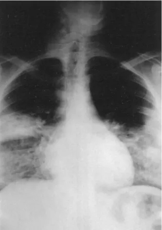
The chest x-ray film revealed pneumonia of the median right lobe (anterior segment), bronchopneumonia in the lower lobes of both right and left lungs (Fig 1). Clinical biology was characterized by an acute inflammatory syndrome. Antibiotics (doxycyline, 200 mg/d) and anxiolytics (alprazolam) were prescribed.
Six weeks later, the patient was readmitted with increased dyspnea. The chest x-ray film was abnormal, suggesting a possible
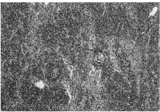
interstitial alveolar pneumopathy and fibrous sequelae of the median right lobe pneumonia. Bronchofibroscopy was performed with BAL. A liver biopsy was obtained in order to exclude sarcoidosis. No Mycoplasma organisms were detected in the BAL fluid. Pulmonary biopsy specimens suggested the possibility of idiopathic pulmonary fibrosis or LLM or interstitial desquama-tive pneumopathy (Fig 2). Treatment with glucocorticoids (meth-ylprednisolone, 16 mg/d) was initiated. No attenuation of the
*From the Laboratory of Biology (Mr. Denoo and Dr. Foidart), Laboratory of Anatomopathology (Dr. Hermans), and Depart-ment of Obstetrics and Gynecology (Dr. Foidart), University of Lie`ge, Lie`ge, Belgium, and the Department of Pneumology, Centre Hospitalier du Bois de l’Abbaye, Seraing, Belgium (Dr. Degives).
This work was supported by a grant from the Fonds de la Recherche Scientifique Me´dicale in Belgium (No. 3.4573.95). Manuscript received December 10, 1997; revision accepted May 19, 1998.
Correspondence to: Professor Jean-Michel Foidart, Laboratory of Tumor and Developmental Biology, University of Lie`ge, Pathol-ogy Tower, B23, Sart Tilman, B-4000 Lie`ge, Belgium; e-mail: jmfoidart@ulg.ac.be
Figure 2. Pulmonary biopsy specimen showing LLM (hematox-ylin-eosin, original3250).
symptoms was obtained after 4 weeks, at which time the dosage was adjusted to 8 mg/d; again, there was no clinical improvement. In the meantime, electron microscopical examination of the lung biopsy specimen indicated the presence of ectopic smooth muscle cells infiltrating the alveolar septae. These cells were immunohistochemically labeled by anti-smooth muscle cell actin and anti-vimentin antibodies; they did not contain factor VIII, HMB45, protein S-100, or the estradiol receptor, but some did express the progesterone receptor. A diagnosis of incipient LLM or benign metastasizing leiomyoma was then advanced. The clinical gynecologic examinations and pelvic echography excluded the presence of uterine fibromas.
Oral contraception (30mg of ethynyl estradiol and 150 mg of desogestrel) was stopped due to the possible effect of sex steroids on the proliferation of smooth muscle cells. Glucocorticosteroid therapy also was discontinued. A regimen specifically designed to suppress the proliferation of the smooth muscle cells was then established: daily administration of an anti-estrogenic agent (tamoxifen citrate, 20 mg/d) in combination with high doses of an antigonadotropic progestin (medroxyprogesterone acetate), 300 mg intramuscularly every 15 days for 2 months and then 500 mg/mo intramuscularly.
The objective parameters of the pulmonary function improved rapidly (Table 1) as well as her general health status. The classic side effects of this type of treatment were noted: artificial menopause and a weight gain of 15 kg over a period of 8 months. Presently, the patient remains asymptomatic with stable cardio-respiratory functions. After 2 years of receiving the initial regi-men, medroxyprogesterone acetate was replaced with norethin-derone acetate (10 mg/d) and tamoxifen therapy was maintained at the same dose. The gynecological and breast status have remained normal.
Discussion
The diagnosis of LLM in this patient was suggested by
the clinical symptoms and by the histologic examination
(conventional histologic findings and
immunohistochemi-cal search for cytoskeletal proteins). The most specific
criterion was the identification of ectopic smooth muscle
cells by electron microscopy.
1,6The tissue required for all
pathologic examinations was obtained by surgical biopsy,
although the amount required for all examinations could
have been provided by transbronchial biopsy.
In this case, the study showed all smooth muscle cells
consistently reacted with smooth muscle actin,
anti-vimentin, and desmin antibodies but were not recognized
by anti-HMB45 or anti-protein S-100 antibodies.
1Bonetti et al
1detected HMB45 in 2 of their 3 cases,
thereby confirming the cellular heterogeneity of LLM
proliferations. This heterogeneity also has been noted by
immunohistochemistry and classic light and electron
mi-croscopic observations. It may reflect different degrees of
differentiation of the same cell types or the coexistence of
several cell types.
The presence of cytoplasmic progesterone receptors
(but not estradiol receptors) in 10% of the cells suggested
a possible benefit of high doses of the progestin
medroxy-progesterone acetate. Uchida et al
9described the
exis-tence of estradiol and progesterone receptors in smooth
muscle cells in one case of benign metastasizing
leiomy-oma. They think that, even if normal lung tissue possesses
hormone receptor, it is rational to consider that multiple
leiomyomatous lesions in the lung should be diagnosed as
benign metastasizing leiomyoma when they possess
posi-tive estrogen and progesterone receptors. The presence of
such functional receptors might explain the efficacy of this
type of treatment in the administration of a luteinizing
hormone-releasing hormone agonist.
3,4Progestin therapy for LLM has been reported in at least
11 patients.
2,5It seems that most women have either
improved or remained stable while taking
medroxyproges-terone acetate whereas the condition of a few worsened
despite the use of this drug or an luteinizing
hormone-releasing hormone agonist. Tamoxifen therapy and
oopho-rectomy were also recommended.
2,5The rationale for combining medroxyprogesterone
ace-tate and tamoxifen, despite the absence of a demonstrable
estradiol receptor, was that no correlation between the
status of progesterone and estrogen receptors and the
response to hormonal therapy has been observed.
5It is
also known that estrogen aggravated this condition.
2Ten years after the initial event, this woman remains
asymptomatic, thus further confirming the hypothesis that
progestins are highly effective and should be prescribed as
the first-line treatment. According to the meta-analysis
conducted by Eliasson et al,
2oophorectomy represents a
valuable alternative.
Because LLM is a very rare disease, it is likely that the
best way to resolve questions of optimal treatment and
prognosis will be through multicenter trials. Adequate
Table 1—Evolution of Pulmonary Function in an LLM Patient*
Pulmonary Function Tests Normal Calculated Values† Before Corticoid Therapy After 2 mo of Corticoid Therapy Under Hormonal Treatment for 2 mo Under Hormonal Treatment for 6 mo Under Hormonal Treatment for 30 mo Under Hormonal Treatment for 7 yr FVC, L 2.96 1.90 1.99 2.74 2.93 2.69 2.82 FEV1, L 2.55 1.65 1.65 2.24 2.36 2.22 2.26 FEV1/FVC, % 80–100 80 83 82 81 79 80
Dlco, % of predicted value 8 mmol/kPa3 min 26 26 55 59 68 58 VC, % of predicted value 3.03 L 80 80 85 89 94 85 TLC, % of predicted value 4.61 L 75 77 82 84 94 87 FRC, % of predicted value 2.58 L 60 60 53 65 75 78 *Dlco5 diffusing capacity of the lung for carbon monoxide; VC 5 vital capacity; TLC 5 total lung capacity; FRC 5 functional residual capacity. †For a normal subject with the same physical characteristics as the patient in this report.
documentation of diagnosis, treatment, and pulmonary
function is, however, essential for meta-analysis evaluation
of treatment.
Conclusion
In one case of LLM, diagnosed on the basis of light and
electron microscopic histologic and immunohistochemical
findings that confirmed the presence of actin, desmin, and
vimentin in the smooth muscle cell proliferation and
detected progesterone receptors in some of the cells,
dramatic clinical improvements of the subjective and
objective respiratory function parameters were obtained
by prescribing a regimen combining an anti-estrogenic
agent (tamoxifen) and a progestin (medroxyprogesterone
acetate).
References
1 Bonetti F, Chiodera PL, Pea M, et al. Transbronchial biopsy in lymphangiomyomatosis of the lung: HMB45 for diagnosis. Am J Surg Pathol 1993; 17:1092–1102
2 Eliasson AH, Phillips YY, Tenholder MF. Treatment of lym-phangioleiomyomatosis: a meta-analysis. Chest 1989; 196: 1352–1355
3 Jacobson TZ, Rainey EJ, Turton CWG. Pulmonary benign metastasising leiomyoma: response to treatment with gosere-lin. Thorax 1995; 50:1225–1226
4 Carney JA, Sheps SG, Go VLW, et al. The triad of gastric leiomyosarcoma, functioning extra-adrenal paraganglioma and pulmonary chondroma. N Engl J Med 1977; 296:1517–1518 5 Taylor JR, Ryu J, Colby TV, et al. Lymphangioleiomyomatosis:
clinical course in 32 patients. N Engl J Med 1990; 323:1254 – 1260
6 White SH, Ibrahim NBN, Forrester-Wood CP, et al. Leiomyo-mas of the lower respiratory tract. Thorax 1985; 40:306 –311 7 Sadoul P. Evaluation du de´ficit fonctionnel respiratoire. Bull
Eur Physiopath Resp 1983; 19:3– 6
8 Comstock GW, Tockman MS, Helsing KJ, et al. Standardized respiratory questionnaire: Comparison of the old with the new. Am Rev Respir Dis 1979; 119:45–53
9 Uchida T, Tokumaru T, Kojima H, et al. A case of multiple leiomyomatous lesions of the lung: an analysis of flow cytom-etry and hormone receptors. Jap J Surg 1992; 22:265–268
Hemoptysis Due to MDI
Therapy in a Patient With
Permanent Tracheostomy*
Treatment With Mask AeroChamber
Michael T. Newhouse, MD, FCCP
Persistent minor hemoptysis resulted from extensive
granulation tissue on the main carina and adjacent
bronchi due to frequent spraying of metered-dose
inhaler (MDI)-generated aerosol medications
di-rectly into a permanent tracheostomy. Salbutamol,
containing oleic acid, was considered the most likely
cause. After an AeroChamber equipped with an
infant mask was interposed between the MDI and
the tracheal stoma, hemoptysis and the pathologic
changes gradually resolved.
(CHEST 1999; 115:279 –282)
Key words: AeroChamber with mask; hemoptysis; granulationtissue; MDI; oleic acid; tracheostomy;
Abbreviations: MDI5 metered-dose inhaler; URT 5 upper respiratory tract
Case Report
H
emoptysis in a 65-year-old laryngectomized man was
found to be due to extensive granulation tissue on the
tracheal carina and adjacent mucosa. This was thought to
have resulted from frequent administration, directly into
the stoma, of large doses of bronchodilator and steroid
aerosols from oleic acid excipient-containing
metered-dose inhalers (MDIs) of albuterol (Ventolin; Glaxo
Well-come; Research Triangle Park, NC) and beclomethasone
(Becloforte; Glaxo Wellcome). The use of an
AeroCham-ber (Monaghan Medical, Inc; Plattsburg, NY) equipped
with an infant (6.5-cm) mask applied to the stoma for
aerosol administration resulted in rapid resolution of the
hemoptysis and marked but incomplete improvement,
after 6 weeks, of the gross changes observed initially at
bronchoscopy. Nine months later, the carina and adjacent
tracheal and bronchial mucosa appeared normal.
This 65-year-old man, a 60-pack-year ex-smoker and
long-retired steel company crane operator with severe
chronic airflow limitation, was seen in January 1995 for
recurring minor episodes of hemoptysis that had been
occurring for about 6 weeks. The episodes occurred
several times a week, with each incidence of hemoptysis
expelling not
. 5 mL of blood, which was often mixed
with very small amounts of a mucoid or slightly purulent
secretion. The patient had undergone a permanent
tra-cheostomy following a laryngectomy for carcinoma of the
larynx in 1983 and had stopped smoking at that time after
having smoked for about 30 to 40 pack-years. During a
physical examination, the patient appeared well, and the
stoma was unremarkable, with no crusting or other
evi-dence of abnormality. The tracheal mucosa visible through
the stoma showed edema and hyperemia. Examination of
his chest revealed hyperinflation, decreased breath
sounds, and rhonchi present only on forced expiration. A
few bibasal crackles were heard.
The chest radiograph showed evidence of marked
hy-perinflation compatible with long-standing and severe
COPD. His current and best postbronchodilator FEV
1and FVC (obtained by means of a stomal adapter to
facilitate connection to the spirometer) during the
previ-ous 3 years were 0.6 and 1.6 L, respectively (predicted, 3.2
and 4.1 L, respectively).
For the previous 10 years, the patient’s regular aerosol
*From the St. Joseph’s Hospital, Firestone Chest/Allergy Clinic, Hamilton, Ontario, Canada.
Manuscript received October 23, 1996; revision accepted July 8, 1998.
Correspondence to: M. Newhouse, MD, FCCP, St. Joseph’s Hospital, Firestone Chest/Allergy Clinic, 50 Charlton Avenue East, Hamilton, Ontario L8N 4A6, Canada
medications included ipratropium bromide 80
mg (4 puffs)
qid, albuterol 200 to 400
mg (2 to 4 puffs) every 4 h as
required, and beclomethasone (250
mg/actuation) 500 mg
bid. He was instructed to administer the aerosol
medica-tion through the stoma by means of a valved holding
chamber equipped with an infant mask (the
AeroCham-ber) and not to spray the medication directly into the
trachea.
A tentative diagnosis of bronchogenic carcinoma was
made.
Fiberoptic bronchoscopy via the stoma showed
consid-erable hyperemia and edema of the tracheal mucosa at
and distal to the stoma. An extensive, irregular, firm,
raised pale ridge of tissue about 2 cm long and 4 mm wide
was seen straddling the carina in an anteroposterior
direction. It extended onto the adjacent tracheal wall. An
oozing hemorrhage was seen intermittently at its junction
with the hyperemic tracheal mucosa, especially along its
right margin. The gross appearance suggested an
inflam-matory or malignant change. No other abnormalities were
noted during the bronchoscopy. Biopsies were performed
at several locations along the carinal tissue ridge, and all of
these showed classical granulation tissue (Fig 1) with some
metaplastic changes but with no evidence of malignancy.
Cytologic examination of a lower tracheal saline lavage was
negative for malignancy and infection.
The cause of the abnormality was not immediately
apparent. However, on further questioning the patient
admitted to having sprayed his MDIs directly into the
stoma for more than 6 months, without using the
Aero-Chamber with mask, because carrying the MDI alone was
more convenient. Thus, the majority of the aerosolized
drugs and excipients, particularly oleic acid (the surfactant
contained in the Ventolin and Becloforte MDIs) would
probably impact in the trachea just distal to the stoma. It
could be anticipated that turbulence at the tracheal carina
would likely encourage the greatest deposition of the
larger aerosol droplets there.
The patient resumed the regular use of the
AeroCham-ber with mask for aerosol inhalation, and over the ensuing
3 to 4 weeks the hemoptysis gradually ceased. Figure 2
shows the patient administering the aerosol therapy to
himself directly into the stoma from an MDI (top) and by
means of the AeroChamber equipped with an infant mask
(bottom).
A repeat bronchoscopy 6 weeks after the patient was
first evaluated showed marked improvement in the
ap-pearance of the carina, with only moderate residual
thick-ening of the mucosa and no bleeding. The rest of the
tracheal mucosa appeared normal. Bronchoscopy about 9
months later revealed a completely normal carina and
adjacent mucosa.
Discussion
This case illustrates an as yet unreported cause of
hemoptysis, probably due to chronic tracheal mucosal
injury by MDI-generated, inhaled aerosols that were
sprayed directly onto the tracheal mucosa through the
stoma.
Spraying the MDI directly into the stoma, thus
bypass-ing the aerodynamic filtration of large particles normally
accomplished by the upper respiratory tract (URT), results
in an approximately 10-fold increase in the deposition of
aerosol medication and excipients onto the mucosa of the
lower respiratory tract. The large particles, comprising
about 60 to 70% of the aerosol discharged from the MDI
mouthpiece, would normally be deposited in the URT if
the aerosol is inhaled po directly or would be retained in
the valved holding chamber of the AeroChamber, if it is
being used to facilitate aerosol therapy.
1Two of the three inhaled medications (Ventolin and
Becloforte) used by the patient regularly contain 7
mg/puff
of the excipient oleic acid for a total dose to the
tracheo-bronchial tree of approximately 250
mg in 24 h. During
inhalation, most of the aerosol mass discharged at a
velocity of about 100 km/h would impact in the trachea
and the first few bronchial divisions, particularly around
the main carina, due to the high flow rate and turbulence.
2Oleic acid has been shown experimentally to cause
muco-sal ulceration in the tracheobronchial tree of rabbits
following the spraying of as few as 5 puffs of Ventolin (500
mg of albuterol), containing 35 mg of oleic acid, onto the
tracheal mucosa directly. In this study, the URT was
bypassed by means of a 0.8
3 15-cm plastic catheter
Figure 1. Top: low-power photomicrograph of carinal biopsy (original3110). Bottom: high-power photomicrograph of a cari-nal biopsy showing features of granulation tissue (hematoxylin-eosin, original355) (see text).
extension attached to the MDI canister, which was passed
into the distal one third of the trachea through an
endotracheal tube. This study supports the pivotal role of
oleic acid in the production of the mucosal injury in the
rabbit model, since similar changes were caused by a
Ventolin placebo (which contained oleic acid but not
albuterol) but not by an albuterol solution or a fenoterol
MDI, which contains lecithin instead of oleic acid as a
surfactant.
3Thus, it is not surprising that spraying large
doses of oleic acid into the tracheostomy stoma might
cause serious mucosal injury over time that gradually
resolved after the patient stopped spraying MDIs directly
into the tracheal stoma.
The AeroChamber with infant mask adapts readily to
the stoma and functions almost like the normal URT to
retain about 70% of the aerosol droplets from the MDI,
containing large drug particles and excipients.
4The efficiency of the AeroChamber in reducing URT
aerosol deposition has been demonstrated clinically, since
in patients who previously had a systemic steroid
depen-dence, and who were treated with beclomethasone
(Be-clovent; Glaxo Wellcome) 800
mg/d by inhalation, the
approximately 12-fold reduction in the oropharyngeal
dose, due to the addition of an Aerochamber to the MDI,
resulted in the elimination of oral candidiasis. This is in
contrast to the 25% prevalence of thrush in patients using
the MDI inserted into the mouth.
5Furthermore, a recent
study of a radiolabeled drug aerosol from our laboratory
showed a 12-fold decrease in the oropharyngeal dose of
albuterol when an Aerochamber was added to the MDI
compared to when an MDI was sprayed directly into the
mouth.
6Resolution of the hemoptysis after 3 to 4 weeks and
regression to a normal mucosa following several months of
the same doses of identical aerosols administered via the
AeroChamber with infant mask applied to the stoma
provides further confirmation that the excipients in
Ven-tolin and Becloforte were the likely cause of the mucosal
injury.
3It is probable that the more than 10-fold decrease
in the deposition of oleic acid excipient onto the tracheal
mucosa resulting from the use of the AeroChamber
1allowed gradual healing of the tracheal mucosa.
This cause of hemoptysis should be considered in
patients with permanent tracheostomies who are taking
aerosol medications dispensed from MDIs if the
medica-tions are sprayed directly into the tracheal stoma. In these
patients, the delivery of the aerosol medication to the
pulmonary airways and the avoidance of tracheal mucosal
injury can be accomplished by delivering MDI-generated
aerosols into the tracheal stoma via the AeroChamber with
a 6.5-cm mask that was designed for use in infants up to 6
months of age. Physicians and tracheostomized patients
needing aerosol therapy with MDIs should be made aware
of this potential problem, and physicians should encourage
the use of the infant holding chambers with mask for
aerosol administration. The use of MDI formulations with
minimal or no oleic acid also would be advisable, where
possible.
ACKNOWLEDGMENTS: My thanks to Dr. Michael Kay of the St. Joseph’s Hospital/McMaster University Pathology Depart-ment for providing the photomicrographs, to Dr. Chris Allen for reviewing the manuscript and providing many helpful sugges-tions, and to Janice Rutgers for preparing the manuscript for publication.
References
1 Dolovich M, Ruffin R, Corr D, Newhouse M. Clinical evaluation of a simple demand inhalation MDI aerosol deliv-ery device. Chest 1983; 84:36 – 41
2 Heistracher T, Balashazy I, Hofman W. The significance of secondary flow for localized particle deposition in bronchial airway bifurcations. J Aerosol Sci 1995; 26(suppl 1):S615– S616
3 Spahr-Schopfer IA, Lerman J, Cutz E, et al. Proximate Figure 2. Top: MDI being sprayed directly into the stoma.
Bottom: inhalation of MDI aerosol via the tracheal stoma by
means of an Aerochamber with the 6.5-cm mask originally designed for aerosol therapy in infants up to 6 months of age.
delivery of a large experimental dose from salbutamol MDI induces epithelial airway lesions in intubated rabbits. Am J Respir Crit Care Med 1994; 150:790 –794
4 Chua HL, Chambers CB, Newhouse MT. Upper respiratory tract (URT) salbutamol (S) deposition following MDI or MDI1Aerochamber: effect of mouth rinsing on oropharyn-geal clearance. J Aerosol Med 1995; 8:89
5 Salzman GA, Pyszczynski DR. Oropharyngeal candidiasis in patients treated with beclomethasone dipropionate delivered by metered-dose inhaler alone and with Aerochamber. J Allergy Clin Immunol 1988; 81:424 – 428
6 Chua HL, Chambers CB, Newhouse MT. Comparison of the effect of four MDI add-on devices on lung deposition and function in asthmatics. Am J Respir Crit Care Med 1994; 149:A217
Two Cases of Repeatedly
Recurrent Atypical Thymoma*
Kazuya Kondo, MD; Syouji Sakiyama, MD;Keiji Takahashi, MD; Tadashi Uyama, MD, FCCP; Yasumasa Monden, MD, FCCP; and
Yukio Shimosato, MD, PhD
Two cases of repeatedly recurrent thymoma with
myasthenia gravis are detailed here. A 41-year-old
woman had 5 recurrent thymomas, including local
recurrences and lumbar and lung metastases; she
was alive at the time of this writing, which was 22
years after her first surgery. A 36-year-old man had
3 recurrent thymomas, including local recurrence,
dissemination, and lung metastasis; he was alive at
the time of this writing, which was 16 years after his
first surgery. Both recurrent lesions were diagnosed
as “atypical thymoma” with moderate nuclear atypia.
The patients with atypical thymoma must be
fol-lowed up carefully due to a possible recurrence.
Surgical treatment with chemoradiotherapy can
lengthen their survival.
(CHEST 1999; 115:282–285)
Key words: atypical thymoma; recurrence; thymic carcinoma;thymoma
L
evine and Rosai
1classified thymic epithelial tumors
into three categories: benign (encapsulated thymoma),
malignant thymoma type 1 (invasive or metastatic
thy-moma without overt cytologic atypia), and malignant
thymoma type 2 (thymic carcinoma with overt cytologic
atypia). Recently, Suster and Moran
2reported 22 cases of
primary thymic epithelial neoplasms showing combined
features of thymoma and thymic carcinoma. The
border-line between thymoma and thymic carcinoma is unclear.
Two cases of thymic epithelial tumors with repeated
recurrences are delineated, and the medical literature is
reviewed.
*From the Second Department of Surgery (Drs. Kondo, Sakiyama, Takahashi, and Uyama), School of Medicine, the University of Tokushima, Kuramoto-cho, Tokushima, and the Department of Pathology (Dr. Shimosato), Keio University School of Medicine, Shinjuku-ku, Tokyo, Japan.
Manuscript received January 21, 1998; revision accepted May 20, 1998.
Correspondence to: Kazuya Kondo, MD, The Second Department of Surgery, School of Medicine, The University of Tokushima, Kuramoto-cho, Tokushima, 770, Japan; e-mail: kondo@clin.med. tokushima-u.ac.jp
Figure 1. Top: thoracic CT scan demonstrating a homoge-neous mass (1.5 3 3.0 cm) in the anterior mediastinum.
Center: thoracic CT demonstrating a heterogeneous mass
(3.0 3 5.0 cm) in the right S-10 region of the lung field.
Bottom: high-power view demonstrating polygonal cells with
moderate nuclear atypia, occasional nucleolar prominence, and more abundant eosinophilic cytoplasm showing squamoid arrangement (atypical thymoma) (hematoxylin-eosin, original 3400).
Case Reports
Case 1
A 41-year-old woman was admitted to the hospital on September 4, 1996. A diagnosis of myasthenia gravis with thymoma was made when she was 20 years old. Thymothymec-tomy was performed. The anterior mediastinal tumor was identified as mixed or epithelial-rich thymoma. When the patient was 28 years old, a mediastinal mass (local recurrence) was detected by chest radiograph. Complete resection of the tumor was performed. A mediastinal tumor (local recurrence) was again detected by chest CT when she was 34 years old. Radiation therapy and surgery were performed. At the age of 35, she developed lumbago. Lumbar MRI demonstrated me-tastases to lumbar vertebrae L2 and L3. Surgery and postop-erative radiation were performed. At the age of 39, a tumor in the left lung (lung metastasis) was detected by chest radio-graph. Left upper lobectomy and postoperative chemotherapy were performed. For 21 years thereafter, symptoms of myas-thenia gravis fluctuated despite the recurrent tumors. At admission, myasthenic symptoms (ptosis, double vision, fa-tigue of the extremities) were present, and values for anti-acetylcholine receptor antibody (47 nmol/L) and SCC tumor marker (4.84 ng/mL) were elevated. A chest x-ray film re-vealed a coin-sized lesion in the lower area of the right lung. A thoracic CT scan demonstrated a homogeneous mass (1.53
3.0 cm) in the anterior mediastinum (Fig 1, top) and a heterogeneous mass (3.03 5.0 cm) in the right S-10 region of the lung (Fig 1, center). Thoracotomy was performed on September 11, 1996. A tumor located in the right S-10 region was completely resected. An anterior mediastinal tumor had invaded the left brachiocephalic vein. The excision of the tumor and a partial resection of the left brachiocephalic vein were performed. The tumor was grayish white and elastic hard, but both necrosis and hemorrhage were absent. Micro-scopically, the tumor showed polygonal cells with moderate nuclear atypia and occasional nucleolar prominence and had more abundant eosinophilic cytoplasm. A diagnosis of “atypi-cal thymoma” was made (Fig 1, bottom). The patient was discharged and received therapy with steroids and anticholin-esterase on November 9.
Case 2
A 36-year-old man was referred to the hospital on June 23, 1994. At the age of 23 years, he had received a diagnosis of myasthenia gravis with thymoma. Thymothymectomy had been performed. On the basis of findings from the anterior mediastinal tumor, a diagnosis of lymphocyte-rich thymoma was made. At the age of 28, he received therapy with steroids and anticholinesterase again, as symptoms of myasthenia gravis had deteriorated. At the age of 30, tumor recurrence in Figure 2. Top left: thoracic CT demonstrating a homogeneous mass (5.03 9.0 cm) protruding into the
right thoracic cavity from right anterior mediastinum. Top right: abdominal CT demonstrating a homoge-neous mass (5.03 6.0 cm) in the upper abdominal wall. Bottom left: High-power view demonstrating an admixture of polygonal cells with moderate nuclear atypia and nucleolar prominence and a moderate amount of lymphocytes (mixed lymphocytic and epithelial thymoma with atypia; hematoxylin-eosin, original 3400). Bottom right: high-power view demonstrating more atypical tumor cells with enlargement of nuclei and nucleolar prominence and abundant eosinophilic cytoplasm without an admixture of the lymphocytes (squamous cell carcinoma; hematoxylin-eosin, original3400).
the anterior mediastinum was detected by chest CT scan. The recurrent tumor was resected, and he received postoperative irradiation therapy (50 Gy). At admission, myasthenia gravis was controlled by steroids and immunosuppressive drugs. Values for anti-acetylcholine receptor antibody (110 nmol/L) and SCC tumor marker (5.0 ng/mL) were elevated. A chest x-ray film revealed a shadow suggestive of a mass in the right hilar region. A thoracic CT scan demonstrated a homogeneous mass (5.03 9.0 cm) protruding into the right thoracic cavity from the right anterior mediastinum (Fig. 2, top left) and a homogeneous mass (5.0 3 6.0 cm) in the upper abdominal wall (Fig 2, top right). Thoracotomy was performed on September 18. As the right anterior tumor invaded the right upper lobe of the lung, a partial resection of the right lung was performed. The abdominal tumor was completely resected. The tumor was grayish white, elastic hard, and partially necrotic. Microscopically, most of the tumor consisted of a biphasic cell population containing polygonal cells with mod-erate nuclear atypia and nucleolar prominence and lympho-cytes. The tumor was identified as “atypical thymoma” (Fig 2,
bottom left). The tumor cells around the necrotic lesion had
severe nuclear atypia, nucleolar prominence, and more abun-dant eosinophilic cytoplasm. Lymphocytes were almost absent in these tumor nests. This lesion was diagnosed as poorly differentiated squamous cell carcinoma. (Fig 2, bottom right). Postoperatively, the patient received chemotherapy (CDDP, ADM, VDR, CPA). Values for the SCC tumor marker normal-ized (1.02 ng/mL). Two years later, he was found to have a nodule of 10 mm in diameter in the right lung; this nodule was resected on October 7, 1996. Histologically, it was similar to the previous atypical thymoma portion of the previous tumor.
Discussion
Recently, several pathologists who have experienced
a considerable number of thymic tumors have
recog-nized relatively aggressive tumors possessing vesicular
nuclei with moderately prominent nuclei, occasional
mitotic figures, perivascular spaces with epithelial
pal-isades, and a mixture of immature thymocytes
(CD1a1).
1,3,4These tumors were named “well
differ-entiated thymic carcinoma” or “atypical thymoma.”
4,5The boundary between thymoma and thymic carcinoma
has become increasingly blurred.
The atypical thymoma cases presented herein showed
an aggressive or malignant course. The patient in case 1
had 5 recurrent thymomas, including local recurrences
and hematogenous metastases, although she was alive at
the time of this writing, which was 22 years after her
first surgery. The patient in case 2 also had 3 recurrent
thymomas, including local recurrence, dissemination,
and hematogenous metastasis, although he was alive at
the time of this writing, which was 16 years after his first
surgery. Kirchner et al
5reported that
well-differenti-ated thymic carcinoma (or atypical thymoma by others)
was more aggressive than thymomas of other subtypes.
Further, Quintanilla-Martinez et al
6demonstrated that
well-differentiated thymic carcinoma was always
inva-sive and had a significantly increased risk of relapse and
death. When the pathologic diagnosis at the time of
initial surgery is atypical thymoma, even if the tumor is
noninvasive or completely resected, patients must be
followed up carefully to detect a possible recurrence
after resection. And if they have a recurrence, they may
survive longer with surgical treatments plus a
combina-tion of radiotherapy and chemotherapy.
The symptoms of myasthenia gravis in these two cases
fluctuated repeatedly, but aggravation of myasthenia
gra-vis was not parallel to thymoma relapse. Relapses of
thymoma without symptoms were discovered by chest
radiograph or chest CT except for the vertebral body
metastasis associated with lumbago in case 1, which was
discovered by lumbar MRI. Because relapse in the
ante-rior mediastinum, where the thymoma frequently
re-curred, is hard to detect by chest radiograph until it
becomes bulky, periodic chest CT examination during
follow-up of atypical thymoma is necessary.
Interestingly, the tumor in case 2 partly transformed to
thymic carcinoma (squamous cell carcinoma). Atypical
thymoma portions consisted of cortical type epithelial cells
and immature lymphocytes, which were immunostained
by anti-CD1a antibody (data not shown). On the other
hand, thymic carcinoma portions consisted of epithelial
cells, which showed eosinophilic cytoplasm with a
ten-dency toward keratinization and no lymphocytes. It is
suggested that some regions in this tumor progressed from
thymoma to thymic carcinoma, and the function of the
thymus disappeared at the same time. This speculation is
supported by a recent report. Suster and Moran
2reported
that a biopsy of 2 anterior mediastinal masses with
myas-thenia gravis showed features of lymphocyte-rich
thy-moma. Thoracotomy with excision was carried out 10 and
14 years later because of an increase in the size of the
lesions. Histologic examination of the resected specimens,
in both cases, showed thymic carcinoma arising from a
thymoma.
2These cases and the two reported here support
the thymoma-thymic carcinoma sequence theory, ie, that
thymoma shifts to thymic carcinoma although most of the
thymic carcinoma may arise de novo. It is important to
treat thymic epithelial tumors as potentially malignant
tumors and to examine histologically the resected tumor in
detail.
In both of the reported cases, values for the tumor
marker SCC for squamous cell carcinoma were elevated
and later became normalized after resection of the
recur-rent tumor. The tumor in case 2 showed some of the
components of squamous cell carcinoma. In case 1, since
the tumor cells in the right S-10 region of the lung have an
epidermoid arrangement, which is one of the features of
atypical thymoma,
4this thymoma may have shown a
tendency towards squamous differentiation. The tumor
marker SCC may be an indicator of the transformation of
thymoma to carcinoma during therapy for the recurrent
thymoma.
ACKNOWLEDGMENTS: We thank Dr. Yukio Shimosato, De-partment of Pathology, Keio University School of Medicine, National Cancer Center Hospital, for his pathologic analysis and helpful discussion.
References
1 Levin GD, Rosai J. Thymic hyperplasia and neoplasia: a review of current concepts. Hum Pathol 1978; 9:495–515
2 Suster S, Moran CA. Primary thymic epithelial neoplasms showing combined features of thymoma and thymic
carci-noma: a clinicopathologic study of 22 cases. Am J Surg Pathol 1996; 20:1469 –1480
3 Marino M, Muller-Hermelink HK. Thymoma and thymic carcinoma: relation of thymoma epithelial cells to the cortical and medullary differentiation of thymus. Virchows Arch [A] Pathol Anat Histopathol 1985; 407:119 –149
4 Shimosato Y, Mukai K. Atlas of tumor pathology. Tumors of the mediastinum. 3rd ed. Washington, DC: Armed Forces Institute of Pathology, 1997
5 Kirchner T, Schalke B, Buchwald J, et al. Well-differentiated thymic carcinoma: an organotypical low-grade carcinoma with relationship to cortical thymoma. Am J Surg Pathol 1992; 16:1153–1169
6 Quintanilla-Martinez L, Wilkins EW Jr, Choi N, et al. Thy-moma: histologic subclassification is an independent prognos-tic factor. Cancer 1994; 74:606 – 617
A Transitional Variant of
Castleman’s Disease
Presenting as a Chylous
Pleural Effusion*
Terri Ten Hoor, MD; Karlene Hewan-Lowe, MD; Joseph I. Miller, MD, FCCP; and Marc Moss, MD
Castleman’s disease is an uncommon
clinicopatho-logic entity that results in unregulated growth of
lymphoid tissue. It may present as benign
involve-ment of one lymph node group or as multicentric
disease with serious systemic symptoms. Pleural
ef-fusions are an uncommon manifestation of
Castle-man’s disease. We present a patient with CastleCastle-man’s
disease who initially presented with a chylous pleural
effusion.
(CHEST 1999; 115:285–288)
Key words: Castleman’s disease; chylothorax; giant lymph node
hyperplasia; pleural effusion; sickle cell anemia
C
astleman’s disease, also known as angiofollicular or
giant lymph node hyperplasia, is a rare disorder that
results in the unregulated growth of lymphoid tissue.
Initially, Castleman’s disease was reported as an indolent
disorder which was usually confined to a single lymph
node group.
1However, recent case reports have described
a multicentric form of Castleman’s that often manifests a
more malignant clinical course. Patients with Castleman’s
disease may present with a variety of signs and symptoms,
but pleural effusions are an uncommon manifestation of
this disease.
2We present a case of the transitional or mixed variant of
Castleman’s disease in a young male with a history of sickle
cell anemia, who initially presented with large bilateral
chylous pleural effusions. To our knowledge, this is the
first case report of Castleman’s disease in association with
a chylous pleural effusion.
Case Report
A 24-year-old man with a history of sickle cell anemia pre-sented to our emergency department with a 10-day history of dyspnea, both with exertion and at rest. He also reported subjective fevers, occasional night sweats, and a mild cough productive of yellow sputum. During the week prior to admis-sion, his shortness of breath progressively worsened until he could no longer sleep in a recumbent position. During the final 2 days prior to admission, he developed his typical sickle cell crisis pain in his chest, back, and legs.
This patient had been hospitalized on many previous occasions for sickle cell crises. He had a remote history of bilateral subclavian vein thrombosis secondary to multiple central line insertions. He did not drink alcohol, smoke, or use illicit drugs. His only medication at the time of admission was folic acid.
On admission, his temperature was 37.7°C, pulse was 72 beats/min, BP was 148/76 mm Hg, and respiratory rate was 34 breaths/min. He was an ill-appearing, thin young man. There was no palpable lymphadenopathy. The jugular veins were distended to 5 cm at 30 degrees of elevation. His heart rate and rhythm were regular. He had a 2/6 systolic murmur along the left sternal border. Chest examination revealed decreased expansion, de-creased breath sounds, and dullness to percussion on the right side. The left side was clear to auscultation and without dullness. The abdominal, neurologic, and skin examinations were unre-markable. The admission chest radiograph is shown in Figure 1. Laboratory examination revealed a hemoglobin of 9.9 g/dL, hematocrit of 28.6%, WBC count of 14.63 109/L, and platelet count of 4333 109/L. His blood chemistries were within normal limits, with a lactate dehydrogenase level of 198 U/L and a total protein level of 6.3 g/dL. The erythrocyte sedimentation rate was 147 mm/h and the reticulocyte count was 7%. An arterial blood gas on 32% supplemental oxygen revealed a pH of 7.45, a Pco2 of 32 mm Hg, and a Po2of 69 mm Hg. The patient underwent a diagnostic thoracentesis, which revealed turbid fluid, 5,944 nucleated cells (1% segmented neutrophils, 96% lymphocytes, and 3% histiocytes), protein 4.3 g/dL, glucose 85 mg/dL, amylase 66 U/L, lactate dehydrogenase 182 U/L, and triglycerides 1,050 mg/dL. Cytologic examination of the pleural fluid revealed no malignant cells. Serum assay revealed an IgG level of 2,730 mg/dL (normal range, 694 to 1,618 mg/dL) and an IgA level of 453 mg/dL (normal range, 63 to 378 mg/dL). The IgM level was normal at 89 mg/dL, and no paraprotein was found. Blood, sputum, and urine cultures revealed no microbial growth. The patient was seronegative for HIV-1 and HIV-2. CT scan of the thorax revealed bilateral pleural effusions, compressive atelecta-sis of the lower lobes bilaterally, and mediastinal adenopathy.
The patient was initially treated with bilateral chest tube thoracostomies and central hyperalimentation. When a signifi-cant decrease in the chylous drainage failed to occur after 14 days of conservative therapy, a surgical approach was deemed neces-sary. The patient underwent a right thoracotomy with ligation of the thoracic duct above the diaphragm, which was successful in diminishing the amount of chylous drainage. Because of adhe-sions, the mediastinum could not be reached to biopsy the mediastinal lymph nodes. In an attempt to determine the etiology of the mediastinal adenopathy, a CT-guided needle biopsy was subsequently performed; however, the tissue was nondiagnostic. *From the Division of Pulmonary and Critical Care Medicine,
Department of Medicine (Drs. Ten Hoor and Moss), Depart-ment of General Thoracic Surgery (Dr. Miller), and DepartDepart-ment of Pathology (Dr. Hewan-Lowe), Crawford Long Hospital and the Carlyle Fraser Heart Center, Emory University School of Medicine, Atlanta, GA.
Manuscript received December 18, 1997; revision accepted April 1, 1998.
Correspondence to: Marc Moss, MD, Crawford Long Hospital of Emory University, Suite 5310, 550 Peachtree St, NE, Atlanta, GA 30365
The patient underwent a Chamberlain procedure to gain access to the mediastinum. The surgeon reported areas of lobulated fat dispersed within a large mass of nodal tissue lying along the thymic region. A biopsy specimen from this nodal tissue revealed rare, small, involuted germinal centers that lacked follicular center cells (Fig 2, 3). The mantle zone of the germinal center was inconspicuous and some of the involuted follicles had a radially oriented, thin-walled blood vessel that originated in the perifollicular tissue and penetrated the germinal center. The remainder of the lymph node revealed sheets of mature plasma cells and increased vascularity of the interfollicular region (Fig 4). The immunohistochemical stain demonstrated positive staining of the interfollicular plasma cells for both kappa and lambda light chains. These morphologic features are consistent with the diagnosis of the transitional or mixed type of Castleman’s disease. The patient was started on high-dose corticosteroid therapy, with an initial dose of 1,000 mg of solumedrol followed by three 125-mg doses of solumedrol. The patient then received 100 mg/d of prednisone. After 2 months of therapy, no response was noted clinically or radiographically on subsequent CT scans of the chest. A total of 2,000 rads of radiation therapy was then delivered to the mediastinum over a 10-day period, followed by chemotherapy with cytoxan 800 mg/m2and vincristine 2 mg. In spite of this aggressive therapy, the patient continued to require increasing amounts of oxygen. A repeat CT scan of the chest demonstrated no change in the size of the mediastinal adenopathy. The patient expired on hospital day 125 after a prolonged and difficult hospital course.
Discussion
Castleman’s disease is an unusual disorder of immune
regulation that results in abnormal proliferation of B
lymphocytes and plasma cells in lymphoid organs.
Histo-logically, it is usually differentiated into three types: a
hyaline vascular variant, a plasma cell variant, and a
transitional (mixed cell) type.
3Pathologically, the hyaline
vascular type features abnormal follicles with a striking
interfollicular vascularity, expanded mantle zones
com-posed of small lymphocytes, and one or more small blood
vessels. These vessels may have thickened walls that form
hyalinized germinal centers. In contrast, the plasma cell
variant displays lymph nodes with an intact mantle zone
surrounded by sheets of mature plasma cells and no
vascular proliferation.
3The transitional or mixed cell
variant is the least common type of Castleman’s disease
and, as in our case, it is characterized by histologic features
predominantly of the hyaline-vascular type with foci of
numerous plasma cells and some large normal-appearing
germinal centers.
4Clinically, patients with Castleman’s disease may
present with an asymptomatic abnormality on chest
radio-graph, localized symptoms caused by compression of the
tracheobronchial tree, or systemic symptoms such as fever
or weight loss. The latter, known as multicentric disease, is
most often associated with the plasma cell variant.
3Typi-cally, the multicentric variant occurs in individuals in their
fourth or fifth decade as an aggressive malignant-like
syndrome with associated features of elevated
sedimenta-tion rate, hepatosplenomegaly, hypergammaglobulinemia,
granulocytosis, and plasmacytosis of the bone marrow. The
multicentric variety has been reported to have mortality
rates as high as 50%.
3The presence of pleural effusions is an uncommon
occurrence in patients with Castleman’s disease.
Pres-ently, only seven cases of Castleman’s disease presenting
as a pleural effusion have been reported in the literature.
Several of these were described as large effusions, but
none were chylous in nature.
2Chylous pleural effusions
result from disruption of the thoracic duct with
subse-quent leakage of lymphatic fluid or chyle into the chest
cavity. It occurs most commonly with malignancy (usually
lymphoma) and trauma, but several other causes have
been described, including sarcoidosis, tuberculosis, yellow
nail syndrome, Turner’s syndrome, Noonan’s syndrome,
and lymphangiomyomatosis. There have been several
re-ported cases of chylous pleural effusions associated with
subclavian vein thrombosis. All of the reported cases have
occurred in newborn infants.
5Although our patient had a
remote history of a subclavian vein thrombosis, the CT
scan shortly after his admission revealed patent subclavian
veins bilaterally.
The diagnosis of a chylous pleural effusion is usually
suspected when cloudy or milky fluid is obtained on
thoracentesis. However, 50% of chylous effusions may not
have the classic milky appearance and the diagnosis should
be suspected if a cloudy, serosanguineous or bloody
effusion does not clear with centrifugation.
6A pleural
effusion is diagnosed as a chylous effusion if the pleural
fluid triglyceride level is greater than 110 mg/dL.
7The
Figure 1. Admission chest radiograph demonstrating bilaterallocation of the pleural effusion is related to the level of
obstruction of the thoracic duct. Normally, the thoracic
duct crosses to the left side of the thoracic cavity between
the fourth and sixth vertebrae.
8Therefore, the critical
obstruction in this patient with a right pleural effusion was
below the level of the fourth thoracic vertebra.
Once the diagnosis of a chylothorax is established, a
course of therapy should be chosen to avoid the known
sequelae of malnutrition, metabolic abnormalities, and an
immunocompromised state.
9By administering total
par-enteral nutrition with a low-fat, calorie, and
high-protein formula, chyle formation will be decreased while
Figure 2. Compact germinal centers are associated with sheets of lymphoid cells in the interfollicular regions (hematoxylin-eosin stain, original3100).
Figure 3. The involuted germinal center is devoid of follicle center cells. A radially oriented artery that originated in the interfollicular region of the lymph node penetrates the small germinal center (hematoxylin-eosin stain, original3400).
an adequate nutritional status is maintained. The
chylo-thorax should also aspirated either by repeated
thoracen-tesis or by tube thoracostomy.
10When appropriate,
ther-apy should also be directed at the specific etiology of the
chylothorax, such as radiation or chemotherapy therapy for
lymphoma involving mediastinal structures. If there is a
loss of chyle of more than 500 mL/d in an adult or more
than 100 mL/d in a child for more than 14 days,
conser-vative therapy should be abandoned and surgical
interven-tion with either thoracic duct ligainterven-tion or pleuroperitoneal
shunt insertion should be considered.
11To our knowledge, this is the first case report of any
variant of Castleman’s disease presenting as a chylous
pleural effusion, and we suggest that Castleman’s disease
be added to the differential diagnosis of a chylous effusion.
References
1 Castleman B, Iverson L, Menendez VP. Localized mediasti-nal lymph-node hyperplasia resembling thymoma. Cancer 1956; 9:822– 830
2 Reynolds SP, Gibbs AR, Weeks R, et al. Massive pleural effusion: an unusual presentation of Castleman’s disease. Eur Respir J 1992; 5:1150 –1153
3 Shahidi H, Myers J, Kvale P. Castleman’s disease. Mayo Clin Proc 1995; 70:969 –977
4 Knowles DM, ed. Neoplastic hematopathology. New York: Williams & Wilkins, 1992
5 Seibert JJ, Golladay ES, Keller C. Chylothorax secondary to superior vena caval obstruction. Pediatr Radiol 1982; 12:252– 254
6 Sassoon CS, Light RW. Chylothorax and pseudochylothorax. Clin Chest Med 1985; 6:163–171
7 Staats B, Ellefson RD, Budahn LL, et al. The lipoprotein profile of chylous and nonchylous pleural effusions. Mayo Clin Proc 1980; 55:700 –704
8 Miller JI. Chylothorax and anatomy of the thoracic duct. In: Shields TW, ed. General thoracic surgery. 3rd ed. Lea and Febiger, 1989; 625– 632
9 Milsom JW, Kron IL, Rheuban KS, et al. Chylothorax: an assessment of current surgical management. J Thorac Cardio-vasc Surg 1985; 89:221–227
10 Fairfax AJ, McNabb WR, Spiro SG. Chylothorax: a review of 18 cases. Thorax 1986; 41:880 – 885
11 Robinson CLN. The management of chylothorax. Ann Thorac Surg 1985; 39:90 –95
Tension Fecopneumothorax
Due to Colonic Perforation in a
Diaphragmatic Hernia
Matthias H. Seelig, MD; Paul J. Klingler, MD; and Klaus Scho¨nleben, MD
A traumatic diaphragmatic hernia is a well-known
complication following blunt abdominal or
penetrat-ing thoracic trauma. Although the majority of cases
are diagnosed immediately, some patients may
present later with a diaphragmatic hernia. A tension
fecopneumothorax, however, is a rarity. We report
on a patient who, 2 years after being treated for a
stab wound to the chest, presented with an acute
tension fecopneumothorax caused by the
incarcera-tion of the large bowel in the thoracic cavity after an
intrathoracic perforation. The etiology and
manage-ment of this condition are discussed.
(CHEST 1999; 115:288 –291)
Figure 4. There is a dense plasmacytosis of the interfollicular region of the lymph node. Interfollicular vessels are increased (hematoxylin-eosin stain, original3400).
Key words: diaphragmatic hernia; pneumothorax; stab wound;
surgical treatment
A
traumatic diaphragmatic hernia is a well-known
com-plication following blunt or penetrating injuries to the
diaphragm. However, the diagnosis may be delayed
be-cause of a lack of symptoms. Clinical signs occur when
diaphragmatic hernias are associated with a small or large
bowel obstruction resulting from incarceration. A
pneu-mothorax resulting from the intrathoracic perforation of
the bowel has only rarely been noticed.
We report on a 20-year-old man with a tension
pneu-mothorax resulting from an intrathoracic perforation of
the colon into the chest after a penetrating stab wound and
a traumatic diaphragmatic hernia.
Case Report
A 20-year-old man was admitted to the hospital with a 2-day history of diarrhea and the acute onset of severe left thoracic pain in combination with vomiting and dyspnea. His medical history was significant for a pneumothorax following a stab wound to his left chest 2 years earlier, which was treated using a chest tube.
Twelve days before the present admission, the patient was admitted to the hospital because of cramping epigastric pain. He was afebrile, and his physical examination was unremarkable. The WBC count was 15.73 109/L. An abdominal ultrasound revealed no pathology, and an upright abdominal radiograph was normal. The patient was put on IV fluids and observed. The pain disappeared over the next few days. An esophagogastroduode-noscopy demonstrated an axial hiatal hernia and grade 1 reflux esophagitis. Omeprazole was prescribed, and the patient was dismissed without complaints. Four days later he was readmitted as an emergency patient.
The patient was in acute distress with dyspnea and jugular venous distention, and he required oxygen. His respiratory rate was 30 beats/min, and his oxygen saturation as determined by pulse oxymetry was 90%. The BP was 100/70, and the pulse rate was 100 beats/min. A physical examination revealed no breath sounds in the left chest. An abdominal examination showed mild diffuse tenderness but no signs of peritonitis. Because a tension pneumothorax was suspected, a large-bore needle was inserted in the second left costal interspace. This maneuver stabilized the patient. Subsequently, a 20 Charriere chest tube (Mallinckrodt Medical; Athlone, Ireland) was inserted in the second left intercostal space, and 700 cm3of feculent fluid was drained. A CT scan of the thorax demonstrated a portion of the large bowel in the left chest cavity (Fig 1). An esophagram could not confirm any leakage. A colonic enema with gastrografin revealed a colopleural fistula from the splenic flexure into the left thorax (Fig 2).
During an explorative laparotomy, the splenic flexure of the large bowel was found to be incarcerated and densely adherent in a traumatic diaphragmatic hernia with multiple perforations. A left hemicolectomy with a primary anastomosis was performed. The left pleura was lavaged transdiaphragmatically, the
dia-phragm was closed in two layers, and two 36 Charriere chest tubes were inserted. The postoperative course was complicated by a pyopneumothorax, requiring a thoracotomy with decortica-tion of the left lung 24 days after the initial operadecortica-tion. The diaphragmatic part of the lung could not be decorticated because of multiple adhesions. The patient recovered well from his surgery. At follow-up 6 months after surgery, the patient was well and active.
*From the Department of Surgery, (Drs. Seelig and Klinger), Mayo Clinic Jacksonville, Jacksonville, FL; and the Department of Surgery (Dr. Scho¨nleben), General Hospital Ludwigshafen, Ludwigshafen am Rhein, Germany.
Manuscript received May 6, 1998; revision accepted July 15, 1998.
Correspondence to: Matthias H. Seelig, MD, Dept. of Surgery, General Hospital Ludwigshafen, Bremerstrasse 79, Ludwig-shafen am Rhine, Germany
Figure 1. A CT scan of the chest demonstrating the bowel in the left thorax.
Figure 2. A colonic enema with gastrografin shows contrast extravasation into the left pleural cavity.