Principles and Practice
of Clinical Research
Brain stimulation in patients with disorders of
consciousness
ǂ
Abstract
Background and Aim: There is a long history of brain stimulation in medical science, and it was tested for years trying to treat several neurological diseases. On the other hand, the treatment choices for patients with severe brain injury resulting in disorders of consciousness (DOC) are still limited and research in this field remains challenging. In the current literature, only a few techniques of brain stimulation were studied in this population of patients. This review describes noninvasive techniques, namely transcranial magnetic stimulation (TMS) and transcranial direct current stimulation (tDCS), which permit to stimulate the brain through the scalp, as well as the current status of deep brain stimulation (DBS) as treatment for patients with DOC. For each technique (i.e. TMS, tDCS and DBS) a systematic search on Pubmed was performed including the term “vegetative state” or “minimally conscious state” or “disorders of consciousness” and 16 articles matched the criteria.
Conclusion: Currently, repetitive TMS (rTMS) and tDCS studies have shown encouraging results, with transient improvements of behavioral signs of consciousness in patients in minimally conscious state (MCS). DBS showed more impressive and extensive behavioral improvement after the implantation of an electrical stimulator in the thalamus. However, this procedure is riskier and the number of patients who can benefit from this intervention is limited. All these therapeutic approaches are still in their infancy. In the years to follow, controlled clinical studies on potential treatments for patients with DOC should multiply and therapeutic measures should be more accessible, controlled and effective.
Key-words: disorders of consciousness; unresponsive wakefulness syndrome/vegetative state; minimally conscious state, transcranial magnetic stimulation; transcranial direct current stimulation; deep brain stimulation, mesocircuit model.
Introduction
Patients with disorders of consciousness (DOC) have great impacts on public health. Two such conditions are the vegetative state (VS), renamed unresponsive wakefulness syndrome (UWS) and the minimally conscious state (MCS). VS/UWS clinically means the patient is awake, but fully unconscious of him/herself and his/her environment (1,2). MCS essentially differs from VS/UWS by the evidence of a partial preservation of awareness (3). This entity can be subdivided in two categories: “MCS minus”
(e.g., visual pursuit, localization of noxious stimulation and/or smiling/crying in contingent relationship to external stimuli) and “MCS plus” (e.g., higher-level behavioral responses such as command following).4
While significant progress has been made in understanding the neural correlates of consciousness disorders, treatment options for patients with altered state of consciousness available today remain poor. Moreover, when these treatments are efficient, the mechanisms
underlying the effects are still almost unknown. However, recent discoveries about brain inherent plastic ability could offer a range of therapeutic possibilities. This could allow brain’s activity to be modulated through its innate properties of plasticity and it could help to increase the chances of recovery of patients with severe brain injury (5). In this review, we will describe the use of noninvasive brain stimulation (i.e., transcranial magnetic stimulation – TMS; transcranial direct current stimulation - tDCS) and deep brain stimulation (DBS) to improve the recovery of patients with DOC. Briefly, the use of non-invasive brain stimulation techniques (TMS and tDCS) to disentangle patients in MCS form patients in VS/UWS will be presented. Finally, we will expose a hypothesis to explain the mechanism of action of these brain stimulation techniques that improve patients’ sign of consciousness, the mesocircuit model.
Methods
Literature search and study selection
In this review we aimed to identify the clinical trials or case reports performed on patients with DOC using brain stimulation techniques. We focused our research on three different techniques: TMS (including rTMS), tDCS and DBS. The medical search engine PubMed was systematically screened to identify articles in English studying brain stimulation in patients with disorders of consciousness. For brain stimulation, the following terms were included: “transcranial magnetic stimulation”, “repeated transcranial magnetic stimulation”, “transcranial direct current stimulation” or “deep brain stimulation”. Each term were associated with terms referring to disorders of consciousness: “vegetative state”, “unresponsive wakefulness syndrome”, “minimally conscious state” or “disorders of consciousness.” We included clinical trials and case report that assessed patients’ consciousness using validated scales and we excluded reviews. For TMS, 1 article matched the inclusion criteria and 3 for rTMS. We identified 4 articles for tDCS and 8 for DBS that matched the inclusion criteria.
Transcranial Magnetic Stimulation (TMS)
TMS has been used since the 1980’s in neurological and psychiatric disorders research. This technique allows
stimulating the cerebral cortex in a noninvasive way by generating a brief but strong magnetic pulse. This pulse is send through a coil applied tangentially to the surface of the scalp. The fast change in magnetic field strength induces a current flow in the tissue, which results in the activation of the neurons, and especially the bent axons underlying the stimulation (6).
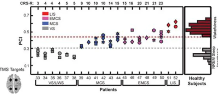
TMS in DOC was historically first applied to the motor cortex with a single pulse protocol. Responses of brain stimulation were recorded by electromyography of the peripheral muscles and behavioral assessment were performed to evaluated the responses derived from the TMS stimulation (7). Since the 2000’s, TMS has been combined with high-density-electroencephalography (EEG), to directly measure the activity of the brain itself. This enables study of cortical excitability under the site of stimulation, and long-range cortical effective connectivity (i.e., causal interactions between distant brain areas) with good spatio-temporal resolution (8,9). Using this combined TMS-EEG approach, teams from Milan, Liège and Madison built the Perturbational Complexity Index (PCI) to classify the level of consciousness of patients or healthy subjects (10). The PCI estimates brain complexity, including both the information content and the integration of brain activations, through algorithmic compressibility. Briefly, using an algorithm that measures the electrophysiological activity induced by TMS, PCI provides information on “how much this evoked activity can be compressed”. PCI values are comprised between 0.1 (not complex activity recorded and high compressibility) and 0.7 (highly complex activity recorded and low compressibility. For example, the PCI is invariably above 0.31 in healthy awake subjects, in patients in MCS or patients in locked-in syndrome, as well as in healthy subjects in REM sleep. In contrast, the PCI is always below a 0.31 threshold during deep sleep, in both VS/UWS patients and in those under general anesthesia using midazolam, propofol or xenon (Figure 1).
Repetitive TMS
Repetitive TMS (rTMS) can influence brain plasticity and cortical organization through stimulation-induced alterations in neuronal excitability. It has been used to induce a sustained inhibition (~1Hz frequency) or activation
(5-20 Hz frequency) of the neuronal population, which
Figure 1: The Perturbational Complexity Index (PCI). PCI measured during consciousness ranged between 0.44 and 0.67, whereas the PCI measured during unconsciousness ranged between 0.12 and 0.31. PCI values in severely brain-injured patients. PCI progressively increases from vegetative state/unresponsive wakefulness (VS/UWS) to minimally conscious (MCS) and to recovery of functional communication (EMCS). PCI attains levels of healthy awake subjects in LIS patients. CRS-R: Coma Recovery Scale-Revised. From Casali et al., 2013.
allowed stimulating brain areas while observing the subsequent behavioral and cognitive changes.11 Higher stimulations at 50 Hz, or theta burst stimulations, were also performed and showed a suppression of specific excitatory circuits in the human motor cortex (12,13). In the literature several studies demonstrated positive effects of rTMS in people with motor disorders and psychiatric conditions (e.g., depression or schizophrenia)(14,15). These findings suggest that rTMS may be a promising therapeutic option for patients with severe brain injury. Pape et al. performed 30 high frequency rTMS sessions on the left dorsolateral prefrontal (DLPF) cortex in a patient with DOC (16). Results were encouraging since the 26 year old patient, who was initially in VS/UWS (286 days after a TBI), improved to MCS after 15 sessions and stay in this state for the rest of the protocol and up to 6 weeks after the end of the stimulations. Another case report performed on a 70 years old patients in MCS for 5 years, showed a behavioral improvement following rTMS over M1 (10 trains of 100stimulat at 20 Hz fro 10 minutes) (17). The authors assessed the effects using the Coma Recovery Scale-Revised (CRS-R) and EEG. The patient showed behavioral improvement after rTMS as well as an increase in alpha, low and high beta activity. A third study investigated the effects of a single rTMS session on 6 chronic (> 12months post insult) patients with DOC, 3 were diagnosed as being in MCS and the other 3 in VS/UWS (18). The stimulation was performed over M1, with 1000 stimuli delivered in 10 trains of 20 Hz, each train lasting 5s with a 20 s inter-train pause. Only one patient in MCS showed behavioral improvement, as measured by the CRS-R. The authors also identified an increase in motor evoked potential (MEP) amplitude in all 6 patients. Even if behavioral improvement was observed in only 1 MCS patient, the results of the physiological outcome are encouraging and may suggest a higher clinical effect for repeated sessions of rTMS.
Thanks to these first studies, rTMS offer a new insight in the treatment of patient with DOC. Nevertheless, it is still necessary to be cautious, since TMS can induce seizures, although the risk is very low as reviewed in the 2009 safety guidelines, that allows to further mitigate this issue.19 Moreover, to confirm the efficacy of the technique, randomized placebo controlled studies should be performed in a wide population of patients.
Transcranial Direct Current Stimulation (tDCS)
In the past fifteen years, it has be shown by many studies that tDCS can modify neuronal excitability and induces behavioral changes (20-23). tDCS involves passing a weak (usually ≤ 2mA) direct current through the brain between two electrodes, the anode (i.e., excitatory) and the cathode (i.e., inhibitory). It is a safe, cheap and easy to use technique that could be easily integrated in rehabilitation programs. Currently, a lot of clinical trials have been conducted to
study the effect of tDCS on post-stroke motor and language deficits, in psychiatric disorders, chronic pain, memory impairment and tinnitus in order to decrease symptoms (24-28). However, its therapeutic effect remains to be more extensively explored (29,30).
Physiologically, anodal tDCS enhances excitability, whereas cathodal tDCS reduces it by decreasing or increasing the action potential threshold (31). The formation of the long-lasting after-effects is not entirely understood but seems to depend on membrane potential changes, modulations of NMDA receptors efficacy as well as modification of ion channels (e.g., calcium) (32). In another word, tDCS does not induce the firing of otherwise resting neurons, such as TMS, but it modulates the spontaneous firing rate of neurons by acting on the membrane potential.
Several studies on patients with brain lesions have shown that a single of tDCS could improve the function of the stimulated area, such as motor function for a stimulation of the primary motor cortex (33) and memory (34,22) or attention (35) when the prefrontal cortex is stimulated. Nevertheless, the effects decrease between one and two hours after the stimulation (36) To solve this problem, researchers performed repeated tDCS protocols using daily stimulation for one (24), two (37), or three weeks (38). Consequently, the effects lasted until 4 weeks after the end of the stimulations.
Nowadays, only a few studies tested the potential therapeutic effects of tDCS in patients with DOC. One of them explored the effect of a single session of anodal tDCS during 20 minutes over the left DLPF cortex on 55 patients with DOC (30 MCS, 25 VS/UWS, 25 post-TBI, 35 chronic) (39). One anodal and one sham stimulations were performed in a randomized order, preceded and followed by a behavioral assessment using the CRS-R (40). 13 (43%) patients in MCS and 2 (8%) patients in VS/UWS further showed post-anodal tDCS related signs of consciousness, which were neither observed during the pre-tDCS evaluation nor during the pre- or post-sham evaluation (i.e., tDCS responder). Out of the 13 MCS responders, 5 were included more than 12 months after injury. This suggests that (i) tDCS could be useful in chronic setting and (ii) that some chronic patients in MCS could still improve even years after the injury.
Another study tested 5 days of anodal tDCS for 20 minutes per day, 5 days per week, for 2 weeks in 10 patients with DOC (7 VS/UWS and 3 MCS) (41). They stimulated the left primary sensorimotor cortex (M1–n=5) or the left dorsolateral prefrontal cortex (DLPC–n=5). The three MCS patients included in this study showed clinical improvement immediately after treatment and the effect lasted one week (2 received a stimulation over left M1 and 1 over left DLPF). On the other hand, no patient in VS/UWS showed immediate enhancement after
stimulation, except for 1 patient who was in VS/UWS for 6 years and showed improvement and change of status to MCS at 12-month follow-up (41).
The outcomes of these two studies showed that tDCS could induce behavioral improvement in severely brain-injured patients with DOC. However, the underlying mechanisms are still poorly understood. Further studies investigating the effect of repeated stimulations of the prefrontal, motor or other cortical areas, supported by neuroimaging, could help clinicians to choose the best area to stimulate according to patients’ brain lesion.
As said above, the way tDCS induces behavioral improvement is only partially understood. Moreover, not all patients are willing to positively respond to tDCS. The proportion of tDCS responders vary from 40 to 80% (42-44). Trying to define the structural and functional brain features of patients in MCS who are likely to respond to tDCS, a multi-modal neuroimaging study was performed to characterize the subgroup of tDCS responders previously describe in Thibaut et al (39). Using Fludeoxyglucose Positron Emission Tomography (FDG-PET) and Magnetic Resonance Imaging (MRI; more specifically Voxel Based Morphometry – VBM), they compared 8 tDCS responders with 13 non-responders (45).
They identified that tDCS responders showed a partial metabolic (FDG-PET) and grey matter (VMB) preservation as compared to tDCS non-responders in three brain regions involved in consciousness processes (46): (i) left DLPF cortex (presumed stimulated area), (ii) precuneus, and (iii) thalamus (Figure 2). These findings highlight the importance of a partial preservation, both structural and functional, of the stimulated area in order to observe an improvement of signs of consciousness following tDCS in patients in MCS. Note that these results are only valid at the group level, and not at the single subject level. Further studies to detect specific patterns to predict the outcome at the individual level are warranted.
Beside treatment purpose, tDCS has been studied as a potential diagnostic tool. Such as for TMS, tDCS was used to study brain response of MCS and VS/UWS patients. In a recent study, TMS was performed before and after tDCS over the orbitofrontal cortex to assess the cortical response to tDCS and the difference between MCS (n=5) and VS/UWS (n=7) patients. The authors identified an increase in MEP amplitude, an intracortical facilitation, as well as a premotor-motor inhibition reduction in patients in MCS. For three VS/UWS patients tDCS had no effect, whereas the other four showed a similar pattern as MCS patients. They also found that high CRS-R total scores were associated with better premotor-motor connectivity and M1 excitability modulation. By means of these results, tDCS seems to be an interesting tool to characterize patients’ brain response to this stimulation and differentiate MCS from VS/UWS. Indeed, anodal tDCS induced an increase
in cortical connectivity and excitability in MCS, while no improvement was observed for patients clinically diagnose as being in VS/UWS, except for some VS/UWS patients who may be misdiagnosed due to an absence of clinical behavior.
Deep Brain Stimulation (DBS)
DBS is widely used to treat several neurological and psychiatric disorders such as motor disorders (e.g., essential tremor, dystonia, Parkinson’s disease), chronic pain, or obsessive-compulsive disorders and is FDA approved (47). However, DBS for patients in MCS is still rarely performed. Basically, DBS encompasses a pulse generator that sends current to a brain electrode that delivers electrical and magnetic impulses in the targeted brain region. For some diseases, like Parkinson`s and Dystonia, DBS inhibits the targeted regions, while for other diseases it has an excitatory purpose. There are two main hypotheses to explain the effect of DBS. The first one supports the idea that the current sends through implanted electrodes can induce a transient blockade of voltage-gated currents and, therefore, limits the neural output in the area near to the electrodes (48). The second hypothesis is the synaptic depression or inhibition (49). In this case, the neural activation or deactivation is regulated indirectly by the activation or deactivation of axon terminals which modulates the synaptic connections with the neurons near the stimulation electrode. Nevertheless, the underlying mechanisms of DBS are not yet fully understood and mainly depend of the target pathology.
By means of neuroimaging techniques, researchers have investigated the effect of DBS on patients’ brain activity. Recent studies highlighted that DBS induces a restoration of normal activity in the network involved in the targeted brain regions, depending on the specific pathology (50).
DBS for patients in MCS aims at stimulating neural circuits responsible for attention, memory, language, or executive functions, by implanting electrodes in the intralaminar nuclei of the thalamus. This area was chosen for several reasons.51 First, the central thalamus is suggested to be altered in regards to the pathophysiological mechanisms linked to the brain injury. Moreover, cellular loss in central thalamus seems to be particularly associated with DOC patients’ level of recovery (52,53) That is why a brain injury can lead to a decrease in forebrain activity. Central and intralaminar nuclei neurons release an excitatory neurotransmitter, glutamate. DBS could facilitate the induction and support the activity in a large network of neurons through the entire brain and thus lead to the recovery of cognitive functions underlined by these networks. Finally, chronic DBS could have long-term behavioral effects, such as those observed in the study of Schiff et al (54). These long-term effects suggest a possible
phenomenon. This last hypothesis has yet to be studied. The first DBS studies in DOC were performed in the 1960’s and 1970’s and focused on the reticular formation, the basal ganglia, and especially, on the thalamus of TBI patients in VS/UWS (55-57) However, their clinical results and the long-term follow-up were incomplete and suboptimal. Moreover the specificity of the stimulation was inaccurate. In the 1980’s a multicentric study was initiated (58). It explored the effect of unilateral DBS electrodes placed either in the centromedian thalamus or dorsal columns of the cervical spinal cord in 25 patients in chronic (3-6 months post insult) VS/UWS following a TBI. The result failed to demonstrate any clinical improvements related to DBS.
In the 1990’s, a Japanese team conducted multiple studies on DBS. In the first one, they stimulated the midbrain reticular formation (cuneiform nucleus) and unspecific thalamic nuclei (median-parafascicular complex) in 8 patients in VS/UWS (2–3 months post injury) (59). Interestingly, 3 patients showed behavioral improvement. However, even though some changes have been observed directly after the activation of the DBS device, most behavioral improvements were recorded only after 3–4 months of treatment. Other studies have shown similar results (60,61) Since the reported improvements appeared within a one-year post injury period, during which a spontaneous recovery is most likely to appear, the observed improvements could also be explained, entirely or partially, by this phenomenon. Moreover, all these studies were not
placebo-controlled. More recently, DBS of the midbrain reticular formation and the median-parafascicular complex was studied in 21 VS/UWS and 5 MCS patients (62) 8 patients in VS/UWS recovered a response to commands (i.e., MCS+) and 4 patients in MCS recovered a functional communication (i.e., emerged from MCS).
In 2007, Schiff and collaborators have reported the case of a chronic posttraumatic patient treated with DBS of thalamic intralaminar nuclei in a double-blind design with recording of several baselines (54) This was the first study that employed standardized reliable and validated outcome measures (such as the CRS-R40) to investigate the effectiveness of DBS. Clinically, the patient was in a minimally conscious state for 6 years and did not show any improvement despite rehabilitation program. Deep brain stimulation was applied bilaterally to the central thalamus and alternated on and off phases in 30-days intervals over 6 months. Intelligible verbalizations and functional object use were directly observed. After a few months of stimulations, responses to command, spontaneous limb movements, oral feeding, and functional communication were objectified during DBS-on periods. When DBS was turned off, behavioral performance decreased significantly but remained above baseline level, suggesting remnant effects. These functional gains were maintained across the 24-months follow-up phase. These findings are very encouraging for the therapy and the recovery of chronic patients with DOC. Even if DBS is very invasive, the post-operative side-effects are limited.
DBS expose the patient to more risks due to the brain surgery than rTMS or tDCS but can stimulate the brain centrally, in the thalamus and activate the thalamo-cortical connectivity, which has a critical role for consciousness recovery (46,63). Finally, let’s not forget that inclusion criteria to receive this stimulation (e.g., preserved metabolism in the thalamus) are very strict and the majority of patients cannot benefit from this therapy.
The Mesocircuit Model
A hypothesis to explain the mechanisms of action of another treatment for patients with DOC, zolpidem (i.e., sedative drugs showing paradoxical responses in rare cases of MCS patients (39), is the mesocircuit model (40,41). The mesocircuit hypothesis supports the idea that, in normal cognitive processing, the striatum disinhibits the central thalamus via the internal globus pallidus (GPi) while the central thalamus promotes activity of associative cortical areas (40). A deafferentation and loss of neurons due to a severe brain injury could induce a reduction of thalamo-cortical and thalamo-striatal connectivity. This will reduce important afferent drive to the striatum and, as a consequence, reduce the activity of the central thalamic and associative areas. According to the mesocircuit hypothesis, Zolpidem could inhibit the GPi and decrease the inhibition
Figure 2: Positron emission tomography (PET): Brain areas showing hypometabolism (in blue), as compared to controls, in patients in a minimally conscious state (FEW corrected): (A) 8 tDCS-responders and (B) 13 non-responders. (C) Regions with less hypometabolism in responders as compared to non-responders (in red). (D) Theoretical (Ruffini, Fox et al. 2014) tDCS induced electric fields. Note that behavioural responsiveness to short duration left dorsolateral prefrontal cortex (DLPFC) tDCS correlates with less impaired metabolism in the areas presumed to be stimulated by tDCS (left DLPFC and mesiofrontal cortices) but also of distant cortical (precuneus) and subcortical (thalamus) regions. From Thibaut et al. 2015.
of the thalamus. If the inhibition is decreased, the frontal area could recover its activity (41). This model provides an explanation of the vulnerability of frontal regions in case of extensive deafferentation with loss of neurons due to severe brain damage observed in patients with DOC (figure 3).
Interestingly, all brain stimulations techniques, rTMS, tDCS and DBS, were performed over brain regions involved in the mesocircuit model. Indeed, rTMS acts on neuronal activity and tDCS increase neuronal excitability of the prefrontal cortex, while DBS directly stimulate the thalamus. These observations are in line with the study of Laureys et al. where a recovery of the connectivity between the thalamus and the frontal area was detected.46 By stimulating the thalamus this reconnection could be restored. Still, prefrontal areas are important in cognitive processes (64) and their stimulation seems to improve consciousness as well, though at a lower level. The mesocircuit model, by integrating this fronto-striato-thalamic loop, seems to explain the effects of several treatments to improve signs of consciousness of patients with DOC and highlights once more the critical role of the thalamus and its connectivity with the frontal areas for consciousness recovery.
Conclusion
All these neuro-stimulation techniques are still at their infancy and many studies need to be done to explore all the potentialities and parameters that would be the most efficient for patient with DOC.
Concerning, rTMS and tDCS further studies investigating the long term effect of these techniques, and their value in clinical practice, are highly required. Others areas of stimulations could also be tested according to patients’ cortical damage. Based on the functional and structural brain signature of tDCS responders, it seems that patients need a partial preservation of the stimulated area to clinically respond to tDCS. Suggesting that, a stimulation of a (partially) preserved area would be more effective than stimulating a damaged brain region. Studies using neuroimaging (MRI, PET and HD-EEG) performed before and after a stimulation should be carried out. This will give the opportunity to investigate the direct effect of tDCS, or rTMS, on patients’ brain and better characterize which area to stimulate according to patients’ cerebral lesion. The final aim would be to develop a patients’ tailored stimulation in order to give them the best chance to recovery a certain degree of autonomy.
Concerning DBS, clinical trials including a larger population of patients with less restrictive inclusion criteria should be performed. This would be the first step to know if DBS could be used as a common treatment for chronic patients with DOC. However, the cost and the risk linked to this technique are important limiting factors.
In conclusion, more work has to be done to strengthen our understanding of potential treatments to promote the recovery of consciousness in patients with DOC. The previously discussed neuro-stimulation techniques are thought to excite mainly the forebrain regions and restore the connectivity between the thalamus and prefrontal cortex. Our understanding of neuronal correlate of consciousness recovery could help neuroscientists and clinicians to find new ways to treat patients. On the other hand, understand the mechanism of how these therapies work may help to understand the phenomena occurring in the process of recovery of consciousness.
Conflict of interest and financial disclosure
The authors followed the International Committee or Journal of Medical Journals Editors (ICMJE) form for disclosure of potential conflicts of interest. All listed authors concur with the submission of the manuscript, the final version has been approved by all authors. The authors have no financial or personal conflicts of interest.
References
1. Jennett, B. & Plum, F. Persistent vegetative state after brain damage. A syndrome in search of a name. Lancet 1, 734–737 (1972).
2. Laureys, S. et al. Unresponsive wakefulness syndrome: a new name for the vegetative state or apallic syndrome. BMC Med 8, 68 (2010). 3. Giacino, J. T. et al. The minimally conscious state: definition and diagnostic criteria. Neurology 58, 349–353 (2002).
4. Bruno, M. A. et al. Functional neuroanatomy underlying the clinical subcategorization of minimally conscious state patients. J Neurol 259, 1087–1098 (2012).
5. Oliveira, L. & Fregni, F. Pharmacological and electrical stimulation in chronic disorders of consciousness: new insights and future directions. Brain Inj 25, 315–327 (2011).
Figure 3: The mesocircuit model underlying forebrain dysfunction and interventions in severe brain injuries. Reduction of thalamocortical and thalamostriatal outflow following deafferentation and loss of neurons from the central thalamus withdraws important afferent drive to the striatum, which may then fail to reach firing threshold because of their requirement for high levels of synaptic background activity. Loss of active inhibition from the striatum allows neurons of the globus pallidus interna to tonically fire and provide active inhibition to their synaptic targets, including relay neurons of the central thalamus. According to this model, Zolpidem seems to act over the striatum by inhibiting it and hence, induces an increase of the thalamic excitatory influence on prefrontal cortices, while DBS directly stimulates the thalamus and tDCS and rTMS performed over the prefrontal cortex, increase the neuronal activity of this brain region.
6. Hallett, M., Epstein, C. M., Berardelli, A., Sackeim, H. & Maccabee, P. Topics in transcranial magnetic stimulation. Suppl Clin Neurophysiol 53, 301–311 (2000).
7. Lapitskaya, N., Coleman, M. R., Nielsen, J. F., Gosseries, O. & de Noordhout, A. M. Disorders of consciousness: further pathophysiological insights using motor cortex transcranial magnetic stimulation. Prog. Brain Res. 177, 191–200 (2009).
8. Massimini, M., Boly, M., Casali, A., Rosanova, M. & Tononi, G. A perturbational approach for evaluating the brain’s capacity for consciousness. Prog Brain Res 177, 201–214 (2009).
9. Casali, A. G., Casarotto, S., Rosanova, M., Mariotti, M. & Massimini, M. General indices to characterize the electrical response of the cerebral cortex to TMS. Neuroimage 49, 1459–68 (2010).
10. Casali, A. G. et al. A theoretically based index of consciousness independent of sensory processing and behavior. Sci. Transl. Med. 5, 198ra105 (2013).
11. Miniussi, C. & Rossini, P. M. Transcranial magnetic stimulation in cognitive rehabilitation. Neuropsychol. Rehabil. 21, 579–601 (2011). 12. Huang, Y. Z., Edwards, M. J., Rounis, E., Bhatia, K. P. & Rothwell, J. C. Theta burst stimulation of the human motor cortex. Neuron 45, 201– 206 (2005).
13. Thut, G. & Pascual-Leone, A. A review of combined TMS-EEG studies to characterize lasting effects of repetitive TMS and assess their usefulness in cognitive and clinical neuroscience. Brain Topogr. 22, 219– 32 (2010).
14. Hovington, C. L., McGirr, A., Lepage, M. & Berlim, M. T. Repetitive transcranial magnetic stimulation (rTMS) for treating major depression and schizophrenia: a systematic review of recent meta-analyses. Ann Med 45, 308–321 (2013).
15. Hsu, W. Y., Cheng, C. H., Liao, K. K., Lee, I. H. & Lin, Y. Y. Effects of repetitive transcranial magnetic stimulation on motor functions in patients with stroke: a meta-analysis. Stroke 43, 1849–1857 (2012). 16. Louise-Bender Pape, T. et al. Repetitive transcranial magnetic stimulation-associated neurobehavioral gains during coma recovery. Brain Stimul. 2, 22–35 (2009).
17. Piccione, F. et al. Behavioral and neurophysiological effects of repetitive transcranial magnetic stimulation on the minimally conscious state: a case study. Neurorehabil. Neural Repair 25, 98–102 (2011). 18. Manganotti, P. et al. Effect of high-frequency repetitive transcranial magnetic stimulation on brain excitability in severely brain-injured patients in minimally conscious or vegetative state. Brain Stimul. 6, 913–21 (2013). 19. Rossi, S., Hallett, M., Rossini, P. M., Pascual-Leone, A. & Safety of, T. M. S. C. G. Safety, ethical considerations, and application guidelines for the use of transcranial magnetic stimulation in clinical practice and research. Clin Neurophysiol 120, 2008–2039 (2009).
20. Boggio, P. S. et al. Effects of transcranial direct current stimulation on working memory in patients with Parkinson’s disease. J Neurol Sci 249, 31–38 (2006).
21. Ferrucci, R. et al. Transcranial direct current stimulation improves recognition memory in Alzheimer disease. Neurology 71, 493–498 (2008). 22. Kang, E. K., Kim, D. Y. & Paik, N. J. Transcranial direct current stimulation of the left prefrontal cortex improves attention in patients with traumatic brain injury: A pilot study. J Rehabil Med 44, 346–350 (2012). 23. Nelson, J. T., McKinley, R. A., Golob, E. J., Warm, J. S. & Parasuraman, R. Enhancing vigilance in operators with prefrontal cortex transcranial direct current stimulation (tDCS). Neuroimage (2012). doi:10.1016/j.neuroimage.2012.11.061
24. Antal, A., Terney, D., Kuhnl, S. & Paulus, W. Anodal transcranial direct current stimulation of the motor cortex ameliorates chronic pain and reduces short intracortical inhibition. J Pain Symptom Manag. 39, 890–903 (2010).
25. Baker, J. M., Rorden, C. & Fridriksson, J. Using transcranial direct-current stimulation to treat stroke patients with aphasia. Stroke 41, 1229– 1236 (2010).
26. Frank, E. et al. Treatment of chronic tinnitus with repeated sessions of prefrontal transcranial direct current stimulation: outcomes from an open-label pilot study. J Neurol 259, 327–333 (2012).
27. Loo, C. K. et al. Transcranial direct current stimulation for depression: 3-week, randomised, sham-controlled trial. Br J Psychiatry 200, 52–59 (2012).
28. Zaehle, T., Sandmann, P., Thorne, J. D., Jancke, L. & Herrmann, C. S. Transcranial direct current stimulation of the prefrontal cortex modulates working memory performance: combined behavioural and electrophysiological evidence. BMC Neurosci 12, 2 (2011).
29. George, M. S. et al. Controversy: Repetitive transcranial magnetic stimulation or transcranial direct current stimulation shows efficacy in treating psychiatric diseases (depression, mania, schizophrenia, obsessive-complusive disorder, panic, posttraumatic stress disorder). Brain Stimul 2, 14–21 (2009).
30. Nitsche, M. A. et al. Transcranial direct current stimulation: State of the art 2008. Brain Stimul 1, 206–223 (2008).
31. Nitsche, M. A. et al. Modulating parameters of excitability during and after transcranial direct current stimulation of the human motor cortex. J Physiol 568, 291–303 (2005).
32. Liebetanz, D., Nitsche, M. A., Tergau, F. & Paulus, W. Pharmacological approach to the mechanisms of transcranial DC-stimulation-induced after-effects of human motor cortex excitability. Brain 125, 2238–2247 (2002).
33. Boggio, P. S. et al. Enhancement of non-dominant hand motor function by anodal transcranial direct current stimulation. Neurosci Lett 404, 232–236 (2006).
34. Jo, J. M. et al. Enhancing the working memory of stroke patients using tDCS. Am J Phys Med Rehabil 88, 404–409 (2009).
35. Kang, E. K., Baek, M. J., Kim, S. & Paik, N. J. Non-invasive cortical stimulation improves post-stroke attention decline. Restor Neurol Neurosci 27, 645–650 (2009).
36. Nitsche, M. A. & Paulus, W. Sustained escitability elevations induces by transcrnaial DC motor cortex stimulation in humans. Neurology 57, 1899–901 (2001).
37. Boggio, P. S. et al. A randomized, double-blind clinical trial on the efficacy of cortical direct current stimulation for the treatment of major depression. Int J Neuropsychopharmacol 11, 249–254 (2008).
38. Polanowska, K. E., Lesniak, M. M., Seniow, J. B., Czepiel, W. & Czlonkowska, A. Anodal transcranial direct current stimulation in early rehabilitation of patients with post-stroke non-fluent aphasia: A randomized, double-blind, sham-controlled pilot study. Restor Neurol Neurosci 31, 761–771 (2013).
39. Thibaut, A., Bruno, M.-A., Ledoux, D., Demertzi, A. & Laureys, S. tDCS in patients with disorders of consciousness. Neurology 82, 1–7 (2014).
40. Giacino, J. T., Kalmar, K. & Whyte, J. The JFK Coma Recovery Scale-Revised: measurement characteristics and diagnostic utility. Arch Phys Med Rehabil 85, 2020–2029 (2004).
41. Angelakis, E. et al. Transcranial direct current stimulation effects in disorders of consciousness. Arch Phys Med Rehabil 95, 283–289 (2014). 42. Song, J. J., Vanneste, S., Van de Heyning, P. & De Ridder, D. Transcranial direct current stimulation in tinnitus patients: a systemic review and meta-analysis. ScientificWorldJournal 2012, 427941 (2012). 43. Goncalves, G. S. et al. Effects of tDCS Induced Motor Cortex Modulation on Pain in HTLV-1: A Blind Randomized Clinical Trial. Clin J Pain (2013). doi:10.1097/AJP.0000000000000037
44. Ferrucci, R. et al. Transcranial direct current stimulation (tDCS) for fatigue in multiple sclerosis. NeuroRehabilitation 34, 121–127 (2014). 45. Thibaut, A. et al. Clinical response to tDCS depends on residual brain metabolism and grey matter integrity in patients with minimally conscious state. Brain Stimul. in press, (2015).
46. Laureys, S. et al. Restoration of thalamocortical connectivity after recovery from persistent vegetative state. Lancet 355, 1790–1791 (2000). 47. Olanow, C. W., Brin, M. F. & Obeso, J. A. The role of deep brain stimulation as a surgical treatment for Parkinson’s disease. Neurology 55, S60–6 (2000).
48. Beurrier, C., Bioulac, B., Audin, J. & Hammond, C. High-frequency stimulation produces a transient blockade of voltage-gated currents in subthalamic neurons. J. Neurophysiol. 85, 1351–6 (2001).
49. Dostrovsky, J. O. et al. Microstimulation-induced inhibition of neuronal firing in human globus pallidus. J. Neurophysiol. 84, 570–4 (2000).
50. Kringelbach, M. L., Green, A. L., Owen, S. L. F., Schweder, P. M. & Aziz, T. Z. Sing the mind electric - principles of deep brain stimulation. Eur. J. Neurosci. 32, 1070–9 (2010).
51. Schiff, N. D., Giacino, J. T. & Fins, J. J. Deep brain stimulation, neuroethics, and the minimally conscious state: moving beyond proof of principle. Arch Neurol 66, 697–702 (2009).
52. Adams, J. H., Graham, D. I. & Jennett, B. The neuropathology of the vegetative state after an acute brain insult. Brain 123 ( Pt 7, 1327–1338 (2000).
53. Maxwell, W. L., MacKinnon, M. A., Smith, D. H., McIntosh, T. K. & Graham, D. I. Thalamic nuclei after human blunt head injury. J Neuropathol Exp Neurol 65, 478–488 (2006).
54. Schiff, N. D. et al. Behavioural improvements with thalamic stimulation after severe traumatic brain injury. Nature 448, 600–603 (2007).
55. Hassler, R., Ore, G. D., Dieckmann, G., Bricolo, A. & Dolce, G. Behavioural and EEG arousal induced by stimulation of unspecific projection systems in a patient with post-traumatic apallic syndrome. Electroencephalogr Clin Neurophysiol 27, 306–310 (1969).
56. McLardy, T., Ervin, F., Mark, V., Scoville, W. & Sweet, W. Attempted inset-electrodes-arousal from traumatic coma: neuropathological findings. Trans Am Neurol Assoc 93, 25–30 (1968).
57. Sturm, V., Kuhner, A., Schmitt, H. P., Assmus, H. & Stock, G. Chronic electrical stimulation of the thalamic unspecific activating system in a patient with coma due to midbrain and upper brain stem infarction. Acta Neurochir 47, 235–244 (1979).
58. Cohadon, F. in Neuromodulation an Overv. (Lazorthes, Y. & Upton, A. R. M.) (Futura Publishers, 1985).
59. Tsubokawa, T. et al. Deep-brain stimulation in a persistent vegetative state: follow-up results and criteria for selection of candidates. Brain Inj 4, 315–327 (1990).
60. Hosobuchi, Y. & Yingling, C. The treatment of prolonged coma with neurostimulation. Adv Neurol 63, 247–251 (1993).
61. Katayama, Y. et al. Characterization and modification of brain activity with deep brain stimulation in patients in a persistent vegetative state: pain-related late positive component of cerebral evoked potential. Pacing Clin Electrophysiol 14, 116–121 (1991).
62. Yamamoto, T. et al. DBS therapy for the vegetative state and minimally conscious state. Acta Neurochir Suppl 93, 101–104 (2005). 63. Laureys, S. & Schiff, N. D. Coma and consciousness: Paradigms (re)framed by neuroimaging. Neuroimage 61, 478–491 (2012).
64. Dehaene, S., Sergent, C. & Changeux, J. P. A neuronal network model linking subjective reports and objective physiological data during conscious perception. Proc Natl Acad Sci U S A 100, 8520–8525 (2003).