HAL Id: tel-01542593
https://tel.archives-ouvertes.fr/tel-01542593
Submitted on 20 Jun 2017HAL is a multi-disciplinary open access archive for the deposit and dissemination of sci-entific research documents, whether they are pub-lished or not. The documents may come from teaching and research institutions in France or abroad, or from public or private research centers.
L’archive ouverte pluridisciplinaire HAL, est destinée au dépôt et à la diffusion de documents scientifiques de niveau recherche, publiés ou non, émanant des établissements d’enseignement et de recherche français ou étrangers, des laboratoires publics ou privés.
edema formation in nephrotic syndrome
Khalil Udwan
To cite this version:
Khalil Udwan. Role of oxidative stress in primary sodium retention and edema formation in nephrotic syndrome. Urology and Nephrology. Université Pierre et Marie Curie - Paris VI, 2015. English. �NNT : 2015PA066331�. �tel-01542593�
Paris VI – Pierre et Marie Curie
Ecole Doctorale de physiologie et physiopathologie Thèse de Doctorat de l’Université Paris VI
Par
KHALIL UDWAN
Pour l’obtention du grade de Docteur de l’Université Paris VI
“Role of oxidative stress in primary sodium retention and edema formation in
nephrotic syndrome”
Thèse dirigée par le Docteur ALAIN DOUCET
Soutenue Publiquement le 18 Juin 2015
Devant le jury composé de :
Prof. JEAN CLAUDE DUSSAULE PRESIDENT DU JURY
Prof. ERIC FERAILLE RAPPORTEUR
Dr. ANNE-LAURE BULTEAU RAPPORTEUR
Prof. JEAN-MARC VERBAVATZ EXAMINATEUR
Table of contents
Acknowledgments Abstract (French) Abstract (English) Introduction 1. Nephrotic syndrome1.1. Definition, signs and symptoms 1.2. Epidemiology
1.3. Complications of NS 1.4. Etiology
1.5. Pathogenesis and consequences 2. Nephrotic edema and ascites
2.1. Sodium retention
2.1.1 Brief description of the mechanisms of sodium handling by the renal tubules
2.1.2. Site of sodium retention
2.1.3. Cellular mechanism and possible etiology of Na+ retention 2.1.4. Our hypothesis regarding edema formation in NS
2.2. Edema and ascites formation
2.2.1. Peritoneum anatomy and structure 2.2.2. The peritoneal filtration barrier 2.2.3. AQP1 in the peritoneum
2.2.4. Our hypothesis regarding edema formation in NS 3. Reactive oxygen species and oxidative stress
3.1. Sources of ROS
3.2. Oxidative Stress and ROS measurements 3.3. NADPH oxidase
3.4. Cellular mechanisms of defence against ROS 3.5. ROS in nephrotic syndrome
3.6. NF- B and peritoneum 3.7. Nrf2 Pathway
4. Objectives Results
1. Pathophysiology of nephrotic ascites: Role of ROS and NF- B 1.1. Introduction
1.2. Article # 1
2. Role of albumin in ROS production and sodium retention in nephrotic syndrome 2.1. Introduction
2.2. Article #2
2.3. Additional results
2.3.1. Sodium handling in DCT from PAN nephrotic rats 2.3.2. Site of sodium storage in NAC-treated PAN rats Summary and perspectives
Acknowledgments
I would never have been able to finish my thesis without the guidance of my director and committee members, help from friends, and support from my family and fiancé.
I would like to express my deepest appreciation to my advisor, Dr. Alain Doucet, who has the attitude and mind of a genius: he continuously and convincingly conveyed a spirit of adventure in regard to research and science. I would like to thank him for his excellent guidance, care, patience, precious suggestions, and for providing me with the perfect atmosphere for research. Without his guidance and persistent help, this thesis work would not have been possible.
I am most grateful to the directors of Equipe 3, Prof. Pascal Houillier and Dr.
Aurelie Edwards for their kind support, ideas, and encouragement.
My special thanks to Dr. Gaëlle Brideau for being a wonderful teacher and for helping and guiding me throughout my PhD period. Her kind, prompt, and humble support has been crucial.
I owe my deepest gratitude to Professor Jean-Claude Dussaule for agreeing to be the president of my thesis jury committee.
It gives me great pleasure in acknowledging the support and help of Prof. Eric
Feraille and Dr. Anne-Laure Bulteau for agreeing to be reporters for my thesis, and
for their brilliant ideas and suggestions to improve my manuscript.
I would also like to extend my thanks to Prof. Jean-Marc Verbavatz for accepting to be the examiner of my thesis jury committee.
It is with immense gratitude that I acknowledge the support and help of Dr. Gilles
I wish to express my sincere thanks to Dr.Lydie Cheval, Dr. Luciana Morla,
Christine Lamouroux, Sylvie Demartz, and Nadia Defontaine for their guidance
and help in teaching me, and performing and discussing the outcome of experiments.
I consider it an honour to thank Dr. Kamel Laghmani, Dr. Jacques Teulon, Dr.
Nicolas Picard, Dr. Olga Andrini, Dr. Gabrielle Planelles, Dr. Stephane Lourdel, Dr. Marc Paulais and Dr. Olivier Lahuna for their valuable scientific discussions.
I wish to express my deepest thanks to Mme. Dalila Haker for her efforts in making my administrative work easier.
I share the credit of my work with my labmates and friends, whom I had the pleasure to work with. I am thankful to Ali, Elie, Virgile, Lucile, Laurent, Suresh, David, Natsuko, Mariem, Yohan, Alexandre, Amel and Ahmed. To all my other friends, you have always been there for me when I needed you.
I thank An-Najah University and UPMC for funding me during my PhD period.
I would also like to thank my lovely parents, sisters, and brothers. They were always supporting me and encouraging me with their best wishes.
Finally, I would like to thank my fiancé, Lais Gorgatti. She was always there for me and stood by my side through the good times and the bad ones. Her support, encouragement, patience and unwavering love were undeniably the bedrock upon which the past four years of my life have been built. I am thankful for her unyielding devotion and love.
Résumé
Le syndrome néphrotique (SN) résulte d’une altération glomérulaire, responsable d’une excrétion urinaire anormale de protéines plasmatiques induisant une hypoalbuminémie. Le SN est toujours associé à une rétention rénale de Na + qui conduit à la génération d'ascite et/ou d'œdèmes. La pathogénie de la rétention de Na + et de la constitution d’œdèmes n’est pas entièrement élucidée. Dans notre étude, nous avons évalué le rôle possible des espèces réactives de l'oxygène (ROS) dans cette pathogénie. Notre étude dans le modèle de rat aminonucléoside puromycine (PAN) de SN fournit des éléments de preuve d'un rôle critique des ROS dans les troubles hydro-électrolytiques associés au SN. Dans le rein, l'endocytose de l'albumine anormalement filtrée dans le néphron distal induit un stress oxydatif qui est responsable de l’augmentation de la Na, K-ATPase. Dans le péritoine, le SN est associé à une augmentation marquée de la perméabilité à l'eau et à une diminution du coefficient de réflexion des protéines de la barrière péritonéale. Ces modifications, déclenchées par le stress oxydatif et l'activation subséquente de NF- B, sont responsables d’environ deux tiers du volume de l'ascite. Enfin, nous avons confirmé que le stress oxydatif participe à la sécrétion de l'aldostérone et est nécessaire à l’apparition de l'hyperaldostéronémie observée chez les rats néphrotiques PAN.
Abstract.
Nephrotic Syndrome (NS) is a nonspecific kidney disorder defined by abnormal urinary excretion of plasma proteins and hypoalbuminemia. NS is always associated with a renal retention of Na+ leading to the generation of ascites and/or edema. The pathogenesis of Na+ retention and edema is not fully elucidated. In our studies we evaluated the possible role of reactive oxygen species (ROS) in this pathogenesis. Our studies in the puromycin aminonucleoside (PAN) rat model of NS provided pieces of evidence for a critical role of ROS in the hydro-electrolytic disorders associated with NS. In the kidney, endocytosis of abnormally filtered albumin in the distal nephron induces an oxidative stress which is responsible for the up-regulation of Na,K-ATPase. In the peritoneum, NS is associated with a marked increase in water permeability and a decrease in the reflection coefficient to proteins of the peritoneal barrier. These changes, which are triggered by oxidative stress and subsequent activation of NF- B, account for approximately two-third of the volume of ascites. Finally, we confirmed that oxidative stress participates in the angiotensin-stimulated secretion of aldosterone and is required for the hyperaldosteronemia observed in PAN-nephrotic rats.
1. Nephrotic syndrome
1.1. Definition, signs and symptoms
Nephrotic syndrome (NS) in humans is a nonspecific kidney disorder defined by abnormal urinary excretion of plasma proteins (>50 mg/kg/day) and hypoalbuminemia (<30 g/l). Diagnosis is made by determination of urine protein/creatinine ratio in a random urine sample or measurement of urinary protein content in a 24-h urine collection [1]. Causes of NS are diagnosed on the basis of history, physical examination, serologic testing, and renal biopsy. Congenital and infantile NS are those that manifest during the first year of life.
In addition to proteinuria and hypoalbuminemia, NS is commonly associated with two additional symptoms: edema and hyperlipidemia. Edema results from excess fluid accumulation in the interstitium. It takes several forms ranging from puffiness around the eye, pitting edema in the extremities, ascites, and severe form of general edema called anasarca. It is classically thought to stem from hypoalbuminemia, vascular fluid leakage towards the interstium, hypovolemia and renal sodium retention. Hyperlipidemia, stems from overproduction of lipoproteins in the liver, secondarily to stimulation of protein synthesis in the liver to compensate for proteinuria, and from decreased lipid catabolism due to the urinary loss of lipoprotein lipase, the main enzyme involved in lipid catabolism [1].
1.2. Epidemiology
Nephrotic syndrome can affect any age depending on its etiology, but in different ways in children and adults. The most frequent form of glomerulopathy in children is minimal change disease (66% of cases), followed by focal and segmental glomerulosclerosis (8%) and mesangiocapillary glomerulonephritis (6%). In adults the most common disease is mesangiocapillary glomerulonephritis (30-40%), followed by focal and segmental glomerulosclerosis (15-25%) and minimal change disease (20%). There are also differences in prevalence between genders, the disease being more common in men than women by a ratio of 2 to 1. The incidence of nephrotic syndrome varies worldwide depending on its primary cause; according to the American Society of Nephrology, diabetic nephropathy is responsible for at least 50 cases per million populations. In children, nephrotic syndrome may occur at a rate of 20 cases per million children [2] .
1.3. Complications of NS
The most common complications of nephrotic syndrome are bacterial infection and thromboembolism. Patients with NS display increased susceptibility to infections due to the presence of edema fluid (which acts as a breeding ground for infections) and urinary loss of plasma immunoglobulins. Peritonitis is the most common infection, followed by lung, skin and urinary infections.
Increased prevalence of thromboembolism is thought to result from increased platelet aggregation, increased fibrinogen concentration, decreased antithrombin III concentrations, increased blood viscosity and decreased blood flow. Venous thrombosis is most common, especially in the renal vein, the deep vessels of the extremities, and the pulmonary arteries (which carry deoxygenated blood from heart to lungs). In patients with refractory nephrosis, low doses of anticoagulants are sometimes used.
In addition, nephrotic syndrome is associated with increased risk of renal failure. The disease responsible NS can damage the glomeruli and alter their ability to clear the blood. Renal failure can either be gradual, leading to chronic renal failure (CRF), or sudden in a form of acute renal failure (ARF) as a result of hypovolemia and decreased blood supply to the kidneys.
1.4. Etiology
Nephrotic syndrome can be caused by diseases that affect only the kidneys, such as membranous nephropathy or focal segmental glomerulosclerosis (FSGS). Diseases that affect only the kidneys are called primary causes of nephrotic syndrome. In addition, nephrotic syndrome can also be caused by systemic diseases, which are diseases that affect many parts of the body, such as diabetes or lupus. Systemic diseases that affect the kidneys are called secondary causes of nephrotic syndrome. More than 50 percent of nephrotic syndrome cases in adults have secondary causes.
The primary forms of the disease, which are usually described according to their histology, correspond to either genetic alterations of proteins involved in the glomerular filtration barrier or to the idiopathic nephrotic syndrome which dstems from an unidentified circulating factor which functionally alters the glomerular barrier. The most common form of primary NS is children is called minimal change disease (MCD) owing to the fact that kidneys
appear normal at optic microscopy level, without glomerular morphologic alterations, and lesions are only visible by electron microscopy. Another important form is focal segmental glomerumosclerosis (FSGS), the most common cause of nephrotic syndrome in adults [3], which is characterized by the presence of tissue scarring in the glomeruli.
Secondary causes of NS usually have the same histological patterns as primary causes, but sometimes they exhibit some differences which suggest a secondary cause, and they are usually described by the underlying disease. The most common causes of secondary NS are: diabetic nephropathy, systemic lupus disease, sarcoidosis, syphilis, HIV and hepatitis B.
For proper treatment of NS it is essential to define the underlying cause as there are numerous causes that can lead to NS or NS range of proteinuria.
1.5. Pathogenesis and consequences
The hallmark of the disease is an increased permeability of the glomerular filtration barrier which leads to high quantities of proteins passing from the blood into the urine. The cause of NS is damage to the glomeruli, which leads to alterations in their capacity to filter the substances transported in the blood.
Although controversy exists regarding the sieving of albumin across the glomerular barrier, it is usually accepted that in healthy individuals, less than 0.1% of plasma albumin crosses the glomerular filtration barrier [4]. In experimental animal studies, it has been proposed that there is ongoing passage of several grams of albumin per day into the urine, with equivalent tubular uptake of albumin, the result being that daily renal excretion of albumin is < 80 mg [5].
The glomerular filtration barrier consists of three successive layers (Figure 1): the capillary endothelium which displays fenestrations allowing the passage of solutes and water but retaining blood cells, the basement membrane which is a negatively charged structure exerting repulsive force on negative proteins, and the vascular sheet of the Bowman’s capsule made of highly differentiated podocytes, the interdigatations of which constitute the filtration diaphragm.
In the glomerular filtration barrier, the podocytes (visceral epithelial cells) are key cell in the selective filtering action of the glomerular capillary wall, they wrap around the capillaries of the glomerulus (Figure 2). Adjacent podocytes interdigitate to cover the glomerular basement membrane which is in contact with glomerular capillaries, but they leave small gaps (the filtration slits). These filtration slits are covered by slit diaphragms (as shown in figure 2).
Figure (1). Filtration barrier (blood-urine) in the kidney. A. The endothelial cells of the glomerulus; 1. Pore (fenestra).
B. Glomerular basement membrane: 1. lamina rara interna 2. Lamina densa 3. Lamina rara externa
C. Podocytes: 1. enzymatic and structural protein 2. Filtration slit 3. Diaphragm
The slit diaphragms are composed of cell surface proteins like nephrin, p-cadherin and podocalyxin, which make sure that large molecules such as albumin and gamma globulins stay in the bloodstream.
Structural and/or functional changes of any of these structures may be the cause of proteinuria. One or more of these targets may be involved in each type of nephrotic syndrome. The importance of the podocytes and the filtration slits is evidenced by genetic diseases. Thus, in congenital nephrotic syndrome of the Finnish type, the gene encoding nephrin, a protein of the filtration slit, is mutated, leading to nephrotic syndrome in infancy. Similarly, podocin, a protein of the podocytes, may be abnormal in a number of children with steroid-resistant focal glomerulosclerosis. In addition, alternation in the glomerular basement membrane (GBM) may alter the glomerular barrier and results in proteinuria and nephrotic syndrome, as seen in mutations in the COL4A3, COL4A4, or COL4A5 genes that encode the type IV collagen α3, α4, and α5 chains, respectively, and cause Alport’s syndrome [6, 7].
Proteinuria may concern only albumin or, in case of greater injury, all plasma proteins. Proteinuria is referred as selective when albumin accounts for > 85% of urine proteins. Because albumin has a net negative charge, it has been proposed that loss of glomerular basement membrane negative charges might be responsible for selective proteinuria. Nonselective proteinuria, would not involve changes in glomerular net charge but rather a generalized defect in permeability. This construct does not permit clear-cut separation of
Figure 2: Diagram of a glomerular capillary, showing the three cell types, as well as the parietal cells of Bowman’s capsule, and their
interrelationship in supporting the glomerulus and forming the filtration barrier. (Modified)
Wen Y. Ding , Moin A. SaleemKidney Research and Clinical Practice,Pages 87 - 93Volume 31, Issue 2, 2012.
causes of proteinuria, except in minimal-change nephropathy, in which proteinuria is selective.
2. Nephrotic edema and ascites
Whatever its etiology, NS is always associated with a renal retention of sodium (Na+) leading to extracellular volume expansion and to the generation of ascites and/or edema. The edema formation and/or ascites in nephrotic syndrome is a serious issue that constraisn the functional activity of patients and make their lives difficult. It ranges from small puffiness and pitting edema to a serious ascites, pleural and cardiac effusion and, in a severe form of edema called anasarca, edema is generalized with a widespread swelling of the skin.
Classically, nephrotic edema was thought to be secondary to hypoalbuminemia, which decreases the gradient of oncotic pressure across capillary wall and increases fluid leakage from vascular towards interstitial compartment. In turn, the resulting hypovolemia stimulates the renin/angiotensin/aldosterone system (RAAS) and promotes renal sodium retention which allows refilling of the vascular compartment. During the past decade, many experimental and clinical observations have ruled out this underfill theory, and it is now admitted that 1) renal
sodium retention is a primary event independent of capillary leakage and activation of the RAAS and 2) capillary leakage stems not only from decreased oncotic gradient but also from intrinsic changes in the properties of the capillary walls. However, neither the
molecular basis of these renal and capillary alterations nor their linkage to proteinuria and/or hypoalbuminemia is fully elucidated. The purpose of our study was to determine the etiology of primary sodium retention and the nature and etiology of the changes in the peritoneal capillaries.
2.1. Sodium retention
2.1.1. Brief description of the mechanisms of sodium handling by the renal tubule
Sodium (Na+) is the predominant cation in the extracellular fluid (ECF), the volume of ECF is directly proportional to the content of Na+ in the body. The body must maintain ECF volume within acceptable limits to maintain tissue perfusion. As demonstrated by Guyton[8], kidney has a major role in controlling arterial blood pressure by maintaining sodium
homeostasis. In humans, more than 25 moles of Na+ in 180 litter of fluid daily are delivered into the glomerular filtrate, most of which (>99%) is reabsorbed while the urine flows along the renal tubule. Tubular sodium reabsorption is a two-step mechanism including active pumping of intracellular sodium across the basolateral membrane by the Na,K-ATPase (except in distal tubule intercalated cells) and passive entry of sodium across the apical membrane. The successive segments of the nephron differ by the mechanism of apical sodium entry.
About 60 to 65% (figure 3) of this sodium load is reabsorbed along the proximal tubules (PCT). In PCT 80% of apical sodium entry is electroneutral and coupled with proton secretion by the exchanger Na+/H+ (NHE3). The loop of Henle (LOH) is responsible for the reabsorption of about 25% of the sodium load, mainly in the thick ascending limb (TAL). In TAL, the apical entrance of Na+ is mediated by the electroneutral cotransporter Na+/K+/2Cl (
NKCC2) fot ~85% and by the exchanger NHE3 (the remaining 15%. Coupled with electrogenic recycling of potassium across the apical membrane and electrogenic exit of chloride across the basoalteral membrane, the transcellular reabsorption of sodium generates a lumen-positive transepithelial voltage which drives paracellular reabsorption of sodium. It is estimated that along the TAL two-third of sodium is reabsorbed via the transcellular pathway and the remaining third by the paracellular patway.
The distal part of the nephron is responsible for the fine tuning of sodium homeostasis. The distal convoluted tubule (DCT) reabsorbs 5 to 7% of the filtered load of sodium. In human DCT, apical uptake of Na+ is mediated by the Na+-Cl- cotransporter (NCC); this reabsorption is electroneutral and sensitive to thiazides. In mouse DCT, it is mediated by NCC in the proximal part and by both NCC and the epithelial sodium channel (ENaC) in the distal part.
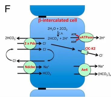
The connecting tubule (CNT) and cortical collecting duct (CCD) are the last sites of homeostatic control of sodium reabsorption which reabsorb 0 to 5% of the filtered sodium. Conversely to other nephron segments, CNT and CCD are made of several cell types: principal and intercalated cells. Classically, sodium was thought to be exclusively reabsorbed by principal cells. In these cells, apical sodium entry is mediated by amiloride-sensitive ENaC, which depolarizes the apical membrane and energizes potassium secretion via ROMK channels. This sodium reabsorption is under the positive control of aldosterone. More recently, it was demonstrated that, under some circumstances associated with high plasma mineralocorticoid levels, type B intercalated cells also contribute to 30-50% of sodium reabsorption in the CCD. In these cells, basolateral exit of sodium is mediated by a Na+ -HCO3- cotransporter, itself energized by the gradient of HCO3- generated by the activity of basolateral H-ATPase. Apical entry of sodium proceeds via the coupling of the pendrine Cl-/ HCO3- exchanger with a Cl-/ Na+-HCO3- exchanger (figure 4). This transport is electroneutral and sensitive to thiazide [9].
Figure 3: Sodium reabsorption along the mammalian nephron.
2.1.2. Site of sodium retention
During the past decades, many studies aimed at determining the site of sodium retention using the puromycin aminonucleoside (PAN) model of nephrotic rats. It is induced by a single injection of PAN: proteinuria and sodium retention develop within less than one week. PAN also induces a NS in humans but mice are resistant to PAN. In a model of unilateral PAN nephrosis, early in vivo micropuncture experiments demonstrated that sodium retention originates beyond the distal convoluted tubule accessible to micropuncture [10]. Later, using in vitro microperfusion and measurements of Na,K-ATPase activity in isolated segments of nephron, our laboratory identified the connecting tubule and cortical collecting duct (CCD) as the main sodium retaining nephron segments in bilateral PAN nephrosis [11, 12]. In addition, in children with idiopathic nephrotic syndrome, amiloride treatment restores the sodium balance to a normal levels [13], suggesting that connecting tubule and cortical collecting ducts, the two nephron segments endowed with amiloride-sensitive sodium channels (ENaC), are also the main sites of sodium retention in the human disease. In addition, it has been shown that nephrotic animals and patients display resistance of their inner medullary collecting ducts to atrial natriuretic peptide [14]. This prevents any compensation of increased sodium reabsorption in connecting tubule and cortical collecting duct.
Figure 4: Schematic description of electroneutral NaCl absorption energized by the H+ V-ATPase. Two cycles of pendrin coupled with one cycle of Ndcbe result in the net uptake of one Na+, one Cl−, and two HCO
3 −
ions, whereas one Cl− ion is recycled across the apical membrane. Then Cl− ion exits the cell through a basolateral chloride channel, while Na+ and bicarbonate exit the cell at the basolateral membrane via Ae4. All these transporters are indirectly energized by the H+ V-ATPase.
Modified, Régine Chambrey et al. PNAS 2013;110:7928-7933
2.1.3. Cellular mechanism and possible etiology of Na+ retention
Our laboratory also showed that sodium retention in PAN nephrotic rats is associated with increased abundance and activity of Na,K-ATPase and of ENaC at the basolateral and apical sides of CCD principal cells respectively [15]. The site of sodium retention and its targets in the CCD resemble those of aldosterone in the kidney. Moreover, PAN nephrotic rats, like some patients with NS, show high plasma aldosterone level [11, 13]. Altogether, these findings are consistent with the underfill theory of nephrotic sodium retention and a role of the RAAS. However, clamping plasma aldosterone at normal level in PAN nephrotic rats prevents neither the retention of sodium nor the generation of ascites [15, 16]. Interestingly, in this aldosterone-clamped model of PAN nephrosis (AC-PAN), sodium retention remains sensitive to amiloride although functional ENaC is no longer detectable [15, 16], suggesting the existence of an alternate pathway for apical sodium uptake. The role of the RAAS in nephrotic sodium retention is further ruled out by the fact that in unilateral PAN nephrosis, sodium retention occurs exclusively in the PAN-injected kidney, excluding the involvement of a systemic factor.
Alternately, it has been proposed that sodium retention during nephrosis is triggered by a factor abnormally present in the luminal fluid that reaches the distal nephron of nephrotic rats and patients secondarily to the alterations of the glomerular filtration barrier. Thus, some groups proposed plasmin to be this local factor, based on the fact that 1) plasmin is detected in the urine of nephrotic patients and PAN nephrotic rats and 2) plasmin activates ENaC through proteolysis in cultured cells [17]. However, we consider it unlikely that primary activation of ENaC by plasmin may be solely responsible for sodium retention in nephrotic syndrome because 1) in the CCD of nephrotic rats there is not only an increased activity of ENaC but also an increase in its abundance at the apical membrane, 2) plasmin-induced activation of ENaC cannot account for the observed over expression of Na,K-ATPase, and 3) studies in AC-PAN rats have shown that sodium retention can occur without activation of ENaC.
2.1.4. Our hypothesis for the etiology of Na+ retention in NS
The most abundant factor abnormally present in the urine of nephrotic patients and animals is albumin. In a healthy individual, less than 0.1% of plasma albumin may cross the glomerular filtration barrier [4]. Thereafter, reabsorption via the megalin/cubulin-dependent
endocytotic pathway in proximal tubules [18, 19] delivers a protein-free luminal fluid to the distal parts of renal tubules [20]. In nephrotic patients, the proximal tubule endocytotic absorption pathway is saturated and the distal part of the nephron is exposed to luminal albumin.
It is well established now that albuminuria has a link in the progression of Chronic Kidney Disease (CKD) [21, 22]. Albumin endocytosis in proximal convoluted tubules (PCT) induces toxicity [23] and triggers an oxidative stress which, in turn, activates different signalling pathways including NF- B and MAP kinases [24]. Moreover, in vitro treatment of proximal tubular cells with albumin induces oxidative stress (OS) and produces numerous reactive oxygen species (ROS) [24-26].
It has been shown recently that apical albumin is also internalized by the collecting duct cells of PAN nephrotic rats and by mCCDc11, a mouse CCD principal cell line[27]. In CCD from PAN rats, albumin endocytosis activates the MAP kinase ERK1,2 ([28], and in mCCD cells, it activates the canonical NF- B pathway leading to expression of pro-inflamatory cytokines [27]. Whether or not albumin endocytosis also triggers OS and ROS production in collecting ducts during NS is not documented, but several published or preliminary results suggest that it does. Thus, our laboratory previously reported or observed that albumin endocytosis in CCDs from PAN nephrotic rats is responsible for activation of ERK and is associated with activation of NF-kB and induction of osteopontin expression (unpublished observation), three targets of ROS [29]. In addition, phospho-ERK is known to increase Na,K-ATPase activity in both CCD and collecting duct cells [30] and may thereby initiate sodium retention.
Several studies showed that ROS are an important modulator of nephron ion transport; Oxidative stress has been shown to regulate BP and sodium handling in various animal models. For example, in the proximal tubules increased oxidative stress influences a number of physiologic processes including renal sodium handling and regulation of Na+,K+-ATPase activity [31, 32] . In the medullar thick ascending limb of the loop of Henle, both exogenous and endogenous superoxides stimulate sodium absorption and enhancement of Na-K-2Cl cotransporter [33, 34]. Moreover, a group of researchers found that ROS production through NADPH oxidase system has a stimulatory effect on ENaC activity in mpkCCDc14 cell line[35]
Based on these data, we hypothesized that luminal endocytosis of albumin in CNT/CCD induces OS and ROS production which in turn increases sodium reabsorption during nephrotic syndrome.
2.2. Edema and ascites formation
Edema and ascites result from an imbalance between capillary filtration and lymphatic drainage. Except for lympho-edema which stem from alteration of lymphatic drainage, all forms of edema result from increased capillary filtration. They are associated with either hypoalbuminemia, as in NS, or increased venous pressure (e.g. gestational edema) or increased capillary permeability (e.g. Quincke’s edema).
Fluid leaking from the capillaries accumulates in the interstitium or, in case of ascites, in the peritoneal cavity. Because the compliance of these compartments is almost infinite, very large volumes of edema or ascites can accumulate without altering much the interstitium hydraulic pressure, which would oppose to the capillary leakage.
2.2.1. Peritoneum anatomy and structure
The peritoneum is a serous membrane that covers most of the intra-abdominal organs. It supports the intra-abdominal organs, provides a small space for them to move, and serves as a conduit for their blood vessels, lymph vessels and nerves. It composed of two major layers: 1) the parietal peritoneum, which is the outer layer and is attached to the abdominal and pelvic walls, and 2) the visceral peritoneum, which is the inner layer and is wrapped around the internal organs. Ultimately these two layers form a continuous sheet.
The virtual space between these two layers constitutes the peritoneal cavity which is normally filled with a small amount of serous fluid, allowing the two layers to freely slide against each other. The peritoneal cavity is the space where ascites accumulates during NS. Ascites is an iso-osmotic extracellular medium mostly devoid of proteins. Because of its important vasculatization and high filtration capacity, the peritoneum is clinically used for dialysis. During peritoneal dialysis, a glucose solution is introduced into the peritoneal cavity,
left there for a specific amount of time to absorb waste products through the mechanism of diffusion, and thereafter aspirated.
2.2.2. The peritoneal filtration barrier
The peritoneal filtration barrier is a complex structure made up of three layers: the capillary endothelium, the interstitium and the mesothelium that lines the inner surface of the peritoneum [36]. The mesothelium is a unistratified squamous epithelium with a basal membrane in contact with the submesothelial interstitium composed of collagen, fibroblasts, and adipocytes and crossed by a rich network of blood and lymph vessels.
Filtration across the peritoneum requires the crossing of five successive resistance barriers: the vascular endothelium of capillaries and post-capillary venules of the peritoneum [37], the endothelium basement membrane, the interstitial matrix, the mesothelium basement membrane and the mesothelium. Transfer across the endothelium and mesothelium proceeds through both transcellular and paracellular pathways. Four simultaneously occurring mechanisms were described for the transport of fluid and solute across the peritoneum: diffusion, convection, osmosis, and fluid absorption.
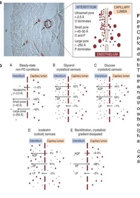
In the context of peritoneal dialysis, this complex filtration structure has been modelled according to the so-called three pore model (TPM) [38] and reviewed by Olivier Devuyst and Bengot Rippe as shown (figure 5). According to this model, the continuous endothelium lining of the peritoneum capillaries are considered as a single membrane and can be functionally described in term of TPM: The large pores (less than ,5% of all pores) allow the passage of molecules with large molecular weight like proteins and contribute to 5-8% of peritoneal ultrafiltration coeffient (LPS), the small pores (99% of total pores) allow the passage of solutes and water and contribute to around 90% of LPS , and finally the ultra-small pores which account only for 2% of LPS.
In the capillary walls of the peritoneal membrane, the small pores represent the major pathway for water and solute transport and they are represented by the space in between individual endothelial cells which called inter-endothelial cleft as shown in figure 4. The radius of these clefts is around 40 A°, whereas the radius of large pores is around 250 A°[39]. The ultra-small, water-only pores of radius around 2,5 A° present in the endothelial cell membrane and account for the transcellular pathway of transport across the peritoneal membrane[40].
More recent works in the modelling of peritoneal transport have been taken beyond the simple TPM. The simplest extension of the regular TPM model is the so-called fiber matrix model or the serial three-pore model, in which includes the interstitial space as a serial transport resistance barrier acting in parallel with the capillary wall[41]. More complicated models included not only the interstitium but also the distributed complexity nature of the peritoneal capillaries in the modeling[42].
Figure 5: Structure of the peritoneal membrane and the three-pore model (TPM). a) Cross-section of the human parietal peritoneum stained for the water channel aquaporin-1 (AQP1), showing different pores in the endothelium and their sizes. b) Transcapillary UF in the TPM using different osmotic solutions in which (A) shows the Fractional fluid flows across the peritoneum under normal conditions with no dialysis. (B) With glycerol. (C) With glucose. (D) In a conventional icodextrin PD solution. (E) Reabsorption of fluid across the small pores occurs when the crystalloid (glucose) osmotic gradient has totally dissipated (usually after 4 h). (Modified).
Olivier Devuyst and Bengt Rippe Kidney Int. Mini review, 85(4):750-8 2014, April.
2.2.3. AQP1 in the peritoneum
AQP1, a 28-kD protein is a water-specific membrane channel, impermeable to urea and glycerol. Its physiological function has been characterized thoroughly in the renal proximal tubules. AQP1 is the first identified water channel, and it distributes in a variety of tissues, including the epithelium of proximal tubules in the kidney, many parts in the eye, lung alveolus, hepatic bile duct, spleen, cardiac endocardium, peritoneum, salivary glands, and erythrocyte membrane [43, 44]. There is accumulating evidence that water channel proteins, aquaporins (AQPs), have a fundamental role in water transport in many forms of tissues [45, 46].
The identification of AQP1 had a major relevance for better understanding the mechanism of water transport and ultrafiltration in the peritoneum. The atomic model of AQP1,derived from studies based on electron and X-ray crystallography, disclosed key features accounting for the high permeation rate and strict selectivity for water transport[47] and several lines of evidence have demonstrated that AQP1 is the ultra-small, water specific pore predicted by TPM.
In human peritoneum, messenger RNA level expression of AQPs revealed that the amount of mRNA level of AQP1 was much greater than AQP3, which was much greater than AQP4 [48]. AQP1 is distributed in the endothelium lining of the peritoneal capillaries and postcapillary venules [49], which is the most important barrier to solute transport during peritoneal dialysis [50]. In addition, immunohistochemistry of rat peritoneal tissues showed the expression of AQP1 in mesothelial cells, venular endothelial cells, and capillary endothelial cells [51] and shown in (figure 6).
Inhibition of AQPs with mercury induced a 66 % inhibition of peritoneal water flow [52] Similarly, peritoneal water flow is reduced by 58% in AQP1 knockout mouse [53].. All these data from literature provide solid evidence that AQP1 plays a major role in water transport across the peritoneal barrier.
2.2.4. Our hypothesis regarding edema formation in NS
Edema formation in NS results from the asymmetry of the extracellular volume expansion resulting from renal sodium retention: the vascular volume is not, or only slightly, modified whereas water and solutes accumulate in the interstitium. As a matter of fact, capillary filtration capacity is increased almost 2-fold in nephrotic patients [54].
According to Starling’s law, water flux across a capillary (Jv) is governed by several parameters:
Jv = Lpx S x [(Pc-Pi) – x ( c- i)]
where Lp is the hydraulic conductivity of capillaries, S the exchange surface, Pc and Pi the hydrostatic pressure of the capillary and interstitium respectively, the reflection coefficient of proteins across the capillary wall, and c and i the oncotic pressure in the capillaries and interstitium respectively. This is a macroscopic modelling of water flux that considers a single pathway with a mean Lp and . With regard to the three-pore model, one should calculate the total water flux as the sum of the fluxes through the three pathways, each one being
A
B
C
D
E
Figure 6 — Immunohistochemical localization of aquaporin-1 (AQP1) in rat peritoneum [(A) to (E)], omentum. Clearstaining was observed in both the visceral peritoneum attached to the liver [(A) (200×) and (B) (1000×)] and the parietal peritoneum attached to the abdominalwall [(D) (200×) and (E) (1000×)], suggesting AQP1 was expressed in thenmesothelial cells. By contrast, staining was negative in the visceral peritoneum without incubation with an anti-AQP1 antibody [(C) (1000×)]. (Modified)
characterized by specific Lp and ; e.g. would equal zero and one for the large and ultra-small pores respectively.
According to the underfill theory, increased peritoneal filtration stems from the decreased c brought about by hypoalbuminemia. However, it is known now that the transcapillary gradient of oncotic pressure ( c- i) is almost unchanged in NS. It has been shown that the transcapillary gradient of oncotic pressure is unchanged in analbuminemic rats, because of a parallel decrease in plasma and interstitium oncotic pressure [55]. Accordingly, there is no edema or ascites formation in analbuminemic rats and patients [55]. .Moreover, diuretic treatments allow the withdrawal of significant amounts of edema without significant change in the transcapillary gradient of oncotic pressure [56]. Thus, decrease in plasma oncotic pressure in animal models as well as in nephrotic patients does not significantly alter the transcapillary gradient of oncotic pressure and is neither a determinant parameter in the genesis of edema, nor a resistance factor to edema withdrawal.
The transcapillary gradient of hydrostatic pressure (Pc-Pi) also is unchanged in NS [54] because soft tissues display an almost infinite compliance.
The 2-fold increase in capillary filtration in nephrotic patients [54] stems mainly from increased capillary hydraulic conductivity and decreased reflexion coefficient for proteins. In other words, it does not result from an increase in the driving force for water filtration but from intrinsic changes of the filtration barrier. However, neither the molecular mechanism responsible for these intrinsic changes of the filtration barrier nor their etiology is known presently.
ROS are known to induce endothelial barrier dysfunction [57] that may lead to tissue edema in the context of inflammation or ischemia [58]. TNF , which may be involved in increased capillary hydraulic conductivity during nephrotic syndrome ( ) is a known target of NF- B and may therefore be induced through production of ROS. We hypothesize that
nephrotic syndrome is associated with endothelial and peritoneal oxidative stress which is responsible for increased capillary permeability and edema and ascites formation via activation of NF B pathway.
Thus, our hypothesis stipulates that a same cellular event, i.e. oxidative stress and ROS production, is responsible for the renal and endothelial alterations leading to edema formation and ascites accumulation
2. Reactive species and oxidative stress
Most stable molecular species have the electrons in their outer orbital arranged in pairs. A molecule with one or more unpaired electrons in its outer orbital is called a free radical. This unpaired electron makes the specie very unstable and able to react with other molecules to pair this electron and thereby generate a more stable specie [59]. The term Reactive Oxygen species (ROS), often used in the literature as a collective term that includes not only oxygen-centred radicals, but also some non-radicals derivatives of oxygen (table 1). A similar term, is Reactive Nitrogen Species (RNS) is also becoming widely used nowadays (table 2).
Table (1): Reactive Oxygen Species (ROS)
Table (2): Reactive Nitogen Species (RNS)
(Source: Oxidative stress and human disease, Taibur Rahman, Ismail Hosen, M. M. Towhidul Islam, Hossain Uddin Shekhar, Advances in Bioscience and Biotechnology Vol.3 No.7A(2012), Article
ROS are chemically active molecules containing oxygen that are continuously produced in vivo in all cells: they are by-products of naturally occurring metabolism of O2 and play a critical role in cellular metabolism, signalling and homeostasis [60]. ROS is a broad term that includes all radicals such as singlet and doublet oxy-radicals and nonoxy-radicals like hydrogen peroxide (H2O2). The different ROS display a wide variety of activity, half-life, mode of production and abundance. Common examples of oxygen free radicals include the hydroxyl radical (OH), superoxide anion (O2−), and hydrogen peroxide (H2O2).The hydroxyl radical, is the most potent oxidant known, and accordingly shows an extremely short half-life. Superoxide O2− is the proximal ROS generated by mitochondria as a result of oxygen reduction by various enzymes in the Krebs cycle and respiratory chain; it acts as a weak oxidizing agent, and it is likely to be more important as a source of OH and H2O2.
It was originally thought that ROS are only produced by phagocytic cells as part of their host cell defense mechanisms. Recent work has demonstrated that ROS have a role in cell signalling role in many biological systems, including; apoptosis; gene expression or suppression; and the activation of cell signaling cascades [61].
3.1. Sources of ROS
Intracellular ROS are generated through various mechanisms, as shown in Figure 7, depending on the cell and tissue types. The major sources of ROS production are the mitochondria, the cell membrane through NADPH oxidase (nicotinamide adenine dinucleotide phosphate-oxidase) system, the perixosome and the endoplasmic reticulum [62].
Mitochondria are the main source of energy production (ATP) in cells. ATP production in the mitochondria is coupled to proton transport across the inner mitochondrial membrane by an electron transport chain in which electrons are passed through a series of proteins via oxidation-reduction reactions. The last acceptor of electron in this chain is an oxygen molecule which gives rise to superoxide anion (O2.-).
Mitochondrial O2·− generation can then lead to the production of ROS such as hydrogen peroxide (H2O2), which can, in the presence of ferrous iron form a highly reactive hydroxyl radical. When mitochondrial enzymatic and non-enzymatic antioxidant systems are overwhelmed by these ROS, oxidative damage and cell death can occur. Mitochondria use approximately the 85–90% of total oxygen, thus representing the major site of oxygen consumption as well as a primary and continuous source of cellular ROS.
3.2. Oxidative stress and ROS measurements
As a result of their unstability and high reactivity, ROS either donate or accept electrons from more stable molecules (lipids, proteins, nucleic acids and
Figure 7: Major sources of ROS production in living cells. This figure summarises the major cellular cities of ROS production, in which it shows the stimuli that induce and activate the corresponding cite to produce ROS. a) Mitochondria. b) NADPH oxidase system. And c) Lipoxygenese.
(Modified).
Erica Novo and Marizio Parola Fibrogenesis Tissue Repair. October ,2008.
carbohydrates are possible targets), which results in a radical chain reaction with the formation of new radicals and a state of oxidative stress in the tissues.
ROS are continuously detoxified by various cellular mechanisms. When an imbalance occurs between ROS production and detoxification, an oxidative stress status manifests which can somehow intensify the pathophysiological mechanisms in several diseases. Thus, during periods of stress, inflammation or infection, ROS level increases dramatically [60] and this may alter the cellular metabolism and cause significant damage to the cell structures. So, in a state of oxidative stress when the production of ROS is massive or the antioxidant defenses are deficient, damage may occur in a variety of tissues.
Unfortunately, direct in vivo quantification of ROS is difficult because of their short half-life; consequently, the evaluation of free radical production and the balance between pro-oxidant and anti-oxidant is based on the evaluation of markers of oxidation reactions, often in in vitro experiments. IOxidative stress can be assessed by measurement of oxidative damage reaction products, for example, lipid peroxidation, DNA oxidation, and protein oxidation. Another approach is based on the measurement of depletion of major antioxidants enzymes and substrate in the cells, such as ~x-tocopherol, vitamin C, and thiol groups.
3.3. NADPH oxidase
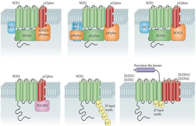
NADPH oxidase (NOX) is a membrane-bound enzyme initially found in the plasma membrane and in the membranes of phagosomes used by neutophils to kill micoorganisms via the generation of superoxide anion. NADPH oxidase was later fonud in most - if not all- mammalian cell types. NADPH oxidases constitute a family of multisubunit enzyme complexes. In most mammals, seven isoforms of NADPH oxidase have been described, each of them consisting of a core catalytic subunit (the so-called NADPH oxidase (NOX) or the dual oxidase (DUOX)) and up to five regulatory subunits (p22phox, p47phox, p40phox, p67phox, Rac1&2) (figure 8). Rodents are missing the NOX5 subunit.
With the exception of NOX4 which is constitutively active, NADPH oxidase activity is controlled by a complex regulatory system including the G-protein Rac [63]. The active enzyme transfers electrons along an electron transport chain from NADPH to the final acceptor oxygen to produce superoxide (O2-). During this reaction, electrons are captured within the cytosol and superoxide is released at the other side of the membrane, either into the extracellular compartment or into organelles lumen. Conversely to other NADPH oxidases, NOX4 does not release superoxide but hydrogen peroxide (H2O2), normally produced from superoxide by superoxide dismutase [64]. NOX4 is highly expressed in kidney, particularly in the tubular system [65].
Figure (8) Subunit composition of the seven mammalian NADPH oxidase isoforms. The catalytic core subunits of the enzymes (NADPH oxidase 1 (NOX1)–NOX5, dual oxidase 1 (DUOX1) and DUOX2) are shown in green; NOX and DUOX maturation and stabilization partners (p22phox, DUOX activator 1 (DUOXA1) and DUOXA2) are shown in red; cytosolic organizers (p40phox, NOX organizer 1 (NOXO1) and p47phox) are shown in orange; cytosolic activators (p67phox and NOX activator 1 (NOXA1)) are shown in green; and small GTPases (RAC1 and RAC2) are shown in blue. Also shown (in pink) is polymerase δ-interacting protein 2 (POLDIP2), which is thought to regulate NOX4 activity and link production of reactive oxygen species by this isoform with cytoskeletal organization.
Modified: Grant R. Drummond, Stavros Selemidis, Kathy K. Griendling & Christopher G. Sobey Nature Reviews Drug Discovery 10, 453-471 (June 2011)
While several enzymes produce ROS, NADPH oxidase is the most significant one [61]. Overproduction of O2- and its downstream catabolism initiate an oxidative stress status which can inactivate critical metabolic enzymes.
3.4. Cellular mechanisms of defence against ROS
In normal healthy conditions, cells detoxify these oxygen radicals by reducing them to oxygen as shown in Figure 9 below. In particular, superoxide dismutases (SOD) are ubiquitous enzymes that catalyze the dismutation of O2- into O2 and hydrogen peroxide (H2O2). Three forms of SOD are present in humans, SOD1 presents in the cytoplasm, SOD2 in the mitochondria, and SOD3 is extracellular. Catalase, which is abundant in the peroxisomes located next to mitochondria, converts the resulting H2O2 into water and oxygen.
Another important mechanism for preventing the accumulation of ROS is to transfer the energy of the reactive peroxides to a small sulfur-containing protein (glutathione) through glutathione peroxidase (GPx). GPxs have several isozymes which encoded by different genes and vary in cellular location. So far 8 isoforms have been identified in humans (GPx1-8)[66]. Of these isoforms, GPx1 is the most abundant form and found in the cytoplasm of almost all mamalians, in which hydrogen perioxide is the preffered substrate.
Figur 9: Pathways of reactive oxygen species (ROS) production and clearance.GSH, glutathione; GSSG, glutathione disulfide.
Oxidative stress occurs when there is excessive free radicals production, low anti-oxidant defence, or both.
3.5. ROS in nephrotic syndrome
ROS production and OS are suspected to be important and thought to be involved in the development of various diseases in humans including cancer [67], cardiovascular diseases [68, 69] and kidney disease [70]. Several lines of evidence indicate that chronic kidney disease (CKD) represents a pro-oxidant state including: 1) increased levels of oxidative markers in the plasma of patients [71, 72], and 2) decrease and defect in the antioxidant defence mechanisms in patients, which leads to a defect in the clearance of excessive ROS production [73].
In the past years, many studies explored the role of ROS in the development of kidney diseases, but several aspects remain unknown. For instance, the relative importance of each different type of ROS, the source of ROS and the exact link between this oxidative stress and the pathogenesis of the disease remain poorly defined.
In nephrotic syndrome, ROS seems to play a role in the etiology and the pathogenesis of nephrotic proteinuria. For instance, ROS was described as a possible mediator of glomelular injury that leads to proteinuria [74]. In addition, it was observed that O2- mediates an oxidative injury of the glomerular basement membrane and increases the glomerular permeability [75, 76]. Moreover, nephrotic patients show evidence of oxidative stress and impaired antioxidant defense in the acute phase of the disease [77]
The exact source of ROS and how it contributes to the pathogenesis of sodium retention and edema formation in NS are not well established. However, NS is associated with decreased serum albumin, and albumin is the main circulatory antioxidant [78, 79] due to its multiple ligand-binding capacities and its scavenging properties for free radicals [80]. The hypothesis of the antioxidant role of albumin is supported by the central role of oxidative stress in some pathology such as liver failure and sepsis [78, 81].
3.6. NF- B and peritoneum
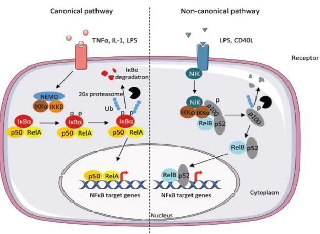
Nuclear factor-kappa B (NF-κB) is a transcription factor known to have pro-inflammatory, mitogenic and anti-apoptotic functions in many cell types [82], and it is thereby involved in several diseases. The NF-κB pathway is modulated in a cell-specific manner and interacts with other transcription pathways, providing a complex variety of cellular responses to its activation [83]. In mammals, there are five members of the transcription factor NF-κB family: RelA (p65), RelB and c-Rel, and the precursor proteins NF-κB1 (p105) and NF-κB2 (p100), which are processed into p50 and p52, respectively. NF-κB transcription factors bind as dimers to κB sites in promoters and enhancers of a variety of genes and induce or repress transcription[84]. NF-κB dimers are bound to inhibitory IκB proteins, which sequester NF-κB complexes in the cytoplasm. To stimulate NF-κB pathway, the degradation of IκB proteins is initiated through phosphorylation by the IκB kinase (IKK) complex, which consists of two catalytically active kinases, IKKα and IKKβ, and the regulatory subunit IKKγ (NEMO).
There are two main NF-κB-activating pathways in cells (figure 8). The canonical pathway (classical) is activated by most physiological NF-κB stimuli; for example, signals from cytokine receptors, such as the tumor necrosis factor receptor (TNFR) and interleukin 1 (IL-1) receptor (IL-1R), antigen receptors and pattern-recognition receptors, including Toll-like receptor 4 (TLR4). This pathway is dependent on IKKβ and leads mainly to the phosphorylation of IκBα and nuclear translocation mainly of p65-containing heterodimers.
In contrast, the non-canonical pathway depends on IKKα-mediated phosphorylation of p100 associated with RelB and leads to partial processing of p100 and the generation of p52-RelB complexes. Non-canonical signalling is induced by specific members of the TNF cytokine family, such as BAFF, CD40 ligand, and lymphotoxin-β2 [83].
After activation, NF- B transcription factor promotes the expression of over 150 target genes, the majority of which participate in the host immune response, in the regulation of the acute phase of stress response such as the inducible nitric oxide synthase (iNOS) and cyclooxygenase-2 (COX-2).
Nephrotic syndrome is associated with a high level of TNFα [85]. Therefore, we tested the possibility of the involvement of NF-κB pathway activation in nephrotic edema, and its dependence on oxidative stress.
Figure 8: Scheme illustrates the canonical and non-canonical pathway leading to the activation of NF-κB. The canonical pathway is induced by cytokines such as Tumor Necrosis Factor (TNFα), Interleukin-1 (IL-1). The activation results in the phosphorylation of IκBα by the IKK complex, leading to its ubiquitylation (Ub) and subsequent degradation by the 26S proteasome. The RelA/p50 complex is free to translocate to the nucleus to activate the transcription of target genes. The non-canonical pathway results in the activation of IKKα by the NF-κB-inducing kinase (NIK) after stimulation by TNF cytokine family. The formation of the complex NIK-IKKα-p100 leads to the phosphorylation of the p100 subunit. This results in 26S proteasome dependent processing of p100 to p52, which can lead to the activation of p52-RelB that target distinct κB element and induce the transcription of target genes. (Modified)
3.7. Nrf2 Pathway
The transcription factor NF-E2 p45-related factor 2 (Nrf2) has a crucial role in maintaining the cellular redox homeostasis by regulating the gene expression of various cytoprotective proteins, including antioxidants and detoxifying enzymes. It regulates the biosynthesis and regeneration of glutathione, thioredoxin and NADPH; control the production of ROS by mitochondria and NADPH oxidase.
Nrf2 itself is controlled primarily at the level of protein stability. At basal levels, Nrf2 has a short half-life of about 20 minutes, and it is subjected to continuous ubiquitination and proteasomal degradation. There are three known ubiquitin ligase systems which contribute to the degradation of Nrf2. In which, Kelch-like ECH associated protein 1 (Keap1) is the first negative regulator of Nrf2 to be discovered [86]. Inducers that block the cycle of Keap-1 mediated degradation of Nrf2 result in the accumulation of the transcription factor, which translates after to the nucleus. Nrf2 binds to antioxidant response elements (AREs), the upstream regulatory regions of its target genes, and initiates transcription of cytoprotective proteins[87].
Nrf2 directly regulates the expression of both the catalytic and the regulatory subunits of γ-glutamyl cysteine ligase (GCL), the rate-limiting enzyme in the biosynthesis of reduced glutathione (GSH)[88]. In addition to its rule on GSH biosynthesisn, Nrf2 provides a mean to maintain the glutathione in its reduced form by the coordinate transcriptional regulation of glutathione reductase 1 (GSR1)[89]. Nrf2 also regulates the gene expression of thioredoxin (TXN), thioredoxin reductase 1 (TXNR1) [90] which are essential for reduction of oxidized protein thiols.
Cells is which Nrf2 has been disrupted showed higher levels of ROS compared to WT cells[91]. Moreover, Nrf2 is also critical for the maintenance of the mitochondrial redox homeostasis. Thus, compared to WT, the total mitochondrial NADH pool is significantly increased in Keap1-KO and dramatically decreased in Nrf2-KO cells[91].
4. Objectives
The aim of our study is to provide further insight in the role of OS and ROS in sodium retention and ascites formation in nephrotic syndrome. Specifically, we addressed the following questions:
Does luminal endocytosis of albumin induce OS in the CCD from nephrotic rats? Is OS in CCD responsible for sodium retention and how does it control the activity of
ENaC and Na,K-ATPase?
Is there OS in the peritoneum of nephrotic rats
Is peritoneal OS responsible for the formation of ascites, and how does it control the water permeability of the peritoneum filtration barrier
What ROS-induce pathway mediates change in peritoneal permeability
Are ROS a possible therapeutic target for the treatment of nephrotic Na+ retention and edema?
1. Pathophysiology of nephrotic ascites: Role of ROS and NF- B 1.1. Introduction
The production of edema and ascites in the nephrotic syndrome was traditionally thought to result from the leakage of intravascular fluid towards the insterstitium brought about by the decrease in plasma oncotic pressure due to hypoalbuminemia. However, it is now proposed that expansion of the insterstitium volume is also accounted for by changes in the intrinsic properties of the vascular endothelium, including increased water permeability and decreased reflection coefficient to proteins. However, the mechanism responsible for these changes is not characterized yet. This first study aimed at characterizing changes in the intrinsic properties of the peritoneal filtration barrier and at determining their pathogenesis. We hypothesized that the changes in water permeability and reflection coefficient for protein stem from oxidative stress.
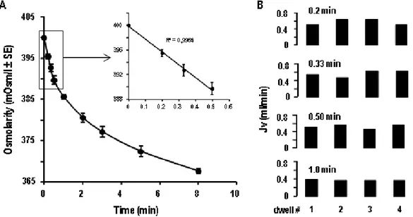
For these purposes, we developed an original dialysis method that allowed us to evaluate the permeability and reflection coefficient of the peritoneum of control and PAN nephrotic rats, and we tested the effect of a ROS scavenger, N-acetylcysteine (NAC), on these parameters. The peritoneal filtration coefficient (Lp x S) was estimated by the slope of the regression line connecting the osmotic pressure gradient (which we induce experimentally) to the water flux (measured by osmotic dilution). These determinations were done either in the absence or the presence of HgCl2, an inhibitor of aquaporin, to evaluate the involvement of the transcellular and paracellular water transport pathways. We also evaluated ROS production and accumulation in the peritoneum and their role in the induction of ascites using NAC. Finally, we tried to characterize the molecular mechanism and signaling pathway by which ROS could initiate the edema in nephrotic syndrome.
First we found a two-fold increase in mannitol-induced water flux across the peritoneum of PAN rats compared to control rats, and this increase was associated with increase in the water filtration coefficient of the paracellular and transcellular pathways, and a decrease in the reflection coefficient to proteins. The increase in transcellular water flux in the peritoneum was associated with increased expression of aquaporin 1 (AQP1). Moreover, we found that the peritoneum of PAN rats displayed a significant oxidative stress, as revealed by dihydroethedium staining and
dysregulation of mRNA expression of NADPH oxidase and ROS detoxifying enzymes.
We found also that NAC treatment decreased the volume of ascites by >60%. This decrease in ascites volume was not associated with an increase in the blood albumin level or with a change in the albuminuria. In addition NAC treatment normalized to control levels the filtration coefficients of the transcellular and paracellular pathways, the reflection coefficient to proteins and the expression of AQP1.
In these experiments we also found an activation of NF- B in the peritoneum of PAN rats attested by increased p50 nuclear labeling and mRNA expression of
NF-B target genes (RANTES and TNF mRNAs). Activation of NF- NF-B was abolished in NAC-treated PAN rats. To evaluate the functional role of NF- B, we treated the PAN nephrotic rats with the NF- B inhibitor JSH-23. JSH-23 treatment mimicked the effects of NAC: the ascites volume decreased significantly, and the increase in water transport and the overexpression of AQP1 were blunted.
In summary, our findings show that:
In the peritoneum of nephrotic PAN rats there is an increase in water permeability and decrease in the reflection coefficient to proteins which account for two-third of nephrotic ascites.
Increased water permeability results from alterations of the paracellular and transcellular pathway, and the later stems from over-expression of aquaporin 1. Increased water permeability and over-expression of aquaporin 1 are mediated by
oxidative stress-induced activation of NF-kB.
It is concluded that changes in the intrinsic properties of the peritoneal permeability barrier are more important than changes in the driving force for water leakage out of the capillaries in the constitution of nephrotic ascites.
1.2. Article #1
Reactive oxygen species and NF B increase peritoneal filtration and contribute to ascites formation in nephrotic syndrome
K. Udwan1,2,3,4, G. Brideau1,2,3,4, M. Fila1,2,3,4,5, A. Edwards1,2,3,4, B. Vogt6 and A. Doucet1,2,3,4
1
Sorbonne Universités, UPMC Univ Paris 06, UMR_S 1138, Centre de Recherche des Cordeliers, F-75006, Paris, France
2
INSERM, UMR_S 1138, Centre de Recherche des Cordeliers, F-75006, Paris, France
3
Université Paris Descartes, Sorbonne Paris Cité, UMR_S 1138, Centre de Recherche des Cordeliers, F-75006, Paris, France
4
CNRS, ERL 8228, Centre de Recherche des Cordeliers, F-75006, Paris, France 5
Department of Pediatric Nephrology, CHU Robert Debré APHP, Université Paris 7 Denis Diderot, F-75020 Paris, France
6
Department of Nephrology and Hypertension, Inselspital, Bern University Hospital, CH-3010, Bern, Switzerland
Running headline: Increased peritoneum water transport in nephrotic syndrome Words count: 4523
Correspondence to: Alain Doucet
CRC, 15 rue de l’Ecole de Médecine, 75270 Paris cedex 6, France
Fax : (33) +144275119 Phone : (33) +44275010
Abstract
Clinical and experimental observations suggest that nephrotic ascites does not solely stem from hypoalbuminemia but also from changes in peritoneal permeability. We investigated the mechanisms underlying these changes in rats with puromycin aminonucleoside (PAN)-induced nephrotic syndrome. The peritoneum of PAN rats displayed an increase in the water filtration coefficient of the paracellular and transcellular pathways, and a decrease in the reflection coefficient to proteins. The transcellular water permeability increase was associated with over-expression of aquaporin 1 (AQP1). The peritoneum of PAN rats displayed oxidative stress, as attested by the accumulation of ROS and the dysregulation of mRNA expression of NADPH oxidase and ROS detoxifying enzymes. Administration of the ROS scavenger N-acetylcysteine (NAC) prevented the changes in water permeability, reflection coefficient, and AQP1 expression, and reduced the volume of ascites by 60% without changing albuminuria and hypoalbuminemia. The peritoneum of PAN rats also displayed activation of NF- B, as attested by increased p50 nuclear labeling and expression of RANTES and TNF mRNAs. Activation of NF- B was blunted in NAC-treated PAN rats. Administration of NF- B inhibitor JSH-23 mimicked the effects of NAC. It is concluded that nephrotic syndrome is associated with an increased filtration coefficient of the peritoneum, accounted in part by increased expression of AQP1, and a decreased reflection coefficient to proteins. These changes are triggered by oxidative stress and subsequent activation of NF- B and account for over half of ascites.
Keywords: nephrotic syndrome, ascites, albuminuria, peritoneum, water


![Figure 6 — Immunohistochemical localization of aquaporin-1 (AQP1) in rat peritoneum [(A) to (E)], omentum](https://thumb-eu.123doks.com/thumbv2/123doknet/2302396.24953/25.892.120.737.55.378/figure-immunohistochemical-localization-aquaporin-aqp-rat-peritoneum-omentum.webp)