http://jcn.sagepub.com/
Journal of Child Neurology
http://jcn.sagepub.com/content/23/12/1460
The online version of this article can be found at: DOI: 10.1177/0883073808318546
2008 23: 1460 originally published online 14 October 2008
J Child Neurol
Joshua L. Bonkowsky, Vincent T. Ramaekers, Edward V. Quadros and Michael Lloyd
Progressive Encephalopathy in a Child with Cerebral Folate Deficiency Syndrome
Published by:
http://www.sagepublications.com
can be found at: Journal of Child Neurology
Additional services and information for
http://jcn.sagepub.com/cgi/alerts Email Alerts: http://jcn.sagepub.com/subscriptions Subscriptions: http://www.sagepub.com/journalsReprints.nav Reprints: http://www.sagepub.com/journalsPermissions.nav Permissions: http://jcn.sagepub.com/content/23/12/1460.refs.html Citations: What is This? - Oct 14, 2008
OnlineFirst Version of Record
- Dec 10, 2008
Version of Record
Case Report
A 17-month-old female was brought to the emergency department following a one-minute generalized tonic-clonic seizure. In the emergency department, the patient had 6 additional generalized tonic-clonic seizures and was treated with lorazepam and phenobarbital. She had a head computed tomography scan, complete blood count, lum-bar puncture, and blood chemistries, all of which were normal. Physical and neurological exam revealed coma, normally reactive pupils, and ankle clonus. Head circum-ference was normal (25% percentile).
The patient remained comatose for 5 days. She had no further seizures during the hospitalization. An electroen-cephalogram (EEG) obtained 2 days after admission showed high amplitude, polymorphic delta and theta activity, with irregular periods of relative attenuation, but no epileptiform features. Her hospitalization was compli-cated by fevers and hyponatremia, both of which were resolved. Brain magnetic resonance imaging (MRI) stud-ies (including with contrast) were performed on hospital days 2 and 9; no abnormalities were noted. Because of the unexplained coma, history of seizures and develop-mental delay, and fevers, an extensive laboratory evaluation was performed, including studies for infectious, endocrine, metabolic, and genetic causes of her encephalopathy (Table 1). A repeat EEG 8 days later consisted of high-amplitude delta activity most prominent over the poste-rior regions, poorly formed sleep spindles during sleep, and no epileptiform features.
At the time of discharge (11 days following admis-sion), the patient had no verbal output, did not follow any commands, would not reach for objects or hold them, and
C
erebral folate deficiency syndrome is a rare but potentially treatable cause of developmental delay and seizures. Although other disorders have been associated with low cerebrospinal fluid (CSF) levels of folate (such as Rett syndrome, Aicardi-Goutieres syndrome, and some mitochondrial disorders),1-5cerebral folate deficiency syndrome is notable for the presence of autoantibodies against membrane-associated folate receptors of the choroid plexus. The blocking antibodies prevent uptake of folate across the blood-spinal fluid bar-rier.6Importantly, successful treatment of cerebral folate deficiency syndrome has been reported using high-dose folinic acid.2,6,7We report a child with a history of seizures and devel-opmental delay who presented with seizures followed by a prolonged episode of coma lasting 5 days. After extensive testing, the diagnosis of cerebral folate deficiency syn-drome was established. Despite treatment with folinic acid, the patient developed intractable epilepsy and pro-found developmental delay.
Progressive Encephalopathy in a Child
With Cerebral Folate Deficiency Syndrome
Joshua L. Bonkowsky, MD, PhD, Vincent T. Ramaekers, MD, PhD, Edward V. Quadros, PhD, and Michael Lloyd, MD
Cerebral folate deficiency syndrome, a recently recognized cause of developmental delay, regression, and seizures, is asso-ciated with autoantibodies against folate receptors. A female child with developmental delay and a history of seizures who presented with seizures and unexplained coma is reported. Extensive testing to evaluate the patient’s coma and subse-quent developmental regression were unrevealing until the results of her cerebrospinal fluid neurotransmitter analysis
returned. These showed low levels of methyltetrahydrofolate, the active metabolite of folate in the cerebrospinal fluid; sub-sequently, elevated titers of autoantibodies against folate receptors were found. Despite treatment with folinic acid, she developed intractable epilepsy and severe developmental delay.
Keywords: encephalopathy; coma; seizures; developmen-tal delay; cerebral folate deficiency
From the Department of Pediatrics, Division of Pediatric Neurology, University of Utah School of Medicine, Salt Lake City, Utah (JLB, ML); Department of Pediatric Neurology, University Hospital Liège, Belgium (VTR); and Department of Biochemistry, State University of New York Downstate Medical Center, Brooklyn, New York (EVQ).
Address correspondence to: Joshua L. Bonkowsky, MD, PhD, Department of Pediatrics, Division of Pediatric Neurology, University of Utah School of Medicine, 295 Chipeta Way/Williams Bldg, Salt Lake City, UT 84108; e-mail: joshua.bonkowsky@hsc.utah.edu.
Bonkowsky JL, Ramaekers VT, Quadros EV, Lloyd M. Progressive encephalopathy in a child with cerebral folate deficiency syndrome.
J Child Neurol. 2008;23;1460-1463. December 2008 1460-1463 © 2008 Sage Publications 10.1177/0883073808318546 http://jcn.sagepub.com hosted at http://online.sagepub.com
Cerebral Folate Deficiency Syndrome / Bonkowsky et al 1461
had to be fed by her parents. However, she was visually attentive and would track objects and could smile or laugh interactively with her parents.
The patient’s medical history was significant for gener-alized tonic-clonic seizures starting at the age of 5 months for which she was treated with phenobarbital (which was stopped by her parents at the age of 12 months). Evaluation at the age of 6 months was normal, including a routine EEG, head computed tomography scan, and brain MRI. Family history, pregnancy and birth history, and other medical history were unremarkable. There was no known consanguinity. She had 2 older healthy siblings. In addition, the patient had a history of mild develop-mental delay; she did not begin to cruise along furniture until the age of 15 months and was not walking independ-ently at the time of admission. Although she babbled, she was not saying any specific words at the time of admission, and her parents felt that her receptive language skills were delayed compared with other children her age.
Cerebrospinal fluid neurotransmitter analysis (for biogenic monoamine metabolites, pterins, and 5-methyl-tetrahydrofolate) was also done during her hospitaliza-tion, and results (obtained after discharge) revealed low levels of 5-methyltetrahydrofolate (28 nmol/L, normal = 40-187 nmol/L). Testing for folate receptor autoantibod-ies in the serum was subsequently performed and showed elevated levels (1.95 pmol folate receptor blocked per mL serum, normal = 0). She was then started on folinic acid replacement at a dose of 5 mg bid (1 mg/kg/day).
Over the next 2 years, the patient developed intractable seizures that were resistant to 5 different antiepileptic drugs, both singly and in combinations (including gabapentin, lev-etiracetam, lorazepam, oxcarbazepine, phenobarbital, phenytoin, and topiramate), and placement of a vagal nerve stimulator. Her seizures included a mixture of par-tial complex, absence, tonic, and tonic-clonic seizures. She had no recovery of her developmental milestones and had continued deterioration in her neurological status. Repeat cerebrospinal fluid 5-methyltetrahydrofolate lev-els were normal (91 nmol/L) while on folinic acid. The patient remained on folinic acid treatment for 6 months and had her dose increased to 2 mg/kg/day before the par-ents discontinued it. During this time, she had 3 admis-sions for status epilepticus. Repeat EEGs showed bitemporal sharp waves with seizures arising from the left temporal or temporal-occipital regions with secondary generalization (Figure 1A). A brain MRI at the age of 2 years was notable only because of mild cerebellar atrophy (Figure 1B). Magnetic resonance spectroscopy showed decreased N-acetylaspartate levels, likely due to loss of neurons. She also required a Nissan and gastrostomy tube placement because of bulbar dysfunction.
The patient’s most recent neurological exam at the age of 3 years showed a severe static encephalopathy and nor-mal head circumference (25th percentile). She has limb spasticity, positive Babinski signs, and axial hypotonia. Her current seizure frequency is >30 seizures/day. The patient functions at the level of a 2-month-old and does not visually track.
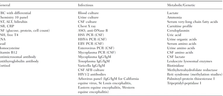
Table 1. List of Testing Which Yielded Normal Results
General Infectious Metabolic/Genetic CBC with differential Blood culture Lactate
Chemistry 10 panel Urine culture Ammonia
AST, ALT, bilirubin CSF culture Serum very-long chain fatty acids ESR, CRP Chest X ray Carnitine profile
CSF (glucose, protein, cell count) ASO, anti-DNase B Ceruloplasmin TSH, free T4 HSV PCR (CSF) Uric acid
ANA HHV6 PCR (CSF) Urine organic acids Lead EBV PCR (CSF) Serum amino acids Homocysteine Enterovirus PCR (CSF) Urine amino acids Vitamin B12 Mycoplasma PCR (CSF) CSF amino acids Antimicrosomal antibody Mycoplasma IgG/IgM CSF lactate
Antithyroglobulin antibody Toxoplasma IgG/IgM Leukocyte lysosomal enzymes Cortisol Varicella IgG/IgM Biotinidase
CSF AFB culture Methyltetrahydrofolate reductase HIV1/2 antibodies Rett syndrome (methylation studies) Arbovirus panel (IgG/IgM for California Palmitoyl-protein thioesterase I equine virus, St Louis encephalitis, Tripeptidyl-peptidase I Eastern equine encephalitis, Western
equine encephalitis)
NOTE: AST = aspartate aminotransferase; ALT = alanine transaminase; ASO = antistreptolysin O; ANA = antinuclear antibody; AFB = acid-fast bacilli; CSF = cerebrospinal fluid; CRP = C-reactive protein; CBC = complete blood count; ESR = erythrocyte sedimentation rate; EBV = Epstein-Barr virus; TSH = thyroid stimulating hormone; HSV = herpes simplex virus; HHV = human herpesvirus; HIV = human immunodeficiency virus; PCR = polymerase chain reaction.
This study was reviewed and a waiver granted by the University of Utah Institutional Review Board.
Discussion
This report describes a child with a progressive encephalopathy and intractable epilepsy. The patient did have a preceding history of mild developmental delay and prior seizures, but her presentation was remarkable for her prolonged unexplained coma. Despite extensive inves-tigations during her initial admission, no explanation for her seizures or coma was found until the results of her cerebrospinal fluid neurotransmitter analysis returned.
The patient was diagnosed with cerebral folate deficiency syndrome based on low cerebrospinal fluid levels of 5-methyltetrahydrofolate, the central nervous system metabolite of folate, and the presence of autoantibodies in the blood against folate receptors. Folate is actively trans-ported from the blood to the cerebrospinal fluid and brain by folate receptors, and the antibodies presumably inhibit trans-port of folate, leading to decreased cerebrospinal fluid levels. Cerebral folate deficiency syndrome has been reported in 30 patients.2,7,8 Typical symptoms include irritability,
developmental delay or regression, hypotonia and ataxia, seizures, and dyskinesias.6 Magnetic resonance imaging findings are variable between patients. Cerebellar atrophy has been reported in 3 of 20 patients with cerebral folate deficiency syndrome2 although cerebellar symptoms are reported in nearly all patients. In some patients, no MRI changes were noted,2,7,8whereas in other cases, moderate demyelination or supratentorial or infratentorial atrophy was found.2
The etiology of cerebral folate deficiency syndrome is unknown, and it is unknown whether it is primarily genetic or is autoimmune with an interaction with envi-ronmental triggers. Some support for the latter hypothe-sis has been shown by a reduction in the titer of folate receptor autoantibodies by adoption of a milk-free diet.9 Marked responses to treatment with folinic acid have been reported,2,6-8and it has been suggested that starting treatment at an earlier age may be associated with an improved response.2 Treatment with immunomodulatory agents has not been reported.
This case underlines the importance of cerebrospinal fluid neurotransmitter analysis in cases of unexplained coma, intractable epilepsy, or episodic progressive devel-opmental regression. Correct and timely diagnosis is important because of the potentially treatable nature of this disorder.6-8Starting folinic acid therapy immediately at diagnosis may be necessary to prevent irreversible cen-tral nervous system sequelae. Unfortunately, our case suggests that folinic acid treatment, even when started early, may not be an adequate treatment for all patients.
Acknowledgments
The authors report no conflicts of interest. Edward V. Quadros holds a patent (US 20060127955 and WO/ 2004/043233) issued to State University of New York Research Foundation describing methods for the detec-tion of folate receptor autoantibodies. This work is sup-ported in part by funding from the National Institutes of Health (HD051880 to Edward V. Quadros and K12 HD001410 to Joshua L. Bonkowsky) and by a grant from the Children’s Health Research Center, University of Utah, to Joshua L. Bonkowsky.
References
1. Pineda M, Ormazabal A, Lopez-Gallardo E, et al. Cerebral folate deficiency and leukoencephalopathy caused by a mito-chondrial DNA deletion. Ann Neurol. 2006;59:394-398. 2. Ramaekers VT, Blau N. Cerebral folate deficiency. Dev Med
Child Neurol. 2004;46:843-851.
3. Ramaekers VT, Hansen SI, Holm J, et al. Reduced folate trans-port to the CNS in female Rett patients. Neurology. 2003;61: 506-515.
Figure 1. (A) Routine electroencephalogram obtained at the age of 3
years, bipolar montage. The pattern in the boxed region and underline are part of the same discharge, consisting of left hemisphere 3-4 Hz spike and wave discharges maximal in the temporal-occipital region. (B) Brain magnetic resonance imaging, sagittal view, T1 series. Mild cerebellar atrophy is seen (arrows).
Cerebral Folate Deficiency Syndrome / Bonkowsky et al 1463
4. Blau N, Bonafe L, Krageloh-Mann I, et al. Cerebrospinal fluid pterins and folates in Aicardi-Goutieres syndrome: a new phe-notype. Neurology. 2003;61:642-647.
5. Ramaekers VT, Weis J, Sequeira JM, Quadros EV, Blau N. Mitochondrial complex I encephalomyopathy and cerebral 5-methyltetrahydrofolate deficiency. Neuropediatrics. 2007;38: 184-187.
6. Ramaekers VT, Rothenberg SP, Sequeira JM, et al. Autoantibodies to folate receptors in the cerebral folate deficiency syndrome.
N Engl J Med. 2005;352:1985-1991.
7. Moretti P, Sahoo T, Hyland K, et al. Cerebral folate deficiency with developmental delay, autism, and response to folinic acid.
Neurology. 2005;64:1088-1090.
8. Hansen FJ, Blau N. Cerebral folate deficiency: life-changing supple-mentation with folinic acid. Mol Genet Metab. 2005;84:371-373. 9. Ramaekers VT, Sequeira JM, Blau N, Quadros EV. A milk-free
diet downregulates folate receptor autoimmunity in cerebral folate deficiency syndrome [published online ahead of print March 19, 2008]. Dev Med Child Neurol. doi: 10.1111/j.1469-8749.2008.02053.x.